Horizon scanning Hypothermic Machine Perfusion & Normothermic Machine Perfusion
Upload
hoangthienCategory
view
224download
0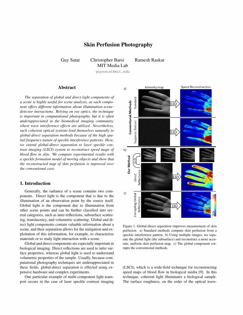
Skin Perfusion Photography
Guy Satat Christopher Barsi Ramesh RaskarMIT Media [email protected]
Abstract
The separation of global and direct light components ofa scene is highly useful for scene analysis, as each compo-nent offers different information about illumination-scene-detector interactions. Relying on ray optics, the techniqueis important in computational photography, but it is oftenunderappreciated in the biomedical imaging community,where wave interference effects are utilized. Nevertheless,such coherent optical systems lend themselves naturally toglobal-direct separation methods because of the high spa-tial frequency nature of speckle interference patterns. Here,we extend global-direct separation to laser speckle con-trast imaging (LSCI) system to reconstruct speed maps ofblood flow in skin. We compare experimental results witha speckle formation model of moving objects and show thatthe reconstructed map of skin perfusion is improved overthe conventional case.
1. IntroductionGenerally, the radiance of a scene contains two com-
ponents. Direct light is the component that is due to theillumination of an observation point by the source itself.Global light is the component due to illumination fromother scene points and can be further classified into sev-eral categories, such as inter-reflections, subsurface scatter-ing, translucency, and volumetric scattering. Global and di-rect light components contain valuable information about ascene, and their separation allows for the mitigation and ex-ploitation of this information, for example, to characterizematerials or to study light interaction with a scene.
Global and direct components are especially important inbiological imaging. Direct reflections are used to infer sur-face properties, whereas global light is used to understandvolumetric properties of the sample. Usually, because com-putational photography techniques are underappreciated inthese fields, global-direct separation is effected using ex-pensive hardware and complex experiments.
One particular example of multi-component light trans-port occurs in the case of laser speckle contrast imaging
Figure 1. Global-direct separation improves measurement of skinperfusion. a) Standard methods compute skin perfusion from aspeckle interference pattern. b) Using multiple images, we sepa-rate the global light (the subsurface) and reconstruct a more accu-rate, uniform skin perfusion map. c) The global component cor-rupts the conventional methods.
(LSCI), which is a wide-field technique for reconstructingspeed maps of blood flow in biological media [9]. In thistechnique, coherent light illuminates a biological sample.The surface roughness, on the order of the optical wave-

length, scatters the light in many directions. The result is ahigh-frequency interference pattern called speckle (Fig. 2,left). Although speckle is often an unwanted effect in imag-ing [19, 27], it can be used for measuring relative speedsof scattering objects. For a fixed object, the speckle patternis time-invariant, and the resulting image has a very highcontrast. For a dynamic object, the speckle pattern variesduring the camera exposure time, washing out the final im-age. Thus, speckle contrast is directly related to the flowvelocity of the scattering materials (Fig. 2, right).
An important application of LSCI is the measurement ofskin perfusion, which determines blood flow in skin, andis used to monitor burns, wounds, and skin lesions. Un-fortunately, however, scattering from the underlying tissuecorrupts the contrast measurement. Here, we present theapplication of global-direct separation methods to coherentillumination to simplify and improve measurement of bloodflow. The technique requires a hardware implementationthat utilizes the natural high-frequency interference patternof laser speckle to separate the desired component from thecorrupting tissue scattering.
1.1. Contributions
Specifically, our contributions include
1. a generalization of global-direct separation to wave-based imaging,
2. an application of global-direct separation in skin per-fusion measurement, and
3. a hardware implementation to demonstrate the princi-ple.
Limitations: Currently, we model speckle scatteringfrom a statistical perspective, rather than with a wave equa-tion. This allows us to model random speckle phenomenawithout having to fully generalize global-direct separationto the wave optics regime, but its extension to deterministicsystems remains an open question.
1.2. Related Work
LSCI theory was proposed by Fercher and Briers [9]after significant statistical analysis [11]. Several modifica-tions and extensions to speckle modeling have been demon-strated [8, 10, 12, 34], but deriving rigorous quantitative re-sults remains an open question [8].
LSCI implementation was first used to analyze mi-crovasculars in the retina [9] and has been applied to cere-bral tissues (usually on rats) [5, 17, 18, 20]. Advancementshave included dual-wavelength systems for studying mi-crovascular activity in burns [25] and multiple exposures touse speckle temporal statistics for improving spatial resolu-tion [6] or contrast of cerebral flow through the skull [18].
Figure 2. Left: speckle formation from a rough static surface hashigh contrast. Right: Speckle formation from a rough dynamicsurface produces low contrast images.
Rege et al. suggest anisotropic numerical processing for in-creased SNR [26]. Our global-direct separation improvesskin perfusion maps and can be integrated with these meth-ods with, e.g., a high-speed camera or projector.
Skin perfusion analysis via LSCI was suggested byBriers [4]. A similar system was suggested later [29]. Anal-ysis of wound healing in pigs was performed recently [30].Commercial devices [24] exist for medical use. A recentlyconducted quantitative comparison of skin perfusion mea-surements between a full field laser Doppler imager (seebelow) and a commercial device concluded that a multi-exposure device offers some advantages [31].
Doppler-based methods are alternatives to LSCI. In thistechnique, time-resolved sensors are used to measure theDoppler shift of scattered light. Briers, however, showedthat LSCI and Doppler methods are equivalent [3], exceptthat LSCI has better sensitivity at low speeds, and thatDoppler methods account for multiple scattering. A recentwork [10] uses laser Doppler for flowmetry in skin and sug-gests a model for quantitative analysis. Doppler sensitivityhas been improved in combination with ultrasound [33].
Global-direct separation can be effected through high-frequency illumination [21] and has been enhanced recently[13, 16]. O’Toole et al. [22] demonstrated a hardware-based global-direct solution using dual coding. Achar [1]addressed the issue of moving objects during global-directseparation by registering frames captured with high fre-quency illumination patterns. Global-direct separation wasused for medical imaging for enhancing veins in skin imag-ing [15]. The present work performs global-direct sepa-ration of coherent light, which naturally produces a high-frequency illumination pattern via wave interference. Thedirect component is used for improved imaging of skin per-fusion.

Figure 3. Global light produces erroneous contrast maps for skinperfusion. Left: a single layer of dynamic scattering can be re-constructed easily. Right: a second layer compounds the contrastimage, and the resulting speed map is incorrect.
2. Theory and Method2.1. Speed Estimation Using Speckle
Diffractive optics considers the wave properties of light.For coherent light propagation, relative phase delays resultin an interference pattern. When coherent light scattersfrom a rough surface (feature size ∼ optical wavelength),many coherent light components add randomly at an obser-vation point. This de-phased sum results in a granular, highfrequency interference pattern called speckle (Fig. 2). Thespeckle contrast at a pixel (i, j) is
Kij =σijµij
, (1)
where µij is the mean and σij is the per-pixel standard devi-ation of the intensity, both calculated over a spatial windowaround a given pixel. Typically, speckle intensities follownegative exponential statistics, so that, theoretically,K = 1.To calculate K for a given image, we calculate µij and σijfor an M ×M window around pixel (i, j). A typical exper-imental contrast map is shown in Fig. 1.
If the scattering object moves, the generated speckle pat-tern will vary dynamically. For short times, the specklepattern changes little, but after a characteristic time τc, thespeckle pattern becomes completely uncorrelated from the
initial configuration. Photographing the speckle image withan exposure time T longer than τc will yield a blurred image(Fig. 2, right). The contrast becomes [2]
K(T ) =
√β
{τcT
+τ2c2T 2
[exp
(−2Tτc
)− 1
]}, (2)
where β is a constant that depends on the ratio of pixel sizeto speckle size. Importantly, the underlying motion of thescattering objects determines τc and, consequently, the con-trast. For example, a fast moving object with speed V willhave a small τc, and the resulting photograph will have verylow contrast (Fig. 2). Because of the underlying assump-tions, the specific form of Eq. 2 is not universally accepted[23], so it is more common to assume [28] that τc � Tand V ∼ λ/τc. This allows us to reconstruct the speed as afunction of contrast and exposure time:
V ∼ (K2T )−1 (3)
In LSCI, we measure blood flow in skin (skin perfu-sion) by observing the speckle patterns produced by mov-ing red blood cells. As blood flows, the cells undergo ran-dom Brownian motion to produce a time-varying specklepattern. Measuring the contrast allows us to reconstruct thespeed map of the flow. However, because light penetratesthe skin, scattering occurs also from tissue and capillariesbeneath the skin. This scattering corrupts the contrast mea-surement and results in an incorrect speed map.
Eq. 3 provides the inverse relationship between scatter-ers’ speeds and image contrast for skin perfusion [28]. Tosummarize, the standard LSCI procedure is: (1) illuminatea sample with coherent light; (2) record a speckle patternfor exposure time T ; (3) use Eq. 1 to calculate the specklecontrast; and (4) reconstruct the speed map via Eq. 3.
2.2. Global-Direct Separation for Coherent Illumi-nation
We utilize global-direct decomposition to improve skinperfusion maps by removing light components scatteredfrom beneath the skin. Consider the simplified schematicin Fig. 3: for a single skin layer, LSCI is straightforward.However, underlying tissue (modeled in Fig. 3 (right) as asecond scattering layer) makes an unwanted speckle contri-bution to the contrast image.
In the context of skin perfusion, therefore, the globalcomponent is due to subsurface and volumetric scattering(Fig. 4). Accepted separation techniques [21] capture mul-tiple images of the scene, which is illuminated with differ-ent high frequency patterns. Thus, a set of intensity valuesare associated with each pixel (i, j). The maximum value(L+) and the minimum value (L−) can be used to separate

Figure 4. Light rays are captured from both the surface and subsur-face. The sensor measures the intensity of D, which is a result ofdirect illumination A and global scattering B + C. Many ray pathscan contribute to global light.
direct (D) and global (G) components as
D =L+ − L−
1− b(4)
G = 2L− − bL+
1− b2(5)
Here, b accounts for background illumination. Note thatglobal-direct separation was formulated using ray optics,whereas speckle is a wave phenomenon.
In order to use Eqs. 4 and 5, and avoid including com-plex diffraction integrals, we apply speckle statistics (de-rived rigorously from the underlying wave physics [12]).We describe our model in Sec. 3, below.
3. Synthetic Results3.1. Simulation Methods
We first evaluate the method with synthetic data. Be-cause rendering dynamic volumetric scattering is challeng-ing, we model a two-layer system. The front layer repre-sents the skin, and the rear layer contains all the scatteringof the underlying tissue.
To model coherent scattering (speckle), we proceed asfollows: first, the front layer is illuminated with a high-frequency speckle pattern, independent of the sample un-der study. This pattern strikes the first layer, and scatteringis modeled by generating a random intensity pattern withnegative exponential statistics. The resulting speckle pat-tern is the direct component. Next, a portion of this patternis transmitted and scattered towards the rear layer. Propa-gation is accounted for by blurring each speckle. The rearlayer forms a second speckle pattern (global component),which is reflected and added to the direct component.
Dynamic speckle is modeled by averaging S realizationsof speckle at a pixel, where S is proportional to the velocityat that pixel [12] and the exposure time: S = vT ∼ T/τc.
Figure 5. Simulated scene with two scattering layers. The blackcharacters indicate areas of high speed. Incident speckle illumina-tion scatters to the first layer, then to the second. Both are recordedsimultaneously for four different incident illumination patterns.
Thus, higher speeds and longer exposure times imply alarger average, which implies a reduced contrast. The simu-lation is repeated with N independent speckle illuminationpatterns, and global-direct decomposition is carried out withthe N resulting images.
The blur kernel size is chosen to approximate diffraction.In the paraxial approximation, a speckle of size w0 blurs tow(z) after a distance z according to
z =πw2
0
λ
√(w(z)
w0
)2
− 1 (6)
For example, for an average speckle size of 5 pixels, a blurkernel of 20 pixels, a wavelength of 0.750 µm, and a camerapixel size of 5 µm with unity magnification, Eq. 6 impliesthat the inter-layer distance is 1 cm, which is a typical quan-tity in biological imaging.
We note here that the high-frequency illumination pat-tern is itself a random speckle field. This intuitively“matches” the system dynamics and allows us to ignorediffraction from, e.g., the edges of the more conventionalcheckerboard pattern. The goal of the method is to calcu-late a relative speed map of the front layer based on N = 4illumination patterns by removing any corruption from thepresence of the rear layer.
3.2. Simulation Results
The synthetic scene is shown schematically in Fig. 5, andresults are shown in Fig. 6. Fig. 6a shows the ground truthspeed maps of each layer and a synthetic measurement ofeach layer in the absence of the other. Note the reduced con-trast within the characters, where the speed is high. Individ-ual measurements for four illumination patterns are shownin Fig. 6b. Though they appear similar, each one is a result

Figure 6. Synthetic results demonstrate benefits of global-direct separation for skin perfusion. (a) Scene ground truth: speed maps indicateareas of relative motion, and reflectance maps indicate the contrast measurement of each layer in the absence of the other. (b) Four mea-surements, illuminated by different speckle patterns. (c) Decomposition to direct component reflectance (top), the sum of measurements asa baseline (bottom). (d) Speed reconstruction based on the corresponding intensity maps. The reconstruction based on direct componentreproduces the speed map of the front layer, whereas the baseline result contains speed contributions from both layers.
of a different speckle illumination pattern. Inputting thesemeasurements into Eqs. 4 and 5, we obtain a direct light im-age. We reconstruct the speed map based on the contrast ofthis image (Fig. 6c,d (top)). For comparison (Fig. 6c,d (bot-tom)), we see that the standard LSCI reconstructs a speedmap that contains information from both layers (“ICCP”and “2014”), whereas the global-direct separation success-fully decouples them.
To quantify the simulation results, we normalize allspeed maps to the total energy of the ground truth speedmap. We define total image energy as the total kinetic en-ergy of the individual pixels:
E =
n∑i,j=1
V 2ij (7)
where Vij is the speed at pixel (i, j), and the image is n×npixels. The error metric is
ek =1
n2
n∑i,j=1
‖V (g)ij − V
(k)ij ‖
2 (8)
where V (k) is the reconstructed speed map (direct or base-line), and V (g) is the front layer ground truth speed map.
Fig. 7 (top) shows the reconstruction error as a func-tion of SNR for different numbers of illumination patterns.We see that both the standard LSCI method (red) and ourmethod (blue) improve for increasing N . For N > 4,
there was no appreciable improvement. With high SNR,our reconstructions produce 50% less error than the base-line LSCI method. Because the direct component is a differ-ence of images (Eq. 4), it is susceptible to noise. Therefore,for low SNR, the two methods are comparable. Interest-ingly, there is a regime of standard LSCI where increasingSNR increases the reconstruction error. We expect that thisis caused by a small amount of noise, which reduces localcontrast and changes the effective local speed.
Fig. 7 (bottom) shows analysis of the reconstruction er-ror for varying distance between layers (a larger distancebetween layers results in more blurring according to Eq. 6).As the distance between the layers increases, the reflectedspeckle field becomes increasingly blurred, which improvesthe global-direct separation. On the other hand, the reflec-tions corrupt a wider area of the image and increase thebaseline reconstruction error. As N increases, the error isreduced and similarly, above N > 4, there is little change.
4. Experimental Validation
4.1. Experimental Prototype
We implement the method in hardware using a Lumen-era monochrome camera without an IR filter to image ob-jects approximately 40 cm away with a 50 mm Nikon lens(f/2.8). To implement four illumination patterns, we usefour light sources: 3 mW lasers of center wavelength 785

Figure 7. Simulation speed reconstruction error for varying num-ber of illumination sources, measurement noise (top) and inter-nal layer depth (bottom). Each plot shows the reconstruction er-ror using the direct component (solid blue), and using the sumof all measurements (dashed red), for various measurement noise(SNR). N is the number of illumination patterns used.
nm, placed around the camera lens. To create illuminationspeckle patterns, each laser passes through a diffusive ma-terial (duct tape). The lasers are controlled by an Arduinomicrocontroller, which is synchronized in real time using aMatlab interface (Fig. 8). The lasers illuminate the scenesequentially, and the camera records an image for each onewith an exposure time of 50 ms. The total acquisition timeis approximately one second. The four recorded images arethen used to separate global and direct light. Contrast anal-ysis is performed on the direct light, and the speed map is
Figure 8. Experimental setup. Top: system block diagram. Bot-tom: picture of the imaging system used.
reconstructed. To calculate contrast, we use an averagingwindow of M = 13 (c.f., Eq. 1), and the background illu-mination is b = 0.01.
4.2. Experimental Results
The first result is shown in Fig. 1, which shows a healthyhuman hand. In Fig. 1a, we calculate the speed map usingtwo LSCI methods. The first is a direct contrast calculationfrom the sum of the four measurements (the sum is used tocreate a uniform illumination over the full field). The sec-ond is normalized by the skin texture. However, both pro-duce similar erroneous results. Skin perfusion in a normalhand should be uniform, whereas the conventional resultshave a highly uneven distribution. The four illuminationmeasurements and the direct component speed reconstruc-tion are shown in Fig. 1b, where we see a far more uniformspeed map.
Our improvement results from the global component(Fig. 1c) being removed and no longer corrupting the skinperfusion measurement. Note that the global componentcontains no speckle, the direct component does containspeckle, and the conventional measurement contains an in-termediate amount. Further inspection of the direct speedmap reveals two outliers, the tip of the second finger fromthe top, and the bottom right part of the palm. These areas

appear to have low speed associated with them. This can beexplained by examining the direct intensity map. We noticethese areas appear dark, which means they were not directlyilluminated during acquisition.
The second result (Fig. 9) shows a finger with a superfi-cial burn on an otherwise healthy finger. Because the burnedskin is dead, perfusion there should be low. Indeed, thespeed map calculated from the direct component corrobo-rates this. Raw measurements are shown in Fig. 9a, withour reconstruction in Fig. 9b. Here, the burned area is blue(low perfusion), and the rest of the (healthy) finger showsconstant perfusion. Interestingly, the burn is almost invisi-ble in the global component (Fig. 9c), and more obvious inthe direct component. Baseline conventional methods (non-normalized and normalized) are shown in Figs. 9d,e. Theburned area has less contrast, and the healthy finger doesnot exhibit constant perfusion.
5. Discussion and Future WorkOur results here indicate that global-direct separation
methods, derived for ray optics, have potential use for co-herent imaging. This can represent a significant advance-ment for LSCI and can improve reconstructions of bloodflow. Here, diffraction is modeled purely as a statisticalmodel, with the resulting high frequency speckle patternsamenable for computational separation, but deterministicscenes (e.g., diffraction from an edge) have not been stud-ied. A first-principles generalization of diffractive global-direct separation, to our knowledge, has yet to be per-formed. Further, though our qualitative results are similar tothose reported in conventional LSCI, we expect that global-direct separation of coherent light can be made quantitative.
The prototype hardware can be improved in severalways. Other optical wavelengths can provide informationabout, e.g., oxygenated and non-oxygenated red blood cells.Furthermore, a single laser steered by galvanometers wouldimprove stability and repeatability compared to the currentmulti-laser approach. Replacing the current 50 mm lenswith an objective will improve spatial resolution (at the costof narrowing the field of view). Computationally, differentspace-time-multiplexing techniques coupled with sparsity-based approaches can be integrated into this approach formore efficient information throughput.
Applying global-direct separation with coherent illumi-nation is not limited to perfusion and can improve otherskin-related analyses, such as extracting biological quanti-ties [32] and improving rendering [14]. It can also improveskin lesion analysis and diagnosis [7].
6. ConclusionsWe have implemented global-direct illumination separa-
tion of coherent speckle patterns for application in skin per-
Figure 9. Experimental results demonstrating improvement of skinperfusion measurement of unhealthy skin. a) Four raw measure-ments of a burned finger (burn is circled in red). The four measure-ments are decomposed to direct (b) and global (c) components, aswell as the sum of measurements for a baseline comparison (d,e).Right column: LSCI analysis results performed on the correspond-ing intensity maps. The burn is most obvious in the direct compo-nent, and is almost invisible in the global component. The directcomponent also provides more uniform results around the burn.
fusion analysis. Our interference model is based on speckleintensity statistics and can be used as a starting point foranalyzing global-direct separation techniques in the pres-ence of diffraction from rough surfaces. This work servesas an indication that ray-based computational photographytechniques have potential for diffraction-based applications.Thus, this contribution offers a bridge between computa-tional photography and biomedical imaging with intuitivemodeling of light scattering. Future efforts in computationalphotography can be applied towards medical imaging of, forexample, wound healing, burn recovery, and skin surgeries.

References[1] S. Achar, S. T. Nuske, and S. G. Narasimhan. Compensating
for Motion During Direct-Global Separation. In 2013 Int.Conf. Comp. Vis., 1481–1488, 2013.
[2] D. Briers, D. D. Duncan, E. Hirst, S. J. Kirkpatrick, M. Lars-son, W. Steenbergen, T. Stromberg, and O. B. Thompson.Laser speckle contrast imaging: theoretical and practicallimitations. J. Biomed. Opt., 18:066018, 2013.
[3] J. D. Briers. Laser Doppler and time-varying speckle: a rec-onciliation. J. Opt. Soc. Am. A, 13:345, 1996.
[4] J. D. Briers and S. Webster. Laser speckle contrast analysis(LASCA): a nonscanning, full-field technique for monitoringcapillary blood flow. J. Biomed. Opt., 1:174–9, 1996.
[5] H. Cheng, Q. Luo, Q. Liu, Q. Lu, H. Gong, and S. Zeng.Laser speckle imaging of blood flow in microcirculation.Phys. Med. Bio., 49:1347–1357, 2004.
[6] H. Cheng, Q. Luo, S. Zeng, S. Chen, J. Cen, and H. Gong.Modified laser speckle imaging method with improved spa-tial resolution. J. Biomed. Opt., 8:559–64, 2003.
[7] S. Cotton, E. Claridge, and P. Hall. A skin imaging methodbased on a colour formation model and its application to thediagnosis of pigmented skin lesions. In Proceedings of Med-ical Image Understanding and Analysis, 99: 49–52, 1999.
[8] D. D. Duncan and S. J. Kirkpatrick. Can laser speckleflowmetry be made a quantitative tool? J. Opt. Soc. Am.A, 25:2088, 2008.
[9] A. Fercher and J. Briers. Flow visualization by means ofsingle-exposure speckle photography. Opt. Comm., 37:326–330, 1981.
[10] I. Fredriksson, M. Larsson, and T. Stromberg. Model-basedquantitative laser Doppler flowmetry in skin. J. Biomed.Opt., 15:057002, 2010.
[11] J. W. Goodman. Laser Speckle and Related Phenomena.Springer, 1975.
[12] J. W. Goodman. Speckle Phenomena in Optics: Theory andApplications. Englewood, CO : Roberts & Co., 2007.
[13] M. Gupta, A. Agrawal, A. Veeraraghavan, and S. G.Narasimhan. Structured light 3D scanning in the presenceof global illumination. In IEEE CVPR, 713–720, 2011.
[14] J. Jimenez, T. Weyrich, T. Scully, N. Barbosa, C. Donner,X. Alvarez, T. Vieira, P. Matts, V. Orvalho, and D. Gutierrez.A practical appearance model for dynamic facial color. ACMTrans. Graph., 29:1, 2010.
[15] A. Kadambi, H. Ikoma, X. Lin, G. Wetzstein, and R. Raskar.Subsurface Enhancement through Sparse Representations ofMultispectral Direct/Global Decomposition. In OSA: Imag-ing and Applied Optics, CTh1B.4, 2013.
[16] T. Kobayashi, M. Gupta, and S. K. Nayar. Multiplexed illu-mination for scene recovery in the presence of global illumi-nation. In IEEE Int. Conf. Comp. Vis., 691–698, 2011.
[17] M. Le Thinh, J. S. Paul, H. Al-Nashash, A. Tan, A. R. Luft,F. S. Sheu, and S. H. Ong. New insights into image pro-cessing of cortical blood flow monitors using laser speckleimaging. IEEE Trans. Med. Imag., 26:833–42, 2007.
[18] P. Li, S. Ni, L. Zhang, S. Zeng, and Q. Luo. Imaging cerebralblood flow through the intact rat skull with temporal laserspeckle imaging. Opt. Lett., 31:1824, 2006.
[19] T. Loupas, W. McDicken, and P. Allan. An adaptiveweighted median filter for speckle suppression in medical ul-trasonic images. IEEE Trans. Circ. Sys., 36:129–135, 1989.
[20] J. K. Meisner, S. Sumer, K. P. Murrell, T. J. Higgins, and R. J.Price. Laser speckle flowmetry method for measuring spatialand temporal hemodynamic alterations throughout large mi-crovascular networks. Microcirculation, 19:619–31, 2012.
[21] S. K. Nayar, G. Krishnan, M. D. Grossberg, and R. Raskar.Fast separation of direct and global components of a sceneusing high frequency illumination. ACM Trans. Graph.,25:935, 2006.
[22] M. O’Toole, R. Raskar, and K. N. Kutulakos. Primal-dualcoding to probe light transport. ACM Trans. Graph., 31:1–11, 2012.
[23] A. B. Parthasarathy, W. J. Tom, A. Gopal, X. Zhang, andA. K. Dunn. Robust flow measurement with multi-exposureimaging. Opt. Expr., 16:1975–1989, 2008.
[24] Perimed (R). PeriCam PSI System http://www.perimed-instruments.com/.
[25] J. Qin, R. Reif, Z. Zhi, S. Dziennis, and R. Wang. Hemo-dynamic and morphological vasculature response to a burnmonitored using a combined dual-wavelength laser speckleand optical microangiography imaging system. Biomed. Opt.Expr., 3:455–66, 2012.
[26] A. Rege, J. Senarathna, N. Li, and N. V. Thakor. Anisotropicprocessing of laser speckle images improves spatiotemporalresolution. IEEE Trans. Biomed. Eng., 59:1272–80, 2012.
[27] J. M. Schmitt, S. H. Xiang, and K. M. Yung. Speckle inoptical coherence tomography. J. Biomed. Opt, 4:95–101,1999.
[28] J. Senarathna, A. Rege, N. Li, and N. V. Thakor. LaserSpeckle Contrast Imaging: theory, instrumentation and ap-plications. IEEE Rev. Biomed Eng., 6:99–110, 2013.
[29] M. S. D. Smith, E. F. Packulak, and M. G. Sowa. Develop-ment of a laser speckle imaging system for measuring rela-tive blood flow velocity. 6343: 634304, 2006.
[30] C. J. Stewart, C. L. Gallant-Behm, K. Forrester, J. Tulip,D. A. Hart, and R. C. Bray. Kinetics of blood flow dur-ing healing of excisional full-thickness skin wounds in pigsas monitored by laser speckle perfusion imaging. Skin Res.Technol., 12:247–53, 2006.
[31] O. Thompson, J. Bakker, C. Kloeze, E. Hondebrink, andW. Steenbergen. Experimental comparison of perfusionimaging systems using multi-exposure laser speckle, single-exposure laser speckle, and full-field laser Doppler. In Dyn.Fluctuations Biomed. Photonics IX, 822204, 2012.
[32] N. Tsumura, N. Ojima, K. Sato, M. Shiraishi, H. Shimizu,H. Nabeshima, S. Akazaki, K. Hori, and Y. Miyake. Image-based skin color and texture analysis/synthesis by extractinghemoglobin and melanin information in the skin. In ACMTrans. Graph. (SIGGRAPH 2003), 770, 2003.
[33] L. Wang, J. Xia, J. Yao, K. I. Maslov, and L. V. Wang. Ul-trasonically encoded photoacoustic flowgraphy in biologicaltissue. Phys. Rev. Lett., 111:204301, 2013.
[34] P. Zakharov, A. Volker, A. Buck, B. Weber, and F. Scheffold.Quantitative modeling of laser speckle imaging. Opt. Lett,31:3465, 2006.