Skin. General outline skin is the heaviest organ of the human body, it constitutes about 16% of...
39
Skin
-
Upload
delphia-mosley -
Category
Documents
-
view
221 -
download
5
Transcript of Skin. General outline skin is the heaviest organ of the human body, it constitutes about 16% of...
Skin

General outline
skin is the heaviest organ of the human body, it constitutes about 16% of body weight
is composed of epidermis , dermis and its appendages
the function of the skin:
barrier and protection sensory reception excretion body temperature regulation absorbing some materials

Layers of skin• Epidermis – outer layer composed of stratified
squamous epithelium.• Dermis – layer mainly composed of dense and
loose irregular connective tissue with lots of appendages in it.
*Hypodermis –Loose connective tissue between skin and deeper structures, lots of adipose cells form adipose tissue

epidermisepidermis
dermisdermis
Epidermis: stratified squamous epithelium of keratinized type
Dermis: dense connective tissue

EpidermisEpidermis
• Contains many layers of stratified squamous epithelium.
• Depending on the thickness of the skin, it is classified as thick or thin skin.
• The cells of thick skin are arranged in five layers
• There are no blood vessels in the epidermis• Abundant never ending

Layers of Thick Skin
From basal to surface:
• Stratum basale • Stratum spinosum • Stratum granulosum• Stratum lucidum• Stratum corneum

stratum corneum
stratum basale
stratum spinosum
stratum granulosum
stratum lucidum

Stratum basale
• A layer of cuboidal or columnar cells
• Cells are actively undergoing mitosis and push previous cells outward. Keeps constant renewal of EC.
• These cells receive nutrients from the blood supply that is found in the dermis next to them.

stratum basale
stratum spinosum
stratum granulosum

Stratum spinosum
• Stratum spinosum are polyhedral cells connected to each other by intercellular bridges (called spinous cells)
• A few lamellated granules are found in the cytoplasm, containing glycolipid and steroid

Stratum granulosum
•Relatively flattened cells, the unclei are undergoing disintegration
•Containing irregularly keratohyalin granules which fill the cell destroying nuclei and other organelles.

Stratum lucidum
• is made up of several layers of homogeneous and translucent cells, the nuclei and organelles of the cells no longer exist.

Stratum corneum
• is the outermost or surface layer.
• It constituted of a series of tightly-connected, flattened, cornified cells
• Cells are filled with keratin, intercellular spaces are filled of material discharged by lamellated granules
•Thickness varies on different parts of body

Cell Types in Epidermis
• Keratinocytes – responsible for production of keratin- makes skin waterproof
• Non-keratinised cells
Melanocytes- responsible for the production of melanin.
Langerhans cells – specialized cells believed to process antigens and deliver to lymph nodes.
Merkel cells – function as mechanoreceptors

Melanocyte:
pigment-producing cell
• large cells with long branched processes, located among the stratum basale cells• derived from neural crest cell• abundant melanosome, containing tyrosinase which turns tyrosine into melanin• The melanin granule content of the skin is responsible for skin color• Melanin granule absorb ultraviolet light, protect the body against ultraviolet irradiation


Langerhans Cell:
dendritic type, located among the spinous cells, presents antigens, -take up antigens in the skin and transport to lymph nodes
-reservoir for the HIV virus

Merkel Cell: - Have short processes, distributed in the basal cell layer- Their functional significance is not clear
It is presumed that they are sensitiveMechanoreceptor because they are in close relation to nerve endings

Dermis:
located beneath the epidermis
Reticular layer:
Papillary layer:a thin layer of LCT which increase the total contact surface between the dermis and epidermis, contain capillary network and nerve ending (tactile corpuscle)
under the papillary layer; consists of irregular DCT; contain large blood vessels, appendages of skin, nerves (lamellated corpuscle). The elastic network is responsible for the elasticity of the skin.
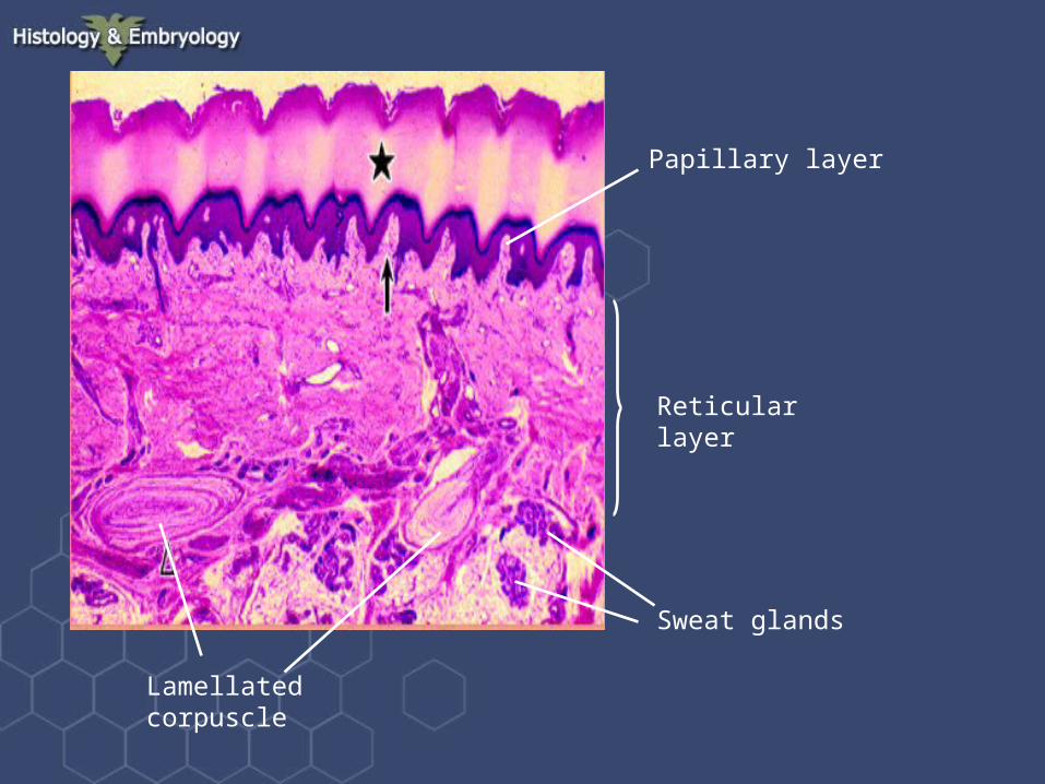
Papillary layer
Reticular layer
Lamellated corpuscle
Sweat glands

Tactile Corpuscle in papillary layer
Tactile Tactile corpusclecorpuscle

Meissner’s corpuscle

Hypodermis (subcutaneous tissue)•A layer of loose connective tissue between the skin and deeper structures
•the fat content is more significant in the hypodermis which not only serves as a reservoir of fat but also protects deeper tissue against pressure or impact

Skin appendages
hair
sebaceous gland
sweat gland

Skin appendages

Hair
• Hair is a thorny thread-like structure.
• Hair shaft is the part that extends above the surface and hair root is part that is embedded in the skin
• Hair resides in a hair follicle.
• Hairs differ in length, thickness and color according to their position on the body


Hair root
Hair follicle

Hair follicleHair root

Hair papilla

Sebaceous Glands
• Associated with the hair follicles• Produce oily substance, which is act as a
lubricant to make the skin soft and protects both skin and the hair from drying out.
• Oil released to outside via the hair follicle. If passage becomes blocked, cause acne.

•Arrector pili – smooth muscle of hair this muscle contracts– “goose pimple”

Sebaceous gland Arrector pili
The secretory potions are round sacs, the cells at the periphery are smaller, toward the centre the cells grow large and become mature which fill with fatty droplet

Arrector pili

Sweat Glands
• Sweat glands are distributed over most of the body.
• Simple tubular glands that transverse from the dermis to the surface of the epidermis.
• Merocrine sweat glands are found on most of the body and produce a thin watery solution and function in heat regulation.
• Apocrine sweat glands are found in axilla, mammary areolae, and circumanal region produce much thicker secretion that is odor producing. Often become activated at puberty

A sweat gland is divided into two parts:
•Secretory portion: is composed of pyramidal secretory cells
•Duct: lined by two layers of cuboidal epithelial cells
Secretory portion
duct