Skeletal effects of RME in the transverse and vertical ... · Conclusions: No significant...
9
© 2017 Dental Press Journal of Orthodontics Dental Press J Orthod. 2017 July-Aug;22(4):61-9 61 original article Skeletal effects of RME in the transverse and vertical dimensions of the nasal cavity in mouth-breathing growing children Mario Cappellette Jr. 1 , Lucia Hatsue Yamamoto Nagai 2 , Raquel Mori Gonçalves 2 , Aparecida Keiko Yuki 2 , Shirley Shizue Nagata Pignatari 1 , Reginaldo Raimundo Fujita 1 Introduction: Maxillary constriction is a dentoskeletal deformity characterized by discrepancy in maxilla/mandible relationship in the trans- verse plane, which may be associated with respiratory dysfunction. Objective: The objective of this study was to evaluate the skeletal effects of RME on maxillary and nasal transverse dimensions and compare the differences between males and females. Methods: Sixty-one mouth- breathers patients with skeletal maxillary constriction (35 males and 26 females, mean age 9.6 years) were included in the study. Posteroanterior (PA) radiographs were taken before expansion (T 1 ) and 3 months after expansion (T 2 ). Data obtained from the evaluation of T 1 and T 2 cepha- lograms were tested for normality with the Kolmogorov-Smirnov method. The Student’s t-test was performed for each measurement to de- termine sex differences. Results: RME produced a significant increase in all linear measurements of maxillary and nasal transverse dimensions. Conclusions: No significant differences were associated regarding sex. The RME produced significant width increases in the maxilla and nasal cavity, which are important for treatment stability, improving respiratory function and craniofacial development. Keywords: Palatal expansion technique. Maxilla. Mouth-breathing. 1 Universidade Federal de São Paulo, Departamento de Otorrinolaringologia e Cirurgia de Cabeça e Pescoço (São Paulo/SP, Brasil). 3 Private practice (São Paulo/SP, Brasil). » The authors report no commercial, proprietary or financial interest in the products or companies described in this article. Patients displayed in this article previously approved the use of their facial and intraoral photographs. Submitted: July 27, 2016 - Revised and accepted: February 03, 2017 DOI: https://doi.org/10.1590/2177-6709.22.4.061-069.oar How to cite: Cappellette Jr. M, Nagai LHY, Gonçalves RM, Yuki AK, Pigna- tari SSN, Fujita RR. Skeletal effects of RME in the transverse and vertical dimen- sions of the nasal cavity in mouth-breathing growing children. Dental Press J Or- thod. 2017 July-Aug;22(4):61-9. DOI: https://doi.org/10.1590/2177-6709.22.4.061-069.oar Contact address: Mario Cappellette Jr. Rua Salete, 200, conj. 101/102 – CEP: 02.016-001, São Paulo/SP, Brasil E-mail: [email protected] Introdução: a constrição maxilar é uma alteração dentoesquelética relacionada à diminuição transversal da arcada superior, que pode correlacionar- -se com problemas respiratórios. O tratamento nos pacientes em crescimento inclui a expansão rápida da maxila (ERM). A correção precoce resulta em maiores alterações esqueléticas e estabilidade dos resultados, podendo evitar desvios de crescimento facial. Objetivo: avaliar comparativamente, por meio de telerradiografias posteroanteriores (PA), as alterações dimensionais da cavidade nasal pré- e pós-ERM em pacientes respiradores bucais dos sexos masculino e feminino. Métodos: realizou-se o estudo de medidas lineares em telerradiografias PA pré- e pós-ERM de uma amostra composta por 61 pacientes (35 do sexo masculino e 26 do feminino), com média de idade de 9,6 anos. Todos os pacientes eram respiradores bucais com constrição maxilar esquelética, e foram tomadas radiografias PA pré- e pós-ERM (3 meses). Os dados obtidos foram avaliados nos tempos T 1 e T 2 a normalidade dos dados foi confirmada por meio do teste de Kolmogorov-Smirnov. O teste t de Student foi aplicado para cada mensuração, para determinar diferenças entre os sexos. Resultados: a ERM promoveu aumento significativo em todas as medidas lineares maxilares e das dimensões da cavidade nasal. Conclusão: nenhuma diferença significativa foi associada ao sexo. A ERM produziu aumentos significativos na largura maxilar e volume da cavidade nasal, que são importantes na estabilidade do tratamento Nos pacientes em crescimento e respiradores bucais com constrição maxilar esquelética da maxila, a ERM promoveu aumento do volume da cavidade nasal que melhorou o fluxo aéreo e possibilitou reconduzir o crescimento facial. As alterações transversais não foram significativas quando relacionadas ao sexo. Palavras-chave: Técnica de expansão palatal. Maxila. Respirador bucal.
Transcript of Skeletal effects of RME in the transverse and vertical ... · Conclusions: No significant...

© 2017 Dental Press Journal of Orthodontics Dental Press J Orthod. 2017 July-Aug;22(4):61-961
original article
Skeletal effects of RME in the transverse and vertical
dimensions of the nasal cavity in mouth-breathing
growing children
Mario Cappellette Jr.1, Lucia Hatsue Yamamoto Nagai2, Raquel Mori Gonçalves2, Aparecida Keiko Yuki2, Shirley Shizue Nagata Pignatari1, Reginaldo Raimundo Fujita1
Introduction: Maxillary constriction is a dentoskeletal deformity characterized by discrepancy in maxilla/mandible relationship in the trans-verse plane, which may be associated with respiratory dysfunction. Objective: The objective of this study was to evaluate the skeletal effects of RME on maxillary and nasal transverse dimensions and compare the differences between males and females. Methods: Sixty-one mouth-breathers patients with skeletal maxillary constriction (35 males and 26 females, mean age 9.6 years) were included in the study. Posteroanterior (PA) radiographs were taken before expansion (T1) and 3 months after expansion (T2). Data obtained from the evaluation of T1 and T2 cepha-lograms were tested for normality with the Kolmogorov-Smirnov method. The Student’s t-test was performed for each measurement to de-termine sex differences. Results: RME produced a significant increase in all linear measurements of maxillary and nasal transverse dimensions. Conclusions: No significant differences were associated regarding sex. The RME produced significant width increases in the maxilla and nasal cavity, which are important for treatment stability, improving respiratory function and craniofacial development.
Keywords: Palatal expansion technique. Maxilla. Mouth-breathing.
1 Universidade Federal de São Paulo, Departamento de Otorrinolaringologia e Cirurgia de Cabeça e Pescoço (São Paulo/SP, Brasil).
3 Private practice (São Paulo/SP, Brasil).
» The authors report no commercial, proprietary or financial interest in the products or companies described in this article. Patients displayed in this article previously approved the use of their facial and intraoral photographs.
Submitted: July 27, 2016 - Revised and accepted: February 03, 2017
DOI: https://doi.org/10.1590/2177-6709.22.4.061-069.oar
How to cite: Cappellette Jr. M, Nagai LHY, Gonçalves RM, Yuki AK, Pigna-tari SSN, Fujita RR. Skeletal effects of RME in the transverse and vertical dimen-sions of the nasal cavity in mouth-breathing growing children. Dental Press J Or-thod. 2017 July-Aug;22(4):61-9. DOI: https://doi.org/10.1590/2177-6709.22.4.061-069.oar
Contact address: Mario Cappellette Jr.Rua Salete, 200, conj. 101/102 – CEP: 02.016-001, São Paulo/SP, BrasilE-mail: [email protected]
Introdução: a constrição maxilar é uma alteração dentoesquelética relacionada à diminuição transversal da arcada superior, que pode correlacionar--se com problemas respiratórios. O tratamento nos pacientes em crescimento inclui a expansão rápida da maxila (ERM). A correção precoce resulta em maiores alterações esqueléticas e estabilidade dos resultados, podendo evitar desvios de crescimento facial. Objetivo: avaliar comparativamente, por meio de telerradiografias posteroanteriores (PA), as alterações dimensionais da cavidade nasal pré- e pós-ERM em pacientes respiradores bucais dos sexos masculino e feminino. Métodos: realizou-se o estudo de medidas lineares em telerradiografias PA pré- e pós-ERM de uma amostra composta por 61 pacientes (35 do sexo masculino e 26 do feminino), com média de idade de 9,6 anos. Todos os pacientes eram respiradores bucais com constrição maxilar esquelética, e foram tomadas radiografias PA pré- e pós-ERM (3 meses). Os dados obtidos foram avaliados nos tempos T1 e T2 a normalidade dos dados foi confirmada por meio do teste de Kolmogorov-Smirnov. O teste t de Student foi aplicado para cada mensuração, para determinar diferenças entre os sexos. Resultados: a ERM promoveu aumento significativo em todas as medidas lineares maxilares e das dimensões da cavidade nasal. Conclusão: nenhuma diferença significativa foi associada ao sexo. A ERM produziu aumentos significativos na largura maxilar e volume da cavidade nasal, que são importantes na estabilidade do tratamento Nos pacientes em crescimento e respiradores bucais com constrição maxilar esquelética da maxila, a ERM promoveu aumento do volume da cavidade nasal que melhorou o fluxo aéreo e possibilitou reconduzir o crescimento facial. As alterações transversais não foram significativas quando relacionadas ao sexo.
Palavras-chave: Técnica de expansão palatal. Maxila. Respirador bucal.

© 2017 Dental Press Journal of Orthodontics Dental Press J Orthod. 2017 July-Aug;22(4):61-962
Skeletal effects of RME in the transverse and vertical dimensions of the nasal cavity in mouth-breathing growing childrenoriginal article
INTRODUCTIONMaxillary constriction is a narrowing of the upper
arch and is one of the most prevalent malocclusions. Some of its typical features are unilateral or bilateral pos-terior crossbite, anterior dental crowding, a high palatal vault, and a decrease in the distance between the lateral walls of the nasal cavity.1 The etiology is believed to be multifactorial2 and the main factors includes physiologi-cal problems, genetic and parafunctional habits3 that could lead to a mixed or mouth-breathing pattern.4,5 Since the airway is assumed to play a role in dentofacial development, the association with respiratory problems, especially nasal obstruction, has been the focus of many researchers who have investigated the possibility that these events are related.6 However, it could be consid-ered erroneous to associate malocclusions only with the breathing pattern.2
The maxillary bones form approximately 50% of the nasal cavity’s anatomic structure.7 RME is a dentofacial orthopedic treatment procedure that has been widely used for correcting maxillary transverse deficiency in young patients and it can change the morphology of the maxillary arch, affecting the geometry and function of the nasal cavity.7 Eysel, in 1886, cited by Haas8,9 was the first rhinologist who studied the effect of RME on nasal cavity function. He found that, in the post-RME peri-od, various changes occurred in the maxilla and adjacent bones, and RME caused an opening of the nasal cavity and reduction in nasal airway resistance. In addition, af-ter the expansion an increase was found in the nasal cav-ity width and in the nasal base adjacent to the midpalatal suture. The maxillary sutures separate the external walls of the nasal cavity laterally, resulting in an increase in the intranasal capacity. Although orthodontic treatment is carried out to correct dental and skeletal discrepancies, some authors showed that RME outcomes could also be effective on naso-respiratory and sleep-disordered breathing problems of growing children.11 Therefore, RME has been suggested in the medical treatment of mouth-breathing, septal deformity, nasal infection, al-lergic rhinitis, and obstructive sleep apnea. However, its clinical use for rhinological effects is controversial.12 Its primary goal is to maximize orthopedic and minimize orthodontic tooth movement13 and there is an agree-ment among orthodontists that a maxillary constriction should be treated early,9 since the proportion of skeletal and dental movement depends on patient’s age and ma-
turity level.14,15 In a study of RME effects, sex differences proved to be important as it is known that the facial skel-eton significantly increases its resistance to expansion with increasing age and maturity.15
The majority of growth studies have used lateral cepha-lometric radiographs to analyze vertical and sagittal dimen-sional changes of the face. However, transverse problems and development of the oronasal area can be better understood by analyzing posteroanterior (PA) cephalometric radio-graphs.5 Structural remodeling of nasal cavity has the effect of increasing nasal patency after RME, and the relevance of PA radiograph is to show this remodeling and quantify the increase in nasal cavity,16 especially in the midface, since its walls are laterally displaced. The purpose of this article was to evaluate the skeletal effects of RME in the transverse and vertical dimensions on nasal cavity in mouth-breathing growing children and to correlate it with both sexes.
MATERIAL AND METHODSThe study sample comprised 61 children (mean age 9.6
years, range 6.5 – 13.10 years; 35 male, 26 female) regardless of facial type or race, who sought treatment at the Depart-ment of Pediatric Otorhinolaryngology of the Universidade Federal de São Paulo, in Brazil. The criteria for selection of the treatment group were as follows: skeletal constricted maxil-lary arches, unilateral or bilateral posterior crossbite requir-ing RME treatment, and mouth-breathing. The exclusion criteria were: no maxillary first molars, metallic restorations, periodontal diseases, previous orthodontic treatment, and genetic disease involving chromosome or mutations.
In order to check for the mouth-breathing pattern, all patients were clinically examined by an experienced oto-rhinolaryngologist that verified the presence of nasal ob-struction after anterior rhinoscopy, oroscopy and nasofi-berendoscopy17. Potential candidates for adenoidectomy or adenotonsillectomy, with complete occlusion of the nasal cavity by nasal turbinates, intranasal tumors or polyps, ad-enoid occupying more than 70% of the choanas were ex-cluded from the study.
The subjects were divided into two groups: males (mean age 10.1 years, range 6.5 – 13.10 years) and females (mean age 9.2 years, range 6.5 – 12.5 years). The study was approved by the Research Ethics Committee of Uni-versidade Federal de São Paulo (protocol # 0907/08) and an informed consent was obtained from the parents or guard-ians, besides the verbal assent from the children before data collection for the study.

© 2017 Dental Press Journal of Orthodontics Dental Press J Orthod. 2017 July-Aug;22(4):61-963
original articleCappellette Jr. M, Nagai LHY, Gonçalves RM, Yuki AK, Pignatari SSN, Fujita RR
Figure 1 - RME appliance, comprising a tooth-anchored device (conventional Hyrax expander).
Figure 2 - Pre-RME and post-RME occlusal radio-graphs.
The orthodontic phase of treatment was undertaken at a local orthodontic clinics under the supervision of the department’s orthodontists.
Pretreatment (T1) orthodontic records, including PA radiographs were taken for all patients. Each patient under-went a standardized RME protocol using a tooth-anchored device activated by means of a modified Hyrax expander with a soldered framework and orthodontic bands on de-ciduous second molars and extended forward to the palatal surfaces of the deciduous canines (Fig 1); or supported by bilateral maxillary first premolars and first molars, in case the first premolars were sufficiently erupted. After the ex-pander was cemented, it was activated six turns. Then, the parents or guardians were instructed to activate the jackscrew one turn (0.25 mm) twice a day until the re-quired expansion was achieved. The degree of expansion
was calculated for each patient, including a bilateral over-expansion and buccal tipping of a half-cusp width. After a mean active expansion period of 15 days (range 7- 21 days), the expander was tied off with a ligature wire and it was kept on the teeth as a passive retainer for at least 90 days (3 months), ranging from 91 to 106 days. This retention period allowed osseous reorganization of the midpalatal suture after expansion. All patients did not receive brackets or wires on the maxillary arch until T2 records were taken.
Postexpansion (T2) PA cephalograms were taken af-ter removal of the expander. The mean interval between T1 and T2 was 98 days (range, 91-105 days). As a result, study sample was composed by 122 PA cephalograms, which were hand traced with a 0.5-mm lead pencil on Ultraphan acetate tracing paper. All tracings were per-formed by the same investigator.
Tables 1 and 2 show the skeletal and dental landmarks, and linear measurements (in millimeters) from Ricketts’ cephalometric analysis.17
Statistics analysisDescriptive statistics including mean, standard devia-
tion (SD), and ranges were calculated for T1 and T2 mea-surements. Independent samples t-test was performed for each measurement, to determine sex differences. Significance for all statistical tests was predetermined at p < 0.05. T1 and T2 measurements and comparisons are presented in Tables 3 to 6. To estimate intraexaminer error, reliability and reproducibility, 20 randomly se-lected records (10 males and 10 females) were reevalu-

© 2017 Dental Press Journal of Orthodontics Dental Press J Orthod. 2017 July-Aug;22(4):61-964
Skeletal effects of RME in the transverse and vertical dimensions of the nasal cavity in mouth-breathing growing childrenoriginal article
Table 1 - Posteroanterior skeletal and dental landmarks.
Table 2 - Posteroanterior planes and linear measurements.
Landmark Description
NC/CN The most lateral point of the nasal cavity
ZL/ZR The most internal point of the frontozygomatic
ANS Anterior nasal spine
JL/JR Deepest point of the alveolar maxillary process
UML/UMR The most prominent lateral point on the buccal surface of permanent first molar crown
Reference plane and line Reference Plane Description
ZL-ZR Z plane- reference line in the horizontal plane
Occlusal plane Occlusal line in the molars
NC-CN Nasal width
ANS-Z plane (NH) Nasal height
JL-JR Maxillary width
IM Intermolar width - distance between UML-UMR parallel to the occlusal plane.
IM/ JL-JR Ratio between intermolar width and maxillary width
JL-JR/ NC-CN Ratio between maxillary width and nasal width
Figure 3 - PA cephalometric radiograph pre-RME. Figure 4 - PA cephalometric radiograph post-RME, showing lines used for measurements.

© 2017 Dental Press Journal of Orthodontics Dental Press J Orthod. 2017 July-Aug;22(4):61-965
original articleCappellette Jr. M, Nagai LHY, Gonçalves RM, Yuki AK, Pignatari SSN, Fujita RR
Table 3 - Comparisons of T1 values between males and females.
NS = nonsignificant; n = number of patients; SD = standard deviation. *Statistically significant (p < 0.05).
Variable Sex Mean n SD P
Age (month)Males 121.5 35 24.8
0.070Females 108.2 26 31.5
Height (mm)Males 53.9 35 3.4
0.025*Females 51.6 26 4.3
Nasal width (mm)Males 28.7 35 2.8
0.782Females 28.5 26 3.2
Área (nasal width x nasal
height) (mm2)
Males 1551.8 35 206.50.153
Females 1473.1 26 214.5
Maxillary width (mm)Males 63.6 35 3.8
0.026*Females 61.4 26 3.6
Intermolar width (mm)Males 58.3 35 3.6
0.239Females 57.2 26 3.7
Intermolar width/ Maxillary
width ratio
Males 1.09 35 0.060.426
Females 1.08 26 0.08
Maxillary width /Nasal widthMales 2.23 35 0.19
0.267Females 2.17 26 0.20
Table 4 - Comparisons of T2 measurements between males and females.
NS = nonsignificant; n = number of patients; SD = standard deviation. Statistically significant, *p < 0.05, **p < 0.01.
Variable Sex Mean n SD P
Height (mm)Males 56.4 35 3.65
0.05*Females 54.2 26 4.79
Nasal width (mm)Males 31.3 35 3.27
0.543Females 31.9 26 3.61
Área (nasal width x nasal
height) (mm2)
Males 1770.7 35 248.60.548
Females 1730.8 26 263.1
Maxillary width (mm)Males 67.4 35 3.32
0.007**Females 64.7 26 4.05
Intermolar width (mm)Males 65.4 35 3.44
0.029*Females 62.9 26 5.08
Intermolar width/
Maxillary width ratio
Males 1.03 35 0.040.996
Females 1.03 26 0.07
Maxillary width / Nasal
width
Males 2.17 35 0.180.014*
Females 2.05 26 0.18
© 2017 Dental Press Journal of Orthodontics Dental Press J Orthod. 2017 July-Aug;22(4):61-966
Skeletal effects of RME in the transverse and vertical dimensions of the nasal cavity in mouth-breathing growing childrenoriginal article
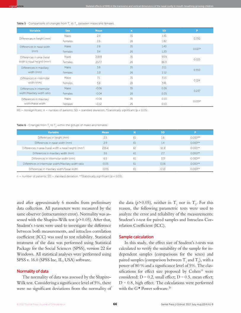
Table 6 - Changes from T1 to T
2 within the groups of males and females.
n = number of patients; SD = standard deviation. **Statistically significant (p < 0.01).
Table 5 - Comparisons of changes from T1 to T
2, between males and females.
NS = nonsignificant; n = number of patients; SD = standard deviation. *Statistically significant (p < 0.05).
Variable Sex Mean n SD P
Differences in height (mm)Males 2.4 35 1.45
0.750Females 2.6 26 1.82
Differences in nasal width
(mm)
Males 2.6 35 1.400.027*
Females 3.4 26 1.20
Differences in area (nasal
width x nasal height) (mm2)
Males 218.8 35 93.90.103
Females 257.7 26 86.0
Differences in maxillary
width (mm)
Males 3.8 35 3.110.550
Females 3.3 26 2.12
Differences in intermolar
width (mm)
Males 7.1 35 3.150.124
Females 5.8 26 3.41
Differences in intermolar
width/Maxillary width ratio
Males -0.06 35 0.060.237
Females -0.04 26 0.05
Differences in maxillary
width/Nasal width
Males -0.06 35 0.100.019*
Females -0.12 26 0.10
Variable Mean N SD P
Differences in height (mm) 2.5 61 1.6 0.000**
Differences in nasal width (mm) 2.9 61 1.4 0.000**
Differences in area (nasal width x nasal height) (mm2) 235.4 61 91.9 0.000**
Differences in maxillary width (mm) 3.6 61 2.7 0.000**
Differences in intermolar width (mm) 6.5 61 3.3 0.000**
Differences in intermolar width/Maxillary width ratio -0.05 61 0.06 0.000**
Differences in maxillary width/Nasal width -0.09 61 0.10 0.000**
ated after approximately 6 months from preliminary data collection. All parameters were measured by the same observer (intraexaminer error). Normality was as-sessed with the Shapiro-Wilk test (p > 0.05). After that, Student’s t-tests were used to investigate the difference between both measurements, and intraclass correlation coefficient (ICC) was used to test reliability. Statistical treatment of the data was performed using Statistical Package for the Social Sciences (SPSS), version 22 for Windows. All statistical analyses were performed using SPSS v. 16.0 (SPSS Inc, IL, USA) software.
Normality of dataThe normality of data was assessed by the Shapiro-
Wilk test. Considering a significance level of 5%, there were no significant deviations from the normality of
the data (p > 0.05), neither in T1 nor in T2. For this reason, the following parametric tests were used to analyze the error and reliability of the measurements: Student’s t-test for paired samples and Intraclass Cor-relation Coefficient (ICC).
Sample calculationIn this study, the effect size of Student’s t-tests was
calculated to verify the suitability of the sample for in-dependent samples (comparisons for the sexes) and paired samples (comparison between T1 and T2), with a power of 80 % and a significance level of 5%. The clas-sifications for effect size proposed by Cohen19 were considered: D = 0.2, small effect; D = 0.5, mean effect; D = 0.8, high effect. The calculations were performed with the G * Power software.20

© 2017 Dental Press Journal of Orthodontics Dental Press J Orthod. 2017 July-Aug;22(4):61-967
original articleCappellette Jr. M, Nagai LHY, Gonçalves RM, Yuki AK, Pignatari SSN, Fujita RR
Under these conditions, the sample allowed to de-tect mean effects (Cohen’s D = 0.65) in the compari-son between the male group (n = 35) and the female group (n = 26). For the comparison between T1 and T2 (n = 61), the sample allowed to detect small effects (Cohen D = 0.32).
Method error analysisThe results obtained through the Student’s t-test for
paired samples showed correspondence between the initial measurements and the repetitions by the same evaluator (Intraclass Correlation Coefficient), both in T1 (p > 0.05 and ICC close to 1.000) and in T2 (p > 0.05 and ICC close to 1.000), indicating the absence of mea-surement error, reliability and reproducibility (Table 7).
RESULTSTable 3 refers to sex differences in T1 and it shows sta-
tistical significance for nasal height and maxillary width variables, which were smaller in female group.
Table 4 compares the sex differences at T2. RME produced greater increases in the male group for nasal height, maxillary width, intermolar width and nasal width/maxillary width ratio.
Table 5 compares the sex differences at T1 and T2. Sig-nificant changes in the nasal width were observed in fe-male group, and the nasal width/maxillary width ratio was smaller in female group.
Table 6 shows changes within the groups (T2-T1). RME produced skeletal, dental and nasal increases in both males and females.
All measurements were considered to be reliable, since the reliability statistics were equal to 1, which in-dicates perfect reliability.
DISCUSSIONIn both males and females groups, the maxilla
showed a significant increase for the linear measure-ments after RME, which is in accordance to the find-ings of other authors that reported post-RME sagittal and transverse increases of skeletal, dental and nasal structures.3,15,21-23 According to Wriedt et al24 and Warren et al,25 the enlargement of the nasal valve with an increase of nasal volume can result in improvement of nasal breathing. More controversial is the question of whether RME can achieve a shift from mouth to nasal breathing patterns and change the subjective sensation of nasal obstruction.26 These effects depend on the existence or not of nasal obstruction and on its cause, location and severity.6
Statistical results in Table 3, which compares the values obtained in the pre-RME (T1) between males and females, show that most of the variables were not significant (p > 0.05) indicating no correla-tion between sex and the linear measurements in T1. The two statistically significant variables — nasal height (p = 0.025) and maxillary width (p = 0.026) —for males are in agreement with other studies in the literature5. Snodell et al5 showed sex differences in maxillary width at 6 years old and higher values for maxillary width and intermolar width at age 12 years in males. In the composition of the mean values of the nasal cavity found by Ricketts et al,18 the propor-tion between nasal height and nasal width represents 60%. In this study, this proportion was 56% in both males and females groups, confirming reduced nasal width in the mouth-breathing sample. Whereas the growth in the vertical direction is larger than in the transverse one, from 6 to 18 years of age — with a
Table 7 - Error analysis: mean and standard deviation (SD), Student’s t-test for paired samples and ICC (n = 20).
SD = standard deviation. ** Statistically significant (p < 0.05).
Variable SexMeasurement T
1
Mean (SD)
Measurement T2
Mean (SD)Student’s t-test ICC
Nasal widthMales 21.78 (1.64) 23.74 (2.68) 0.531 0.989
Females 22.22 (1.71) 22.12 (1.56) 0.671 0.907
HeightMales 42.77 (3,99) 45.94 (4.29) 0.477 0.999
Females 41.0 (4.09) 40.83 (4.01) 0.267 0.993
Intermolar widthMales 102.55 (5.41) 104.16 (5.19) 0.443 0.999
Females 101.21 (4.19) 101.3 (4.42) 0.653 0.991
Maxillary widthMales 58.72 (2,86) 61.93 (3.42) 0.067 0.998
Females 58.79 (3.03) 58.71 (3.01) 0.070 0.998

© 2017 Dental Press Journal of Orthodontics Dental Press J Orthod. 2017 July-Aug;22(4):61-968
Skeletal effects of RME in the transverse and vertical dimensions of the nasal cavity in mouth-breathing growing childrenoriginal article
increased percent in males — and by evaluating the mean age of the sample, the proportion between the values of transverse and vertical measures was below the one found in longitudinal studies of facial growth.5 These results are consistent with studies reporting a de-crease in measures of the nasal cavity in mouth-breath-ing patients.6
The results in Table 4 refer to the sex difference at T2. RME produced greater increases in the male group for nasal height (p = 0.05), maxillary width (p = 0.026), intermolar width (p = 0.029) and nasal width/maxillary width ratio (p = 0.014). The maxil-lary width increase correlates with intermolar width, according to longitudinal cephalometric studies, and the nasal width was also correlated with maxillary width and maxillary intermolar width.5 The greatest increase in the maxillary width and in the intermo-lar width in males can be understood by the higher bone resistance of females, which complete puberty earlier than males, and the higher maxillary growth rate in males, which may affect resistance to the ex-pansion forces,27 leading to increased expression of dental effects in males. For this reason, timing orth-odontic treatment to coincide with growth may be a considerable factor in the stability of the dentition, and studies agree that the use of orthopedic maxil-lary expansion should be cautiously applied past the age of 15 years for females and 17 years for males.28
The nasal width was correlated with maxillary width for both males and females, indicating the rela-tionship between airway and maxillary width.5 More than 80% of the maxillary growth is completed at age 6 to 11 years, with a higher rate in females; excluding nasal width — with a higher rate in males, which is only 75% complete in females,5 justifying the greater increase in nasal width in females found in the present study. The significant maxillary width/nasal width ratio value shows transverse growth of the nasal cavity and control of maxillary constriction, which are im-portant for dental and skeletal relationships and nasal airway increase. The arch width and arch depth are dependent on each other;29 thereby, the increase of the nasal floor close to the midpalatal suture in width after RME and the growth in the vertical direction with a higher rate in males5 — according to longi-tudinal studies of facial growth — could explain their nasal height increase.
The analysis between males and females at T1 and T2 is shown in Table 5, which compares the mean values of all variables. The only two significant variables were: nasal width (p = 0.027) and nasal width/maxillary width ratio (p = 0.019). Negative values in the nasal width/max-illary width ratio and the radiographic evaluation in fron-tal and occlusal norms confirm that the maxilla was later-ally moved, creating a pyramidal shape with the fulcrum located near frontonasal suture and PNS, with a larger opening in the anterior maxillary base.8
Studies have shown the benefits of RME for the en-largement of the nasal cavity and improvement of nasal permeability through otorhinolaryngologist evaluation, radiographic examination, rhinomanometry, acoustic rhinometry,7,11,12,30-35 and subjective evaluation conduct-ed among patients reporting improved breathing.6,17
Many studies have emphasized the ability of RME to produce lateral expansion of the nasal cavity and to decrease nasal resistance6,7. According to the present study results, the use of RME showed favorable results in the treatment of maxillary constriction and increased nasal cavity for both sexes; thereby, a possibility is of-fered that some potential may exist for a young patient to “outgrow” a breathing problem.
The Table 6 shows the effects of RME on the skel-etal, dental and nasal structures at T1 and T2. The results show that both males and females were significantly fa-vored (p < 0.01) by the transverse increase of the maxilla and nasal cavity. After statistical analysis and compar-ison of measurements in males and females, all linear variables were not significant. The transversal changes were not significantly different between sexes in the treatment of maxillary constriction with RME for a sample with a mean age of 9 years and 7 months.
As this was a short-term study, the alterations ob-served in the pretreatment and immediate posttreatment radiographs represent the effects produced by the ex-pander. It is important to point out that the time inter-val between the first and the second radiograph in this study did not exceed three months, which minimizes growth as a variable in the interpretation of the results. Therefore, studies of facial growth had to be considered in the interpretation of the differences between sexes.
CONCLUSIONThere was no evidence that maxillary constriction in
growing subjects is in any way associated with sex.

© 2017 Dental Press Journal of Orthodontics Dental Press J Orthod. 2017 July-Aug;22(4):61-969
original articleCappellette Jr. M, Nagai LHY, Gonçalves RM, Yuki AK, Pignatari SSN, Fujita RR
1. Magnusson A, Bjerklin K, Nilsson P, Jönsson F, Marcusson A. Nasal cavity
size, airway resistance, and subjective sensation after surgically assisted
rapid maxillary expansion: a prospective longitudinal study. Am J Orthod
Dentofacial Orthop. 2011 Nov;140(5):641-51.
2. El H, Palomo JM. Airway volume for different dentofacial skeletal patterns.
Am J Orthod Dentofacial Orthop. 2011 June;139(6):e511-21.
3. Cappellette M Jr, Beaini TL. A tomografia computadorizada no diagnóstico e
análise de pacientes submetidos a disjunção maxilar – revisão de literatura e
apresentação de caso clínico. Sci Pract. 2014;7(27):134-40.
4. McNamara JA. Influence of respiratory pattern on craniofacial growth. Angle
Orthod. 1981 Oct;51(4):269-300.
5. Snodell SF, Nanda RS, Currier GF. A longitudinal cephalometric study of
transverse and vertical craniofacial growth. Am J Orthod Dentofacial
Orthop. 1993 Nov;104(5):471-83.
6. Ramires T, Maia RA, Barone JR. Alterações da cavidade nasal e do
padrão respiratório após expansão maxilar. Rev Bras Otorrilaringol.
2008;74(5):763-9.
7. De Felippe NLO, Bhushan N, Silveira AC, Viana G, Smith B. Long-term effects
of orthodontic therapy on the maxillary dental arch and nasal cavity. Am J
Orthod Dentofacial Orthop. 2009 Oct;136(4):490.e1-8; discussion 490-1.
8. Haas AJ. Rapid expansion of the maxillary dental arch and nasal cavity by
opening the midpalatal suture. Angle Orthod. 1961;31(2):73-90.
9. Haas AJ. Palatal expansion: just the beginning of dentofacial orthopedics.
Am J Orthod. 1970 Mar;57(3):219-55.
10. Basciftci FA, Mutlu N, Karaman AI, Malkoc S, Küçükkolbasi H. Does the timing
and method of rapid maxillary expansion have an effect on the changes in
nasal dimensions? Angle Orthod. 2002;72(2):118-23.
11. Kiliç N, Oktay H. Effects of rapid maxillary expansion on nasal breathing
and some naso-respiratory and breathing problems in growing children:
A literature review. Int J Pediatr Otorhinolaryngol. 2008 Nov;72(11):1595-601.
12. Palaisa J, Ngan P, Martin C, Razmus T. Use of conventional tomography
to evaluate changes in the nasal cavity with rapid palatal expansion. Am J
Orthod Dentofacial Orthop. 2007;132(4):458-66.
13. Lamparski DG Jr, Rinchuse DJ, Close JM, Sciote JJ. Comparison of skeletal
and dental changes between 2-point and 4-point rapid palatal expanders.
Am J Orthod Dentofacial Orthop. 2003 Mar;123(3):321-8.
14. Ciambotti C, Ngan P, Durkee M, Kohli K, Kim H. A comparison of dental and
dentoalveolar changes between rapid palatal expansion and nickel-titanium
palatal expansion appliances. Am J Orthod Dentofacial Orthop. 2001
Jan;119(1):11-20.
15. Cross DL, McDonald JP. Effect of rapid maxillary expansion on skeletal,
dental and nasal structures: a postero-anterior cephalometric study. Eur J
Orthod. 2000 Oct;22(5):519-28.
16. Cameron CG, Franchi L, Baccetti T, McNamara JA Jr. Long-term effects of
rapid maxillary expansion: a posteroanterior cephalometric evaluation. Am J
Orthod Dentofacial Orthop. 2002 Feb;121(2):129-35; quiz 193.
17. Izuka EN, Feres MFN, Pignatari SSN. Immediate impact of rapid maxillary
expansion on upper airway dimensions and on the quality of life of mouth
breathers. Dental Press J Orthod. 2015 May-June;20(3):43-9.
18. Ricketts RM, Roth RH, Chaconas SJ, Schulhof RJ, Engel GA. Orthodontic
diagnosis and planning. Denver: Rocky Mountain Data Systems; 1982.
19. Cohen J. A power primer. Psycol Bull. 1992;112(1):155-9.
REFERENCES
20. Faul F, Erdfelder E, Lang AG, Buchner A. G*Power 3: A flexible statistical
power analysis program for the social, behavioral, and biomedical sciences.
Behav Res Methods. 2007 May;39(2):175-91.
21. Habeeb M, Boucher N, Chung CH. Effects of rapid palatal expansion on the
sagittal and vertical dimensions of the maxilla: a study on cephalograms
derived from cone-beam computed tomography. Am J Orthod Dentofacial
Orthop. 2013 Sept;144(3):398-403.
22. Phatouros A, Goonewardene MS. Morphologic changes of palate after rapid
maxillary expansion: A 3-dimensional computed tomography evaluation.
Am J Orthod Dentofacial Orthop. 2008 July;134(1):117-24.
23. Hassan B, van der Stelt P, Sanderink G. Accuracy of three-dimensional
measurements obtained from cone beam computed tomography surface
rendered images for cephalometric analysis: influence of patient scanning
position. Eur J Orthod. 2009 Apr;31(2):129-34.
24. Wriedt S, Kunkel M, Zentner A, Wahlmann UW. Surgically assisted rapid
palatal expansion. An acoustic rhinometric, morphometric and sonographic
investigation. J Orofac Orthop. 2001 Mar;62(2):107-15.
25. Warren DW, Hershey HG, Turvey TA, Hinton VA, Hairfield WM. The nasal
airway following maxillary expansion. Am J Orthod Dentofacial Orthop. 1987
Feb;91(2):111-6.
26. Vig KW. Nasal obstruction and facial growth: the strength of evidence
for clinical assumptions. Am J Orthod Dentofacial Orthop. 1998
June;113(6):603-11.
27. Cross DL, McDonald JP. Effect of rapid maxillary expansion on skeletal,
dental, and nasal structures: a postero-anterior cephalometric study. Eur J
Orthod. 2000 Oct;22(5):519-28.
28. Bishara SE, Staley RN. Maxillary expansion: clinical implications. Am J Orthod
Dentofacial Orthop. 1987 Jan;91(1):3-14.
29. Lindsten R, Ogaard B, Larsson E, Bjerklin K. Transverse dental and dental arch
depth dimensions in the mixed dentition in a skeletal sample from the 14th
to the 19th century and Norwegian children and Norwegian Sami children
of today. Angle Orthod. 2002 Oct;72(5):439-48.
30. Cappellette M Jr, Carlini D, Pignatari SSN, Cruz OLM, Weckx LLM. Rinometria
acústica em crianças submetidas à disjunção maxilar. Rev Dental Press
Ortod Ortop Facial. 2006;11(2):84-92.
31. Muniz RFL, Cappellette M Jr, Carlini D. Alterações no volume nasal de
pacientes submetidos a disjunção da maxila. Rev Dental Press Ortod Ortop
Facial. 2008;13(1):54-9.
32. Cappellette M Jr. Disjunção maxilar. 1ª ed. São Paulo: Ed. Santos; 2014.
432 p.
33. Garrett BJ, Caruso JM, Rungcharassaeng K, Farrage JR, Kim JS, Taylor GD.
Skeletal effects to the maxilla after rapid maxillary expansion assessed with
cone-beam computed tomography. Am J Orthod Dentofacial Orthop. 2008
July;134(1):8-9.
34. Enoki C, Valera FC, Lessa FC, Elias AM, Matsumoto MA, Anselmo-Lima
WT. Effect of rapid maxillary expansion on the dimension of the nasal
cavity and on nasal air resistance. Int J Pediatr Otorhinolaryngol. 2006
July;70(7):1225-30.
35. Haralambidis A, Ari-Demirkaya A, Acar A, Küçükkeleş N, Ateş M, Ozkaya S.
Morphologic changes of the nasal cavity induced by rapid maxillary
expansion: a study on 3-dimensional computed tomography models. Am J
Orthod Dentofacial Orthop. 2009 Dec;136(6):815-21.
The treatment of maxillary constriction with RME in mouth-breathing patients without nasal obstruc-tion showed significant improvement in the nasal width/maxillary width ratio, representing a transverse increase of the nasal cavity.
Author contributionsConception or design of the study: MCJ. Data ac-
quisition, analysis or interpretation: MCJ, LHYN. Writing the article: MCJ, LHYN. Critical revision of the article: MCJ, LHYN, SSNP, RRF. Final approval of the article: MCJ, LHYN, RMG, AKY, SSNP, RRF.