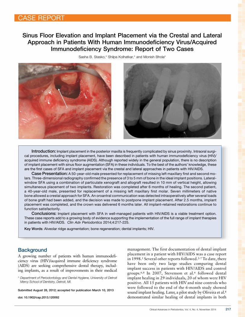
Sinus Floor Elevation and Implant Placement via the Crestal and Lateral Approach in Patients With...
9
CASE REPORT Sinus Floor Elevation and Implant Placement via the Crestal and Lateral Approach in Patients With Human Immunodeficiency Virus/Acquired Immunodeficiency Syndrome: Report of Two Cases Sasha B. Stasko,* Shilpa Kolhatkar,* and Monish Bhola* Introduction: Implant placement in the posterior maxilla is frequently complicated by sinus proximity. Intraoral surgi- cal procedures, including implant placement, have been described in patients with human immunodeficiency virus (HIV)/ acquired immune deficiency syndrome (AIDS). Although reported widely in the general population, there is no description of implant placement with sinus floor augmentation (SFA) in these individuals. To the best of the authors’ knowledge, these are the first cases of SFA and implant placement via the crestal and lateral approaches in patients with HIV/AIDS. Case Presentation: A 50-year-old male presented for replacement of missing left maxillary first and second mo- lars. Three-dimensional radiography confirmed the presence of 3 to 5 mm of bone in the ideal implant positions. Lateral- window SFA using a combination of particulate xenograft and allograft resulted in 10 mm of vertical height, allowing simultaneous placement of two implants. Restoration was completed after 6 months of healing. The second patient, a 40-year-old male, presented for replacement of a missing left maxillary first molar. Seven millimeters of native bone allowed a crestal approach for SFA. An oroantral communication was detected intraoperatively after several loads of bone graft had been added, and the decision was made to postpone implant placement. After 2.5 months, implant placement was completed, and the crown was delivered 6 months later. All implant-retained restorations continue to function satisfactorily. Conclusions: Implant placement with SFA in well-managed patients with HIV/AIDS is a viable treatment option. These case reports add to a growing body of evidence supporting the implementation of the full range of implant therapies in patients with HIV/AIDS. Clin Adv Periodontics 2014;4:217-225. Key Words: Alveolar ridge augmentation; bone regeneration; dental implants; HIV. Background A growing number of patients with human immunodefi- ciency virus (HIV)/acquired immune deficiency syndrome (AIDS) are seeking comprehensive dental therapy, includ- ing implants, as a result of improvements in their medical management. The first documentation of dental implant placement in a patient with HIV/AIDS was a case report in 1998. 1 Several other reports followed. 2-7 To date, there have been only two large studies comparing dental implant success in patients with HIV/AIDS and control groups. 6,8 In 2007, Stevenson et al. 6 followed dental implant healing in 29 individuals, 20 of whom were HIV positive. All 15 patients with HIV and nine controls who were followed to the end of the 6-month study showed sound implant healing. Later, a pilot study by Oliveira et al. 8 demonstrated similar healing of dental implants in both * Department of Periodontology and Dental Hygiene, University of Detroit Mercy School of Dentistry, Detroit, MI. Submitted August 28, 2012; accepted for publication March 10, 2013 doi: 10.1902/cap.2013.120092 Clinical Advances in Periodontics, Vol. 4, No. 4, November 2014 217
Transcript of Sinus Floor Elevation and Implant Placement via the Crestal and Lateral Approach in Patients With...
CASE REPORT
Sinus Floor Elevation and Implant Placement via the Crestal and LateralApproach in Patients With Human Immunodeficiency Virus/Acquired
Immunodeficiency Syndrome: Report of Two CasesSasha B. Stasko,* Shilpa Kolhatkar,* and Monish Bhola*
Introduction: Implant placement in the posterior maxilla is frequently complicated by sinus proximity. Intraoral surgi-cal procedures, including implant placement, have been described in patients with human immunodeficiency virus (HIV)/acquired immune deficiency syndrome (AIDS). Although reported widely in the general population, there is no descriptionof implant placement with sinus floor augmentation (SFA) in these individuals. To the best of the authors’ knowledge, theseare the first cases of SFA and implant placement via the crestal and lateral approaches in patients with HIV/AIDS.
Case Presentation: A 50-year-old male presented for replacement of missing left maxillary first and second mo-lars. Three-dimensional radiography confirmed the presence of 3 to 5mm of bone in the ideal implant positions. Lateral-window SFA using a combination of particulate xenograft and allograft resulted in 10 mm of vertical height, allowingsimultaneous placement of two implants. Restoration was completed after 6 months of healing. The second patient,a 40-year-old male, presented for replacement of a missing left maxillary first molar. Seven millimeters of nativebone allowed a crestal approach for SFA. An oroantral communication was detected intraoperatively after several loadsof bone graft had been added, and the decision was made to postpone implant placement. After 2.5 months, implantplacement was completed, and the crown was delivered 6 months later. All implant-retained restorations continue tofunction satisfactorily.
Conclusions: Implant placement with SFA in well-managed patients with HIV/AIDS is a viable treatment option.These case reports add to a growing body of evidence supporting the implementation of the full range of implant therapiesin patients with HIV/AIDS. Clin Adv Periodontics 2014;4:217-225.
Key Words: Alveolar ridge augmentation; bone regeneration; dental implants; HIV.
BackgroundA growing number of patients with human immunodefi-ciency virus (HIV)/acquired immune deficiency syndrome(AIDS) are seeking comprehensive dental therapy, includ-ing implants, as a result of improvements in their medical
management. The first documentation of dental implantplacement in a patient with HIV/AIDS was a case reportin 1998.1 Several other reports followed.2-7 To date, therehave been only two large studies comparing dentalimplant success in patients with HIV/AIDS and controlgroups.6,8 In 2007, Stevenson et al.6 followed dentalimplant healing in 29 individuals, 20 of whom were HIVpositive. All 15 patients with HIV and nine controls whowere followed to the end of the 6-month study showedsound implant healing. Later, a pilot study byOliveira et al.8
demonstrated similar healing of dental implants in both
* Department of Periodontology and Dental Hygiene, University of DetroitMercy School of Dentistry, Detroit, MI.
Submitted August 28, 2012; accepted for publication March 10, 2013
doi: 10.1902/cap.2013.120092
Clinical Advances in Periodontics, Vol. 4, No. 4, November 2014 217

TABLE 1 Patient Medical History and Dental Information
Patient Information Case 1 Case 2
Age (years) 50 40
Sex Male Male
Year HIV diagnosed 2008 2008
Smoking status Non-smoker Non-smoker
HIV-related medications Combination of 600 mg efavirenz, 200 mgemtricitabine, and 300 mg tenofovirdisoproxil fumarate* once daily
300 mg atazanavir sulfate once daily; 100 mgritonavir once daily; and 300 mg tenofovir
disoproxil fumarate once daily
Other medical conditions History of stomach ulcer (surgically treated in 1987) Hypertension
Penicillin allergy (reaction: hives) Leg spasms
Anxiety
Non-HIV medications None 10 mg hydrochlorothiazide þ amlodipineonce daily; 5 mg cyclobenzaprine once daily;5 mg diazepam three times daily; 500/20 mg
naproxen once daily; and 20 mgesomeprazole once daily
Laboratory values
CD4þ T lymphocyte (cells/mm3) 391 530
Neutrophil count (absolute) (� 103/mm) 3.3 1.8
Platelet count (per mL) 207,000 278,000
Viral load (copies/mL) Undetectable 103
Dental history
Chief complaint “I need more teeth to chew with.” “I need a dental checkup.”
Missing teeth #14, #15 #14
Date and reason for tooth loss March 30, 2009: severe non-restorablecarious lesions
November 30, 2009: severe non-restorablecarious lesions
Residual ridge height 3 to 5 mm 7 mm
Periodontal diagnosis Generalized slight gingivitis Generalized slight gingivitis
Other treatment alternatives No action, removable partial denture,fixed partial denture
No action, removable partial denture,fixed partial denture
Location sinus/implant surgery performed University of Detroit Mercy GraduatePeriodontics Department
University of Detroit Mercy GraduatePeriodontics Department
Dates of treatment
Sinus augmentation December 2011 July 2011
Implant placement December 2011 October 2011
Uncovery April 2012 March 2012
Restorations August 2012 March 2012
* Atripla, Bristol-Meyers Squibb, New York, New York.
C A S E R E P O R T
218 Clinical Advances in Periodontics, Vol. 4, No. 4, November 2014 Sinus Floor Elevation in Patients With HIV/AIDS

patients with HIV/AIDS and controls. This study furtherdemonstrated no differences in healing or postoperativeperi-implant bone loss when comparing patients withHIV/AIDS receiving different highly active antiretroviraltherapy (HAART) regimens.8
To aid in implant placement, sinus floor augmentation(SFA) is frequently required in the atrophic posterior max-illa. Simultaneous implant placement with SFA via thecrestal or lateral approach offers the advantage of shortertreatment time. Although widely reported in the generalpopulation, there is no description of implant placementwith SFA in patients with HIV/AIDS. To the best of theauthors’ knowledge, the first such cases are presentedhere.
Clinical PresentationCase 1A 50-year-old male presented to the University of DetroitMercy, School of Dentistry Graduate Periodontics, Detroit,Michigan, for replacement of missing teeth #14 and #15.A detailed medical history, including medications and labora-tory values, was obtained (Table 1). An extraoral and intraoralclinical and radiographic examinationwas completed. Cone-beam computed tomography analysis confirmed the pres-ence of adequate horizontal dimension, a lack of septae,and local pathology yet inadequate vertical height (3 to5 mm) in the ideal implant positions (Fig. 1).
Case 2A40-year-oldmalepresented to theUniversityofDetroitMercy,School of Dentistry Graduate Periodontics for replacement ofmissing tooth #14. Table 1 summarizes his pertinent medicaland dental information. A thorough clinical and radiographicexamination was performed and reduced vertical height (z7mm) in the region of the planned implant was diagnosed(Fig. 2).
Case Management and Clinical OutcomesCase 1On the day of surgery, the patient’s medical history was re-viewed, vital signs were taken, written informed consentwas obtained, and postoperative instructions were reviewed.Table 2 summarizes perioperative medications and regimens.After local infiltration of anesthesia, incisions (crestal, sulcular,and vertical) were made, and a full-thickness mucoperiostealflap (FTMPF)was elevated. The lateral windowwas outlined,
FIGURE 1 Case 1. Lateral approach. Presurgicalcone-beam computed tomography of edentu-lous left maxillary sinus showing minimal residualbone height.
FIGURE 2 Case 2. Crestal approach. Preoperative periapical radiograph ofplanned implant #14 region.
C A S E R E P O R T
Stasko, Kolhatkar, Bhola Clinical Advances in Periodontics, Vol. 4, No. 4, November 2014 219

TABLE 2 Perioperative Medication Regimens
Medication Class Medication Regimen
Antihistamine Loratadine Begin 1 week before and continue 2 weeks after surgery
Antibiotic 500 mg azithromycin on day 1and then 250 mg
Begin 3 days before and continue for 1 week after surgery
Analgesics 600 mg ibuprofen Before surgery and every 6 hours for 3 days and then as needed for pain
5 mg hydrocodone/500 mgacetaminophen
Post-surgery, one to two tablets every 6 hours as needed for breakthrough pain
Oral rinse 0.12% chlorhexidine gluconate Preoperative rinse and then twice daily for 2 weeks post-surgery
FIGURE 3 Case 1. Lateral-window osteotomy with sinus membraneelevation. Note the Class I perforation (arrow).
FIGURE 4 Case 1. Composite bone graft of particulate xenograft anddemineralized allograft in lateral window and cover screw over implants.
FIGURE 5 Case 1. Resorbable collagen membrane over lateral window.
FIGURE 6 Case 1. Final periapical radiograph of two 13-mm implants insites #14 and #15. Note tack between the root of tooth #13 and implant #14used to stabilize the resorbable barrier.
C A S E R E P O R T
220 Clinical Advances in Periodontics, Vol. 4, No. 4, November 2014 Sinus Floor Elevation in Patients With HIV/AIDS

FIGURE 7 Case 1. 7a Clinical photograph of FTMPF to attach healingabutments. 7b Mineralized cortical bone allograft around implant #14 withhealing abutments on both implants. 7c Soft-tissue closure aroundexposed implants.
FIGURE 8 Case 1. Soft-tissue healing before restoration at 7 months afterimplant placement.
FIGURE 9 Case 1. Final clinical photograph of restored implants.
FIGURE 10 Case 1. Final periapical radiograph of implants #14 and #15.
FIGURE 11 Case 2. Direction indicator before SFA and placement ofimplant at site #14.
C A S E R E P O R T
Stasko, Kolhatkar, Bhola Clinical Advances in Periodontics, Vol. 4, No. 4, November 2014 221
and elevation of the sinus lining was initiated. A Class Iperforation9 of the Schneiderian membrane was noted onthe anterior superior aspect (Fig. 3, arrow). The perforationwas repairedwith a collagenmembrane† and a small amountofbonegraft‡wasplaced to stabilize it.Themaxillary sinusos-teotomies were then prepared using the manufacturer’s recom-mended drill sequence and the dental implantsx were placed.Additional bone grafts‖{ were packed in small increments untilsufficient elevation of the sinus floor was obtained (Fig. 4). Asecond collagen membrane# (Fig. 5) was then placed over thelateral wall and secured on the mesial aspect with a titaniumtack. Periosteal-releasing incisions were performed to ensuretension-free primary closure. Final periapical (Fig. 6) and pano-ramic radiographs were taken and postoperative instructionswere reinforced. The patient returned 1, 2, and 4 weeks aftersurgery, throughout which his healing was uneventful (nosigns of infection, delayed healing, or prolonged bleeding).
Placement of healing collars was scheduled 4 monthsafter surgery. Updated laboratory values showed minimalchange from preoperative measurements. A clinical exam-ination revealed a papule on the buccal aspect corresponding
to the vertical incision made during surgery. The patientreported no discomfort. No swelling or erythema werepresent, and no exudate was expressed. A periapical radio-graph revealed an absence of peri-implant radiolucency.The patient was placed on an antibiotic and returned 1week later for a scheduled exploratory procedure. At thistime, an FTMPF (Fig. 7a) was elevated and no mobilitywas observed in either implant when cover screws were
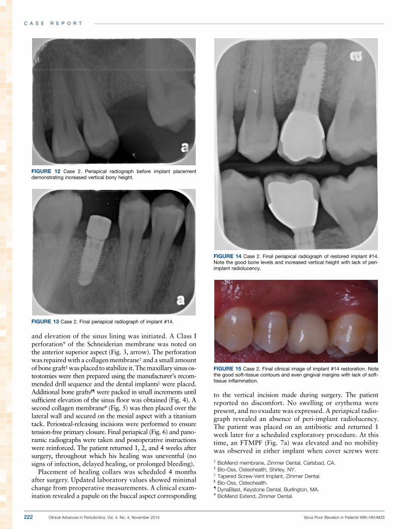
FIGURE 12 Case 2. Periapical radiograph before implant placementdemonstrating increased vertical bony height.
FIGURE 13 Case 2. Final periapical radiograph of implant #14.
FIGURE 14 Case 2. Final periapical radiograph of restored implant #14.Note the good bone levels and increased vertical height with lack of peri-implant radiolucency.
FIGURE 15 Case 2. Final clinical image of implant #14 restoration. Notethe good soft-tissue contours and even gingival margins with lack of soft-tissue inflammation.
† BioMend membrane, Zimmer Dental, Carlsbad, CA.‡ Bio-Oss, Osteohealth, Shirley, NY.x Tapered Screw-Vent Implant, Zimmer Dental.‖ Bio-Oss, Osteohealth.{ DynaBlast, Keystone Dental, Burlington, MA.# BioMend Extend, Zimmer Dental.
C A S E R E P O R T
222 Clinical Advances in Periodontics, Vol. 4, No. 4, November 2014 Sinus Floor Elevation in Patients With HIV/AIDS

replaced with healing collars. Minimal circumferential crestalbone was absent around implant #14 without implant threadexposure. Bone levels were otherwise unchanged. The gran-ulation tissue surrounding implant #14 was removed andreplaced with allograft** (Fig. 7b), and the area was closed(Fig. 7c). Seven months after implant placement, a pre-restorative clinical examination (Fig. 8) and periapical radio-graph confirmed that the implants were osseointegrated.Final restoration was completed at this time (Figs. 9 and 10).
Case 2On the day of surgery, the patient’s medical history was re-viewed, vital signs were taken, written informed consentwas obtained, and postoperative instructions were re-viewed. Table 2 summarizes perioperative medications andregimens. After local infiltration of anesthesia, a crestalincision was made, and the FTMPF was elevated to exposethe bony crest. An osteotomy was created to end just shortof the sinus floor (Fig. 11) followed by use of osteotomesand bone grafts††‡‡ to infracture and lift the sinus floor.The Valsalva maneuver was performed on multiple occa-sions and was negative. However, a positive Valsalva ma-neuver was elicited at the end of the SFA procedure. Thedecision was made to delay implant placement, an extracel-lular matrix membranexx was placed over the osteotomy,and primary closure was achieved. Three months later,a new periapical radiograph (Fig. 12) showed adequatebone height for placement of a 10-mm implant.‖‖ Theimplant demonstrated primary stability after placementand a 5-mmhealing collar was placed (Fig. 13). Restorationwas completed 6 months later (Figs. 14 and 15).
DiscussionThe literature describing oral surgical procedures in pa-tients with HIV/AIDS is sparse. Of the few documentedcases, implants have been placed most often in the mandi-ble or inmaxillary regionswith sufficient bone to accept animplant.1-8 However, in clinical practice, it is common thatimplant placement in the posterior atrophic maxilla re-quires SFA to augment or obtain additional bone volume.
Both crestal and lateral window approaches have provedinstrumental in achieving this goal.
According to the most recent systematic review andmeta-analysis, the 3-year survival rate of implants placedin the maxillae simultaneously with augmentation viathe lateral approach is 90.1%.10 This success rate increasesto 98.3% when only rough surface implants are consid-ered, with a membrane covering the lateral window.10
In case 1, implants could be placed simultaneously withlateral-window SFA (LWSFA) as a result of increasedstability from the more proximal bone. Perforation ofthe sinus membrane was managed easily. This is the mostcommon complication reported with this procedure(19.5%10), yet it does not seem to affect the success of finalimplant placement.11,12 Implants placed in a sinus elevatedvia the crestal technique bear a relatively high estimatedsurvival rate of 92.8%.13 In case 2, it was decided to delayplacement of the implant because radiographs did not showa well-contained bone graft. The finding of a Schneiderianmembrane perforation in case 2 was disappointing butnot uncommon. As in the lateral approach, membraneperforation is listed as the most frequent surgical com-plication using the transalveolar technique.13
One reason practitioners may hesitate to perform inva-sive oral surgical procedures in patients with HIV/AIDS isthe possibility of postoperative complications. However,Campo et al.14 documented a relatively low complicationrate of 2.2% (overall) to 4.8% (invasive) when dental pro-cedures were performed on patients with HIV.
In both of the present cases, despite finding Schneiderianmembrane perforations, implant healing proceeded un-eventfully. All implant-retained restorations continue tofunction satisfactorily. The present authors acknowledgethat this report demonstrates only a short-term outcome.Long-term controlled clinical trials are needed to confirmthat SFA in combination with implant placement is as pre-dictable in patients with HIV/AIDS as it is in the generalpopulation. This is important given the increasing numberof people with HIV/AIDS enjoying longer lives as a resultof better medical management, which includes HAART. n
**Mineralized cortical bone, Musculoskeletal Transplant Foundation,Edison, NJ.
†† Bio-Oss, Osteohealth.‡‡ DynaBlast, Keystone Dental.xx DynaMatrix, Keystone Dental.‖‖ Tapered Screw-Vent Implant, Zimmer Dental.
C A S E R E P O R T
Stasko, Kolhatkar, Bhola Clinical Advances in Periodontics, Vol. 4, No. 4, November 2014 223

Summary
Why are these cases newinformation?
j To the best of the authors’ knowledge, this is the first documentationof simultaneous dental implant placement and LWSFA in patients withHIV/AIDS.
j Also to the best of the authors’ knowledge, this is the firstdocumentation of SFA via the crestal approach in a patient withHIV/AIDS.
What are the keys to successfulmanagement of these cases?
j Collaboration with medical colleagues to ensure stable pertinentlaboratory values
j Management of sinus perforation in the LWSFA approach witha resorbable barrier
j Primary stabilization of implants placed simultaneously with LWSFAj Ensuring graft containment before implant placement (case 2)
What are the primary limitations tosuccess in these cases?
j Sinus perforation (case 2)j Lack of ability to visualize sinus lining and bone graft (case 2)
AcknowledgmentsThe authors thank Dr. Shayna Sanchez, third-year periodon-tics resident, University of Detroit Mercy, Detroit, Michigan,for her surgical execution of case 2; Mr. Kian Sahba andMs. Mira Diora, senior dental students, University of DetroitMercy, for restoration of cases 1 and 2, respectively; andMr. Eric Jacobs, media specialist, University of DetroitMercy, for his assistance with the intraoperative photog-raphy. Dr. Bhola is a consultant for Keystone Dental,Burlington,Massachusetts, and Zimmer Dental, Carlsbad,California. Drs. Stasko and Kolhatkar report no conflicts ofinterest related to this case report.
CORRESPONDENCE:Dr. Sasha B. Stasko, Department of Periodontology and Dental Hygiene,University of Detroit Mercy School of Dentistry, 2700Martin Luther King Jr.Blvd., Detroit, MI 48208. E-mail: [email protected].
C A S E R E P O R T
224 Clinical Advances in Periodontics, Vol. 4, No. 4, November 2014 Sinus Floor Elevation in Patients With HIV/AIDS
References1. Rajnay ZW, Hochstetter RL. Immediate placement of an endosseous
root-form implant in an HIV-positive patient: Report of a case. JPeriodontol 1998;69:1167-1171.
2. Baron M, Gritsch F, Hansy AM, Haas R. Implants in an HIV-positivepatient: A case report. Int J Oral Maxillofac Implants 2004;19:425-430.
3. Shetty K, Achong R. Dental implants in the HIV-positive patient —Case report and review of the literature. Gen Dent 2005;53:434-437,quiz 438, 446.
4. Strietzel FP, Rothe S, Reichart PA, Schmidt-Westhausen AM. Implant-prosthetic treatment in HIV-infected patients receiving highly activeantiretroviral therapy: Report of cases. Int J Oral Maxillofac Implants2006;21:951-956.
5. Achong RM, Shetty K, Arribas A, Block MS. Implants in HIV-positivepatients: 3 case reports. J Oral Maxillofac Surg 2006;64:1199-1203.
6. Stevenson GC, Riano PC, Moretti AJ, Nichols CM, Engelmeier RL, FlaitzCM. Short-term success of osseointegrated dental implants in HIV-positiveindividuals: A prospective study. J Contemp Dent Pract 2007;8:1-10.
7. Kolhatkar S, Khalid S, Rolecki A, Bhola M, Winkler JR. Immediatedental implant placement in HIV-positive patients receiving highlyactive antiretroviral therapy: A report of two cases and a review of theliterature of implants placed in HIV-positive individuals. J Periodontol2011;82:505-511.
8. Oliveira MA, Gallottini M, Pallos D, Maluf PS, Jablonka F, Ortega KL.The success of endosseous implants in human immunodeficiency virus-positive patients receiving antiretroviral therapy: A pilot study. J AmDent Assoc 2011;142:1010-1016.
9. Fugazzotto PA, Vlassis J. A simplified classification and repair systemfor sinus membrane perforations. J Periodontol 2003;74:1534-1541.
10. Pjetursson BE, Tan WC, Zwahlen M, Lang NP. A systematic review ofthe success of sinus floor elevation and survival of implants inserted incombination with sinus floor elevation. J Clin Periodontol 2008;35(Suppl. 8):216-240.
11. Schwartz-Arad D, Herzberg R, Dolev E. The prevalence of surgicalcomplications of the sinus graft procedure and their impact on implantsurvival. J Periodontol 2004;75:511-516.
12. Karabuda C, Arisan V, Ozyuvaci H. Effects of sinus membraneperforations on the success of dental implants placed in the augmentedsinus. J Periodontol 2006;77:1991-1997.
13. Tan WC, Lang NP, Zwahlen M, Pjetursson BE. A systematic review ofthe success of sinus floor elevation and survival of implants inserted incombination with sinus floor elevation. Part II: Transalveolar tech-nique. J Clin Periodontol 2008;35(Suppl. 8):241-254.
14. Campo J, Cano J, del Romero J, Hernando V, Rodrıguez C,Bascones A. Oral complication risks after invasive and non-invasivedental procedures in HIV-positive patients. Oral Dis 2007;13:110-116.
indicates key references.
C A S E R E P O R T
Stasko, Kolhatkar, Bhola Clinical Advances in Periodontics, Vol. 4, No. 4, November 2014 225