Sinonasal Osteosarcoma in a Horse
5
ISSN 1679-9216 1 CASE REPORT Pub. 364 Acta Scientiae Veterinariae, 2019. 47(Suppl 1): 364 DOI: 10.22456/1679-9216.90022 Received: 26 September 2018 Accepted: 12 January 2019 Published: 29 January 2019 1 Universidade Federal de Uberlândia (UFU), Uberlândia, MG, Brazil. 2 Departamento de Patologia, Faculdade de Medicina de Botucatu, Universidade Estadual de São Paulo (UNESP), SP, Brazil. 3 Centro Universitário do Triângulo (UNITRI), Uberlândia, MG. CORRESPONDENCE: D.J.Z. Delfiol [djzdelfi[email protected] - Tel.: + 55 (34) 3225-8250]. Hospital Veterinário da Universidade Federal de Uberlândia. Av. Mato Grosso n. 3289 - Bloco 2S. Bairro Umuarama. CEP 38405-314 Uberlândia, MG, Brazil. Sinonasal Osteosarcoma in a Horse Raíssa Oliveira Leite 1 , Viciany Erique Fabris 2 , Elisa Sant’Anna Monteiro da Silva 3 , Geison Morel Nogueira 1 & Diego José Zanzarini Delfiol 1 ABSTRACT Background: Osteosarcomas are common malignant bone tumors described in dogs, humans and cats. However, there are rare reports in horses. The tumor etiology has not been fully elucidated. Clinical signs are associated to the tumor size, location and growth characteristics. When located on the face, the most common findings are facial distortion, nasal discharge and inspiratory dyspnea. The aim of this study was to report an osteoblastic osteosarcoma in a horse, located on the right maxillary region with projections into the corresponding nostril. Case: A 6-year-old mixbreed saddle gelding, presenting bay coat was admitted to a University Hospital with a chronic sinusitis history. The animal was previously examined and treated by other veterinarians, who suspected of odontogenic maxillary sinusitis, and therefore a repulsion of the fourth premolar from the right maxilla hemiarcade was performed. However, an improvement of the clinical signs was not observed and subsequently the gelding was referred to a Veterinary Hospital. During physical examination the patient presented dyspnea, tachypnea and tachycardia. An increase on the face size was observed, together with a fetid and purulent nasal discharge. During oral cavity inspection, a diagonal wear was detected at the occlusal surface of the right hemiarcade; food accumulation was seen at the dental extraction site, and a communication with the rostral maxillary sinus was evidenced, from where a purulent fetid discharge was draining. At the radiographic exam, well defined margins of a tumor were observed, with adjacent bone lysis and the presence of a central nucleus showing a gross granular mineral radiopacity, distorting the frontal and nasal bones. During the endoscopic exam, a mass partially occluding the right nasal cavity close to the nasal opening was seen, which was blocking the progression of the endoscopy. However, during the left cavity inspection, at the end of the nasal septum a mass with irregular surface was detected emerging from the border of the right choana, which extended up to the nasopharynx region. The diagnosis was established based on clinical evaluation and histopathological findings of the tumor, which confirmed osteoblastic osteosarcoma. The animal was euthanized due to poor clinical conditions and prognosis. There was no evidence of metas- tasis to other organs during necropsy. Discussion: The majority of reported osteosarcoma cases in horses do not define the tumor histopathological subtype. The occurrence of metastasis in equine osteosarcoma is not well established, however it seems to be uncommon. It is important to emphasize the relevance of performing a necropsy in patients presenting osteosarcoma, in order to establish a pattern concerning the metastasis incidence in the species. The predominant osteosarcoma location is the mandible, although there are few reports in the paranasal sinuses and appendicular skeleton. The tumor usual location makes the treatment difficult because of the impossibility of performing a complete surgical resection, which leads to a euthanasia decision. Although osteosarcoma has been little reported in horses, it should be a differential diagnosis for facial alterations and paranasal sinuses abnormalities, which would contribute to an early diagnosis and increase the chances of a favorable prognostic. Keywords: nasal cavity, facial distortion, neoplasm, osteoblastic, tumor, sinusitis.
Transcript of Sinonasal Osteosarcoma in a Horse
ISSN 1679-9216
1
CASE REPORTPub. 364
Acta Scientiae Veterinariae, 2019. 47(Suppl 1): 364
DOI: 10.22456/1679-9216.90022Received: 26 September 2018 Accepted: 12 January 2019 Published: 29 January 2019
1Universidade Federal de Uberlândia (UFU), Uberlândia, MG, Brazil. 2Departamento de Patologia, Faculdade de Medicina de Botucatu, Universidade Estadual de São Paulo (UNESP), SP, Brazil. 3Centro Universitário do Triângulo (UNITRI), Uberlândia, MG. CORRESPONDENCE: D.J.Z. Delfiol [[email protected] - Tel.: + 55 (34) 3225-8250]. Hospital Veterinário da Universidade Federal de Uberlândia. Av. Mato Grosso n. 3289 - Bloco 2S. Bairro Umuarama. CEP 38405-314 Uberlândia, MG, Brazil.
Sinonasal Osteosarcoma in a Horse
Raíssa Oliveira Leite1, Viciany Erique Fabris2, Elisa Sant’Anna Monteiro da Silva3, Geison Morel Nogueira1 & Diego José Zanzarini Delfiol1
ABSTRACT
Background: Osteosarcomas are common malignant bone tumors described in dogs, humans and cats. However, there are rare reports in horses. The tumor etiology has not been fully elucidated. Clinical signs are associated to the tumor size, location and growth characteristics. When located on the face, the most common findings are facial distortion, nasal discharge and inspiratory dyspnea. The aim of this study was to report an osteoblastic osteosarcoma in a horse, located on the right maxillary region with projections into the corresponding nostril.Case: A 6-year-old mixbreed saddle gelding, presenting bay coat was admitted to a University Hospital with a chronic sinusitis history. The animal was previously examined and treated by other veterinarians, who suspected of odontogenic maxillary sinusitis, and therefore a repulsion of the fourth premolar from the right maxilla hemiarcade was performed. However, an improvement of the clinical signs was not observed and subsequently the gelding was referred to a Veterinary Hospital. During physical examination the patient presented dyspnea, tachypnea and tachycardia. An increase on the face size was observed, together with a fetid and purulent nasal discharge. During oral cavity inspection, a diagonal wear was detected at the occlusal surface of the right hemiarcade; food accumulation was seen at the dental extraction site, and a communication with the rostral maxillary sinus was evidenced, from where a purulent fetid discharge was draining. At the radiographic exam, well defined margins of a tumor were observed, with adjacent bone lysis and the presence of a central nucleus showing a gross granular mineral radiopacity, distorting the frontal and nasal bones. During the endoscopic exam, a mass partially occluding the right nasal cavity close to the nasal opening was seen, which was blocking the progression of the endoscopy. However, during the left cavity inspection, at the end of the nasal septum a mass with irregular surface was detected emerging from the border of the right choana, which extended up to the nasopharynx region. The diagnosis was established based on clinical evaluation and histopathological findings of the tumor, which confirmed osteoblastic osteosarcoma. The animal was euthanized due to poor clinical conditions and prognosis. There was no evidence of metas-tasis to other organs during necropsy.Discussion: The majority of reported osteosarcoma cases in horses do not define the tumor histopathological subtype. The occurrence of metastasis in equine osteosarcoma is not well established, however it seems to be uncommon. It is important to emphasize the relevance of performing a necropsy in patients presenting osteosarcoma, in order to establish a pattern concerning the metastasis incidence in the species. The predominant osteosarcoma location is the mandible, although there are few reports in the paranasal sinuses and appendicular skeleton. The tumor usual location makes the treatment difficult because of the impossibility of performing a complete surgical resection, which leads to a euthanasia decision. Although osteosarcoma has been little reported in horses, it should be a differential diagnosis for facial alterations and paranasal sinuses abnormalities, which would contribute to an early diagnosis and increase the chances of a favorable prognostic.
Keywords: nasal cavity, facial distortion, neoplasm, osteoblastic, tumor, sinusitis.
2
R.O. Leite, V.E. Fabris, E.S.M. da Silva, G.M. Nogueira & D.J.Z. Delfi ol. 2019. Sinonasal Osteosarcoma in a Horse. Acta Scientiae Veterinariae. 47(Suppl 1): 364.
INTRODUCTION
Osteosarcoma is a primary malignant tumor of mesenchymal tissues, notably of the bone tissue, showing an infi ltrative growth pattern and leading to and extensive destruction of the adjacent areas [10]. The tumor is classifi ed according to the histological patterns: poorly differentiated, osteoblastic, chondro-blastic, fi broblastic, telangiectatic and giant cells type [19]. The most affected regions are the mandible bones, while there are rare reports on the paranasal sinuses [1,4] and appendicular skeleton [1].
The objective of this study was to report an osteoblastic osteosarcoma in the nasal cavity and the maxillary paranasal sinuses of a horse, describing the clinical, radiographic, hematological and histopa-thological alterations, as well as the endoscopy and necropsy fi ndings.
CASE
A mixbreed saddle gelding, presenting bay coat and aging 6-year-old was admitted to a University Hospital with a chronic sinusitis history. The animal was previously examined and treated by other vete-rinarians, who suspected of odontogenic maxillary sinusitis. After radiographic evaluation, they perfor-med a repulsion of the fourth premolar from the right maxilla hemiarcade. However, an improvement of the clinical signs was not observed after 90 days of treatment and therefore the gelding was referred to a Veterinary Hospital.
During physical examination the patient was alert, presenting inspiratory dyspnea, uneven airfl ow in the nostrils (less airfl ow in the right nostril), pink mucous membranes, capillary refi ll time of 2 s and a rectal temperature of 38.0ºC. During the hospitalization period, the gelding showed tachycardia (mean 52 ± 5 bpm) and respiratory rate of 10 ± 6 rmm.
Upon face inspection, the right maxillary region was increased, showing a hard consistency at palpation with low sensitivity to pain (Figure 1-A). There was a tumor with irregular surface and hard consistency close to the opening of the nasal cavity, which was partially obstructing the nostril, making breathing diffi cult. In addition, a foul smelling and purulent nasal discharge was observed. There were no alterations at tracheal and pulmonary auscultation, and during paranasal sinuses percussion, the sound was solid at the right side.
During oral cavity inspection, a diagonal wear was observed at the occlusal surface of the right he-miarcade; food accumulation was detected at the dental extraction site, and after its removal, a communication with the rostral maxillary sinus was evidenced, from where a purulent fetid discharge was draining. Mo-reover, an alveolar bone fragment was easily removed from the same location.
The following complementary exams were requested: complete blood count, radiography and endoscopy. There was a decrease in the total count of red blood cells (5.03x10³/mm3), hemoglobin (8.7 g/dL) and hematocrit (25.5%). However, there were no changes on MCV (50.7 fl ), HCM (17.3 pg) and MCHC (34.1 g/dL), characterizing a moderate normocytic normochromic anemia. There were no changes on the leukogram.
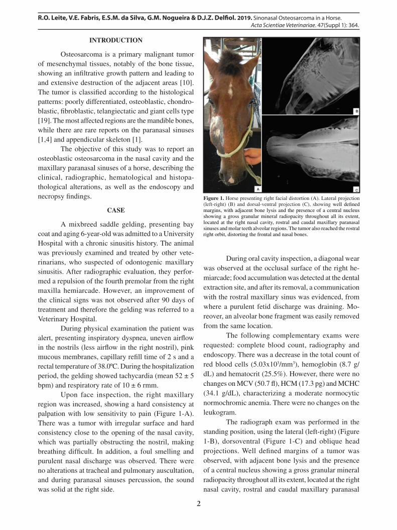
The radiograph exam was performed in the standing position, using the lateral (left-right) (Figure 1-B), dorsoventral (Figure 1-C) and oblique head projections. Well defi ned margins of a tumor was observed, with adjacent bone lysis and the presence of a central nucleus showing a gross granular mineral radiopacity throughout all its extent, located at the right nasal cavity, rostral and caudal maxillary paranasal
Figure 1. Horse presenting right facial distortion (A). Lateral projection (left-right) (B) and dorsal-ventral projection (C), showing well defi ned margins, with adjacent bone lysis and the presence of a central nucleus showing a gross granular mineral radiopacity throughout all its extent, located at the right nasal cavity, rostral and caudal maxillary paranasal sinuses and molar teeth alveolar regions. The tumor also reached the rostral right orbit, distorting the frontal and nasal bones.

3
R.O. Leite, V.E. Fabris, E.S.M. da Silva, G.M. Nogueira & D.J.Z. Delfi ol. 2019. Sinonasal Osteosarcoma in a Horse. Acta Scientiae Veterinariae. 47(Suppl 1): 364.
sinuses and molar teeth alveolar regions. In addition, the tumor reached the rostral right orbit, distorting the frontal and nasal bones.
At the endoscopy exam, a partial occlusion of the right nasal cavity was observed close to the nasal opening, which showed to be a mass, with irregular surface and redish to black color. The mass was blo-cking the progression of the endoscopy and therefore it was not possible to complete that antimere evalua-tion. There were no changes observed at the left nasal cavity, however, at the end of the nasal septum a mass with irregular surface was detected emerging from the border of the right choana, which extended up to the nasopharynx region.
Because of the location, extension and degree of damage caused by the tumor, surgical resection was not feasible. Moreover, the horse presented respiratory diffi culty. Therefore, a euthanasia followed by necrop-sy was indicated.
During necropsy, the head cut was performed at the median sagittal plan and a transverse cut was performed at the rostral maxillary paranasal sinus. A hard mass presenting smooth surface was observed, which occupied all the right nasal cavity extension and was caudally projected, affecting the nasal shells until the nasopharynx. When the mass was cut open, it showed an irregular aspect, hard consistency, with white-yellowish soft tissue areas. During the inspec-tion, there were no signifi cant changes on other organs.
Fragments were obtained from the inside of the right dorsal nasal shell, from rostral paranasal sinus region in a mucosal transition area, and from tumor tissue. The fragments were irregular bone tissue with membranous regions (mucosa), measuring 8.0 x 6.5 x 2.6 cm and weighing 55 g. They showed yellowish color, hard consistency, as well as soft consistency areas. Nine representative fragments were fi xed in 10% formalin (Formoldeído P.A.)1, submitted to de-calcifi cation, and stained with hematoxylin and eosin.
At microscopic analysis a malignant tumor composed by indistinct cytoplasm osteoblastic cells was detected, which was eosinophilic, presented large nuclei of vesicular aspect, loose chromatin and pro-minent nucleolus. In many foci, these cells produced homogeneous pink material, compatible to osteoid (Figure 2-B), a fi nding that strongly suggests osteo-sarcoma diagnosis. Fibroblastic areas were minimum. Necrotic areas were sparse and within small groups of
cells. The tumor was infi ltrating normal bone tissue, also compromising the nasal sinus mucosa (Figures 2-A & 2-D). A dense infl ammatory infi ltration of neutrophils, plasmocytes and macrophages with re-active bone in between was observed in the affected areas, which indicate sinusitis with osteomyelitis (Figure 2-C). Considering the aforementioned fi n-dings, the diagnosis of osteoblastic osteosarcoma was confi rmed.
DISCUSSION
The majority of tumors that affect equine paranasal sinuses are found in older animals, with exception of osteomas, fi brosarcomas, angiosarcomas, lymphosarcomas and osteosarcomas, which have been reported in foals and young horses [20]. According to the literature, osteosarcoma affected horses age between 7 weeks to 27 years old [1,5,12,17], with an average of 10 years old. The present study describes a 6-year-old gelding diagnosed with osteosarcoma, which is in agreement with the literature descriptions of the condition in younger animals. Similar fi ndings are described in dogs, in which the tumor affect both young and older animals, however, the condition is described only in older animals in the feline species [16,18].
The tumor clinical signs are usually related to the affected location [3]. When located in the head, the tumor leads to facial distortion, nasal discharge and obstruction of the normal airfl ow at the affected site. These clinical signs are often related to the tumor
Figure 2. A- Neoplasia (N) infi ltrating the mucosa (M) of the nasal cavity. Observe the respiratory epithelium (arrow) and normal bone (*). B- Osteoid (OT), the most important fi nding for diagnosing osteosarcoma, produced by neoplastic osteoblasts (NO) showing nuclear polymorphism, with round nuclei and conspicuous nucleolus. C- Area of secondary contamination (sinusitis/osteomyelitis) showing non-neoplastic reparative bone (RB), with appositional calcifi ed lines, and reactive to chronic infl ammation (CIP). D- Osteosarcoma cells (OS) infi ltrating normal bone (*).
4
R.O. Leite, V.E. Fabris, E.S.M. da Silva, G.M. Nogueira & D.J.Z. Delfiol. 2019. Sinonasal Osteosarcoma in a Horse. Acta Scientiae Veterinariae. 47(Suppl 1): 364.
size, location and growth characteristics. Nonetheless, there is no clinical sign considered as pathognomonic in case of an osteosarcoma [11,13,15,17].
In the present case, the right nasal cavity was partially obstructed by the tumor, leading to dyspnea. In addition, the lesion caused by the tumor mass in expansion caused chronic sinusitis, with purulent and fetid nasal discharge at the affected site. In a retro-spective study in which 5,558 horses were evaluated, sinonasal diseases were identified in 1.4% of the cases, and from these, 0.14% were nasal cavity tumors, all of them showing nasal discharge [8].
As the patient of the present study, which was first diagnosed with chronic sinusitis and treated without success, Springer et al. [17] reported a case of a horse presenting dyspnea, which was treated with bronchodilators and steroids for three years. The condition got worse until the tumor could be observed by the owner through the nasal cavity. These studies show the need of considering nasal cavity and para-nasal sinuses tumors as possible causes of respiratory disorders in horses, which would contribute to an early diagnosis and increase the chances of a favor-able prognosis.
During the hematological analysis, a normo-cromic normocytic anemia was evidenced. The finding is in agreement with the report of Koch et al. [14], in which a mare presenting osteosarcoma showed a moderate decrease in hemoglobin and hematocrit values. Anemia is associated to chronic diseases and tumor cases in humans and animals [14]. Typically, the erythrogram alterations observed in inflammatory, infectious or tumor diseases correspond to a light to moderate normocromic, normocytic anemia [2].
The radiographic analysis of the lesion re-vealed an extensive area of bone lysis. At palpation, a bone fragility was observed at the moment the alveolar fragments were easily removed during inspection of the oral cavity. Jenne et al. [12] also observed such fragility during a radiographic exam of the calcaneus bone, in which an extensive poorly delimitated area of osteolysis was seen, resulting in pathological fracture.
Despite the limitations, the endoscopic exam was helpful to establish a prognostic to the present reported case. An equine osteosarcoma restricted to the nasal cavity was described, in which the endoscopic exam was not able to be performed at the affected nostril, being conducted at the other nostril, where a
caudal delimitation of the tumor was visualized, sup-porting the therapeutic approach [17].
In horses, the osteosarcoma is predominantly localized in the head, making the treatment difficult because of the impossibility of performing a complete surgery resection of the tumor, leading to an euthanasia decision [1]. Although the unfavorable prognostic, there are some exceptions in the literature, where the authors have succeeded at the surgical resection of the osteosarcoma when the tumor was located at accessible surgical sites [5,17]. Herein, surgical ablation was not indicated due to the unfavorable clinical conditions showed by the patient. In addition, the extension and location of the lesion, together with the adjacent tis-sue damage, would make the complete tissue removal difficult and would bring permanent consequences to the animal wellbeing.
With regards to the osteosarcoma classifica-tion identified in the present report, the histopatho-logical analysis demonstrated compatible features to the osteoblastic type. An osteosarcoma human study demonstrated that the tumor histopathological subtype significantly interferes on the chemotherapy response [9]. The majority of reported osteosarcoma cases in horses do not define the tumor histopathological subtype, which demonstrates the need of performing additional studies on the species, mainly in the osteo-sarcoma cases, where the prognostic usually vary due to multiple factors.
In the present study metastasis to other organs were not identified. The occurrence of metastasis due to osteosarcoma in horses is not well established, however, it seems to be uncommon [11]. Eight cases of osteosarcoma were reported in horses [1], wherein necropsies were performed in two of them and no evidence of metastasis was found. Because of the lack of clinical and pathological evidences of metastasis incidence on the animals they have evaluated, the au-thors suggested that the metastatic pattern in equine osteosarcoma could be similar to that in cats, which metastasis-developing rate is relatively slow.
However, another study reported an osteo-sarcoma case in the proximal region of a mule radial bone [13], which showed a rapid progression and the development of liver and regional lymph node me-tastasis. In canine species, the presence of metastasis varies between 80 to 90% of the cases. In feline, the reports are between 5 to 10% [6]. The speculation on

5
R.O. Leite, V.E. Fabris, E.S.M. da Silva, G.M. Nogueira & D.J.Z. Delfiol. 2019. Sinonasal Osteosarcoma in a Horse. Acta Scientiae Veterinariae. 47(Suppl 1): 364.
http://seer.ufrgs.br/ActaScientiaeVeterinariaeCR364
metastasis occurrence in equine species is limited due to the low incidence of the tumor and to limitations on the post mortem exams described [14].
The present report points out the importance of considering the occurrence of tumors in the nasal cavity and paranasal sinuses, in cases of face tumors, chronic sinusitis and inspiratory dyspnea in horses,
which would contribute to an early diagnosis and better prognosis.
MANUFACTURER1Labsynth Produtos para Laboratório Ltda. Diadema, SP, Brazil.
Declaration of interest. The authors report no conflicts of interest. The authors alone are responsible for the content and writing of the paper.
REFERENCES
1 Bush J.M., Fredrickson R.L. & Ehrhart E.J. 2007. Equine osteosarcoma: a series of 8 cases. Veterinary Pathology. 44(2): 247-249.
2 Cançado R.D. & Chiattone C.S. 2002. Anemia de Doença Crônica. Revista Brasileira de Hematologia e Hemoterapia. 24(2): 127-136.
3 Cilliers I., Williams J., Carstens A. & Duncan N.M. 2008. Three cases of osteoma and an osseous fibroma of the paranasal sinuses of horses in South Africa. Journal of the South African Veterinary Association. 79: 185-193.
4 Cissell D.D., Wisner E.R. & Textor J. 2012. Computed tomographic appearance of equine sinonasal neoplasia. Vet-erinary Radiology and Ultrasound. 53 (3): 245-251.
5 Cousty M. & Tricaud C. 2014. Oncology Virtual Issue Articles. Equine Veterinary Education. 26(5): 269-269. 6 Dimopoulou M., Kirpensteij J., Moens H. & Kik M. 2008. Histologic prognosticators in feline osteosarcoma: a
comparison with phenotypically similar canine osteosarcoma. Veterinary Surgery. 37(5): 466-471. 7 Fowles J.S., Brown K.C., Hess A.M., Duval D.L. & Gustafson D.L. 2016. Intra- and interspecies gene expression
models for predicting drug response in canine osteosarcoma. BMC Bioinformatics. 17 (1): 1-14. 8 Hanna A., Stieger-Vanegas S.M. & Heidel J.R. 2015. Nasal Adenocarcinoma in a Horse with Metastasis to Lung,
Liver, and Bone and Review of Metastasis in Nine Horses with Sinonasal Tumors. Case Reports in Veterinary Medicine. (2015). Article ID 845870.
9 Hauben E.I., Weeden S., Pringle J. & Marck E.A. 2002. Does the histological subtype of high-grade central osteosar-coma influence the response to treatment with chemotherapy and does it affect overall survival ? A study on 570 patients of two consecutive trials of the European Osteosarcoma Intergroup. European Journal of Cancer. 38: 1218-1225.
10 Head K.W. & Dixon P.M. 1999. Equine nasal and paranasal sinus tumours. Part 1: review of the literature and tumour classification. Veterinary Journal. 157(3): 261-278.
11 Jenner F. 2010. Osteosarcoma in Equidae. Equine Veterinary Education. 22(3): 130-131. 12 Jenner F., Solano M., Gliatto J., Lavallee S. & Kirker-Head C. 2003. Osteosarcoma of the tarsus in a horse. Equine
Veterinary Journal. 35(2): 214-216. 13 Kilcoyne I., Wilson M., Terzo E. & David F. 2010. Osteosarcoma of the proximal radius in a donkey. Equine Veteri-
nary Education. 22(3): 125-129. 14 Koch E., Pack L., Zwicker L.A., Lopez-Mendez C. & Aburto E.M. 2014. Osteosarcoma in the proximal humerus
of a mare. Equine Veterinary Education. 26(8): 410-415. 15 Lee E.S. 1975. Osteosarcoma: a reconnaissance. Clinical Radiology. 26: 5-25. 16 Meyer F.R.L. & Walter I. 2016. Establishment and Characterization of New Canine and Feline Osteosarcoma Primary
Cell Lines. Veterinary Sciences. 3(2): 1. 17 Springer T., Elce Y.A. & Green E. 2010. Treatment of an osteoblastic osteosarcoma in an aged gelding. Equine
Veterinary Education. 22(4): 159-162. 18 Vanel M., Blond L. & Vanel D. 2013. Imaging of primary bone tumors in veterinary medicine: Which differences?.
European Journal of Radiology. 82(12): 2129-2139. 19 Vieira C.R., Fredo G. & Boabaid F.M. 2014. Osteossarcoma osteoblástico na cavidade nasal de um equino. In: VII
Encontro Nacional de Diagnóstico Veterinário (Cuiabá, Brasil). pp.2-3. 20 Witte T.H. & Perkins J.D. 2011. Early diagnosis may hold the key to the successful treatment of nasal
and paranasal sinus neoplasia in the horse. Equine Veterinary Education. 23(9): 441-447.