Single-tube heminested PCR coupled with ‘touchdown’ PCR for the analysis of the walleye dermal...
9
ELSEVIER Journal of Virological Methods 60 (1996) 29 37 Journal of Virological Methods Single-tube heminested PCR coupled with ‘touchdown’ PCR for the analysis of the walleye dermal sarcoma virus env gene Abstract The nucleotide sequences of geographically distant isolates of a newly characterized retrovirus of fish, the walleye dermal sarcoma virus (WDSV) were compared. Primers were designed based on the nucleotide sequence of a single molecular clone isolated from a tumor collected from a fish in New York state. USA. Conventional polymerase chain reaction (PCR) did not allow the satisfactory amplification of WDSV isolates from a different geographic region (Quebec, Canada), probably owing to the high genetic variability of retroviruses. To allow the successful amplification of Quebec WDSV isolates. heminested PCR and ‘touchdown’ PCR were combined in a highly sensitive. short and simple protocol taking place in a single tube. thus minimizing cross contamination. This technique allowed the first unambiguous detection of a fish retrovirus in Canada and the phylogenetic analysis of various geographic isolates of this virus. Other potential applications for this technique include PCR amplification using degenerate primers. and amplification of templates with some expected degree of genetic variability. K~IJ~II.oI.L/.(.: Fish: Nested PCR; Nucleic acid; ‘Touchdown’ PCR: Retrovirus; Sarcoma; Tumor ~. 1. Introduction Nested polymerase chain reaction (N-PCR) is the amplification of an extended nucleotide se- quence followed by the amplification of a region located within the first amplicon. Two internal (double nested) or a single internal primer (hem- inested (HN-PCR)) may be used. In both cases, * Corresponding author. Tel.: + I 514 7738521 ext. 8307: fax: + I S 14 7788 I 13; e-mail: martincn~~crc.Llmontreal.ca the sensitivity and specificity obtained are greater than those offered by conventional PCR amplifi- cation. For this reason, N-PCR is now used widely to detect specific viral DNA sequences in clinical specimens (Albert and Fenyo, 1990). Touchdown PCR was originally developed to cir- cumvent spurious priming during gene amplifica- tion (Don et al., 1991). This technique has been employed when the degree of complementarity between primers and template is uncertain, for instance when primers are derived from amino acid sequences or when some sequence variability 0166.0934:96’$15.00 s(: 1996 Elscvier Science B.V. All rights reserved PII SO 166.0934(96)0X24- I
-
Upload
zhiying-zhang -
Category
Documents
-
view
215 -
download
1
Transcript of Single-tube heminested PCR coupled with ‘touchdown’ PCR for the analysis of the walleye dermal...

ELSEVIER Journal of Virological Methods 60 (1996) 29 37
Journal of Virological Methods
Single-tube heminested PCR coupled with ‘touchdown’ PCR for the analysis of the walleye dermal sarcoma virus env gene
Abstract
The nucleotide sequences of geographically distant isolates of a newly characterized retrovirus of fish, the walleye dermal sarcoma virus (WDSV) were compared. Primers were designed based on the nucleotide sequence of a single molecular clone isolated from a tumor collected from a fish in New York state. USA. Conventional polymerase chain reaction (PCR) did not allow the satisfactory amplification of WDSV isolates from a different geographic region (Quebec, Canada), probably owing to the high genetic variability of retroviruses. To allow the successful amplification of Quebec WDSV isolates. heminested PCR and ‘touchdown’ PCR were combined in a highly sensitive. short and simple protocol taking place in a single tube. thus minimizing cross contamination. This technique allowed the first unambiguous detection of a fish retrovirus in Canada and the phylogenetic analysis of various geographic isolates of this virus. Other potential applications for this technique include PCR amplification using degenerate primers. and amplification of templates with some expected degree of genetic variability.
K~IJ~II.oI.L/.(.: Fish: Nested PCR; Nucleic acid; ‘Touchdown’ PCR: Retrovirus; Sarcoma; Tumor ~.
1. Introduction
Nested polymerase chain reaction (N-PCR) is
the amplification of an extended nucleotide se-
quence followed by the amplification of a region
located within the first amplicon. Two internal
(double nested) or a single internal primer (hem-
inested (HN-PCR)) may be used. In both cases,
* Corresponding author. Tel.: + I 514 7738521 ext. 8307:
fax: + I S 14 7788 I 13; e-mail: martincn~~crc.Llmontreal.ca
the sensitivity and specificity obtained are greater than those offered by conventional PCR amplifi- cation. For this reason, N-PCR is now used widely to detect specific viral DNA sequences in clinical specimens (Albert and Fenyo, 1990). Touchdown PCR was originally developed to cir- cumvent spurious priming during gene amplifica- tion (Don et al., 1991). This technique has been employed when the degree of complementarity between primers and template is uncertain, for instance when primers are derived from amino acid sequences or when some sequence variability
0166.0934:96’$15.00 s(: 1996 Elscvier Science B.V. All rights reserved
PII SO 166.0934(96)0X24- I

is expected between different templates. In TD- PCR, the annealing temperature starts several de- grees above the annealing temperature calculated for the specific primer pair and it is gradually lowered every other cycle. In this manner, the first hybridization events occur between primer and genomic DNA sequences with the greatest com- plementarity, thus minimizing the number of spu- rious bands due to mispriming.
The walleye dermal sarcoma virus (WDSV) is a newly characterized fish retrovirus (Holzschu et al., 1995; Martineau et al., 1992). It is associated etiologically with a cutaneous tumor, the walleye dermal sarcoma. WDS is highly prevalent throughout North America: up to 27% of walleyes collected from Oneida Lake, NY, are affected (Bowser et al., 1988). We were interested in com- paring the nucleotide sequences of geographically distant WDSV isolates. Primer complementarity with the templates was difficult to predict owing to the high genetic variability of retroviruses, and because the only sequence information about this virus has been derived from a single molecular clone (Martineau et al., 1992). To improve PCR specificity and sensitivity, TD-PCR was combined with a single-tube heminested PCR method in which the two successive amplification rounds were conducted in the same tube. This novel technique has been successfully employed to analyse the sequence diversity of WDSV collected from different geographical locations.
2. Material and methods
Six tumors (WDS), livers and spleens, were removed after euthanasia with methane tricaine sulphonate from four different adult walleyes col- lected in 1995 from Baskatong reservoir, Quebec, Canada. The neoplasm and tissue samples were immediately frozen in liquid nitrogen. For DNA isolation, neoplasm and tissue samples were thawed in a water bath at 37°C. Proteinase K was added at a concentration of 0.1 mg/ml and diges- tion was carried out at 50°C overnight. After digestion, the samples were subjected to three extractions with phenol and chloroform. Follow- ing ethanol precipitation, the DNA pellet was resuspended in TE buffer (pH 8.0).
The 5’ end of the WDSV mc gene, which encodes the surface protein (SU) of the viral envelope, was amplified and sequenced with five DNA oligonucleotide primers whose sequences were based on the sequence data obtained from a single molecular clone of WDSV isolated from WDS tissue collected in Oneida Lake, New York (Holzschu et al., 1995; Martineau et al., 1992). The primers (synthesized by General Synthesis and Diagnostics, Toronto, Ontario) included two forward (sense) primers (ENV Al and ENV A2) and three reverse (antisense) primers (ENV B 1, ENV B2 and ENV B3). Primer locations and sequences are shown in Fig. 1 and Table 1. Each primer consisted of 20 or 24 nucleotides and had a T, between 41.2 and 58.5”C. The lengths of the expected amplicons are listed in Fig. 1.
The Opti-PrimeTM PCR Optimization Kit (Stratagene, Cat. No.200422) was used to deter- mine the optimal buffer conditions. The optimiza- tion reaction was carried out according to the protocol provided by the company in a DNA thermal cycler (Ericomp, Twin Block, Easy cycler series, 6044 Cornerstone Court West, Suite E, San Diego, CA 92121).
<
ENVBJ
Fig. I. Schematic illustration of the physical map of the
WDSV and primers used for nested PCR. The positions of the primers and the target site of PCR amplification arc given
relative to the sequence derived from a molecular clone of WDSV. Forward primers: ENV Al and ENV A2. Reverse
primers: ENV Bl, ENV B2 and ENV B3. The beginning
(5974) and the end (9651) of the ent: ORF are shown.
Z. Zhang, D. Martineau 1 Journal of Virologicul Methods 60 (1996) 29-37 31
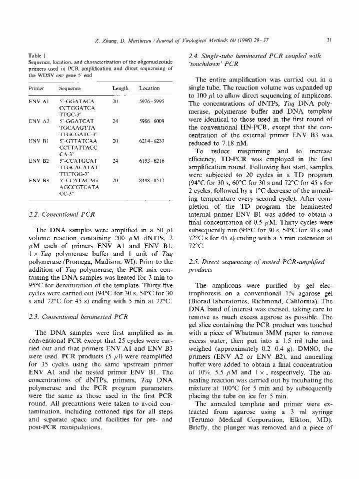
Table 1 Sequence, location, and characterization of the oligonucleotide
primers used in PCR amplification and direct sequencing of
the WDSV UZL’ gene 5’ end
Primer Sequence Length Location
ENV Al 5”-GGATACA 20 59165995
CCTGGATCA TTGC-3”
ENV A2 5”-GGATCAT 24 5986-6009
TGCAAGTTA
TTGCGATC-3”
ENV Bl 5”.GTTATCAA 20 6214-6233
CCTTATTACC
CA-3”
ENV B2 5”CCATGCAT 24 619336216
TTGCACATAT
TTCTGG-3”
ENV B3 5”CCATACAG 20 8498-8517
AGCCGTCATA
cc-3”
2.2. Conventional PCR
The DNA samples were amplified in a 50 ~1 volume reaction containing 200 ,uM dNTPs, 2 p M each of primers ENV Al and ENV B 1, 1 x Tuq polymerase buffer and 1 unit of Tuq
polymerase (Promega, Madison, WI). Prior to the addition of Taq polymerase, the PCR mix con- taining the DNA samples was heated for 3 min to 95°C for denaturation of the template. Thirty five cycles were carried out (94°C for 30 s, 54°C for 30 s and 72°C for 45 s) ending with 5 min at 72°C.
2.3. Conventional heminested PCR
The DNA samples were first amplified as in conventional PCR except that 25 cycles were car- ried out and that primers ENV Al and ENV B3 were used. PCR products (5 ~1) were reamplified for 35 cycles using the same upstream primer ENV Al and the nested primer ENV Bl. The concentrations of dNTPs, primers, Tuq DNA polymerase and the PCR program parameters were the same as those used in the first PCR round. All precautions were taken to avoid con- tamination, including cottoned tips for all steps and separate space and facilities for pre- and post-PCR manipulations.
2.4. Single-tube heminested PCR coupled with
‘touchdown’ PCR
The entire amplification was carried out in a single tube. The reaction volume was expanded up to 100 ,ul to allow direct sequencing of amplicons. The concentrations of dNTPs, Taq DNA poly- merase, polymerase buffer and DNA template were identical to those used in the first round of the conventional HN-PCR, except that the con- centration of the external primer ENV B3 was
reduced to 7.18 nM. To reduce mispriming and to increase
efficiency, TD-PCR was employed in the first amplification round. Following hot start, samples were subjected to 20 cycles in a TD program (94°C for 30 s, 60°C for 30 s and 72°C for 45 s for 2 cycles, followed by a 1°C decrease of the anneal- ing temperature every second cycle). After com- pletion of the TD program the heminested internal primer ENV Bl was added to obtain a final concentration of 0.5 ,uM. Thirty cycles were subsequently run (94°C for 30 s, 54°C for 30 s and 72°C s for 45 s) ending with a 5 min extension at 72°C.
2.5. Direct sequencing of nested PCR-ampliJied products
The amplicons were purified by gel elec- trophoresis on a conventional 1% agarose gel (Biorad laboratories, Richmond, California). The DNA band of interest was excised, taking care to remove as much excess agarose as possible. The gel slice containing the PCR product was touched with a piece of Whatman 3MM paper to remove excess water, then put into a 1.5 ml tube and weighed (approximately 0.220.4 g). DMSO, the primers (ENV A2 or ENV B2), and annealing buffer were added to obtain a final concentration of lo%, 5.5 PM and 1 x , respectively. The an- nealing reaction was carried out by incubating the mixture at 100°C for 5 min and by subsequently placing the tube on ice for 5 min.
The annealed template and primer were ex- tracted from agarose using a 3 ml syringe (Terumo Medical Corporation, Elkton, MD). Briefly, the plunger was removed and a piece of

32
M 1 2 3 4 5 6 7 8 gio1112 M
Fig. 2. Agaroae gel electrophoresis of the products obtained by the amplilicat~on of walleye dermal sarcoma DNA from Quebec.
using primer!, ENV Al :ENV RI. Direct visualization by ethidium bromide Iluorescence. LT. lambda DNA Hiutllll standard: L;unea
I. 2. 3 and 4: two tumour. one spleen and one liver DNA samples of ~alleqt’ 9i-0306-01. re\pccti\cly: Lane 5: turnour DNA sample
of B second uallcyc (94-0907-01): Lane? 6. 7 and 8: turnour. qleen and li\cr DN4 urnpIes of a third wallqc (950206-02).
respectively: Lanes 9, IO. I I and 12: tfio turnours. spleen and liter DN4 samples of a fourth \%alleqc (c)S-0206-01).
Whatman 3MM paper was dropped onto the bottom of the syringe and rinsed with deionized water. The DNA-containing gel slice was trans- ferred into the syringe. the plunger was reinserted and pushed firmly to expel the template-primer solution into a microcentrifuge tube until no more liquid was driven out. The template-primer solu- tion (15 /11) was collected into a fresh tube. The labelling and termination reactions were carried out using ‘% dATP and T7 DNA polymerase (Pharmacia Biotech. Uppsala, Sweden) according to the protocol provided by the manufacturer, except that the labelling reaction was carried out on ice.
3. Results
3.1. Conrmtionui PCR
Primers ENV Al and ENV Bl allowed the amplification and sequencing of the 5’ C/II’ region
of WDSV from Oneida lake (data not shown) by conventional PCR. Optimal conditions for ENV Al and ENV Bl were as follows: buffer pH 8.3, 1.5 mM MgCIZ, 75 mM KCI, IO mM Tris-HCI.
In contrast. when samples from Quebec were analyzed with ENV Al and ENV Bl. a mixture of products of heterogenous lengths was visualized as a high molecular weight smear (Fig. 2). Reopti- mizations of PCR parameters failed to improve the specificity or the sensitivity of the amplifica- tion.
To characterize the Quebec WDSV strains, HN-PCR \vas used first. ENV Al and ENV B3 wcrc used as cxtcrnal primers for the first ampliti- cation whereas ENV BI was used as a single internal primer. Primers ENV A2 and ENV B2 were used as nested sequencing primers.
Conventional HN-PCR was carried out as de- scribed in Material and methods. Amplification
Z. Zhuq, D. Mnrtineau : Jouwtal of Virologid Metlds 60 (1996) 29-17 33
I’A 1 2 3 4 5 6 7 8 91011 12 fl
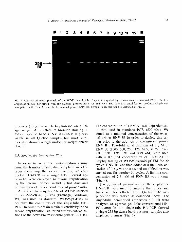
Fig. 3. A&arose gel clectrophoresis of the WDSV OK 258 bp fragment amplified by conventional heminested PCR. The first
amplification was performed with the external primers ENV Al and ENV B3. THe first amplification products (5 1~1) was
reamplitied with ENV Al and the heminested primer ENV BI. Templates are the same as depicted in Fig. 2.
products (10 ,~l) were electrophoresed on a 1% agarose gel. After ethidium bromide staining, a 258-bp specific band (ENV Al-ENV Bl) was visible in all Quebec samples but most sam- ples also showed a high molecular weight smear (Fig. 3).
3.3. Single-tube hetninrstrd PCR
In order to avoid the contamination arising from the transfer of amplified templates into the tubes containing the second reaction, we con- ducted HN-PCR in a single tube. Several ap- proaches were employed to favour amplification by the internal primer, including hot start and optimization of the external:internal primer ratio.
A 12.7 kb full-length clone of WDSV inserted in pGEM-SZf( + ) (3 kb) (Promega, Madison, WI) was used as standard (WDSV-pGEM) to optimize the conditions of the single-tube HN- PCR. In order to obtain maximal sensitivity in the second amplification, we tested various concentra- tions of the downstream external primer ENV B3.
The concentration of ENV Al was kept identical to that used in standard PCR (500 nM). We aimed at a minimal concentration of the exter- nal primer ENV B3 in order to deplete this pri- mer prior to the addition of the internal primer ENV Bl . Two-fold serial dilutions of 1 ,uM of ENV B3 (1000, 500, 250, 125, 62.5, 31.25, 15.63, 7.81, 3.91, 1.95 0.98 and 0.49 nM) were used with a 0.5 ,uM concentration of ENV Al to amplify 820 ng of WDSV plasmid pGEM for 20 cycles. ENV Bl was then added at a final concen- tration of 0.5 PM and a second amplification was carried out for another 30 cycles. A limiting con- centration of 7.81 nM of ENV B3 was optimal (Fig. 4).
The optimized parameters for the single-tube HN-PCR were used to amplify the tumor and tissue samples collected from Quebec. The am- plification was carried as described above. The single-tube heminested amplicons (IO 1111) were analyzed on agarose gel. Like conventional HN- PCR amplification, single-tube HN-PCR showed a single 258-bp dense band but most samples also displayed a smear (Fig. 5).

34
258---c- BP
Zhung, D. Martinrau )lI Journal of Virolo~ical Mctlds 60 (1996) 29- 37
M 1 2 3 4 5 6 7 8 9 10 11 12 M
Fig. 4. Influence of different concentrations of the external primer ENV B3 on the sensitivity of conventional heminested PCR using
820 ng of WDSV-pGEM as template. Two-fold serial dilutions of a I /IM solution of ENV B3 were used with primer ENV Al as
external primer, the primer ENV Bl was added after 20 cycles. M, lambda DNA/Hind111 standard: final concentrations of ENV B3:
1000, 500, 250, 125, 62.5, 31.25, 15.63, 7.81, 3.91, 1.95. 0.98 and 0.49 nM (lanes I 12).
3.4. Single-tube heminested PCR coupled with ‘touchdown ’ PCR
In order to improve the specificity and sensitiv- ity of the single-tube HN-PCR, the ‘touchdown’ program was used for the first amplification round in single-tube heminested PCR. The am- plification was carried out under conditions iden- tical to those of single-tube HN-PCR except that in the first round, the annealing temperature started 4°C above the calculated T, value and gradually decreased in the following cycles. This modification greatly improved the specificity of single-tube HN-PCR without ‘touchdown’ PCR, as shown by minimal smearing (Fig. 6).
3.5. Relative sensitivity of the single-tube heminested PCR with ‘touchdown’ PCR
To determine the sensitivity of this method, the optimized parameters determined for the single- tube HN-PCR with TD-PCR were used to am- plify serial dilutions of the plasmid WDSV-pGEM (dilution factor: IO). Fig. 7 shows a representative assay where amplicons resulting from the amplification of a single proviral copy (WDSV-pGEM) were detected.
3.6. Direct sequencing oj’ wqdicons
The single-tube heminested amplicons were di- rectly sequenced with the nested primers ENV A2 and ENV B2 as described in Section 2. Sequence alignment showed many differences between each individual sample (Fig. 8). These findings confi- rmed the specificity of the single-tube nested PCR and indicated that the amplicons were not the results of cross-contamination.
4. Discussion
Nested PCR requires that the second reaction contains a several fold excess of internal primer over external primers. Conventional methods ac- complish this by transferring an aliquot of the first reaction into another tube for the second amplification. This transfer enhances the risks for cross contamination since tubes containing high concentrations of the first amplicon must be opened and manipulated to set up the second amplification reactions.
To avoid cross contamination, single-tube nested PCR has been carried out using a strategy termed ‘drop-in/drop-out’ in which longer exter-

35
M 1 2 3 4 5 6 7 8 9 10 11 12 M
Fig. 5. Amplification of the WDSV e,x 258 bp fragment by single-tube heminested PCR under optimal conditions. The heminested
primer ENV Bl was directly introduced after 20 cycles of amplification with primers ENV Al and ENV B3. The PCR reaction (IO
1~1) were loaded on an 1% agarose gel and stained with ethidium bromide. M, lambda DNA!Hi/zdIII standard; templates of lanes
1~~ 12 are the same as in Fig. 2.
258 BP
Fig. 6. Amplification of the WDSV erx 258 bp fragment with the external primer ENV Al and ENV B3 and the internal primer
ENV Bl by the single-tube heminested PCR coupled with ‘touchdown’ PCR. Amplified products (IO ~11) were electrophoresed in I’%
agarose gel. The templates are as in Fig. 2. The remaining 90 /tl of the amplification reaction from three tumors (95-0907-01.
95-0206-02 and 95-0306-01) were used to directly sequence the amplified products (see Fig. 8).
nal primers (approximately 30 bases) have a anneal. In the second amplification, the annealing higher T,, than the internal primers. The first temperature is decreased to allow the internal
amplification is undertaken at a higher annealing primer(s) to anneal. The reaction tube does not
temperature to allow only the external primers to need to be opened once the reaction has begun

Fig. 7. Determination of the sensitivit) of the single-tube heminested P<‘R coupled with ‘touchdown’ PCR for the detection ol
cloned WDSV (WDSV-pGEM) in I”o agarose c -cl stained with ethidium bromide. ENV Al and ENV B3 were used as external
primers to run 20 cycles followed b) the addition of the internal primer and ENV Bl. A second amplilication was carried out for
another 30 cycle?. T’en-fold serial dilutions of WDSV-pGEM plasmid wel-e used as tcmplatcs. M, lambda DNA:Hi~t/lll standard:
the amounts of WDSV-pGEM are cqui\alent to IO‘. IO”. IO’. IO’. IO’. IO’. IO. I, 0. I. and 0.0 I molecule? in each reaction
trespecti\ely (law5 I IO).
CONSENSUS: AAGTTATTGCGATCATCTCTCTCCTCCTCGTTGGGGGGGCCAGTCAACCA 95-0907-01: AAGTTATTGCGATCATCTCTCTCCTCCTCGTTGGGGGGGCCAGTCAACCA 95-0206-02: AACTTATTGCGATCATCGCTCTCCTCCTCGTTGGGAAACCA 95-0306-01: AAGTTATTGCGATCATCGCTCTCCTCCTCGTTGGGGGAACCAGTCAACCA
*****************.********************..~~~~~*~*~~~
CONSENSUS: GCTACTTTCTTAGAGAAAGCACTTCCGACTGATGGTCCCAGCCTGGAGAC 95-0907-01: GCTACTATCATCGTGAATGCACTCCCCACTAATGGTCCTAGTCTGGAGAC 95-0206-02: GCTACTATCTTCGTGAATGCACTCCCGACTGACGGTCCCAGCCTGGAGAC 95-0306-01: GCTACTATCATCGTGAATGCACTCCCCCACT~CGGTCCCAGTCTGGAGAC
******.*t.* *.***.***** ** ***.t ***** l * ********
CONSENSUS: CATAGAACATAAGACAGAAATGGTTAATACAACCAGGAGCGAAGAACAAA 95-Ogo7-01: CATAGAACAAAJsGACAGCAATGG'l'.rlUICATAACATAACAATGAACCAAGAACAAA 95-0206-02: CATAGAACAAUGACAGCAATCGTTAACAAGGA 95-0306-01: CATAGAACAAAAGACAGCAATGGTTAACATAACARTGAAA
*****.a***.******* ********* * *** *.**.* *********
CONSENSUS: GTCCGGTTAGACCATCAAAAACTAGACAACAATTAATAGACGAAACACCA 95-0907-01: ACCCGGTTAGATCATTTACTAGACAACAATTAATAGACGAAACACCA 95-0206-02: AGCCGGTTAGATCATTTAAAACTAGACAACAATTMTAGATAAAACACCA 95-0306-01: ACCCGGTTAGATCATTTAAAACTAGACAACAATTMTAGATAAAACACCA
. ********* *** .t********************** .*******t
CONSENSUS: GAAATATGTGCAAATGCA 218 95-0907-01: GAAATATGTGCAAATGCA 218 95-0206-02: GAAATATGTGCAAATCCA 218 95-0306-01: GAAATATGTGCAAATGCA 218
***?a**************
consensus lengti,: 218 :iimt1ty : 189 ( 86.78)
50 50 50 50
100 100 100 100
150 150 150 150
200 200 200 200
Fig. 8. Direct sequencing of slnglc-tube hemine~ted PCR products coupled uith ‘touchdown’ PCR. The alignment shows the
sequences of the WDSV cwr 5’ end in thrw tumor\ (c)j-0907-()I. ‘95-0206-02 and 95-0306-01 ). (*). &xtical: (.). different. The
consensus sequence is from a WDSV clone isolated ft-om a walleye tumor in Onclda lake. NY. LISA.
(Pecharatana et al.. 1995). This method has been avoid cross contamination (Ulrich et al., 1995). successfully employed in the diagnosis of several However. unavoidable primer mismatch occurs viral infections to achieve higher sensitivity and to when primers are derived from amino acid se-
quences or when some sequence variability is ex- pected among the samples to be tested. In these cases, TD-PCR would be useful but it cannot be incorporated in ‘drop-in/drop-out’ technique since the long external primers would require a starting annealing temperature above 72°C which is sub- optimal for Tq polymerase activity. In addition, the higher costs involved by synthesizing longer primers for ‘drop-in/drop-out’ PCR are not negli- gible.
The combination of TD-PCR with single-tube HN-PCR was extremely useful for the study of WDSV genetic diversity. First, a phylogenetic analysis of WDSV geographically distant isolates could be carried out. Secondly, this method helped in determining unambiguously that WDSV, the only fish retrovirus molecularly char- acterized thus far, is present in Canada. Third. it greatly improved the specificity and sensitivity of conventional HN-PCR and of single-tube HN- PCR without prior ‘touchdown’. Fourth, this technique decreased the potential for cross con- tamination since manipulations were limited to the addition of the internal primer. Fifth, the amount of reagents required was reduced by nearly 50% and results were achieved 3-4 h ear- lier than in conventional N-PCR.
Potential applications for this method include PCR amplification of a specific template using degenerate primers, and PCR amplification of templates with some expected degree of genetic variability such as viral isolates or uncharacter- ized but related genes.
Acknowledgements
This work was supported by Grant No. OGPO 138236 from the Natural Sciences and Engineer- ing Research Council of Canada (NSERC), by the Collaborative NSERC-DFO project CRD 174902 and by Grant No. 95NC-1238 from FCAR.
References
Albert. J. and Fenyo, E.M. (1990) Simple. sensitive. and specific
detection of human immunodcficiency virus type I in clinical
specimens by polymerase chain reaction with nested primers.
J. Clin. Microbial. 28. 1560- 1564.
Bowser, P.R.. Wolfe, M.J., Forney. J.L. and Wooster. G.A.
(1988) Seasonal prevalence of skin tumors from walleye
(.Sti~o.srerh ~irrcwn) from Oneida Lake. New York. J. Wildl.
Dis. 24. 293 -298.
Don. R.H.. Cox. P.T., Wainwright. B.J.. Baker. K. and Mattick.
J.S.( 199l)‘Touchdoun’PCRtocircumve~ltspuriouspriming
during gent amplification. Nucl. Acids Res. l9( 14). 4008.
Holzschu. D.L., Martineau, D., Fodor. S.K., Vogt. V.M..
Bowser. P.R. and Case]. J.W. (1995) Nucleotide scqucnce
and protein analysis of a complex piscine retrocirus. Wallcyc
Dermal Sarcoma Virus. J. Virol. 69. 5320 5331.
Martineau, D.. Bouser. P.R.. Renshaw. R.R. and Casey. J.W.
(1992) Molecular characterization of a unique retrovirus
associated with a fish tumor. J. Viral. 66. 596 599.
Pecharatana. S.. Pickett. M.A.. Watt. P.J. and Ward. M.E. (I 99.5) Genotyping ocular strains of C‘/rkrr~~~&r t~~~+ornorit by
single-tube nested PCR. PCR Methods Applic. 3, 200-204.
Ulrich. P.P.. Romeo. J.M.. Daniel, L.J. and Vyas. G.N. (1995)
An improved method for the detection of hepatitis C virus
RNA in plasma utilizing heminestcd primers and internal
control RNA. PCR Methods Applic 7. 141 249.