sinclair Blood Collection in Swine · miniature breeds for blood collection procedures. There may...
4
www.sinclairbioresources.com [email protected] PO Box 658 Columbia, MO 65205 Tel: (573)387-4400 ▪ Fax: (573)387-4404 Sample Collection Series Blood Collection in Swine M. Michael Swindle, DVM, Professor and Chairman, Department of Comparative Medicine, Medical University of SC, Charleston, SC 29425 © 2010 Sinclair BioResources, LLC. All Rights Reserved. 1 sinclair bio-resources Introduction Blood collection from swine, especially larger animals, has been described as difficult. However, methods of blood collection and guidelines for sampling volumes applicable to research have been developed. 1 The purpose of this manuscript is to provide guidance on the collection of blood samples from swine used for biomedical research projects. There are minimal differences between domestic and miniature breeds for blood collection procedures. There may be variations in values caused by the following factors: nutrition, disease-status, genetics, age, gender, environmental, and analytical methodologies. Basically swine of the same weight will have similar sized blood vessels and blood volumes regardless of the breed. Pigs raised indoors develop an iron deficient microcytic, hypochromic anemia in the first week postpartum and are routinely injected with iron dextran as prophylaxis shortly after birth. 1, 2 Values for hematocrit, erythrocytes and hemoglobin tend to increase from birth to maturity. Reference values for various breeds with age ranges from fetal to adult have been published. 1 The hematocrit in adult swine ranges between 35-40% in most breeds, however, it can be as low as 32% and as high as 45% in some breeds. Clinically relevant anemia is generally apparent when the hematocrit is <25%. Hemoglobin values range between 11.4-13.5 gm/dl. The total blood volume is 65 ml/kg BW (range 61-68 ml/kg). A safe maximum one time bleeding volume for survival procedures is 6.6 ml/kg. 1,3 In hemorrhagic shock models withdrawal of 37.5 ml/kg over 60 minutes is uniformly fatal. 4 If repeated blood samples are required then the recovery time between bleedings varies depending upon the % of blood that is collected. Recovery times between bleedings of various percentages of the total blood volumes are as follows: 7.5%-1 wk, 10%-2 wk, 15%-3 wk. 3 Maximum recommended administration volumes for various routes including IV have been published. 1,3 Recommended administration volumes (ml/kg) are as follows: IM-0.05-0.5, IP-10.0-20.0, PO-10-15, SC-2, IV bolus-2.5-5.0, IV infusion 5.0-15.0 mg/kg/hr. Various vessels are amenable to collection of different volumes of blood due to their size and location and are discussed below. Recommended needle sizes are also discussed for each blood vessel. Restraint for Venipuncture Agricultural methods of restraint such as snout tying and squeeze chutes should be avoided in research swine. These methodologies are stressful to the pig and make it more difficult for personnel to interact with the animals when they are on a chronic study. Vocalization during this type of restraint is also stressful to personnel who are working with the animals. Stress response also causes variability in many blood values. Swine may be restrained in humane restraint slings or with use of minimal manual restrain in smaller animals. These methods are discussed in other bulletins on this website and have been published with illustrations. 1 If animals need to be chemically restrained, several methods may be utilized. Administration of diazepam 0.5-5 mg/kg PO in a food treat or by SC injection will provide relaxation for most swine in approximately 15 minutes post administration. Diazepam sedation will last for 4-6 hours. Swine may also be induced under gas anesthesia with isoflurane or sevoflurane for blood sampling. In large and/or fractious animals intranasal administration (IN) of 0.2-0.4 mg/kg midazolam is very effective in providing short term sedation in <5 minutes after administration. The sedation lasts approximately 20 minutes. For IN administration the injectable agent is pulled into a syringe, the needle is removed and the solution is rapidly injected into a nostril while the pig is being distracted with a hand-held food treat or toy. The object for distraction is held above the level of the pig’s head causing the snout to be elevated. Lidocaine patches can be used to desensitize the skin to repeated needle sticks. Venipuncture Sites and Techniques Bleeding sites may be rotated between the vessels discussed in this section. For chronic projects involving frequent blood samples the implantation of catheters or vascular access ports may be indicated. Short term catheterization with intracaths may also be performed for up to 3 days. Those techniques have been published 1, 5 and summaries may be found other bulletins on this website. Retroorbital bleeding has been described in the agricultural literature but it should be avoided because it is both distressing to the pig and may cause ocular complications. Vacutaners can be used for the larger vessels but they will not function well for the cephalic, coccygeal and auricular veins. Alcohol applied to the skin over the superficial veins will facilitate the identification and dilation of the vessels. Cranial Vena Cava: The cranial vena cava (precava) is the largest vessel that is commonly accessed for blood samples. The sample size from this vessel is only limited by the amount of blood which can be safely removed without causing harm to the animal. Animals may be positioned in a restraint sling or placed in dorsal recumbency to access this vessel. The vessel is always accessed from the right side of the
Transcript of sinclair Blood Collection in Swine · miniature breeds for blood collection procedures. There may...
PO Box 658 Columbia, MO 65205Tel: (573)387-4400 ▪ Fax: (573)387-4404
Sample Collection SeriesBlood Collection in Swine
M. Michael Swindle, DVM, Professor and Chairman, Department of Comparative Medicine, Medical University of SC, Charleston, SC 29425
© 2
010
Sin
clai
r Bio
Res
ourc
es, L
LC.
All
Rig
hts
Res
erve
d.
1
sinclairbio-resources
IntroductionBlood collection from swine, especially larger animals, has been described as difficult. However, methods of blood collection and guidelines for sampling volumes applicable to research have been developed.1 The purpose of this manuscript is to provide guidance on the collection of blood samples from swine used for biomedical research projects.
There are minimal differences between domestic and miniature breeds for blood collection procedures. There may be variations in values caused by the following factors: nutrition, disease-status, genetics, age, gender, environmental, and analytical methodologies. Basically swine of the same weight will have similar sized blood vessels and blood volumes regardless of the breed. Pigs raised indoors develop an iron deficient microcytic, hypochromic anemia in the first week postpartum and are routinely injected with iron dextran as prophylaxis shortly after birth.1, 2 Values for hematocrit, erythrocytes and hemoglobin tend to increase from birth to maturity. Reference values for various breeds with age ranges from fetal to adult have been published.1
The hematocrit in adult swine ranges between 35-40% in most breeds, however, it can be as low as 32% and as high as 45% in some breeds. Clinically relevant anemia is generally apparent when the hematocrit is <25%. Hemoglobin values range between 11.4-13.5 gm/dl. The total blood volume is 65 ml/kg BW (range 61-68 ml/kg). A safe maximum one time bleeding volume for survival procedures is 6.6 ml/kg.1,3 In hemorrhagic shock models withdrawal of 37.5 ml/kg over 60 minutes is uniformly fatal.4 If repeated blood samples are required then the recovery time between bleedings varies depending upon the % of blood that is collected. Recovery times between bleedings of various percentages of the total blood volumes are as follows: 7.5%-1 wk, 10%-2 wk, 15%-3 wk.3
Maximum recommended administration volumes for various routes including IV have been published.1,3 Recommended administration volumes (ml/kg) are as follows: IM-0.05-0.5, IP-10.0-20.0, PO-10-15, SC-2, IV bolus-2.5-5.0, IV infusion 5.0-15.0 mg/kg/hr. Various vessels are amenable to collection of different volumes of blood due to their size and location and are discussed below. Recommended needle sizes are also discussed for each blood vessel.
Restraint for VenipunctureAgricultural methods of restraint such as snout tying and squeeze chutes should be avoided in research swine. These methodologies are stressful to the pig and make it more difficult for personnel to interact with the animals when they are on a chronic study. Vocalization during
this type of restraint is also stressful to personnel who are working with the animals. Stress response also causes variability in many blood values. Swine may be restrained in humane restraint slings or with use of minimal manual restrain in smaller animals. These methods are discussed in other bulletins on this website and have been published with illustrations.1
If animals need to be chemically restrained, several methods may be utilized. Administration of diazepam 0.5-5 mg/kg PO in a food treat or by SC injection will provide relaxation for most swine in approximately 15 minutes post administration. Diazepam sedation will last for 4-6 hours. Swine may also be induced under gas anesthesia with isoflurane or sevoflurane for blood sampling. In large and/or fractious animals intranasal administration (IN) of 0.2-0.4 mg/kg midazolam is very effective in providing short term sedation in <5 minutes after administration. The sedation lasts approximately 20 minutes. For IN administration the injectable agent is pulled into a syringe, the needle is removed and the solution is rapidly injected into a nostril while the pig is being distracted with a hand-held food treat or toy. The object for distraction is held above the level of the pig’s head causing the snout to be elevated. Lidocaine patches can be used to desensitize the skin to repeated needle sticks.
Venipuncture Sites and TechniquesBleeding sites may be rotated between the vessels discussed in this section. For chronic projects involving frequent blood samples the implantation of catheters or vascular access ports may be indicated. Short term catheterization with intracaths may also be performed for up to 3 days. Those techniques have been published1, 5 and summaries may be found other bulletins on this website. Retroorbital bleeding has been described in the agricultural literature but it should be avoided because it is both distressing to the pig and may cause ocular complications. Vacutaners can be used for the larger vessels but they will not function well for the cephalic, coccygeal and auricular veins. Alcohol applied to the skin over the superficial veins will facilitate the identification and dilation of the vessels.
Cranial Vena Cava: The cranial vena cava (precava) is the largest vessel that is commonly accessed for blood samples. The sample size from this vessel is only limited by the amount of blood which can be safely removed without causing harm to the animal. Animals may be positioned in a restraint sling or placed in dorsal recumbency to access this vessel. The vessel is always accessed from the right side of the

PO Box 658 Columbia, MO 65205Tel: (573)387-4400 ▪ Fax: (573)387-4404
Sample Collection SeriesBlood Collection in Swine
M. Michael Swindle, DVM, Professor and Chairman, Department of Comparative Medicine, Medical University of SC, Charleston, SC 29425
© 2
010
Sin
clai
r Bio
Res
ourc
es, L
LC.
All
Rig
hts
Res
erve
d.
2
sinclairbio-resources
thoracic inlet to avoid the vagus and recurrent laryngeal nerves which are more prominent on the left side. The needle is advanced at a 450 angle towards the left shoulder of the pig. Repeated blood sampling from this area will result in hematoma or blood clot formation in the thoracic inlet because it is not possible to stop leakage from the venipuncture with digital pressure. In swine <50 kg the vessel can be accessed using a 20 g, 1.5” needle. For animals >50 kg needles as large as 18 g, 2.5” may have to be used. (Figure 1)
External Jugular Vein: The external jugular vein can be accessed in the jugular furrow which is visualized after extending the neck and retracting the foreleg caudally. The amount of blood that can be withdrawn from this vessel is also unlimited, but generally it is used for samples <20 ml because of the manipulations of the pig that are required. This is a large blood vessel which can be accessed with a 20 g, 1.0-1.5” needle in most swine. (Figure 2)
Internal Jugular Vein and Carotid Artery: The internal jugular vein, carotid artery and vagus nerve are all located in a sheath on either side of the trachea on the ventral surface of the cervical vertebrae. Positioning of the head and neck are the same as for the external jugular vein. The carotid pulse can be used to locate these vessels. The vein will be slightly lateral to the
artery and the nerve. The carotid artery is frequently used as the vascular access site for terminal bleeding under general anesthesia. These vessels can be accessed with a 20 g, 1.0-1.5” needle. Bradycardia or arrhythmias may occur if the vagus nerve is accidentally stimulated with the needle. (Figure 3)
Cephalic Vein: The cephalic vein can be accessed along the cranial surface of the radius on the foreleg and also as it crosses the ventral surface of the neck to enter the external jugular vein at the thoracic inlet. A tourniquet is placed around the foreleg at the level of the proximal radius and the elbow (ulnar head) is pressed to extend the leg. In order to access the vessel on the ventral surface of the neck the animal is placed in dorsal recumbency and digital pressure is applied to the thoracic inlet alongside the trachea. The cephalic
vein can then be visualized. This vessel can be accessed with a 20 g, 1.0” needle. The amount of blood that can be withdrawn is usually < 5 ml. (Figures 4 & 5)
Figure 1. Cranial vena cava venipuncture
Figure 2. External jugular vein access
Figure 3. Internal jugular vein and carotid artery access
Figure 4. Cephalic vein access on the foreleg
PO Box 658 Columbia, MO 65205Tel: (573)387-4400 ▪ Fax: (573)387-4404
Sample Collection SeriesBlood Collection in Swine
M. Michael Swindle, DVM, Professor and Chairman, Department of Comparative Medicine, Medical University of SC, Charleston, SC 29425
© 2
010
Sin
clai
r Bio
Res
ourc
es, L
LC.
All
Rig
hts
Res
erve
d.
3
sinclairbio-resources
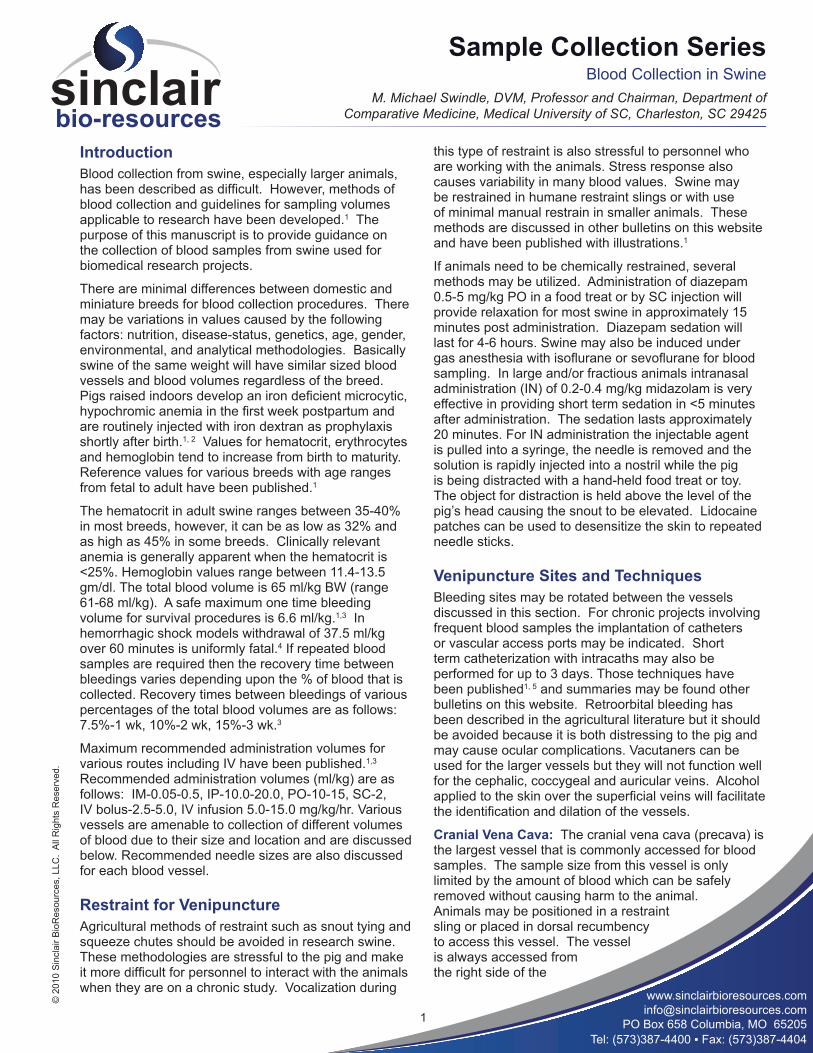
Femoral Vein and Artery: The femoral artery and vein are located in the femoral groove formed by the borders of the sartorius and gracilis muscles. They may be located by palpation of the pulse or by following the pulse of the medial saphenous artery which disappears as this superficial vessel disappears into the femoral groove. The vessels are located under the medial edge of the gracilis muscle rather than directly in the groove. The pig is placed in dorsal recumbency and the rear leg is gently extended to access this site. Sedation is required for this procedure. The femoral vessels can be
accessed with 20 g, 1.0-1.5” needles. The amount of blood that can be withdrawn is usually 5-10 ml. (Figures 6 & 7))
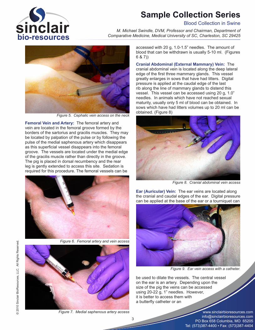
Cranial Abdominal (External Mammary) Vein: The cranial abdominal vein is located along the deep lateral edge of the first three mammary glands. This vessel greatly enlarges in sows that have had litters. Digital pressure is applied at the caudal edge of the last rib along the line of mammary glands to distend this vessel. This vessel can be accessed using 20 g, 1.0” needles. In animals which have not reached sexual maturity, usually only 5 ml of blood can be obtained. In sows which have had litters volumes up to 20 ml can be obtained. (Figure 8)
Ear (Auricular) Vein: The ear veins are located along the cranial and caudal edges of the ear. Digital pressure can be applied at the base of the ear or a tourniquet can
be used to dilate the vessels. The central vessel on the ear is an artery. Depending upon the size of the pig the veins can be accessed using 20-22 g, 1” needles. However, it is better to access them with a butterfly catheter or an
Figure 5. Cephalic vein access on the neck
Figure 6. Femoral artery and vein access
Figure 7. Medial saphenous artery access
Figure 8. Cranial abdominal vein access
Figure 9. Ear vein access with a catheter.
PO Box 658 Columbia, MO 65205Tel: (573)387-4400 ▪ Fax: (573)387-4404
Sample Collection SeriesBlood Collection in Swine
M. Michael Swindle, DVM, Professor and Chairman, Department of Comparative Medicine, Medical University of SC, Charleston, SC 29425
© 2
010
Sin
clai
r Bio
Res
ourc
es, L
LC.
All
Rig
hts
Res
erve
d.
4
sinclairbio-resources
intravascular catheter. Use of 18” intracaths will allow you to pass a catheter into the maxillary or external jugular vein in most animals. For this technique, the catheter is passed to the base of the ear and then the ear is pulled cranially and a finger is used to facilitate passage of the catheter beyond the base of the ear into the larger vessels. Catheters placed in the ear veins can be maintained successfully for up to 3 days. Only 1-2 ml of blood can usually be obtained from the ear vein itself, however, if the method of passing a catheter is used then the amount of blood that can be obtained is unlimited. (Figure 9)
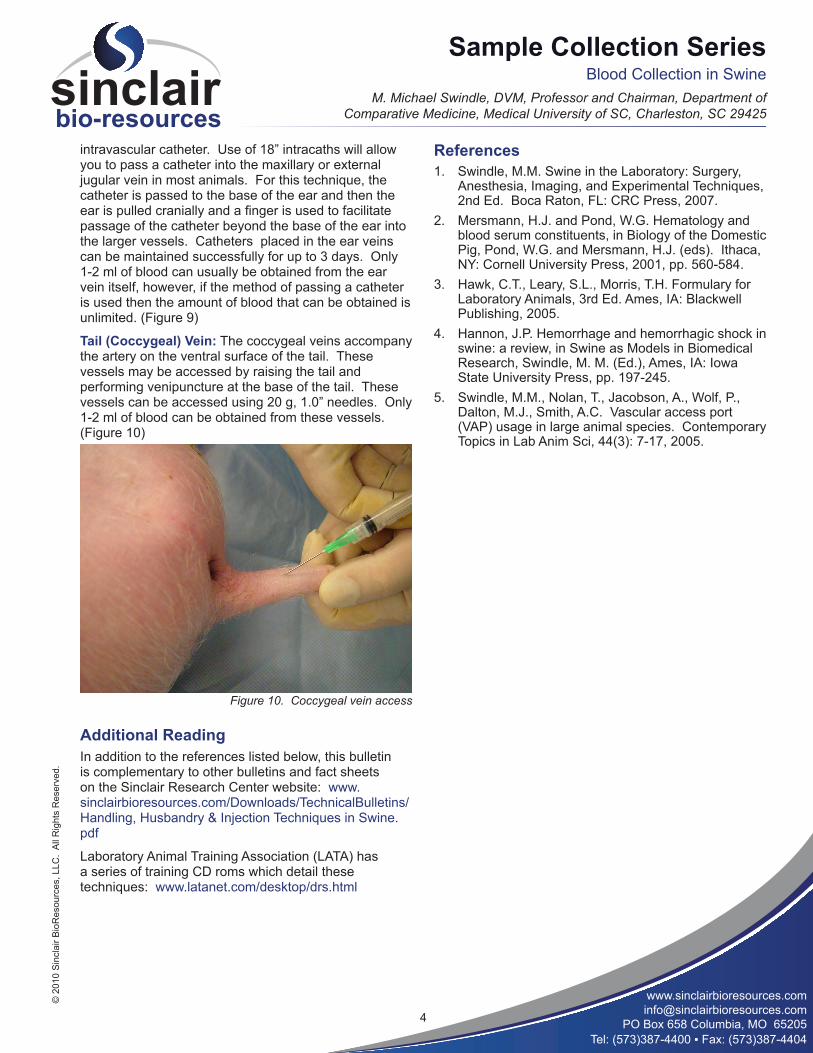
Tail (Coccygeal) Vein: The coccygeal veins accompany the artery on the ventral surface of the tail. These vessels may be accessed by raising the tail and performing venipuncture at the base of the tail. These vessels can be accessed using 20 g, 1.0” needles. Only 1-2 ml of blood can be obtained from these vessels. (Figure 10)
Additional ReadingIn addition to the references listed below, this bulletin is complementary to other bulletins and fact sheets on the Sinclair Research Center website: www.sinclairbioresources.com/Downloads/TechnicalBulletins/Handling, Husbandry & Injection Techniques in Swine.pdf
Laboratory Animal Training Association (LATA) has a series of training CD roms which detail these techniques: www.latanet.com/desktop/drs.html
ReferencesSwindle, M.M. Swine in the Laboratory: Surgery, 1. Anesthesia, Imaging, and Experimental Techniques, 2nd Ed. Boca Raton, FL: CRC Press, 2007. Mersmann, H.J. and Pond, W.G. Hematology and 2. blood serum constituents, in Biology of the Domestic Pig, Pond, W.G. and Mersmann, H.J. (eds). Ithaca, NY: Cornell University Press, 2001, pp. 560-584. Hawk, C.T., Leary, S.L., Morris, T.H. Formulary for 3. Laboratory Animals, 3rd Ed. Ames, IA: Blackwell Publishing, 2005. Hannon, J.P. Hemorrhage and hemorrhagic shock in 4. swine: a review, in Swine as Models in Biomedical Research, Swindle, M. M. (Ed.), Ames, IA: Iowa State University Press, pp. 197-245.Swindle, M.M., Nolan, T., Jacobson, A., Wolf, P., 5. Dalton, M.J., Smith, A.C. Vascular access port (VAP) usage in large animal species. Contemporary Topics in Lab Anim Sci, 44(3): 7-17, 2005.
Figure 10. Coccygeal vein access