Silybum marianum Treatment of Liver Fibrosis in … F. Salama, et al.pdfInt.J.Curr.Microbiol.App.Sci...
14
Int.J.Curr.Microbiol.App.Sci (2015) 4(3): 557-570 557 Original Research Article Effect of Egyptian Plant Silybum marianum on the Kidney during the Treatment of Liver Fibrosis in Female Albino Rats Induced by Alcohol in Comparison to the Medical Silymarin from China Afrah F. Salama 1 , Ehab Tousson 2 , Engy M. A. Elfetoh 1 *, M. A. Elhaak 3 and M. A. Elawni 4 1 Biochemistry Section, Department of Chemistry, Faculty of Science, Tanta University, Egypt 2 Department of Zoology, Faculty of Science, Tanta University, Egypt 3 Botany Department, Faculty of Science, Tanta University, Egypt 4 Organic Section, Department of Chemistry, Faculty of Science, Cairo University, Egypt *Corresponding author ABSTRACT Introduction Liver regulates many important metabolic functions, so the hepatic injury was associated with distortion of these functions (Wolf, 1999). Liver considered as the key organ of metabolism, excretion and variedly exposed to xenobiotics because of its strategic placement in the body. Toxins absorbed from the intestinal tract gain access first to the liver resulting in a variety of liver ailment (Ramachandra et al., 2007). Thus liver fibrosis considered as a one of the common problems on the human health (Krishnan et al., 2012). Liver damage ranged from acute hepatitis to hepatocellular carcinoma, through apoptosis, necrosis, inflammation, immune response, fibrosis, ISSN: 2319-7706 Volume 4 Number 3 (2015) pp. 557-570 http://www.ijcmas.com Liver fibrosis is one of the common problems on the human health. Many herbal and medicinal plants and their extracts are widely studied. The medical Chinese silymarin got a bright reputation in relieve of the liver fibrosis. A total of 72 female Albino rats were divided into six groups; G 1 (Control), G 2 (Fibrosis), G 3 (Chinese silymarin), G 4 (Fibrosis + Chinese silymarin), G 5 (Egyptian silymarin extract) and, G 6 (Fibrosis + Egyptian silymarin extract). Results showed that, serum creatinine and tissues MDA were significantly decreased in G 6 as compared to G 2 . Also, blood urea was significantly decreased in G 6 compared to those in G 2 and G 4 . Tissues CAT enzyme activity, total thiol and TAC were significantly increased in G 6 as compared to G 2 and G 4 . The results revealed that, the Egyptian plant extract improved kidney functions and their oxidative stress parameters in comparison with the Chinese silymarin, in case of liver fibrosis. Also, GC-MS showed that the Egyptian silymarin has more peaks than that of Chinese silymarin. The smaller peak area in the Egyptian silymarin extract was about one third the big one in the Chinese silymarin. The Egyptian silymarin extract have extra components that needs further study for its identification. Keywords Egyptian silymarin extract, Chinese silymarin, Liver, Kidney, Oxidative stress
Transcript of Silybum marianum Treatment of Liver Fibrosis in … F. Salama, et al.pdfInt.J.Curr.Microbiol.App.Sci...

Int.J.Curr.Microbiol.App.Sci (2015) 4(3): 557-570
557
Original Research Article
Effect of Egyptian Plant Silybum marianum on the Kidney during the Treatment of Liver Fibrosis in Female Albino Rats Induced by Alcohol in
Comparison to the Medical Silymarin from China
Afrah F. Salama1, Ehab Tousson2, Engy M. A. Elfetoh1*, M. A. Elhaak3 and M. A. Elawni4
1Biochemistry Section, Department of Chemistry, Faculty of Science, Tanta University, Egypt 2Department of Zoology, Faculty of Science, Tanta University, Egypt
3Botany Department, Faculty of Science, Tanta University, Egypt 4Organic Section, Department of Chemistry, Faculty of Science, Cairo University, Egypt
*Corresponding author
A B S T R A C T
Introduction
Liver regulates many important metabolic functions, so the hepatic injury was associated with distortion of these functions (Wolf, 1999). Liver considered as the key organ of metabolism, excretion and variedly exposed to xenobiotics because of its strategic placement in the body. Toxins absorbed from the intestinal tract gain access
first to the liver resulting in a variety of liver ailment (Ramachandra et al., 2007). Thus liver fibrosis considered as a one of the common problems on the human health (Krishnan et al., 2012). Liver damage ranged from acute hepatitis to hepatocellular carcinoma, through apoptosis, necrosis, inflammation, immune response, fibrosis,
ISSN: 2319-7706 Volume 4 Number 3 (2015) pp. 557-570 http://www.ijcmas.com
Liver fibrosis is one of the common problems on the human health. Many herbal and medicinal plants and their extracts are widely studied. The medical Chinese silymarin got a bright reputation in relieve of the liver fibrosis. A total of 72 female Albino rats were divided into six groups; G1 (Control), G2 (Fibrosis), G3 (Chinese silymarin), G4 (Fibrosis + Chinese silymarin), G5 (Egyptian silymarin extract) and, G6 (Fibrosis + Egyptian silymarin extract). Results showed that, serum creatinine and tissues MDA were significantly decreased in G6 as compared to G2. Also, blood urea was significantly decreased in G6 compared to those in G2 and G4. Tissues CAT enzyme activity, total thiol and TAC were significantly increased in G6 as compared to G2 and G4. The results revealed that, the Egyptian plant extract improved kidney functions and their oxidative stress parameters in comparison with the Chinese silymarin, in case of liver fibrosis. Also, GC-MS showed that the Egyptian silymarin has more peaks than that of Chinese silymarin. The smaller peak area in the Egyptian silymarin extract was about one third the big one in the Chinese silymarin. The Egyptian silymarin extract have extra components that needs further study for its identification.
K e y w o r d s
Egyptian silymarin extract, Chinese silymarin, Liver, Kidney, Oxidative stress

Int.J.Curr.Microbiol.App.Sci (2015) 4(3): 557-570
558
ischemia due to altering gene expression and regeneration (Shaker et al., 2010). All processes that involved hepatocyte, Kupffer, stellate and endothelial cells which induced liver disease that related to the crucial role of reactive oxygen and nitrogen species. The main sources of free radicals represented by hepatocyte mitochondria and cytochrome P450 enzymes, by endotoxin activated macrophages (Kupffer cells) and by neutrophils (Loguercio and Federico, 2003).
Alcohol has been implicated in the genesis of liver disease. Both its consumption and metabolism promoted the production of inflammatory mediators that resulted in hepatotoxicity and fibrogenesis. Finally, led to progressively sever liver injury and then caused cirrhosis (O'Shea et al., 2010). Toxic substances generated during the metabolism of alcohol in the liver and then contributed to the development of alcoholic liver disease (ALD).
Alcohol consumption increases the intestinal permeability of endotoxin. The endotoxin mediated inflammatory signaling plays a major role in alcoholic liver fibrosis (Abhilash et al., 2014). These substances included highly reactive molecules that could damage vital cell components through oxidation (Das and Vasudevan, 2006).
The kidney seemed to be the only vital organ generally spared in chronic alcoholics without advanced alcoholic liver disease or hepato renal syndrome. But, regular alcohol consumption raised the blood pressure, which considered as a risk factor for renal damage (Shanmugam et al., 2010). Some studies suggested that chronic ethanol ingestion per se was not nephrotoxic (Das and Vasudevan, 2005). Oxidative stress recognized to be a key step in the pathogenesis of ethanol-associated liver injury and kidney damage (Gramenzi et al., 2006).
Oxidative stress and associated cellular injury promoted inflammation. Antioxidants could have beneficial effects in reducing the incidence of ethanol induced changes in cellular lipids, proteins and nucleic acids. They could act by reducing free radical production (Chelators of redox-active iron derivatives), trapping free radicals themselves, interrupting the peroxidation process and reinforcing the natural antioxidant defense (Gupta, 2005 and Das and Vasudevan, 2006).
Many herbal, medicinal and pharmaceutical plants and their extracts were widely studied by many researches. Silybum marianum (Milk thistle) plant got a bright reputation in relieve of the liver fibrosis and kidney damage and that might be for the potent silymarin mixture. Mechanism of action for silymarin conducted mainly to the antioxidant, anti-inflammatory, antifibrotic and antilipidemic roles. The extract of the seeds of Silybum marianum has been used for centuries to treat liver and kidney disorders (Rainone, 2005).
Silymarin considered as a mixture of flavonolignan compounds isolated from the seeds of Silybum marianum plant. The most constituents are silibinin, isosilibinin, silicristin and silidianin (Agarwal et al., 2013). These compounds type and quantity differed by the natural environments where the plant was collected (Ibrahim et al., 2013). Therefore the present work aimed to study the chemical and medicinal effect of naturally growing Silybum marianum plant in Egypt (Gharbya Governorate) compared with the Chinese silymarin (Commercial medication) which considered the most widespread in the medical filed these days and the commonly used one, as well as capacity of protection of kidney damage during liver fibrosis, via estimation of biochemical and oxidative stress markers in addition to the histopathological changes.

Int.J.Curr.Microbiol.App.Sci (2015) 4(3): 557-570
559
Materials and methods
Chemicals:
Silymarin (Chinese medication) was obtained from Sedico Co. Egypt (Chinese origin) the other fine chemicals were obtained from Sigma Chemical Co., U.S.A. All other chemicals and reagents were of analytical grade.
Sample preparation and extraction process
The seeds of the Egyptian plant (Silybum marianum) were crushed to fine powder in a coffee grinder for two minutes, but at 15 sec. intervals. The crushed seeds were stored at - 20°C until the extraction was performed. A weight of 40 g of crushed seeds (powder) was soaked in 300 ml hexane overnight for 3 days for purification of fat (Defatting process). The hexane is recovered under vacuum at room temperature by using BUCH Rotavapor and then collected the defatted components.
The defatted components were then extracted by soaking in 70% alcohol (500 ml) for 3 days and then stirring for 3 hrs. The solvent was recovered by using BUCH Rotavapor at very deep vacuum without any thermal application to keep the components off any changes that may happened by thermal application. The extracted components were filtered through whatman filters 0.2 µm filter (Chemiton, Spain) and finally the purified components were lyophilized by using lyophilized system (Telstar model, Spain). The dried components (The Egyptian silymarin) were stored at -20°C until utilized (El-Shafeey et al., 2012).
GC-MS analysis
The analysis of the Egyptian silymarin extract and the silymarin (Chinese
medication) were done using a Hewlett Packard GC-6890 (Shimadzu, Japan), column (30 m length, 0.32 mm ID, 0.25 m film thickness), Restec, USA. The oven temperature was maintained at 50°C, raised 20°C / min to 280°C and held for 2 min. Also, identification of the volatile extract of both compounds was performed using gas chromatography
mass spectrometry according to the linear retention indices and mass spectra of separated compounds (Ka konien et al., 2011).
Animal s experimental design:
The experiments were performed on 72 female Albino rats weighing 120 g (± 20 g) obtained from Faculty of Vertinary, Cairo University, Egypt. The rats were housed in the laboratory for 1 week before the experimental work and maintained on the standard diet and water available in the animal research house of Zoology Department Faculty of Science, Tanta University. The temperature in the animal room was maintained at 23 ± 2 °C with a relative humidity of 55 ± 5% and at a 12:12 h light dark cycle. The experimental protocol was approved by Local Ethics Committee and Animals Research. The rats were randomly and equally divided into six groups (12 animals each).
Group 1 (G1): Control group in which rats free access to food and water ad libtum, then intragastrically intubated with saline (1 ml/100 g B.W) for 8 weeks.
Group 2 (G2): Fibrosis group in which rats treated with ethanol (10%, vol. /vol.) as the sole source of drinking for 8 weeks for induction of liver fibrosis, (the induction for liver fibrosis takes 4 weeks, continuing for another 4 weeks to meet the rest groups when received the treatment after induction of liver fibrosis) (Faremi et al., 2008).

Int.J.Curr.Microbiol.App.Sci (2015) 4(3): 557-570
560
Group 3 (G3): Chinese silymarin group in which the rats left at normal life as G1. Each rat received 200 mg Chinese silymarin /kg body weight/ day for 4 weeks by oral gavage (Shaker et al., 2010).
Group 4 (G4): Rats received ethanol for 4 weeks as in G2 (for induction of fibrosis), the treatment started from the beginning of the 5th week till the 8th weeks, in which each rat received 200 mg Chinese silymarin /kg body weight/ day by oral gavage with continuing drinking ethanol.
Group 5 (G5): Egyptian silymarin extract group in which the rats left at normal life as G3. Each rat received 200 mg a crude extract of Silybum marianum /kg body weight/ day for 4 weeks by oral gavage (Shaker et al., 2010).
Group 6 (G6): Like G4, but rats were treated with Egyptian silymarin extract instead of Chinese silymarin.
At the end of the experimental period, after an overnight fast, the rats were euthanized for collecting the blood samples and kidney tissues.
Histological investigation:
The kidney tissues were immediately removed and fixed in 10% neutral-buffered formalin for 24 h. The fixed specimens were then dehydrated, cleared and embedded in paraffin. Serial sections of 5-mm thick were cut by means of rotary microtome (Litz, Wetzlar, Germany). Sections were processed for haematoxylin and eosin staining (Bancroft and Cook, 1994). All stained slides were viewed using Olympus microscope and images were captured by a digital camera (Cannon 620). Brightness and contrast were adjusted using Adobe
Photoshop software (version 4.0.1; Adobe Systems, Mountain View, California).
Blood collection:
Blood samples from each rat were collected from the eyes by retro-orbital puncture from orbital plexus using blood capillary tubes. Blood was incubated at room temperature for 10 minutes, and then centrifuged at 3000 r.p.m for 10 min and the sera were collected; serum separated and kept in clean stopper plastic vials at 80°C until analysis of serum parameters.
Estimation of kidney function markers:
Serum creatinine and blood urea concentrations were assayed colorimetrically by using kits from Biodiagnostics Co. (Fawcett and Soctt, 1960 and Larsen, 1972).
Tissues preparation:
The kidney tissues were immediately isolated, cleaned from blood adhering matters, washed in ice-cold saline solution, then dried on a filter paper, weighed and frozen at 80°C. The liver and kidney tissues were homogenized (10% W/V) in potassium phosphate buffer (0.01 M pH 7.4) for estimation of glutathione S-transferase (GST) and catalase (CAT) enzymes activities, total antioxidant capacity (TAC), total thiol and total protein (TP), KCl solution (1.15 M) was used for estimation of malondialdehyde (MDA) using homogenizer (Hettich model EBA 12R, Germany).
GST enzyme activity was estimated through the formation of adduct, due to conjugation of GSH with 1-chloro-2, 4-dinitrobenzene (CDNB) according to the method described by Habig et al. (1974). CAT enzyme activity

Int.J.Curr.Microbiol.App.Sci (2015) 4(3): 557-570
561
was measured by monitoring H2O2
decomposition at 240 nm according to the method described by Xu et al. (1967). TAC was measured using the ferric reducing antioxidant power (FRAP) by the method described by Benzie and Strain (1999). Total thiol was measured using DTNB according to the method described by Sedlak and Lindsay (1968). The protein content in the tissues was determined by the method of Tsuyosh and James (1978).
Malondialdehyde (MDA) level is one of the terminal products, formed at the time of the decomposition of the polyunsaturated fatty acids mediated by free radicals. MDA was measured by the method of Lahouel et al. (2004).
Statistical analysis:
The data obtained in the experiment was expressed in terms of mean ± SEM. Statistical significance of data variations were assessed by one way analysis of variance (ANOVA) followed by a comparison between different groups using Tukey-Kramer multiple comparison t-test,
which compare between all groups and showed the significant effect of treatment (Graph pad Instate software). A value of P<0.05 was considered to be statistically significant.
Results and Discussion
Gas chromatography
mass spectrometry (GC-MS) of Egyptian silymarin extract and Chinese silymarin:
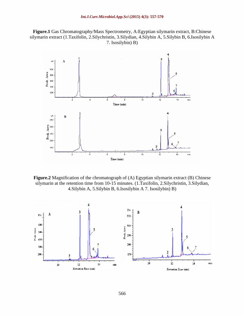
Analyses of Egyptian silymarin extract and the Chinese silymarin were present in Figure 1 (A and B). All compounds were identified by their retention time. The chromatograms were shown on the same abundance scale for the ease of visual comparison. Results were highly sensitive and reproducible. The
resulting chromatograms showed that there are seven major compounds in Egyptian silymarin extract as well as in the Chinese silymarin. By the retention time, the peaks could be for taxifolin, silychristin, silydian, silybin A, silybin B, isosilybin A and isosilybin B. The Egyptian silymarin extract chromatogram showed also a marked peak at least at 6-8 retention times which disappeared in the Chinese silymarin.
The peak area indicated greater counter of the identified compounds in the Egyptian silymarin extract in comparison with the Chinese silymarin Figure 2 (A and B). Peak 3 is greater in the Egyptian silymarin extract and peak 4 (For silybin) is separated into two peaks (4 and 5) in both compounds, the two peaks approximately were similar both in the Egyptian extract silymarin and the Chinese medication but the smaller peak was about one third the big one in the Chinese silymarin (i.e. higher peak in the Egyptian silymarin extract). These differences in the contents of the components of silymarin in both the Egyptian extract and the Chinese one, i.e. The Egyptian silymarin extract have extra components that need further studies for its identification.
Relative organ weight, kidney function test and its oxidative stress:
The kidney relative weight, Table 1 showed that G2 (Fibrosis group) decreased significantly (p<0.01) than the control group (G1). Table 1 showed serum creatinine of groups G2, G4 and G6 increased significantly than that of the control (G1), (p<0.001), (p<0.05) and (p<0.01) respectively. Also, serum creatinine of G2 increased significantly (p<0.05) than G3 and (p<0.001) of G5. Also, serum creatinine of G5
decreased significantly (p<0.05) of G4 and (p<0.01) of G6.

Int.J.Curr.Microbiol.App.Sci (2015) 4(3): 557-570
562
On the other hand, blood urea of G2
increased significantly than the other groups, (p<0.001). Also, blood urea of G4 increased significantly (p<0.01) and G6 (p<0.001) than the control (G1). While, groups G3 and G5
decreased significantly (p<0.001) than groups G4 and G6 (Table 1).
Table 1 showed kidney oxidative stress markers. GST enzyme activity of groups G2, G4 and G6 decreased significantly (p<0.001) than other groups. While, groups G3 and G5
increased significantly (p<0.001) than groups G4 and G6.
CAT enzyme activity of kidney tissues showed that groups G2 and G4 decreased significantly (p<0.001) than that of the control (G1). Also, CAT of G3 increased significantly (p<0.05) of G6 and (p<0.001) of G4. While, CAT of G4 decreased significantly (p<0.001) than G5 and G6
(Table 1).
Relative to the total antioxidant capacity, groups G2, G4 and G6 decreased significantly (p<0.001) of G1 while, groups G3 and G5 increased significantly (p<0.05) of G2. On the other hand, G3 and G5
increased significantly (p<0.001) than groups G4 and G6 and also, G5 increased significantly (p<0.01) of G1 and (p<0.05) than G3 (Table 1).
MDA of groups G2, G6 and G4 increased significantly (p<0.001) and (p<0.05) than G1, respectively. While, MDA of groups G3
and G5 decreased significantly (p<0.01) and (p<0.001) than the control (G1). Also, MDA of groups G3 and G5 decreased significantly (p<0.001) than that of groups G4 and G6
(Table 1).
Kidney total thiol of groups G2, G4 and G6
decreased significantly than that of the control (G1), (p<0.001) and (p<0.01), respectively. While, groups G3, G5 and G6
increased significantly (p<0.001) and (p<0.05) than G2, respectively. On the other hand, G3 increased significantly (p<0.01) than groups G4 and also, G5 increased significantly (p<0.001) of G4 and (p<0.05) of G6 (Table 1).
Kidney total protein of G2 decreased significantly (p<0.001) than G1 and (p<0.01) than G5. Also, total protein of groups G4 and G6 decreased significantly (p<0.01) than that of the control (G1). On the other hand, G5 showed a significant (p<0.01) increase than groups G4 and G6
(Table 1).
Histological examination of kidney
Histological examination of the kidney section in control, Chinese silymarin and Egyptian silymarin extract groups revealed entirely normal structures of the renal cortex which comprised renal corpuscles, proximal and distal convoluted tubules (Figure 3A, 3D and 3F). Kidney sections in alcoholic rats showed variable histopathological changes in glomeruli and some parts of the urinary tubules. These changes were in the Malpighian corpuscles, which lost their characteristic configuration and the renal tubules appeared with wide lumen and degenerated epithelium. Some glomeruli seemed to have lost their attachments and mesangial stroma and others were atrophied with dilatation in the subcapsular space (Figure 3B and 3C). Also, kidney sections in alcoholic rat cotreated with the Chinese silymarin revealed congestion of renal blood vessels and degeneration of some tubules (Figure 3E). However, kidney sections in alcoholic rat cotreated with the Egyptian silymarin extract showed better organized tubular and glomerular structures with well-established epithelia which resembled that of the control group except mild inflammatory infiltration (Figure 3G and 3H).

Int.J.Curr.Microbiol.App.Sci (2015) 4(3): 557-570
563
Analyzing the Egyptian silymarin extract and the Chinese silymarin by GC-MS showed general similarity in the number of peaks of compounds but, those compounds was found in a higher concentration in the Egyptian silymarin extract than the Chinese silymarin. Also, those data was compatible with the data obtained by Ka konien
et al. (2011). It was also important to note that the Egyptian silymarin extract acquired additional peaks between 6-8 and 12-14 retention times. Liu Yan-Ze and Lee (2012) found 18 compounds in silymarin, ten flavonolignan and eight small molecules including adenine, adenosine, uridine, trihydroxychromone. Those eight compounds are between 5 and 10 distance. Since the Egyptian silymarin extract contained additional peaks, that needs further study to identify then, which in turn reflected the high remediation effect of the Egyptian silymarin extract. The result of the present study showed a reduction in relative kidney weights in animals treated with ethanol in comparison with those of the control. Das et al. (2008) didn t observe any significant change in relative kidney weights in ethanol treated groups compared to the control group.
Serum creatinine and blood urea levels increased in the ethanol treated group as was found by Saravanan and Nalini (2007). Treatments with the Chinese and the Egyptian silymarin extract decreased their level, but with any significant change between the Chinese silymarin and the Egyptian silymarin extract. These findings were in accordance with those reported by Karimi et al. (2005), Kaur et al. (2010) and El-Shafeey et al. (2012).
Catalase present in the peroxisomes of nearly all aerobic cells, served to protect the cell from the toxic effects of hydrogen peroxide by catalyzing its decomposition into molecular oxygen and water without the
production of free radicals. The detoxification of 4-hydroxynonenal is compromised when Glutathione S-transferase (GST) level is reduced. Thus, ethanol or its metabolic products might specifically target GST isoenzymes and the reduction in enzyme activity or expression may contribute to ethanol hepatotoxicity (Hiratsuka et al., 2001). Cytochrome P450 2E1 (CYP2E1) could be the sole catalyst of fetal ethanol oxidation produces 1-hydroxy ethyl radicals, which have been shown to inactivate several proteins including antioxidant enzyme system (Epstein, 1996). In consistent with these reports, present results showed a decreased activities of GST and CAT in kidney tissues on the group treated with ethanol as compared to the control group.
Treatment with the Chinese and the Egyptian silymarin extract increased the activities of GST and CAT in rat's kidney tissues. These results were in the same line as with those results reported by Das et al. (2006) and El-Gazayerly et al. (2014). This may happen due to the essential activity of silymarin is an antioxidant effect of its flavonolignan and of other poly-phenolic substituent, which is attributed to the radical scavenging ability of both free radicals and reactive oxygen species (ROS) (Nencini et al., 2007).
Brighenti et al (2005) reported that moderate alcohol drinking can increase TAC, whereas daily and higher ingestion of alcoholic beverages reduce blood TAC. Ethanol consumption daily for 60 days by rats caused a reduction in TAC in kidney tissues as compared to the control group. In contrast, groups treated with the Chinese and the Egyptian silymarin extract elevated TAC in kidney tissues when compared to ethanol group. This was in accordance with results reported by Amin et al. (2012). The GST activity and TAC on the kidney tissues

Int.J.Curr.Microbiol.App.Sci (2015) 4(3): 557-570
564
were increased by both silymarin treatments but with greater effect by the Egyptian silymarin extract. The tissue damage produced by high level of MDA in kidney due treatment with ethanol as compared with the control group. Radosavljevi
et al.
(2011) reported that the ethanol increase the levels of MDA content in kidney. Significant decreased in total thiol in kidney was detected in ethanol group as compared with the control group. As well, the Chinese and the Egyptian silymarin extract treated groups had significant decrease in MDA levels and significant increase in total thiol level as antioxidant content when compared with ethanol group. These data are agreed the reported one by Raja et al. (2007) and Toklu et al. (2008). These results suggested the protective effects of both types of silymarin, which led to antioxidation, prevention of lipid peroxidation (Basaga et al., 1997) and retarding glutathione depletion (Alidoost et al., 2006). The Egyptian silymarin extract was found to be more effective than the Chinese silymarin.
As has been found the kidney total protein were significantly decreased in ethanol treated group (Hessien et al., 2010; Reddy et al., 2010). Treatment with the Chinese and the Egyptian silymarin extract countered this effect and raised the protein level suggesting the stabilization of endoplasmic reticulum required for protein synthesis. These findings were in accordance with the results of Noorani and Kale (2012). The differences in the kidney total protein didn t vary significantly between the Chinese silymarin and the Egyptian extracted one.
The current study, we found that the consumption of ethanol for 8 weeks induced many histopathological changes in rat's kidney which had been detected by the distortion of Bowman s capsule architecture. The Bowman s space considered as signs of cell death. Proximal and distal convoluted
tubules are sparsely distributed. These results were in agreement with those of Wilson et al. (2011). Our histological study confirms our biochemical result where the Egyptian silymarin extract improve both the kidney tissues better than the Chinese silymarin. Shaker et al. (2010) and El-Shafeey et al. (2012) evaluated the protective role of the Egyptian silymarin extract against the toxicity of ethanol and the histological changes in the kidney. Some improvements especially for the Egyptian silymarin have been shown in in renal tubules and renal corpuscles in the kidney. These results are in agreement with Karimi et al. (2005) who reported that Silybum marianum extract had a protective role against acute cisplatin nephrotoxicity.
The Egyptian silymarin extract could be extended for the isolation and structure determination of nephroprotective principle. Silymarin is considered as a mixture of flavonolignan compounds isolated from the seeds of Silybum marianum plant. These compounds type and quantity differed by the natural environments where the plant was collected. This confirms the increased number and amounts of compounds in the Egyptian silymarin extract as compared with the Chinese one, so now it is better than the medical Chinese silymarin. Silymarin caused marked alteration in some biochemical parameters induced oxidative damage and inhibited the activities of antioxidant enzymes. While, silymarin administrated in combination with ethanol had a beneficial effect, in the therapy of ethanol-induced liver fibrosis, silymarin alone prove to be beneficial in decreasing the levels of free radicals and lipid, and increasing antioxidant enzymes in healthy rats. Results proved the efficiency of the Egyptian silymarin extract exceeds the Chinese silymarin in treatment of kidney damage during liver fibrosis.

Int.J.Curr.Microbiol.App.Sci (2015) 4(3): 557-570
565
Table.1 Relative organ weights (g/100 g body weight) of the kidney, kidney function tests {Serum creatinine and blood urea (mg/dl)} and kidney tissues oxidative stress parameters {Glutathione S-transferase (GST) and catalase (CAT) enzymes activities (mole/min/g tissue) and concentrations of MDA (nmole/ g tissue), total thiol (mM/ g tissue), total antioxidant capacity (µmole Fe+2/g tissue) and total protein (mg/g tissue)} of female rats treated with vehicle G1 (Control), G2 (Ethanol), G3 (Chinese silymarin only), G4 (Ethanol + Chinese silymarin), G5 (Egyptian silymarin extract only) and G6 (Ethanol + Egyptian silymarin extract)
Groups
Parameters
G1 G2 G3 G4 G5 G6
Kidney weight
0.621 ± 0.02a 0.513 ± 0.01a 0.512 ± 0.01a 0.521 ± 0.02a 0.510 ± 0.02a 0.512 ± 0.01a
Serum creatinine 0.7±0.07acb 1.5±0.15adf 1.0 ± 0.07f 1.2 ± 0.13cl 0.7 ± 0.04dln 1.3 ±0.11bn
Blood urea 25 ± 1.3ab 56 ± 1.4 ad 15 ± 1.0 dg 41 ± 2.2bdgj 15 ± 4.6djm 38 ± 2.3adgm
GST activity 0.86±0.023a 0.40±0.02ad 0.89±0.014dg 0.64±0.019adgj 0.87±0.018djm 0.61±0.02adgm
CAT enzyme activity
0.96±0.02a 0.17±0.01ad 1.0±0.02dgi 0.45± 0.02adgj 0.98± 0.09dj 0.82±0.02dij
Total antioxidant power capacity
1.96±0.10ab 0.82 ± 0.06adf 2.0 ± 0.10dgi 1.0 ± 0.06agj 2.37 ± 0.09bdijm 1.15 ± 0.01afgm
MDA 79.1 ±1.85abc 160.98±1.02ad 65.79 ± 0.8bdg 90.0 ± 2.0cdgj 60.56 ± 2.43adjm 94.38 ± 3.95adgm
Total thiol 4.02 ± 0.13ab 2.53 ± 0.23adf 3.64 ±0.08dh 2.74 ± 0.12ahj 3.88 ±0.15djo 3.19 ± 0.16bfo
Total protein 38.22± 2.1ab 25.37 ± 3.2ae 30.2 ± 1.73 27.62 ± 1.25bl 36.16 ± 1.86elo 26.8 ± 0.81bo
Values are expressed as mean ± SEM; n = 12. - a, d, g, j, m (P<0.001) - b, e, h, k, n (P<0.01) - c, f, i, l, o (P,0.05)
Int.J.Curr.Microbiol.App.Sci (2015) 4(3): 557-570
566
Figure.1 Gas Chromatography/Mass Spectrometry, A:Egyptian silymarin extract, B:Chinese
silymarin extract (1.Taxifolin, 2.Silychristin, 3.Silydian, 4.Silybin A, 5.Silybin B, 6.Isosilybin A 7. Isosilybin) B)
Figure.2 Magnification of the chromatograph of (A) Egyptian silymarin extract (B) Chinese silymarin at the retention time from 10-15 minutes. (1.Taxifolin, 2.Silychristin, 3.Silydian,
4.Silybin A, 5.Silybin B, 6.Isosilybin A 7. Isosilybin) B)

Int.J.Curr.Microbiol.App.Sci (2015) 4(3): 557-570
567
Figure.3 Photomicrographs of rat kidney stained by haematoxylin and eosin. (A, D and F): Kidney sections in control, Chinese silymarin, Egyptian silymarin groups showed normal structures of the renal cortex which comprised renal corpuscles (arrows), proximal and distal convoluted tubules. (B and C): Kidney sections in alcoholic rat showed variable histopathological changes in renal corpuscles (glomeruli; arrows) and urinary tubules. Some glomeruli lost their characteristic configuration and others were atrophied with dilatation in the subcapsular space. (E, G and H): Kidney sections in alcoholic rat cotreated with the Chinses silymarin and the Egyptian silymarin extract showed congestion of renal blood vessels in the Chinese silymarin, while the Egyptian silymarin extract showed better organized tubular and glomerular structures with well-established epithelia which resembled that of the control group except mild inflammatory infiltration
Abbreviation
ALD, alcoholic liver disease; TP, total protein; DTNB, 5,5'-dithiobis-(2-nitrobenzoic acid); MDA, malondialdehyde; CAT, catalase; GST, glutathione-s-
transferase; SEM, standard error deviation; GC-MS, gas chromatography-mass spectrometry; CYP2E1, cytochrome P450 2E1; GSH, glutathione; CCl4, carbon tetra chloride; TAC, total antioxidant capacity; ROS, reactive oxygen species.

Int.J.Curr.Microbiol.App.Sci (2015) 4(3): 557-570
568
References
Abhilash, P.A., Harikrishnan, R., Indira M. 2014. Ascorbic acid suppresses endotoxemia and NF- B signaling cascade in alcoholic liver fibrosis in guinea pigs: a mechanistic approach. Toxicol. Appl. Pharmacol. 274: 215-224.
Agarwal, C., Wadhwa, R., Deep, G., Biedermann, D., Ga ák, R., K en V., Agarwal R. 2013. Anti-cancer efficacy of silybin derivatives - a structure-activity relationship. PLoS One. 8: 1-11.
Alidoost, F., Gharagozloo, M., Bagherpour, B., Jafarian, A., Sajjadi, S.E., Hourfar, H., Moayedi, B. 2006. Effects of silymarin on the proliferation and glutathione levels of peripheral blood mononuclear cells from beta-thalassemia major patients. Int. Immunopharmacol. 6: 1305-1310.
Amin, Z.A., Bilgen, M., Alshawsh, M.A., Ali, H.M., Hadi, A.H., Abdulla, M.A. 2012. Protective role of Phyllanthus niruri extract against thioacetamide-induced liver cirrhosis in ratmodel. Evidence Based Complement. & Alternat. Med. doi: 10.1155/2012/241583.
Bancroft, J.D., Cook, H.C. 1994. In: Manual of Histological Techniques and their Diagnostic Application. Bancroft and Cook (Eds.). Edinburgh, London, New York, Tokyo: Churchill Livingstone, pp: 23 26
Basaga, H., Poli, G., Tekkaya, C., Aras, I. 1997. Free radical scavenging and antioxidative properties of 'silibin' complexes on microsomal lipid peroxidation. Cell Biochem. Funct. 15: 27-33.
Benzie, I.F., Strain, J.J. 1999. Ferric reducing/antioxidant power assay: direct measure of total antioxidant activity of biological fluids and modified version for simultaneous measurement of total antioxidant power and ascorbic acid concentration. Meth. Enz. 299: 15 27.
Brighenti, F., Valtuena, S., Pellegrini, N., Ardigo, D., Del Rio, D., Salvatore, S., Piatti, P.M., Serafini, M., Zavaroni, I. 2005. Total plasma antioxidant capacity of the diet is inversely and independently related to plasma concentration of high-sensitivity C-reactive protein in adult Italian subjects. Brit. J. Nutr. 93: 619625.
Das, S.K., Vasudevan, D.M. 2005. Biochemical diagnosis of alcoholism. Ind. J. Clin. Biochem. 20: 35-42.
Das, S.K., Vasudevan, D.M. 2006. Protective effects of silymarin, a milk thistle (Silybum marianum) derivative on ethanol- induced oxidative stress in liver. Ind. J. Biochem. Biophys. 43: 306-311.
Das, S.K., Varadhan, S., Dhanya, L., Mukherjee, S., Vasudevan, D.M. 2008. Effects of chronic ethanol exposure on renal function tests and oxidative stress in kidney. Ind. J. Clin. Biochem. 23: 341-344.
El-Shafeey, M., El-Adawi, H., El-Azhary, D., Abd El-Wahab, A., Abdel-Mohsen, M. 2012. Protective effect of milk thistle and grape seed extracts on fumonisin B1 induced hepato- and nephro-toxicity in rats. Egypt. Acad. J. Biolog. Sci. 4: 63-85.
El-Gazayerly, O.N., Makhlouf, A.I., Soelm, A.M., Mahmoud, M.A. 2014. Antioxidant and hepatoprotective effects of silymarin phytosomes compared to milk thistle extract in CCl4
induced hepatotoxicity in rats. J. Microencapsul. 31: 23-30.
Epstein, M. 1996. Renal sodium handling in liver disease. In: The Kidney in liver disease. Epestin M. (Ed.) 4th ed. Hanley and Belfus Publication, Philadelphia, pp: 1-31
Faremi, T.Y., Suru, S.M., Fafunso, M.A., Obioha, U.E. 2008. Hepatoprotective potentials of Phyllanthus amarus against ethanol-induced oxidative stress in rats. Food Chem. Toxicol. 46: 2658-2664.

Int.J.Curr.Microbiol.App.Sci (2015) 4(3): 557-570
569
Fawcett, J.K., Soctt, J.E. 1960. A rapid and
precise method for the determination of urea. J. Clin. Path. 13: 156-159.
Gramenzi, A., Caputo, F., Biselli, M., Kuria, F., Loggi, E., Andreone, P., Bernardi, M. 2006. Review article: alcoholic liver disease--pathophysiological aspects and risk factors. Aliment Pharmacol. Ther. 24: 1151-1161.
Graph pad Instate software. www. graphpad. Gupta, S., Pandey, R., Katyal, R., Aggarwal,
H.K., Aggarwal, R.P., Aggarwal, S.K. 2005. Lipid peroxide levels and antioxidant status in alcoholic liver disease. Ind. J. Clin. Biochem. 20: 67-71.
Habig, W.H., Pabst, M.J., Jakoby, W.B. 1974. Glutathione-s-transferases: the first enzymatic step in mercapturic acid formation. J. Biol. Chem. 249: 7130-7139.
Hessien, M.H., El-Sharkawi, I.M., El-Barbary, A.A., El-Beltagy, D.M., Snyder, N. 2010. Non-invasive index of liver fibrosis induced by alcohol, thioacetamide and Schistosomal infection in mice. BMC Gastroenterol. doi: 10.1186/1471-230X-10-53.
Hiratsuka, A., Tobita, K., Saito, H., Sakamoto, Y., Nakano, H., Ogura, K., Nishiyama, T., Watabe, T. 2001. (S)-preferential detoxification of 4-hydroxy-2(E)-nonenal enantiomers by hepatic glutathione S-transferase isoforms in guinea-pigs and rats. Biochem. J. 355: 237-244.
Ibrahim, M., Elhaak, M.A., Mehama, H. 2013. Allelopathic potential of Silybum marianum and its utilization ability as a bioherbicide. Abstract of the annual international conference of the Egyptian Society of Experimental Biology, February, 2013 Cairo University Egypt, pp: 30.
Karimi, G., Ramezani, M., Tahoonian, Z. 2005. Cisplatin nephrotoxicity and protection by milk thistle extract in rats. Evid. Based Complement Alternat. Med. 2: 383-386.
Ka konien , V., Bimbirait -Survilien , K., Ratautait , V., Stankevi ius, M.,
Korny ova, O., Raga inskien , O., Proscevi ius, J., Maru ka, A. 2011. Investigation of phytochemical composition of biotechnologically modified medicinal plants. Sodininkyst Ir. Dar ininkyst . 30: 23-34.
Kono, H., Wheeler, M.D., Rusyn, I., Lin, M., Seabra, V., Rivera, C.A., Bradford, B.U., Forman, D.T., Thurman, R.G. 2000. Gender differences in early alcohol-induced liver injury: role of CD14, NF-kappaB and TNF-alpha. Am J. Physiol. Gastrointest. Liver Physiol. 278: 652-661.
Kaur, G., Athar, M., Alam, M.S. 2010. Dietary supplementation of silymarin protects against chemically induced nephrotoxicity, inflammation and renal tumor promotion response. Invest. New Drugs, 28: 703-713.
Krishnan, A., Li, X., Kao, W.Y., Viker, K., Butters, K., Masuoka, H., Knudsen, B., Gores, G., Charlton, M. 2012. Lumican, an extracellular matrix proteoglycan, is a novel requisite for hepatic fibrosis. Lab. Invest. 92: 1712-1725.
Lahouel, M., Boulkour, S., Segueni, N., Fillastre, J.P. 2004. The flavonoids effect against vinblastine, cyclophosphamide and paracetamol toxicity by inhibition of lipid-peroxydation and increasing liver glutathione concentration. Pathol. Biol. 52: 314-322.
Larsen, K. 1972. Creatinine assay by a reaction-kinetic principle. Clin. Chem. Acta. 41: 209-217.
Liu, Yan-ze, Lee David, Yue-wei 2012. Standardization and Identification of Minor Components of Silymarin (MK-001).Chin. Herb. Med. 4: 237-244.
Loguercio, C., Federico, A. 2003. Oxidative stress in viral and alcoholic hepatitis. Free Radical Biol. Med. 34: 1 10.
Nencini, C., Giorgi, G., Micheli, L. 2007. Protective effect of silymarin on oxidative stress in rat brain. Phytomedicine. 14: 129 135.
Noorani, A.A., Kale, M.K. 2012. Pretreatment of albino rats with methanolic fruit

Int.J.Curr.Microbiol.App.Sci (2015) 4(3): 557-570
570
extract of Randia dumetorum (L.) protects against alcohol induced liver damage. Korean J. Physiol. Pharmacol. 12: 125-130.
O'Shea, R.S., Dasarathy, S., Mc-Cullough, A.J. 2010. Alcoholic Liver Disease. Hepatol. 51: 307-328.
Radosavljevi , T., Mladenovi , D., Ninkovi , M., Vu evi , D., Bori i
I , Je i -Vuki evi , R., ljivan anin, T., Lopi i , S., Todorovi , V. 2011. Oxidative stress in rat liver during acute cadmium and ethanol intoxication. J. Serb. Chem. Soc., 76: 1 21.
Rainone, F. (2005) Milk thistle. Am Family Phys. 72: 1285- 1288.
Raja, S., Ahmed, K., Kumar, V., Mukherjee, K., Bandyopadhyay, A., Mukherjee, P. 2007. Antioxidant effect of Cytisus scoparius against carbon tetrachloride treated liver injury in rats. J. Ethnopharm. 109: 41 47.
Ramachandra Setty, S., Quereshi, A.A., Viswanath Swamy, A.H., Patil, T., Prakash, T., Prabhu, K., Veeran Gouda, A. 2007. Hepatoprotective activity of Calotropis procera flowers against paracetamol-induced hepatic injury in rats. Fitoterapia. 78: 451-454.
Reddy, V.D., Padmavathi, P., Paramahamsa, M., Varadacharyulu, N.C. 2010. Amelioration of alcohol-induced oxidative stress by Emblica officinalis (Amla) in rats. Ind. J. Biochem. Biophys. 47: 20-25.
Saravanan, N., Nalini, N. 2007. Impact of Hemidesmus indicus R.Br.extract on ethanol-mediated oxidative damage in rat kidney. Redox Rep. 12: 229-235.
Sedlak, J., Lindsay, R.H. 1968. Estimation of total, protein-bound and non-protein sulfhydryl groups in tissue with Elman's reagent. Anal. Biochem. 24: 192-205.
Shaker, E., Mahmoud, H., Mnaa, S. 2010. Silymarin, the antioxidant component and Silybum marianum extracts prevent liver damage. Food Chem. Toxicol. 48: 803 806.
Shanmugam, K.R., Ramakrishna, C.H., Mallikarjuna, K., Reddy, K.S. 2010. Protective effect of ginger against alcohol-induced renal damage and antioxidant enzymes in male albino rats. Ind. J. Exp. Biol. 48: 143-149.
Toklu, H., Akbay, T., Velioglu-Ogunc, A., Ercan, F., Gedik, N., Keyer-Uysal, M., Sener, G. 2008. Silymarin, the antioxidant component of Silybum marianum, prevents Sepsis-induced acute lung and brain injury. J. Surgical Res. 145: 214 222.
Tsuyosh, O., James, K.B. 1978. A simplified method of quantitating protein using the biuret and phenol reagents. Anal. Biochem. 86: 193-200.
Wilson, J.I., Emonido, O.F., Akpulu, S.P., Igbigbi P.S. 2011. Effect of ethanol and sialidase activities on the developing kidney of Wistar rats. J. Cell Animal Biol. 5: 200-205.
Wolf, P. 1999. Biochemical diagnosis of liver diseases. Ind. J. Clin. Biochem., 14: 5990.
Xu, J.B., Yuan, X.F., Lang, P.Z. 1997. Determination of catalase activity and catalase inhibition by ultraviolet spectrophotometry. Chin. Environ. Chem. 16: 73 76.