Signaling Organogenesis in Parasitic Angiosperms: Xenognosin Generation, Perception, and Response
15
Signaling Organogenesis in Parasitic Angiosperms: Xenognosin Generation, Perception, and Response W. John Keyes, Ronan C. O’Malley, Dongjin Kim, and David G. Lynn* Searle and Jones Chemistry Laboratory, 5735 Ellis Avenue, The University of Chicago, Chicago, IL 60637, USA ABSTRACT Parasitic strategies within the angiosperms generally succeed by tightly coupling developmental transi- tions with host recognition signals in a process re- ferred to as xenognosis. Within the Scrophulari- aceae, Striga asiatica is among the most studied and best understood parasitic member with respect to the processes of host recognition. Specific xenog- nosins regulate seed germination, the development of the host attachment organ, the haustorium, and several later stages of host-parasite integration. Here we discuss the signals regulating the development of the haustorium, the critical vegetative/parasitic transition in the life cycle of this obligate parasite. We provide evidence for the localized production of H 2 O 2 at the Striga root tip and suggest how this oxi- dant is used to exploit host peroxidases and cell wall pectins to generate a simple benzoquinone signal. This benzoquinone xenognosin proves to be both necessary and sufficient for haustorial induction in cultured seedlings. Furthermore, evidence is pro- vided that benzoquinone binding to a redox active site completes a “redox circuit” to mediate signal perception. This redox reaction regulates the time- dependent expression of specific marker genes criti- cal for the development of the mature host attach- ment organ. These studies extend the emerging se- ries of events necessary for the molecular regulation of organogenesis within the parasitic plants and sug- gest novel signaling features and molecular mecha- nisms that may be common across higher plants. Key words: Striga asiatica; Parasitic angiosperms; Signal transduction; Organogenesis; Haustorial de- velopment; Benzoquinones; Xenognosin INTRODUCTION All organisms must acquire and process information about their environment. Even the simplest unicel- lular organisms must detect nutrients, mediate dor- mancy, and navigate physical and biologic dangers. The Escherichia coli genome contains as many as 50 so-called two-component signal transmitter/ response regulator systems (Chang and Steward 1998) through which environmental cues may be monitored. Because of the simplicity of these sys- tems, the molecular mechanisms of perception and response are likely to be first understood in bacteria. Higher eukaryotes require further cellular coor- dination. Probably the most dramatic example is found in the sessile higher plants, which alter form throughout their life cycle to exploit their environ- mental niche. This ability to sculpt one’s architec- *Corresponding author; e-mail: [email protected] J Plant Growth Regul (2000) 19:217–231 DOI: 10.1007/s003440000024 © 2000 Springer-Verlag 217
-
Upload
w-john-keyes -
Category
Documents
-
view
212 -
download
0
Transcript of Signaling Organogenesis in Parasitic Angiosperms: Xenognosin Generation, Perception, and Response

Signaling Organogenesis in ParasiticAngiosperms: XenognosinGeneration, Perception,
and Response
W. John Keyes, Ronan C. O’Malley, Dongjin Kim, and David G. Lynn*
Searle and Jones Chemistry Laboratory, 5735 Ellis Avenue, The University of Chicago, Chicago, IL 60637, USA
ABSTRACT
Parasitic strategies within the angiosperms generallysucceed by tightly coupling developmental transi-tions with host recognition signals in a process re-ferred to as xenognosis. Within the Scrophulari-aceae, Striga asiatica is among the most studied andbest understood parasitic member with respect tothe processes of host recognition. Specific xenog-nosins regulate seed germination, the developmentof the host attachment organ, the haustorium, andseveral later stages of host-parasite integration. Herewe discuss the signals regulating the development ofthe haustorium, the critical vegetative/parasitictransition in the life cycle of this obligate parasite.We provide evidence for the localized production ofH2O2 at the Striga root tip and suggest how this oxi-dant is used to exploit host peroxidases and cell wallpectins to generate a simple benzoquinone signal.
This benzoquinone xenognosin proves to be bothnecessary and sufficient for haustorial induction incultured seedlings. Furthermore, evidence is pro-vided that benzoquinone binding to a redox activesite completes a “redox circuit” to mediate signalperception. This redox reaction regulates the time-dependent expression of specific marker genes criti-cal for the development of the mature host attach-ment organ. These studies extend the emerging se-ries of events necessary for the molecular regulationof organogenesis within the parasitic plants and sug-gest novel signaling features and molecular mecha-nisms that may be common across higher plants.
Key words: Striga asiatica; Parasitic angiosperms;Signal transduction; Organogenesis; Haustorial de-velopment; Benzoquinones; Xenognosin
INTRODUCTION
All organisms must acquire and process informationabout their environment. Even the simplest unicel-lular organisms must detect nutrients, mediate dor-mancy, and navigate physical and biologic dangers.The Escherichia coli genome contains as many as50 so-called two-component signal transmitter/
response regulator systems (Chang and Steward1998) through which environmental cues may bemonitored. Because of the simplicity of these sys-tems, the molecular mechanisms of perception andresponse are likely to be first understood in bacteria.
Higher eukaryotes require further cellular coor-dination. Probably the most dramatic example isfound in the sessile higher plants, which alter formthroughout their life cycle to exploit their environ-mental niche. This ability to sculpt one’s architec-*Corresponding author; e-mail: [email protected]
J Plant Growth Regul (2000) 19:217–231DOI: 10.1007/s003440000024
© 2000 Springer-Verlag
217

ture to a particular physical, chemical, and biologicniche is powerful and has obviously been successfulbut requires mechanisms for coupling external sig-nals with coordinated cell growth and cellular dif-ferentiation. In animals the body plan is typically setin the embryo, but plants continue to grow andchange their form by exploiting the specialized, per-petually embryonic regions known as meristems.Both the presence of a cell wall, which prevents cel-lular migration, and the absence of efficient circula-tion require the plant to maintain a greater cellularautonomy than seen in animals. The meristemstherefore empower the plant with the ability to re-spond to diverse environmental stimuli and do soboth through enhanced cellular plasticity and agreater response pleiotropy. Although this plasticityhas enabled agricultural manipulation of plantgrowth, the increased response pleiotropy hasgreatly complicated our understanding of the natureof the signals perceived, the mechanisms of signalperception, and the signal-transducing cascades thatmediate the diversity of known cellular responses.
In the most obvious and best-characterized sys-tems, for example, the light-mediated responsescontrolled by phytochrome, the actual molecules in-volved in signal transduction, other than the photo-receptor itself, remain poorly characterized (Nagyand Schafer 1999). Even more striking, the modes ofaction of plant hormones, at least those controllingcrucial events in growth and development, eventhose whose structures have been known for morethan half a century, are virtually unknown (Arteca1996; Brault and Maldiney 1999; Coenen and Lo-max 1997). Although it is clear that appropriateplant tissues respond to either light or hormones byaltering the transcription of genes within minutes(Guilfoyle and others 1989; Guilfoyle and others1993; Nagy and Schafer 1999; Silverthorne and To-bin 1987), the signaling cascades that mediate hor-monal induction of gene expression are just nowbeing explained (Abel and Theologis 1996; Braultand Maldiney 1999; Hooley 1999; Jones 1994).Even though the characterization of these pathwaysis critical, their definition remains only the first criti-cal step in understanding the hormones’ mecha-nisms of action. For example, rapid responses toboth the plant hormones auxin and cytokinin havebeen documented (Fowler and others 1999), but thefirst division is seen in isolated tobacco pith tissueafter 3–5 days of continual culture.
It is a combination of this confusion about therequired time for signal exposure, the apparentpleiotropic effects of the known signals, and the lackof a simple and rapid developmental system wherethe initiating signal is well understood that has so
stymied our efforts to define the molecular controlsregulating plant growth. Here we discuss the para-sitic plants as a model system to dissect the eventsnecessary for plant organogenesis. In these plants,the signals controlling the development of the hostattachment organ, the haustorium, originate in thehost plant. The obligate parasites have a limited life-time without attachment, and the parasite meriste-matic tissue must respond immediately to the signal.Such an immediate homogeneous commitment, andacross multiple individuals, synchronizes the re-sponse to allow the time dependence of gene ex-pression to be analyzed during organogenesis. Herewe discuss the specific model for signal perceptionand response that has emerged from studies on theAfrican witchweed, Striga asiatica.
The Parasitic Plants
One percent of all flowering plants are not solitarybut search out and form parasitic associations withother plants (Cronquist 1988; Kuijt 1969; Kuijt1977; Musselman 1980). Parasitism occurs broadlyacross the angiosperms, being found in at least 16families, in the form of trees, shrubs, vines, or herbs,and in habitats that range from the poles to the dryand humid tropics (Molau 1995). This broad distri-bution is a manifestation of the repeated appearanceof parasitism throughout angiosperm evolution (De-Pamphilis 1995; Musselman and Press 1995).
Searcy was the first to seriously consider the mo-lecular evolution of the parasitic plants and pro-posed three general phases (Searcy 1970; Searcy andMacInnis 1970). The first phase consisted of devel-oping the specialized organ that forms the physi-ologic bridge to the host, the haustorium. After thisphase, the second and third transitions were ones ofspecialization; the second phase being the loss ofboth biochemical pathways and morphologic struc-tures that become redundant with host attachment,and the third phase being the accrual of more com-plex adaptations specific to an obligate parasite, in-cluding host specialization and mechanisms to over-come host defenses.
Over the last 10 years, several groups have beenable to test predictions made by Searcy’s proposal(DePamphilis 1995). Analyses of the nonphotosyn-thetic holoparasite Epifagus virginiana provided strik-ing support for evolutionary reduction. The plastidDNA (ptDNA) of this organism was approximately athird the size of a nonparasitic relative (Nicotianatobacum). Essentially all the plastid-encoded photo-synthetic genes—the NADPH dehydrogenase genes,all four RNA polymerase genes, 13 of the 30 plastidencoded tRNAs, and 6 of the 22 plastid-encoded ri-
218 W. J. Keyes and others

bosomal protein genes—were either missing com-pletely or existed as pseudogenes (DePamphilis andPalmer 1990; Wolfe and others 1992). Acceleratedrates of molecular evolution of both the remainingplastid genes and the rDNA of these parasites havealso been documented and explained by molecularpopulation genetic models of mutation, selection,and drift (DePamphilis 1995; Nickrent and Starr1994). In addition, significant biochemical evidencenow exists for highly specialized processes in hostselection (Boone and others 1995; Lynn and Chang1990), distance regulation (Chang and others 1986;Fate and Lynn 1996; Fate and others 1990), andovercoming host resistance (Olivier and others1991; Riopel and Timko 1995).
By Searcy’s proposal, the critical transition to theparasitic mode would be phase one. Histologic stud-ies of mature haustoria are available, and althoughclear differences in anatomic features are presentacross the parasitic plants, the early initiation eventsappear similar (Atsatt 1983; Kuijt 1969; Riopel andTimko 1995). The development of lateral haustoriain the rhinanthoid Scrophulariaceae consist of earlyrounds of cell division and radial swelling by thepericycle and inner cortex to give rise to the haus-torial primordia. Lateral roots and root nodules arealso absorptive tissues, and the early stages of theirformation are quite similar to those of the haustoria(DeKlerk and others 1999). Therefore, a simplifyinghypothesis would maintain that parasitism arosefrom the loss of some signaling function that wascomplemented externally by another plant. It wouldthen follow that the events responsible for inducingthese organs have similar biochemical and mecha-nistic origins. In the case of the haustorium, both theinducing signal and some understanding of signalperception are available (Lynn and Chang 1990) andcan be exploited to dissect the signaling pathway. Atthe very least, biochemical dissection of the signal-ing pathway for haustorial induction could provideinsight into both the induction of plant organogen-esis and the early steps in parasite evolution.
Haustorial Development
Early rounds of cell division by the pericycle andinner cortex give rise to the lateral haustorial pri-mordia in many of the Scrophulariaceae parasites(Kuijt 1977; Riopel and Musselman 1979). Thispostembryonic origin is similar in both lateral root(Malamy and Benfey 1997) and legume nodulationformation (Crespi and Galvez 2000; Mylona andothers 1995) where coordinated cell expansion anddivisions occur along the primary root proximal tothe vascular tissue in the pericycle layer. The devel-
opmental programs of all three are induced by en-vironmental signals and respond to hormones.Haustoria-like structures (Riopel and Baird 1987)and the early stages of nodule development (Crespiand Galvez 2000; Mylona and others 1995) are in-duced by cytokinins, whereas IAA appears to be im-portant for initiation, morphogenesis, and contin-ued viability of lateral roots (DeKlerk and others1999; Malamy and Benfey 1997). Compounds thatinhibit auxin transport (Hirsch and others 1989) alsoinduce nodulelike structures.
In the terminal haustoria formed in S. asiatica,organogenesis is manifested primarily in the redirec-tion of cellular swelling events. The cells distal to themeristem switch from longitudinal to radial growth,and the circumscribed pre-epidermal cells formhaustorial hairs as shown in Figure 1. The extent ofswelling required for the new organ can be signifi-cant, with an increase in diameter from twofold tofourfold, and the rate dramatic, development beingcomplete within 24 h of induction. The swollen cellscreate minimally a larger surface area likely to becritical for host attachment, and it is within thisswollen tip that the haustorial primordia form, giv-ing rise to the infection peg and ultimately the ma-
Figure 1. Two-day-old S. asiatica seedling was incubatedat 30°C in 1 mL of H2O containing 10 µM DMBQ. Thephotograph shows a typical swollen root tip with hausto-rial hair formation at the site of the arrows, after 24 hunder these conditions.
Signaling Organogenesis 219

ture host interface (Kuijt 1977; Riopel and Baird1987; Riopel and Timko 1995).
Results from radioactive thymidine incorpora-tion, as shown in Figure 2, have established thathaustorial development in S. asiatica is the result of adominant, if not exclusive, redirection of cellular ex-pansion and is independent of new cell division.Moreover, incorporation is arrested immediately af-ter exposure to the xenognostic quinone, with littlesignificant uptake detectable for at least 12 h. Im-mediately after removal of the inducing signal, 2,6-dimethoxy-1,4-benzoquinone (DMBQ), DNA syn-thesis recovers to the steady-state level seen for nor-mal root growth. The role of the existing meristeminitials in this recommitment is not yet clear.
Kinetin has been shown to induce haustorial de-velopment in parasitic Schrophulariceae (Riopel andMusselman 1979), and as shown in Figure 3A, mi-cromolar concentrations are sufficient for inductionin S. asiatica. However, haustorial hair growth is
more exaggerated and the radial swelling not as pro-nounced as with DMBQ induction. Later stages ofdevelopment also appear somewhat compromisedby continual kinetin exposure, giving morphologi-cally distorted haustoria, but the induction processappears normal. In contrast, auxins are very potentinhibitors of haustorial induction. Figure 3B estab-lishes that nanomolar concentrations of a-naphtholacetic acid or indole acetic acid (data not shown) aresufficient to inhibit xenognosin induction, but thestructurally similar b-naphthol acetic acid, whichhas no auxinlike activity, has no effect on haustorialorganogenesis. The cells of the root meristem of S.asiatica therefore appear poised for the transition tohaustorial organogenesis and are generally respon-sive to plant hormones.
Xenognostic Signaling
The angiosperm parasitic strategy generally succeedsby tightly coordinating early developmental stageswith chemical signals from the host (Smith and oth-
Figure 2. Two-day-old S. asiatica seedlings (100) were la-beled at 30°C in 1 h in 200 µL media containing 1 µCi of[3H]-thymidine after each indicated incubation time with(d) or without (■) 2 µM DMBQ. At 12 h, a portion of theseedlings incubated with DMBQ were washed (3×) andfurther incubated in water with the indicated time pointsused for [3H]-thymidine incorporation (m). After the[3H]-thymidine incubation, the seedlings were groundinto a fine powder in liquid nitrogen and transferred to atube with 500 µL of the extraction buffer (100 mM EDTA,250 mM NaCl, 100 µg proteinase K) and the mixture wasincubated at 55°C for 1 h. After centrifugation at 12,000 ×g for 5 min, the insoluble pellets were removed, and thesame volume of 10% TCA was added to the supernatant.After incubation on ice for 10 min, the suspension wasfiltered through a Whatman GF/C glass filter, and the filterwas washed with 3 mL of a 5% TCA solution. The radio-activity of the filters was determined by scintillationcounting. Data were acquired in triplicate and expressedas ± SD.
Figure 3. Two-day-old S. asiatica seedlings (20–30 perwell) were incubated at 30°C in 1 mL of H2O containingeither (A) kinetin or (B) 10 µM DMBQ with a-IAA. Seed-lings were scored at 24 h. The induction percentage wasdetermined in triplicate and expressed as ± SD.
220 W. J. Keyes and others
ers 1990). S. asiatica, probably the best studied interms of its signaling chemistry, is chlorophyllous asan adult but requires host-derived signal com-pounds for both germination (Boone and others1995; Lynn and Chang 1990; Worsham 1987) andthe development of the haustorium (Albrecht andothers 1999; Chang and Lynn 1986; Lynn 1985;Riopel and Timko 1995). Consistent with Searcy’sgeneral evolutionary model of structural and bio-chemical minimization, the young seedlings have nolateral roots or root hairs. With the initiation of thehaustorium, the vegetative phase of Striga’s life cycleceases, and the parasitic phase begins. In the absenceof viable host attachment, the apical meristem neverdevelops, and because of its limited seed reserves,the seedling survives for less than 5 days.
The first structural characterization of specifichost-derived molecules that cued the vegetative toparasitic growth transition came from work withhost exudates. Xenognosin A (Lynn and others1981) and B (Steffens and others 1982) providedboth the origin for the term xenognosin, or hostrecognition signal, and established that specific phe-nols could play a necessary, and possibly sufficient,role in haustorial induction. Later work with S. asi-atica uncovered evidence that the activity was asso-ciated with phenolics of the host cell wall, leading tothe actual finding that specific quinones oxidativelyreleased from the wall pectins constituted the activesignal (Chang and Lynn 1986; Lynn 1985). Thesefindings led to the hypothesis that oxidative en-zymes released from the parasite generated a suffi-cient xenognostic quinone concentration only at aviable host surface rich in wall pectins (Lynn andChang 1990). This mechanism was appealing in thatit regulated parasitic commitment only in closeproximity with a viable host so as to increase thelikelihood of successful attachment.
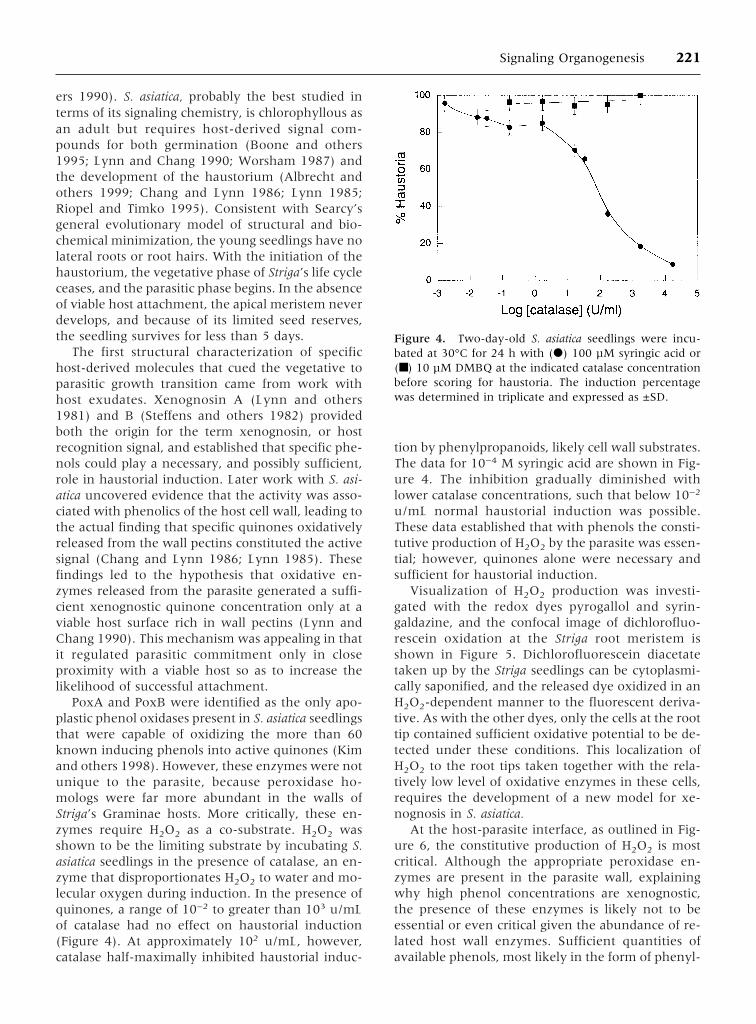
PoxA and PoxB were identified as the only apo-plastic phenol oxidases present in S. asiatica seedlingsthat were capable of oxidizing the more than 60known inducing phenols into active quinones (Kimand others 1998). However, these enzymes were notunique to the parasite, because peroxidase ho-mologs were far more abundant in the walls ofStriga’s Graminae hosts. More critically, these en-zymes require H2O2 as a co-substrate. H2O2 wasshown to be the limiting substrate by incubating S.asiatica seedlings in the presence of catalase, an en-zyme that disproportionates H2O2 to water and mo-lecular oxygen during induction. In the presence ofquinones, a range of 10−2 to greater than 103 u/mLof catalase had no effect on haustorial induction(Figure 4). At approximately 102 u/mL, however,catalase half-maximally inhibited haustorial induc-
tion by phenylpropanoids, likely cell wall substrates.The data for 10−4 M syringic acid are shown in Fig-ure 4. The inhibition gradually diminished withlower catalase concentrations, such that below 10−2
u/mL normal haustorial induction was possible.These data established that with phenols the consti-tutive production of H2O2 by the parasite was essen-tial; however, quinones alone were necessary andsufficient for haustorial induction.
Visualization of H2O2 production was investi-gated with the redox dyes pyrogallol and syrin-galdazine, and the confocal image of dichlorofluo-rescein oxidation at the Striga root meristem isshown in Figure 5. Dichlorofluorescein diacetatetaken up by the Striga seedlings can be cytoplasmi-cally saponified, and the released dye oxidized in anH2O2-dependent manner to the fluorescent deriva-tive. As with the other dyes, only the cells at the roottip contained sufficient oxidative potential to be de-tected under these conditions. This localization ofH2O2 to the root tips taken together with the rela-tively low level of oxidative enzymes in these cells,requires the development of a new model for xe-nognosis in S. asiatica.
At the host-parasite interface, as outlined in Fig-ure 6, the constitutive production of H2O2 is mostcritical. Although the appropriate peroxidase en-zymes are present in the parasite wall, explainingwhy high phenol concentrations are xenognostic,the presence of these enzymes is likely not to beessential or even critical given the abundance of re-lated host wall enzymes. Sufficient quantities ofavailable phenols, most likely in the form of phenyl-
Figure 4. Two-day-old S. asiatica seedlings were incu-bated at 30°C for 24 h with (d) 100 µM syringic acid or(■) 10 µM DMBQ at the indicated catalase concentrationbefore scoring for haustoria. The induction percentagewas determined in triplicate and expressed as ±SD.
Signaling Organogenesis 221

propanoid esters decorating the pectins (insert A),provide the co-substrate for the peroxidases. Asshown in insert B, oxidative cleavage of model phe-nols generates the quinone. A sustained quinoneconcentration is critical, because the quinone mustbe present for many hours for terminal commitmentto organogenesis (see later). This time restrictionplaces a critical threshold on the necessary H2O2 andphenol concentrations. Most critically, Striga cellwalls are low in phenols and probably devoid ofphenols at the root tip. In this way, the parasite isable to provide H2O2 to the host wall as the limitingreagent for sustained quinone production.
How general might this mechanism for signalgeneration be among the parasitic plants? Themodel requires the acquisition of reaction pathwayscapable of continually producing low levels of hy-drogen peroxide. H2O2 is produced ubiquitouslyamong both plants and animals as a first line of bi-otic defense, and several known pathways exist forits generation (Kim and others 1998; Lamb andDixon 1997; Mehdy 1994). Each one, in principle,should be capable of constitutive H2O2 generation atthe root apex, and the possibilities are not easilynarrowed by this restriction. The cells involved in
this localized production of the reactive oxygen spe-cies (ROS) must also avoid general oxidative toxic-ity, as well as the ability of ROS to activate otherdefense pathways (Baker and Orlandi 1995; Lamband Dixon 1997). S. asiatica is somewhat unusualamong the parasitic plants in that it is a dicot para-sitizing monocots and may be capitalizing on differ-ences in sensitivities and/or responses to the ROS inits monocot hosts. The responses to the generatedquinones may also be very different in monocotsand dicots; the same quinone distribution should bereadily produced in most plant tissues by simplewounding events. Finally, age differences, given the5-day window for Striga seedling viability, are al-most certainly critical for this obligate parasite, butsuch age differences are not likely to be as pro-nounced in the facultative parasites.
Modifications to this mechanism, or completelydifferent strategies for xenognosin generation, aretherefore likely to exist across the parasitic an-giosperms, but an important principle of parasitismmay have emerged from the work on Striga. The“offensive” xenognosis pathway is minimally usingcomponents of reaction pathways necessary for pro-tection of the plant from biotic stresses. Clearly ele-ments of self/non-self recognition are essential tothe establishment of the haustorial interface (Atsatt1983) and are apparently functionally connected torecognition. Further definition of the mechanisms ofxenognosis in this and other parasites should pro-vide a unique opportunity to uncover genes criticalto the early steps in cell-cell recognition and non-self resistance.
Xenognosin Perception
The quinones expected to be generated from theoxidation of the common cell wall phenolics includebenzo-1, 4-quinone (BQ), methoxybenzo-1,4-quinone (MBQ), and DMBQ, all three of which in-duce haustorial formation in both New and OldWorld parasitic angiosperms (Albrecht and others1999; Lynn and Chang 1990). Attempts in S. asiaticato establish a functional correlation among syntheticvariants of these quinones and haustorial inductionrevealed a dependence on electromotive potential(Smith and others 1996), Em, the energy required toadd a single electron reductively to the quinonenucleus. As shown in Figure 7 (Smith and others1996), no quinone whose Em potential lay outsideof a defined redox range, −250 to 0 mV relative toSCE, was active, and the quinones at the redoxboundaries show only partial induction of haustorialdevelopment. The heavily substituted benzoqui-nones, all trisubstituted and most disubstituted,were inactive regardless of redox potential.
Figure 5. One microliter of 100 mM dichlorofluorescein-diacetate in DMSO was added to S. asiatica seedlings in 1mL of water buffered at pH 5.5. After 15 min, the stainingof the tissue was detected with a Zeiss LSM 510 confocalmicroscope. A green argon-ion laser (488 nm) was usedfor excitation, and fluorescence was detected at 525 nm.No staining was detected along the root axis or around theseed coat.
222 W. J. Keyes and others

The observation that the active range is definedby the first half-volt potential and bounded by bothoxidative and reductive extremes is consistent withthe quinones serving as single electron carriers. Thisfinding was interesting in light of earlier studies thathad established a precise time dependence for theinduction process (Smith and others 1990). Theseobservations were incorporated into a model inwhich the inducing quinone signal was perceived bymeans of an electron transport chain. This putativeredox circuit would require quinone binding to becompleted, and the time dependence of depolariza-tion resulting from the electron flow between tworedox pools could be functionally coupled with thetime dependence of the commitment to haustorialdevelopment (Smith and others 1996). Severalphysical tests of this model for signal perception nowexist.
Based on other more well-characterized biologicredox circuits, including oxidative phosphorylationand photosynthesis where quinones function aselectron carriers, the reactive one-electron reducedsemiquinone is expected to be bound tightly with-in a redox binding site. Structural analogs dis-tributed at the extremes of the redox window were
used to confirm this prediction. Tetrafluorobenzo-1,4-quinone (TFBQ) is of similar steric size to BQ, andbecause it is strongly electropositive, should bereadily reduced within the defined redox window tothe semiquinone (Figure 8). Reoxidation, however,would be thermodynamically restricted as a result ofthe oxidative limits placed on the active window.Therefore, if TFBQ were bound at a site within thewindow, further reaction would be arrested at theone-electron reduced semiquinone. Consistent withthe prediction, TFBQ inhibited induction and wasvery potent, half-maximally inhibiting 10−5 MDMBQ induction at 10−7 M TFBQ (Smith and others1996). Two important observations can be derivedfrom the TFBQ experiments. First, both one-electron oxidative and reductive reactions are im-portant for induction, that is, if either are blocked,haustorial induction does not continue. Second, theinhibition by TFBQ is freely reversible and not theresult of general toxicity resulting from the genera-tion of a reactive TFBQ semiquinone. Therefore, theTFBQ semiquinone most probably binds tightlywithin an active site, blocking the second requiredelectron transfer step, and by doing so, inhibitshaustorial induction.
Figure 6. The model for the generation of the haustorial inducing quinones envisions H2O2 generated at the parasiticseedling root tip accumulating at the host interface. The H2O2 is used as an oxidation cosubstrate, together with wallpectins, by the host cell wall peroxidases to generate the xenognostic quinones. Insert A shows sorghum wall fractionationsand establishes that the pectins contain the peroxidase substrate as measured by haustorial inducing activity. Insert B showsthe oxidative cleavage of the model substrate, syringic acid.
Signaling Organogenesis 223

Further support for the existence of a semi-quinone intermediate was derived from the devel-opment of quinone analogs containing specific ben-zylic leaving groups. Several compounds, at leastconceptually derived from the naturally occurringbioreductively activated antitumor antibiotics (Zengand others 1996), were developed and shown toinhibit haustorial induction specifically (Smith andothers 1996). The most specific inhibitors were thesimple oxirane and cyclopropane benzoquinoneanalogs. Cyclopropyl benzoquinone (CPBQ) isunique in that the cyclopropane ring opens readilyby radical delocalization from the semiquinone state(Figure 8). The lifetime of the ring-opened cyclopro-pane was shown to be sufficient for bond rotationand racemization (Zeng and others 1996), generat-
ing a quinone methide ring-opened intermediate.Quinone methides are strong alkylating agents, andit was proposed that CPBQ alkylated the bindingsite. Consistent with the prediction, and in contrastwith TFBQ inhibition, CPBQ inhibition was irrevers-ible (Smith and others 1996).
Accepting that the limiting redox range is impor-tant and that the semiquinone intermediate exists,what is this redox reaction? The redox range inhaustorial induction is similar to that seen for thequinone cofactors in both photosynthesis and oxi-dative phosphorylation. In these processes, thequinone is reduced to a semiquinone intermediateby an electron that is generated at a donor site byeither photochemical excitation or NADH oxidation.The resulting semiquinone is re-oxidized when the
Figure 7. The quinone analogs are plotted as a function of their Em potential (lower scale). The upper scale is an expansionof the redox range in the active window and contains the active haustorial inducers (9–17). The half-wave reductivepotential of the quinones was measured with a saturated Calomel reference electrode, and the relative potentials arecompared with that of benzoquinone, which was set at zero (Smith and others 1996). The abscissa represents the haustorialinduction percentage referenced to DMBQ at the maximal inducing concentration. The solid arrows indicate 100% ofcontrol (85% with DMBQ). Dashed arrows indicate partial haustoria induction between 40 and 50% of the DMBQcontrols.
224 W. J. Keyes and others

electron passes to the next acceptor in the electroncarrier chain. As in haustorial induction, both reduc-tive and oxidative steps are required for electronflow, and only quinones within a specific redox win-dow serve as carriers. The similarities between theseorganellar pathways and the observed events inhaustorial induction are striking and suggest thatcellular organelles might be involved in the process.At high concentrations, benzoquinones are meta-bolic toxins that can cause oxidative cellular dam-age, but there is no evidence that unsubstituted ben-zoquinones interfere in the microsomal electrontransport chains at lower concentrations. Neverthe-less, the plastid genomes of parasitic plants haveevolved under very different pressures (DePamphilis1995), and further evaluation of the functional rolethese organelles may play in haustorial developmentwill be important.
More generally, there is a long history of animaland plant cell redox processes in addition to those ofthe cellular organelles, and models have been pre-sented suggesting that altered electron flow throughdifferent pathways directs cell growth (Crane andothers 1988). Moreover, transcriptional factors fromviral, bacterial, and mammalian sources are knownto be under redox control (Abate and others 1990;Allen 1993; McBride and others 1992; Staal and oth-ers 1990; Storz and others 1990; Tagaya and others1994), and in plants, chloroplast genome expressionis regulated by redox reactions (Allen 1994; Bucha-nan 1991; Danon and Mayfield 1994). Therefore, amechanistic involvement of redox reactions with
the more complex cellular processes of growth anddevelopment has been recognized.
Early studies detected NAD(P)H oxido-reductasesin all plant membranes investigated (Moller and Lin1986). The plasma membrane (PM)–associatedoxido-reductases have been associated with an elec-tron transport system in roots (Rubinstein and oth-ers 1984), leaves (Marre and others 1998), indi-vidual cells (Barr and others 1985; Misra and others1984), protoplasts (Lin 1982; Lin and others 1984;Thom and Maretzki 1985), isolated membranes(Misra and others 1984), and purified plasma mem-brane fractions (Barr and others 1985b; Buckhoutand Hrubec 1986; Qui and others 1985). These PM-localized electron transport systems have been stud-ied for many years with impermeable electron ac-ceptors. In fact, the initial studies associated redoxevents with the control of cell growth (Baron andHoffman 1929; Brooks 1947). Impermeable oxi-dants like ferricyanide and diferric transferrin pro-mote growth in animal cells (Crane and others1985) and generally inhibit growth in plants (Barrand others 1985a), presumably in both cases bysome alteration of the transmembrane redox state(Boss and Morre 1988; Crane and Barr 1989; Craneand others 1988).
Some of the enzymes involved in these redoxprocesses have been identified. NAD(P)H-K3Fe(CN)6
and NAD(P)H-duroquinone reductases were foundin the plasma membranes of several plants (Asardand others 1987; Askerlund and others 1988; Buck-fout and Hrubec 1986; De Luca and others 1984)and have been purified from zucchini microsomes(Guerini and others 1987; Valenti and others 1990)and corn root purified PMs (Luster and Buckhout1988). In the corn PMs, two enzymes were found, aferricyanide reductase and a 28-kDa protein, whichhas both duroquinone and ferricyanide reductaseactivity. The 28-kDa enzyme was reported to have aflavin requirement (Luster and Buckhout 1989),and monoclonal antibodies directed to this proteinwere prepared (Buckhout and Luster 1988).
More recently, NADPH oxidoreductase in plantshas been immunologically correlated with the bet-ter-characterized human neutrophil machinery re-sponsible for the defensive oxidative burst Desikanand others 1996; Dwyer and others 1996; Vera-Estrella and others 1994; Xing and others 1997).This oxidative complex consists of a heterodimericflavocytochrome b-558 of subunit mass approxi-mately 22 and 91 kDa (p22-phox and gp91-phox)(Segal 1989), a Rac GTPase of ∼22 kDa (Knaus andothers 1991; Kwong and others 1993), and twonovel polypeptides of approximately 47 and 67 kDa
Figure 8. One-electron redox process between TFBQ,CPBQ, and their corresponding semiquinones. Notice thering-opened cyclopropane structure of the CPBQ semi-quinone.
Signaling Organogenesis 225

(p47-phox and p67-phox) (Nunoi and others 1988;Volpp and others 1989). This complex is predicted toparticipate in defensive oxidative burst reactions inplant cells (Lamb and Dixon 1997; Mehdy 1994), asit does in animals.
Earlier studies had identified a soybean NADHoxidase that is stimulated by auxin and inhibited byactinomycin D, mimicking the effects seen ongrowth in vivo, and the activity was isolated fromhypocotyl plasma membranes (Brightman and oth-ers 1988). The enzyme appeared to be localized inthe regions of most rapid elongation (Qui and others1985), and the auxin response was not detected inthe mature portions of the hypocotyl (Brightmanand others 1988). Three proteins purify with theactivity, 36, 52, and 72 kDa, but whether one or allof these components are required for activity is notknown. The connection between the NADH andNAD(P)H activities, the identified proteins, and therelationship between growth and defensive path-ways needs to be clarified.
Earlier models involving two separate pathwayshave been presented (Boettger and Hilgendorf 1988;Crane and Barr 1989) where the e− flow is chan-neled either through the NADH oxidase to O2 orthrough the ferricyanide/duroquinone oxido-reductase. Auxin stimulates NADH oxidase and cellelongation, whereas ferricyanide or duroquinonedrain e−flow through the reductase and inhibit cellelongation. At this point it is not clear how the path-ways would be antagonistic given that the normalterminal acceptor, O2, is apparently the same. Thepossibility that an NADH oxidase is a site of auxinaction (Brightman and others 1988) further arguesthat the oxidoreductases may provide a molecularlinkage between cellular defense (Lamb and Dixon1997; Mehdy 1994) and the control of cell growthand differentiation (Crane and Barr 1989).
No current evidence correlates these enzymaticactivities, which occur widely in plants, with theactivities required for haustorial induction in theparasitic members. However, the information avail-able on the specific signals regulating haustorial in-duction and the insight gained from these chemicaland mechanistic studies provide the necessary toolsto begin to define the genetic loci involved.
Signal Response
The developmental program mediating organogen-esis of the haustorium in S. asiatica is certainlystreamlined, and apparently the inherent controlsnormally placed on developmental commitmentsare simplified. Nevertheless, precision in the hostresponse of these obligate parasites is critical, and for
that reason, it is important that the functional rea-sons for redox reactions being selected for xenogno-sis remain a critical part of our thinking. In this con-text, the response to different concentrations of thexenognostic quinones is most remarkable. Figure 9shows a series of exposure/removal experimentsand the corresponding response time for haustorialinduction with both 2 and 10 µM DMBQ. Both con-centrations quantitatively induce the transition tothe parasitic mode, an unusual circumstance forsimple signal/receptor activation. Equally unusual,the exposure times required for both inducers arevery long, and the lower concentration requires 4additional hours.
The exposure/removal experiments in Figure 9evaluated the terminal irreversible commitment tohaustorial development. With premature removal ofthe xenognostic signal, haustorial development isaborted, and meristematic growth is re-established(Smith and others 1990; 1996). From the perspec-tive of the parasite, a weaker signal provides a lessreliable marker of a viable host, and the ability torespond more slowly to a lower signal concentrationcould enhance the precision in the commitment tothe host. Seedling resources not committed to haus-torial development are available for further rootelongation, continuing the search for a viable host.
The first insight into the response to this apparent“internal clock” regulating haustorial commitmentcame from observations of the morphologic changesinherent in haustorial development. The initial re-
Figure 9. Two-day-old S. asiatica seedlings (20–30 perwell) were incubated at 30°C in 1 mL of H2O containingeither 2 µM (■) or 10 µM (d) DMBQ. At the indicatedtime points the seedlings were washed and incubated withH2O. At 24 h the seedlings were scored for haustorial in-duction, with each time point taken in triplicate and ex-pressed as ± SD.
226 W. J. Keyes and others

sponse of Striga to xenognosin exposure is a radialswelling of the cells just distal to the meristem. Theextent of swelling was found to be linearly depen-dent on both the concentration of the signal and thelength of time of its exposure (Smith and others1996). This notion of cell size has been a critical partof our understanding of cell cycle control and differ-entiation for many years (Fowler and others 1999).The recent discovery of the expansins (Cosgrove1999), a set of highly conserved plant proteinswidely involved in plant cell swelling, suggested agenetic locus whose expression may be an early re-sponse to haustorial induction. Of the almost onedozen expansins found in S. asiatica, three, SaExp1,SaExp2 and SaExp3, were regulated during hausto-rial induction (O’Malley and Lynn 2000). ForSaExp3, the sequence most similar to the knownseedling expansins (Cho and Kende 1997), a steady-state message level that is maintained during veg-etative growth was very rapidly depleted. The othertwo RNAs gradually accumulated over the xenog-nosin exposure time. Curiously, the rate of accumu-lation of both SaExp1 and SaExp2 was dependenton the xenognosin concentration; higher DMBQconcentrations gave larger rates. The relative con-centration-dependent rate of root tip swelling wasthe same as that observed for SaExp1 and SaExp2message accumulation (Smith and others 1996).
With regard to specificity, SaExp message accu-mulation is regulated by the same xenognostic sig-nals that regulate haustorial induction. Kinetin at 10µM requires the same time dependence for hausto-rial induction as 1 µM DMBQ and induces bothSaExp1 and SaExp2 accumulation and SaExp3depletion at the same rate and to the same level asthe quinone (O’Malley and Lynn 2000). Heavilysubstituted quinones, even those that fall within theactive redox window such as t-butyl benzoquinone(tBuBQ), are not haustorial inducers (Smith andothers 1996) and do not stimulate the accumulationof SaExp1 and SaExp2. DMBQ (10 µM)-induceddepletion of SaExp3 and accumulation of SaExp1and SaExp2 transcripts were completely blocked byco-incubation of the seedlings with either TFBQ (1µM) or auxin (0.1 µM). Therefore, the chemical sig-nals that mediate haustorial development also regu-late the expression of these transcripts.
A further series of timed exposure/removal, re-exposure/removal experiments were developed toevaluate the additivity of multiple exposures (Smithand others 1990). If seedlings were exposed toDMBQ for 4 h, washed, and then re-exposed toDMBQ, this subsequent re-exposure required an ad-ditional 2 h (Figure 10). Therefore the total 6-h ex-posure time necessary to reach the threshold was
necessary and was independent of the interveningsignal removal. However, if the re-exposure was de-layed for up to 6 h before re-exposure, the 6-h clockis reset, and a full exposure time was required forcommitment to haustoria. A gradual increase in therequired re-exposure time is seen as the delay in-creases between 2 h and 6 h.
A corresponding time dependence is seen in ex-pansin SaExp1 and SaExp2 accumulation (Figure10). A 4-h exposure to DMBQ leads to a significantincrease in SaExp1 and SaExp2 message accumula-tion. After signal removal, there was an approximate2-h delay or overshoot in which little change occursin the message level. By 6 h, the SaExp1 and SaExp2message had returned to the basal level seen beforesignal exposure. The response time of SaExp1 andSaExp2 message accumulation is slow, but on thesame order as the time for commitment to haustorialdevelopment. These expansins mark the accumula-tion toward a threshold that must be reached beforecommitment to haustorial development. In addi-tion, message instability correlates with the loss ofmemory of previous xenognosin exposures. There-fore, expansin message provides a reliable molecularmarker of the accumulating threshold that must bereached before terminal commitment to haustorialdevelopment and the parasitic phase of Striga’s lifecycle.
CONCLUSIONS
At this point, it is not clear how generally the sig-naling events described for Striga asiatica apply to the
Figure 10. Two-day-old S. asiatica seedlings were incu-bated with DMBQ for 4 h. The DMBQ solution was re-moved and the seedlings washed three times and incu-bated in water for a variable delay time, t, before beingeither re-exposed to DMBQ (■) or having their mRNAextracted and SaExp1 transcripts quantified by RT-PCR(d) (O’Malley and Lynn 2000). The time t on the abscissarepresents the delay after the first 4-h exposure.
Signaling Organogenesis 227

diverse members of the parasitic angiosperms. Cer-tainly, the importance of the plant cell wall, the out-ermost boundary of the cell, provides the critical res-ervoir of signal molecules in Striga parasitism. Theuse of localized H2O2 generation by Striga to liberatethe xenognostic quinones would cleverly exploitboth the existing defensive host peroxidases andaromatic components of the pectins, both critical de-terminants of a viable host. Is it a robust and generalstrategy for xenognosin generation and host detec-tion? It may be a difficult strategy for the host toavoid given the importance of ROS to defense.
Given the common release of H2O2 from the plantcell as a first line of defense, these same benzoqui-nones must be commonly released from the wall,certainly during cell wounding. With the redox per-ception mechanisms of Striga and the abundance ofredox machinery already known to be present in allplants, it will be important to determine the functionof benzoquinone detection both to host and parasite.Clearly both rapid and long-term responses to thesexenognositic quinones occur in Striga. The inherentphysical instability of quinones in plant tissues mayfurther complicate these experiments and may havemasked general responses to quinones already. Aswith the parasites, the precursor phenols could beimportant, and in that context, specific cell wall phe-nols capable of being oxidatively converted intobenzoquinones have already been shown to mediatecell growth in tobacco (Lynn and Chang 1990;Tamagnone and others 1998).
The time dependence and additive effects of mul-tiple exposures to the quinones are certainly themost interesting and novel aspects of this signalingprocess. Haustorial induction has been likened to aleaky molecular capacitor (O’Malley and Lynn2000). As long as the quinone is present, the cellscontinue to build up the SaExp1 and SaExp2 charge.When this charge reaches a defined threshold, the“discharge” is manifested in the terminal commit-ment to haustorial organogenesis. If the circuit isbroken, by premature removal of the xenognosticquinone, the SaExp1 and SaExp2 charge slowly dis-sipates and seedling growth resumes. As shown inFigure 10, the time dependence for re-charging is afunction of the extent of this dissipation. Such time-dependent responses, and the way in which theycontrol the terminal commitment to organogenesis,are likely to be important for all facets of plantgrowth and development, particularly those eventsregulated by external environmental factors. Themodels developed will be important to our under-standing of topics as diverse as plant cell-cell signals,the mechanisms of their generation and perception,commitment to organogenesis, strategies of patho-
genesis, and angiosperm evolution. The acquiredmechanistic and biochemical understanding shouldnot only enrich our understanding of the form andfunction of higher plants but could also contribute tothe worldwide constraints these parasites place onagricultural productivity.
ACKNOWLEDGMENTS
We are indebted to Rebecca Norris at the USDAlaboratory in Oxford, NC, for providing seeds ofStriga asiatica, and we are grateful to the US Depart-ment of Energy ER20024, NIH GM47369, and theRockefeller Foundation for financial support.
REFERENCES
Abate C, Patel L, Rauscher III FJ, Curran T. 1990. Redox regula-tion of Fos and Jun DNA-binding in vitro. Science 249:1157–1161.
Abel S, Theologis A. 1996. Early genes and auxin action. PlantPhysiol 111:9–17.
Albrecht H, Yoder JI, Phillips DA. 1999. Flavonoids promotehaustoria formation in the root parasite Triphysaria versicolor.Plant Physiol 119:585–591.
Allen JF. 1993. Redox control of transcription: sensors, responseregulation, activators and repressors. FEBS Letters 332:203–207.
Allen JF. 1994. Redox control of gene expression and the func-tion of chloroplast genomes: an hypothesis. Photosynth Res36:95–102.
Arteca RN. 1996. Plant growth substances. Principles and appli-cations. Paris: Chapman & Hall, Thomson Publishing.
Asard H, Caubergs R, Renders D, De Greef JA. 1987. Duro-quinone-stimulated NADH oxidase and B type cytochromes inthe plasma membrane of cauliflower and mung beans. PlantSci 53:109–112.
Askerlund P, Larsson C, Widell S. 1988. Localization of donor andacceptor sites of NADH dehydrogenase activities using inside-out and right-side-out plasma membrane vesicles from plants.FEBS Lett 239:23–28.
Atsatt PR. 1983. Encyclopedia of plant physiology. In: Lange OL,Nobel PS, Osmond CB, Ziegler H, editors. Vol. 12C New Series.Berlin: Springer-Verlag. p 519–535.
Baker CJ, Orlandi EW. 1995. Active oxygen in plant pathogen-esis. Ann Rev Phytopathol 33:299–321.
Baron ESG, Hoffman LA. 1929. The catalytic effect of dyes on theoxygen consumption of living cells. J Gen Physiol 13:483–494.
Barr R, Craig TA, Crane FL. 1985. Transmembrane ferricyanidereduction in carrot Daucus-Carota cells. Biochim Biophys Acta812:49–54.
Barr R, Sanselius AS, Crane FL, Morre DJ. 1985. Oxidation ofreduced pyridine nucleotides by plasma membranes of soybeanhypocotyls. Biochem Biophys Res Commun 131:943–948.
Boettger M, Hilgendorf F. 1988. Hormone action on transmem-brane electron and proton transport. Plant Physiol 86:1038–1043.
Boone LS, Fate G, Chang M, Lynn DG. 1995. Seed germination.In: Press MC, Graves JD, editors. Parasitic plants. London:Chapman & Hall. p 14–38.
228 W. J. Keyes and others

1988. Second messengers. In: Boss W, Morre DJ, editors. Plantgrowth. New York: Alan R. Liss.
Brault M, Maldiney R. 1999. Mechanisms of cytokinin action.Plant Physiol Biochem 37:403–412.
Brightman AO, Barr R, Crane FL, Morre DJ. 1988. Auxin-stimulated NADH oxidase purified from plasma membrane ofsoybean. Plant Physiol 86:1264–1269.
Brooks MM. 1947. Activation of eggs by oxidation-reduction in-dicators. Science 106:320
Buchanan BB. 1991. Regulation of CO2 assimilation in oxygenicphotosynthesis: the ferredoxin/thioredoxin system. Arch Bio-chem Biophys 288:1–9.
Buckhout TJ, Hrubec TC. 1986. Pyridine nucleotide-dependentferricyanide reduction associated with isolated plasma mem-branes of maize Zea mays L. roots. Protoplasma 135:144–154.
Buckhout TJ, Luster DG. 1989. Purification of NADH-ferricyanideand NADH-duroquinone reductases from maize (Zea mays L.)root plasma membrane oxidoreductases in control of animaland plant growth NATO ASI Series, Vol 157. New York: Ple-num Press. p 81
Chang M, Lynn DG. 1986. The haustorium and the chemistry ofhost recognition in parasitic angiosperms. J Chem Ecol 12:551–561.
Chang M, Netzly DH, Butler LG, Lynn DG. 1986. Chemical regu-lation of distance: characterization of the first natural host ger-mination stimulation for Striga asiatica. J Am Chem Soc108:7858–7860.
Chang C, Stewart RC. 1998. The two-component system. Regu-lation of diverse signaling pathways in prokaryotes and eu-karyotes. Plant Physiol 117:723–731.
Cho HT, Kende H. 1997. Expression of expansin is correlated withgrowth in deepwater rice. Plant Cell 9:1661–1667.
Coenen C, Lomax TL. 1997. Auxin-cytokinin interactions inhigher plants: old problems and new tools. Trends Plant Sci2:351–356.
Cosgrove DJ. 1999. Enzymes and other agents that enhance cellwall extensibility. Ann Rev Plant Physiol Plant Mol Biol50:391–417.
Crane FL, Barr R. 1989. Plasma membrane oxidoreductases. CritRev Plant Sci 8:273–307.
Crane FL, Sun IL, Clark MG, Grebing C, Low H. 1985. Trans-plasma membrane redox systems in growth and development.Biochim Biophys Acta 811:233
Crespi M, Galvez S. 2000. Molecular mechanisms in root noduledevelopment. J Plant Growth Regul 19:155–166.
Cronquist A. 1988. The evolution and classification of parasiticplants. Bronx: New York Botanical Garden.
Danon A, Mayfield SP. 1994. Light-regulated translation of chlo-roplast messenger RNAs through redox potential. Science266:1717–1719.
DeKlerk G-J, Van der Krieken W, DeJong JC. 1999. The forma-tion of adventitious roots: new concepts, new possibilities. CellDev Biol Plant 35:189–199.
De Luca L, Bader U, Hertel R, Pupillo P. 1984. Detergent activityof NADH oxidase in vesicles derived from the plasma mem-brane of Cucurbita pepo. Plant Sci Lett 36:93–98.
DePamphilis CW. 1995. Genes and genomes In: Press MC, GravesJD, editors. Parasitic plants. London: Chapman & Hall. p 177–205.
DePamphilis CW, Palmer JD. 1990. Loss of photosynthetic and
chlororespiratory genes from the plastid genome of a parasiticflowering plant. Nature 348:337–339.
Desikan R, Hancock JT, Coffey MJ, Neill SJ. 1996. Generation ofactive oxygen elicited cells of Arabidopsis thaliana is mediatedby a NADPH oxidase-like enzyme. FEBS Lett 382:213–221.
Dwyer SC, Legendre L, Low PS, Leto TL. 1996. Plant and humanneutrophil oxidative burst complexes contain immunologicallyrelated proteins. Biochim Biophys Acta 1289:231–237.
Fate G, Chang M, Lynn DG. 1990. Control of germination inStriga asiatica: chemistry of spatial definition. Plant Physiol93:201–207.
Fate G, Lynn DG. 1996. Xenognosin methylation is critical indefining the chemical potential gradient that regulates the spa-tial distribution in Striga pathogenesis. J Am Chem Soc118:11369–11371.
Fowler MR, Eyre S, Scott NW, Slater A, Elliott MC. 1999. Theplant cell cycle in context. Mol Biotech 10:123–153.
Guerrini F, Valenti V, Pupillo P. 1987. Solubilization and purifi-cation of NADPH dehydrogenase of Cucurbita microsomes.Plant Physiol 85:828–834.
Guilfoyle T, McClure B, Hagen G, Brown C, Wright D, Gee M.1989. Rapid activation of a gene cluster by auxin. In: UCLAsymposia on molecular and cellular biology. New York: WileyLiss Inc. p 203–210.
Guilfoyle TJ, Hagen G, Li Y, Ulmasov T, Liu Z, Strabala T, Gee M.1993. Auxin-regulated transcription. Aust J Plant Physiol20:489–502.
Hirsch AM, Bhuvaneswari TV, Torrey JG, Bisseling T. 1989. Earlynodulin genes are induced in alfalfa root outgrowths elicited byauxin transport inhibitors. Proc Nat Acad Sci 86:1244–1248.
Hooley R. 1999. A role for G proteins in plant hormone signaling.Plant Physiol Biochem 37:393–402.
Jones AM. 1994. Auxin-binding proteins. Annu Rev Plant PhysiolPlant Mol Biol 45:393–420.
Kim D, Kocz R, Boone L, Keyes WJ, Lynn DG. 1998. On becom-ing a parasite: evaluating wall oxidases in parasitic plant de-velopment. Chem Biol 5:103–117.
Knaus UG, Heyworth PG, Evans T, Curnutte JT, Bokoch GM.1991. Regulation of phagocyte oxygen radical production bythe GTP-binding protein Rac2. Science 254:1512–1515.
Kuijt J. 1969. The biology of parasitic flowering plants. Berkeley,CA: University of California Press.
Kuijt J. 1977. Haustoria of phanerogamic parasites. Annu RevPhytopath 17:91–118.
Kwong CH, Malech HL, Rotrosen D, Leto TL. 1993. Regulation ofthe human neutrophil NADPH oxidase by rho-related G-proteins. Biochemistry 32:5711–5717.
Lamb C, Dixon RA. 1997. The oxidative burst in plant diseaseresistance. Annu Rev Plant Physiol Plant Mol Biol 48:251–275.
Lin W. 1982. Responses of corn (Zea mays cultivar Pioneer Hybrid3320) root protoplasts to exogenous NADH: Oxygen consump-tion, ion uptake and membrane potential. Proc Natl Acad SciUSA 79:3773–3776.
Lin W, Schmitt MR, Hitz WD, Giaquinta RT. 1984. Sugar trans-port in isolated corn Zea mays root protoplasts. Plant Physiol76:894–897.
Lynn DG. 1985. The chemistry of allelopathy: Biochemical inter-actions among plants. In: Thompson AC, editor. ACS Sympo-sium Series No. 2268. Washington DC: American ChemicalSociety. p 55.
Lynn DG, Chang M. 1990. Phenolics signals in cohabitation: im-
Signaling Organogenesis 229

plications for plant development. Ann Rev Plant Physiol PlantMol Biol 41:497–526.
Lynn DG, Steffens JC, Kamat VS, Graden DW, Shabanowitz J,Riopel JL. 1981. Isolation and characterization of the first hostrecognition substance for parasitic angiosperms. J Am ChemSoc 103:1868–1870.
Luster DG, Buckhout TJ. 1988. Characterization and partial pu-rification of multiple electron transport activities in plasmamembranes from maize Zea mays roots. Physiol Plant 73:339–347.
Luster DG, Buckhout TJ. 1989. Purification and identification ofa plasma membrane associated electron transport protein frommaize Zea mays L. roots. Plant Physiol 91:1014–1019.
Malamy JE, Benfey PN. 1997. Down and out in Arabidopsis: theformation of lateral roots. Trends Plant Sci 2:390–396.
Marre MT, Moroni A, Albergoni FG, Marre E. 1998. Plasmalem-ma redox activity and proton extrusion I. Activation of theproton-pump by ferricyanide-induced potential depolarizationand cytoplasm acidification. Plant Physiol 87:25–29.
McBride AA, Klausner RD, Howley PM. 1992. Conserved cys-teine residue in the DNA-binding domain of the bovine papil-lomavirus type 1 E2 protein confers redox regulation of theDNA-binding activity in vitro. Proc Natl Acad Sci USA89:7531–7735.
Mehdy MC. 1994. Active oxygen species in plant defense againstpathogens. Plant Physiol 105:467–472.
Misra PC, Craig TA, Crane FL. 1984. A link between transport andplasma membrane redox system(s) in carrot cells. J BioenergBiomembr 16:453
Molau U. 1995. Reproductive ecology and biology. In: Press MC,Graves JD, editors. Parasitic plants. London: Chapman & Hall.p 141–176.
Moller IM, Lin W. 1986. Membrane bound NADPH dehydroge-nases in higher plant cells. Annu Rev Plant Physiol 37:309
Musselman LJ. 1980. The biology of Striga, Orobanche, and otherroot parasitic weeds. Annu Rev Phytopathol 18:463–489.
Musselman LJ, Press MC. 1995. Introduction to parasitic plants.In: Press MC, Graves JD, editors. Parasitic plants. London:Chapman & Hall. p 1–13.
Mylona P, Pawlowski K, Bisseling T. 1995. Symbiotic nitrogenfixation. Plant Cell 7:869–885.
Nagy F, Schafer E. 1999. Nuclear and cytosolic events of light-induced, phytochrome-regulated signaling in higher plants.EMBO J 19:157–163.
Nickrent DL, Starr EM. 1994. High rates of nucleotide substitu-tion in nuclear small-subunit (18S) rDNA from holoparasiticflowering plants. J Mol Evol 39:62–70.
Nunoi H, Rotrosen D, Gallin JI, Malech HL. 1988. Two forms ofautosomal chronic granulomatous disease lack distinct neutro-phil cytosol factors. Science 242:1298–1301.
Olivier A, Benhamou N, Leroux GD. 1991. Cell surface interac-tions between sorghum roots and the parasitic weed Striga her-monthica. Cytochemical aspects of cellulose distribution in re-sistant and susceptible host tissues. Can J Bot 69:1679–1690.
O’Malley RC, Lynn DG. 2000. Expansion message regulation inparasitic angiosperms: marking time in development. Plant Cell12:1455–1466.
Qui ZS, Rubinstein B, Stern AI. 1985. Evidence for electron trans-
port across the plasma membrane of Zea mays root cells. Planta165:383–393.
Riopel JL, Baird WV. 1987. Striga. In: Musselman LJ, editor. Para-sitic weeds in agriculture v.1. Boca Raton, FL: CRC Press. p107–126.
Riopel JL, Musselman LJ. 1979. Experimental initiation of haus-toria in Agalinis purpurea (Scrophulariaceae). Am J Bot 66:570
Riopel JL, Timko MP. 1995. Haustorial initiation and differentia-tion. In: Press MC, Graves JD, editors. Parasitic plants. London:Chapman & Hall. p 39–79.
Rubinstein B, Stern AI, Stout RG. 1984. Redox activity at thesurface of oat Avena sativa cultivar Garry root cells. Plant Phys-iol 76:386–439.
Searcy DG. 1970. Measurements by DNA hybridization in vitro ofthe genetic basis of parasitic reduction. Evolution 24:207–219.
Searcy DG, MacInnis AJ. 1970. Measurements of DNA renatur-ation of the genetic basis of parasitic reduction. Evolution24:796–806.
Segal AW. 1989. The electron transport chain of the microbicidaloxidase of phagocytic cells and its involvement in the molecu-lar pathology of chronic granulomatous diseases. J Clin Invest83:1785–1793.
Silverthorne J, Tobin EM. 1987. Phytochrome regulation ofnuclear gene expression. BioEssays 7:18–23.
Smith CE, Dudley MW, Lynn DG. 1990. Vegetative-parasitic tran-sition control and plasticity in Striga development. Plant Phys-iol 93:208–221.
Smith CE, Ruttledge T, Zeng Z, O’Malley RC, Lynn DG. 1996. Amechanism for inducing plant development: The genesis of aspecific inhibitor. Proc Natl Acad Sci USA 93:6986–6991.
Staal FJT, Roederer M, Herzenberg LA. 1990. Intracellular thiolsregulate activation of nuclear factor kappa-B and transcriptionof human immunodeficiency virus. Proc Natl Acad Sci USA87:9943–9947.
Steffens JC, Lynn DG, Kamat VS, Riopel JL. 1982. Structuralspecificity of host recognition in Agalinis purpurea. Ann Bot50:1–7.
Storz G, Tartaglia LA, Ames BN. 1990. Transcriptional regulatorof oxidative stress-inducible genes direct activation by oxida-tion. Science 248:189–194.
Tagaya Y, Maeda Y, Mitsui A, Kondo N, Matsui H, Hamuro J,Brown N, Arai K, Yokota T, Wakasugi H. 1994. ATL-derivedfactor ADF. An IL-2 receptor-TAC inducer homologous tothioredoxin. Possible involvement of dithiol-redution in theIL-2 receptor induction. EMBO J 13:2244
Tamagnone L, Merida A, Stacy N, Plaskitt K, Parr A, Chang C-F,Lynn DG, Dow JM, Roberts K, Martin C. 1998. Inhibition ofphenolic acid metabolism results in precocious cell death andaltered cell morphology in leaves of transgenic tobacco plants.Plant Cell 10:1801–1816.
Thom M, Maretzki A. 1985. Evidence for a plasmalemma redoxsystem in sugarcane in sugarcane Saccharum-SP. Plant Physiol77:873–876.
Valenti V, Guerrini F, Pupillo P. 1990. NADPH-Duroquinone re-ductase in the plant plasma membrane. J Exp Bot 41:183–192.
Vera-Estrella R, Higgins VJ, Blumwald E. 1994. Plant defenseresponse to fungal pathogens: II. G-protein-mediated changesin host plasma membrane redox reactions. Plant Physiol106:97–102.
230 W. J. Keyes and others

Volpp BD, Nauseef WM, Clark RA. 1989. Subcellular distributionand membrane association of human neutrophil substratesfrom ADP-ribosylation by pertussis toxin and cholera toxin. JImmun 142:3206–3212.
Wolfe KH, Morden CW, Palmer JD. 1992. Function and evolu-tion of a minimal plastid genome form a nonphotosyn-thetic parasitic plant. Proc Natl Acad Sci USA 89:10648–10852.
Worsham AD. 1987. Germination of witchweed seeds. In: Mus-
selman LJ, editor. Parasitic weeds in agriculture. Boca Raton,FL: CRC Press. p 45.
Xing T, Higgins VJ, Blumwald E. 1997. Race-specific elicitors ofCladosporium fulvum promote translocation of cytosolic compo-nents of NADPH oxidase to the plasma membrane of tomatocells. Plant Cell 9:249–259.
Zeng Z, Cartwright CH, Lynn DG. 1996. Chemistry of cyclopro-pyl-p-benzoquinone: a specific organogenesis inhibitor inplants. J Am Chem Soc 118:1233–1234.
Signaling Organogenesis 231


![Direct Organogenesis from Cotyledonary Node Explants of ... · shoot organogenesis in C. peporeported [19] direct organogenesis in Cucumis sativus [20] and reported L. cy-lindrica](https://static.fdocuments.us/doc/165x107/5fac27dc76c37d66627b9b5d/direct-organogenesis-from-cotyledonary-node-explants-of-shoot-organogenesis.jpg)