Signaling in Innate Immunity and...
20
Signaling in Innate Immunity and Inflammation Kim Newton and Vishva M. Dixit Department of PhysiologicalChemistry, Genentech, Inc., South San Francisco, California 94080 Correspondence: [email protected] SUMMARY Inflammation is triggered when innate immune cells detect infection or tissue injury. Surveil- lance mechanisms involve pattern recognition receptors (PRRs) on the cell surface and in the cytoplasm. Most PRRs respond to pathogen-associated molecular patterns (PAMPs) or host- derived damage-associated molecular patterns (DAMPs) by triggering activation of NF-kB, AP1, CREB, c/EBP, and IRF transcription factors. Induction of genes encoding enzymes, che- mokines, cytokines, adhesion molecules, and regulators of the extracellular matrix promotes the recruitment and activation of leukocytes, which are critical foreliminating foreign particles and host debris. A subset of PRRs activates the protease caspase-1, which causes maturation of the cytokines IL1b and IL18. Cell adhesion molecules and chemokines facilitate leukocyte ex- travasation from the circulation to the affected site, the chemokines stimulating G-protein- coupled receptors (GPCRs). Binding initiates signals that regulate leukocyte motility and effector functions. Other triggers of inflammation include allergens, which form antibody complexes that stimulate Fc receptors on mast cells. Although the role of inflammation is to resolve infection and injury, increasing evidence indicates that chronic inflammation is a risk factor for cancer. Outline 1 Introduction 2 DAMPs and PAMPs Trigger The Innate Immune Response 3 Toll-like Receptors (TLRs) 4 RIG-I-Like Receptors (RLRs) 5 Nod-Like Receptors (NLRs) 6 The Pro-Inflammatory Cytokine Tumor Necrosis Factor (TNF) 7 Selectins and Integrins 8 G-Protein-Coupled Receptors (GPCRs) 9 Fc Receptors 10 Inflammation as a Risk Factor for Cancer 11 Concluding Remarks References Editors: Lewis Cantley, Tony Hunter, Richard Sever, and Jeremy Thorner Additional Perspectives on Signal Transduction available at www.cshperspectives.org Copyright # 2012 Cold Spring Harbor Laboratory Press; all rights reserved; doi: 10.1101/cshperspect.a006049 Cite this article as Cold Spring Harb Perspect Biol 2012;4:a006049 1 on January 25, 2019 - Published by Cold Spring Harbor Laboratory Press http://cshperspectives.cshlp.org/ Downloaded from
Transcript of Signaling in Innate Immunity and...
Signaling in Innate Immunityand Inflammation
Kim Newton and Vishva M. Dixit
Department of Physiological Chemistry, Genentech, Inc., South San Francisco, California 94080
Correspondence: [email protected]
SUMMARY
Inflammation is triggered when innate immune cells detect infection or tissue injury. Surveil-lance mechanisms involve pattern recognition receptors (PRRs) on the cell surface and in thecytoplasm. Most PRRs respond to pathogen-associated molecular patterns (PAMPs) or host-derived damage-associated molecular patterns (DAMPs) by triggering activation of NF-kB,AP1, CREB, c/EBP, and IRF transcription factors. Induction of genes encoding enzymes, che-mokines, cytokines, adhesion molecules, and regulators of the extracellular matrix promotesthe recruitment and activation of leukocytes, which are critical for eliminating foreign particlesand host debris. A subset of PRRs activates the protease caspase-1, which causes maturation ofthe cytokines IL1b and IL18. Cell adhesion molecules and chemokines facilitate leukocyte ex-travasation from the circulation to the affected site, the chemokines stimulating G-protein-coupled receptors (GPCRs). Binding initiates signals that regulate leukocyte motility andeffector functions. Other triggers of inflammation include allergens, which form antibodycomplexes that stimulate Fc receptors on mast cells. Although the role of inflammation is toresolve infection and injury, increasing evidence indicates that chronic inflammation is arisk factor for cancer.
Outline
1 Introduction
2 DAMPs and PAMPs Trigger The InnateImmune Response
3 Toll-like Receptors (TLRs)
4 RIG-I-Like Receptors (RLRs)
5 Nod-Like Receptors (NLRs)
6 The Pro-Inflammatory Cytokine TumorNecrosis Factor (TNF)
7 Selectins and Integrins
8 G-Protein-Coupled Receptors (GPCRs)
9 Fc Receptors
10 Inflammation as a Risk Factor for Cancer
11 Concluding Remarks
References
Editors: Lewis Cantley, Tony Hunter, Richard Sever, and Jeremy Thorner
Additional Perspectives on Signal Transduction available at www.cshperspectives.org
Copyright # 2012 Cold Spring Harbor Laboratory Press; all rights reserved; doi: 10.1101/cshperspect.a006049
Cite this article as Cold Spring Harb Perspect Biol 2012;4:a006049 1
on January 25, 2019 - Published by Cold Spring Harbor Laboratory Press http://cshperspectives.cshlp.org/Downloaded from
1 INTRODUCTION
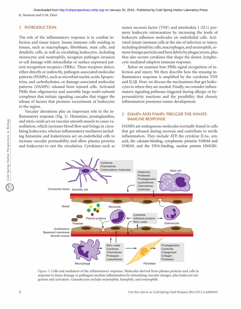
The role of the inflammatory response is to combat in-fection and tissue injury. Innate immune cells residing intissues, such as macrophages, fibroblasts, mast cells, anddendritic cells, as well as circulating leukocytes, includingmonocytes and neutrophils, recognize pathogen invasionor cell damage with intracellular or surface-expressed pat-tern recognition receptors (PRRs). These receptors detect,either directly or indirectly, pathogen-associated molecularpatterns (PAMPs), such as microbial nucleic acids, lipopro-teins, and carbohydrates, or damage-associated molecularpatterns (DAMPs) released from injured cells. ActivatedPRRs then oligomerize and assemble large multi-subunitcomplexes that initiate signaling cascades that trigger therelease of factors that promote recruitment of leukocytesto the region.
Vascular alterations play an important role in the in-flammatory response (Fig. 1). Histamine, prostaglandins,and nitric oxide act on vascular smooth muscle to cause va-sodilation, which increases blood flow and brings in circu-lating leukocytes, whereas inflammatory mediators includ-ing histamine and leukotrienes act on endothelial cells toincrease vascular permeability and allow plasma proteinsand leukocytes to exit the circulation. Cytokines such as
tumor necrosis factor (TNF) and interleukin 1 (IL1) pro-mote leukocyte extravasation by increasing the levels ofleukocyte adhesion molecules on endothelial cells. Acti-vated innate immune cells at the site of infection or injury,including dendritic cells, macrophages, and neutrophils, re-move foreign particles and host debris by phagocytosis, plusthey also secrete cytokines that shape the slower, lympho-cyte-mediated adaptive immune response.
Below we examine how PRRs signal recognition of in-fection and injury. We then describe how the ensuing in-flammatory response is amplified by the cytokines TNFand IL1b. Next, we discuss the mechanisms that get leuko-cytes to where they are needed. Finally, we consider inflam-matory signaling pathways triggered during allergic or hy-persensitivity reactions and the possibility that chronicinflammation promotes tumor development.
2 DAMPs AND PAMPs TRIGGER THE INNATEIMMUNE RESPONSE
DAMPs are endogenous molecules normally found in cellsthat get released during necrosis and contribute to sterileinflammation. They include ATP, the cytokine IL1a, uricacid, the calcium-binding, cytoplasmic proteins S100A8 andS100A9, and the DNA-binding, nuclear protein HMGB1.
EndotheliumBasement membrane
Smooth muscle
Mast cell
Fibroblast
Granulocyte
MonocyteVessel
Plasma proteins
Connective tissue
Macrophage
Dendritic cell
HistamineProteasesProstaglandinsLeukotrienesChemokinesCytokines
CytokinesAdhesive proteinsNitric oxide
Platelets
ProstaglandinsCytokinesCollagenaseCollagenProteases
Nitric oxideCytokinesChemokinesProteasesLeukotrienes
CytokinesChemokinesCostimulatory molecules
Figure 1. Cells and mediators of the inflammatory response. Molecules derived from plasma proteins and cells inresponse to tissue damage or pathogens mediate inflammation by stimulating vascular changes, plus leukocyte mi-gration and activation. Granulocytes include neutrophils, basophils, and eosinophils.
K. Newton and V.M. Dixit
2 Cite this article as Cold Spring Harb Perspect Biol 2012;4:a006049
on January 25, 2019 - Published by Cold Spring Harbor Laboratory Press http://cshperspectives.cshlp.org/Downloaded from
Amyloid b fibrils associated with Alzheimer’s disease havealso been shown to be pro-inflammatory. PAMPs, in con-trast, are pathogen-derived, often essential for microbesurvival, and, like DAMPs, structurally diverse. PAMPsinclude bacterial and viral nucleic acids, fungal b-glucanand a-mannan cell wall components, the bacterial proteinflagellin, components of the peptidoglycan bacterial cellwall, and lipopolysaccharide (LPS) from Gram-negativebacteria.
3 TOLL-LIKE RECEPTORS (TLRs)
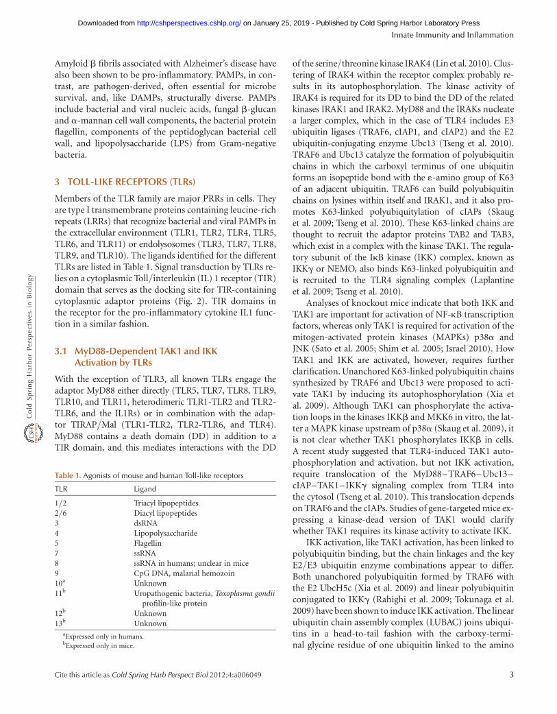
Members of the TLR family are major PRRs in cells. Theyare type I transmembrane proteins containing leucine-richrepeats (LRRs) that recognize bacterial and viral PAMPs inthe extracellular environment (TLR1, TLR2, TLR4, TLR5,TLR6, and TLR11) or endolysosomes (TLR3, TLR7, TLR8,TLR9, and TLR10). The ligands identified for the differentTLRs are listed in Table 1. Signal transduction by TLRs re-lies on a cytoplasmic Toll/interleukin (IL) 1 receptor (TIR)domain that serves as the docking site for TIR-containingcytoplasmic adaptor proteins (Fig. 2). TIR domains inthe receptor for the pro-inflammatory cytokine IL1 func-tion in a similar fashion.
3.1 MyD88-Dependent TAK1 and IKKActivation by TLRs
With the exception of TLR3, all known TLRs engage theadaptor MyD88 either directly (TLR5, TLR7, TLR8, TLR9,TLR10, and TLR11, heterodimeric TLR1-TLR2 and TLR2-TLR6, and the IL1Rs) or in combination with the adap-tor TIRAP/Mal (TLR1-TLR2, TLR2-TLR6, and TLR4).MyD88 contains a death domain (DD) in addition to aTIR domain, and this mediates interactions with the DD
of the serine/threonine kinase IRAK4 (Lin et al. 2010). Clus-tering of IRAK4 within the receptor complex probably re-sults in its autophosphorylation. The kinase activity ofIRAK4 is required for its DD to bind the DD of the relatedkinases IRAK1 and IRAK2. MyD88 and the IRAKs nucleatea larger complex, which in the case of TLR4 includes E3ubiquitin ligases (TRAF6, cIAP1, and cIAP2) and the E2ubiquitin-conjugating enzyme Ubc13 (Tseng et al. 2010).TRAF6 and Ubc13 catalyze the formation of polyubiquitinchains in which the carboxyl terminus of one ubiquitinforms an isopeptide bond with the 1-amino group of K63of an adjacent ubiquitin. TRAF6 can build polyubiquitinchains on lysines within itself and IRAK1, and it also pro-motes K63-linked polyubiquitylation of cIAPs (Skauget al. 2009; Tseng et al. 2010). These K63-linked chains arethought to recruit the adaptor proteins TAB2 and TAB3,which exist in a complex with the kinase TAK1. The regula-tory subunit of the IkB kinase (IKK) complex, known asIKKg or NEMO, also binds K63-linked polyubiquitin andis recruited to the TLR4 signaling complex (Laplantineet al. 2009; Tseng et al. 2010).
Analyses of knockout mice indicate that both IKK andTAK1 are important for activation of NF-kB transcriptionfactors, whereas only TAK1 is required for activation of themitogen-activated protein kinases (MAPKs) p38a andJNK (Sato et al. 2005; Shim et al. 2005; Israel 2010). HowTAK1 and IKK are activated, however, requires furtherclarification. Unanchored K63-linked polyubiquitin chainssynthesized by TRAF6 and Ubc13 were proposed to acti-vate TAK1 by inducing its autophosphorylation (Xia etal. 2009). Although TAK1 can phosphorylate the activa-tion loops in the kinases IKKb and MKK6 in vitro, the lat-ter a MAPK kinase upstream of p38a (Skaug et al. 2009), itis not clear whether TAK1 phosphorylates IKKb in cells.A recent study suggested that TLR4-induced TAK1 auto-phosphorylation and activation, but not IKK activation,require translocation of the MyD88–TRAF6–Ubc13–cIAP–TAK1–IKKg signaling complex from TLR4 intothe cytosol (Tseng et al. 2010). This translocation dependson TRAF6 and the cIAPs. Studies of gene-targeted mice ex-pressing a kinase-dead version of TAK1 would clarifywhether TAK1 requires its kinase activity to activate IKK.
IKK activation, like TAK1 activation, has been linked topolyubiquitin binding, but the chain linkages and the keyE2/E3 ubiquitin enzyme combinations appear to differ.Both unanchored polyubiquitin formed by TRAF6 withthe E2 UbcH5c (Xia et al. 2009) and linear polyubiquitinconjugated to IKKg (Rahighi et al. 2009; Tokunaga et al.2009) have been shown to induce IKK activation. The linearubiquitin chain assembly complex (LUBAC) joins ubiqui-tins in a head-to-tail fashion with the carboxy-termi-nal glycine residue of one ubiquitin linked to the amino
Table 1. Agonists of mouse and human Toll-like receptors
TLR Ligand
1/2 Triacyl lipopeptides2/6 Diacyl lipopeptides3 dsRNA4 Lipopolysaccharide5 Flagellin7 ssRNA8 ssRNA in humans; unclear in mice9 CpG DNA, malarial hemozoin10a Unknown11b Uropathogenic bacteria, Toxoplasma gondii
profilin-like protein12b Unknown13b Unknown
aExpressed only in humans.bExpressed only in mice.
Innate Immunity and Inflammation
Cite this article as Cold Spring Harb Perspect Biol 2012;4:a006049 3
on January 25, 2019 - Published by Cold Spring Harbor Laboratory Press http://cshperspectives.cshlp.org/Downloaded from

Tpl2
TLR4
LPS
M
MD2
Mal
TIR
AP
Ubc13Uev1a
PIP2
TAB2/3
IKKα/β
NF-κBp105
MyD
88
IRAK4
IRAK1/2TRAF6
cIAP1/2
TAK1IKKγ
UbcH5cRelAp65
MEK1
ERK1/2
Cytoplasm
Transcription
Nucleus
MSK1/2
SCFβ-TrCP
CREBATF
Proteasome
TAB2/3
MyD
88
IRAK4
IRAK1/2TRAF6
cIAP1/2
TAK1
IKKγMKK3/6
MKK4
JNK1/2
p38αRelAp65
NF-κB1p50
RelAp65
NF-κB1p50
Ubc13Uev1a
IκBα
JNK1/2p38αERK1/2
AP1
Endolysosome
c/EBPβ
TR
AM
TIC
AM
2
TR
AM
TIC
AM
2
TR
IFT
ICA
M1
RIP1Peli1
TRADDTRAF3
TBK1
IRF3
IRF3 IRF3
IKKα/βIKKγ
RelAp65
SCFβ-TrCP
Proteasome
IκBα
B
A
Transmembranedomain
H2N CO2HTIR
Leucine-rich repeats
Carboxy-terminal leucine-rich repeat
C OutputLeukocyte recruitmentCell adhesionCell survivalRemodeling of extracellular matrixVascular effectsSynthesis of inflammatory mediatorsInflammatory cytokinesAntiviral responseIntracellular signaling (positive)Intracellular signaling (negative)PRRsRegulators of adaptive immune response
GenesCcl2, Ccl3, Ccl4, Ccl5, Cxcl1, Cxcl2, Cxcl5, Cxcl10, Ccrl2Icam1, Vcam1Bcl2a1, CflarMmp13Edn1Hdc, Nos2, Ptges, Ptgs2Il1a, Il1b, Il6, Il18, TnfIfnbBirc2, Birc3, Casp4, Mefv, NfkbizBcl3, Dusp1, Nfkbia, Socs3, Tnfaip3, Zc3h12aFpr1, Nlrp3Ch25h, Icosl, Il10, Il12a, Il12b, Il15, Tnfsf9
Figure 2. Signaling by TLR4. (A) Domain structure of human TLR4. (B) Binding of LPS to TLR4 and the coreceptorMD2 triggers interactions between the cytoplasmic TIR domain of TLR4 and TIR-containing adaptor proteins(Mal, MyD88, and TRAM). MyD88 binds IRAK4, which requires its kinase activity to bind the kinases IRAK1and IRAK2 sequentially. The MyD88–IRAK complex also engages the ubiquitin ligase TRAF6 to make polyubiqui-tin chains that activate the IKK complex for NF-kB- and ERK-dependent gene transcription. Ubiquitin ligasescIAP1 and cIAP2 recruited to the TLR4 signaling complex regulate translocation of a subset of signaling componentsto the cytoplasm, where TAK1 activation initiates a MAPK cascade that stimulates gene expression. TLR4 activated atthe plasma membrane is endocytosed but can signal within the endosomal compartment via the adaptors TRAMand TRIF. The kinase and ubiquitin ligase combination of RIP1 and Peli1 interacts with TRIF to signal NF-kB acti-vation, whereas TBK1 and TRAF3 stimulate IRF3-dependent transcription. (C) Functional outputs of some of thegenes upregulated by TLR4 signaling.
K. Newton and V.M. Dixit
4 Cite this article as Cold Spring Harb Perspect Biol 2012;4:a006049
on January 25, 2019 - Published by Cold Spring Harbor Laboratory Press http://cshperspectives.cshlp.org/Downloaded from

terminus of another ubiquitin. E2 enzymes that supportLUBAC activity in vitro include UbcH5c and UbcH7 (Ger-lach et al. 2011; Ikeda et al. 2011; Tokunaga et al. 2011).Binding of the carboxy-terminal UBAN domain in IKKgto linear ubiquitin chains may produce conformationalchanges necessary for IKK activation. The importance ofIKKg binding to polyubiquitin is supported by the identi-fication of mutations in the UBAN domain that impairNF-kB activation and cause immunodeficiency in humans(Table 2) (Doffinger et al. 2001). These studies may explainwhy NF-kB activation is largely normal in the absence ofUbc13 while MAPK activation is compromised (Yamamo-to et al. 2006).
The MAPKs JNK and p38a, like TAK1, are activateddownstream from TLR2 and TLR4 in a cIAP-dependentmanner. The TAK1-containing signaling complex translo-cates into the cytosol and recruits the kinase MKK4 tophosphorylate and activate JNK (Tseng et al. 2010). It prob-ably also recruits MKK3 and MKK6 to activate p38a be-cause both kinases associate with TRAF6 in response toLPS (Wan et al. 2009). p38a is required for activation ofthe transcription factors CREB and c/EBPb, and it con-tributes to the induction of several genes, including thoseencoding chemokines (Cxcl1, Cxcl2), cytokines (IL10,IL12b, IL1a, and IL1b), and regulators of extracellular ma-trix remodeling (Mmp13) and cell adhesion (Vcam1)(Kang et al. 2008; Kim et al. 2008). JNK regulates the activityof the AP1 transcription factor and stimulates expression ofpro-inflammatory mediators such as TNF (Das et al. 2009).
Activation of the IKK complex is required for NF-kB-dependent transcription as well as transcriptional re-sponses downstream from the MAPK ERK. IKKb sub-strates include p105, the precursor of the p50 NF-kB1transcription factor, as well as the IkB proteins that seques-ter NF-kB transcription factors in the cytosol. Phosphory-lation by IKKb targets these substrates for K48-linkedpolyubiquitylation by the E3 ubiquitin ligase SCFb-TrCP
and subsequent proteasomal degradation (Kanarek et al.2010). Degradation of p105, which exists in a complex
with the kinase Tpl2, activates a Tpl2–MEK1–ERK kinasecascade that leads to the induction of genes such as Ptgs2by the CREB/ATF family of transcription factors (Banerjeeand Gerondakis 2007). The cyclooxygenase 2 (COX2)enzyme encoded by Ptgs2 is involved in the synthesis ofprostaglandins, which are important mediators of pain,inflammation, and fever. IkB degradation allows dimericNF-kB transcription factors composed largely of RelA(p65) and NF-kB1 (p50) subunits to accumulate in the nu-cleus and drive expression of a large number of pro-inflam-matory genes (Table 3 lists a subset of these genes).
NF-kB also induces genes that limit the duration andmagnitude of the inflammatory response, such as Tnfaip3and Nfkbia (the latter encodes IkBa and thus forms anegative-feedback loop). These genes prevent the inflam-matory response from causing more tissue damage thanthe initial injury. For example, mice lacking the A20 de-ubiquitylating enzyme encoded by Tnfaip3 die from un-checked inflammation. A20 is thought to switch off TLRsignaling by countering ubiquitylation by TRAF6 or cIAPs(Newton et al. 2008; Skaug et al. 2009; Shembade et al.2010). Somatic mutation of TNFAIP3 occurs frequentlyin some human B-cell lymphomas, which suggests thatA20 may function as a tumor suppressor, and polymor-phisms within the TNFAIP3 locus have been associatedwith autoimmune disorders, including systemic lupus er-ythematosus, rheumatoid arthritis, Crohn’s inflamma-tory bowel disease, and psoriasis (Table 2) (Vereecke et al,2011). The tumor suppressor gene CYLD, which is mutatedin familial cylindromatosis, also encodes a deubiquitylat-ing enzyme that limits NF-kB signaling. CYLD cleavesboth linear and K63-linked polyubiquitin efficiently in vi-tro (Komander et al. 2009), but mice lacking CYLD do notdevelop the multi-organ inflammation seen in A20-defi-cient mice, perhaps because CYLD normally is phosphory-lated and inactivated by IKK (Reiley et al. 2005). AnotherNF-kB target gene that suppresses TLR signaling isCd200, which encodes the membrane glycoprotein ligandfor CD200R1 (Mukhopadhyay et al. 2010). TAM (Tyro3,Axl, and Mer) receptor tyrosine kinases are further exam-ples of receptor systems that negatively regulate TLR signal-ing (Rothlin et al. 2007).
3.2 MyD88-Dependent Type I Interferon (IFN)Induction by TLRs 7 and 9
The MyD88–IRAK4–IRAK1–TRAF6 signaling complexrequired for pro-inflammatory cytokine production byTLR7 and TLR9 also stimulates synthesis of interferon a
(IFNa) and IFNb in plasmacytoid dendritic cells (pDCs).The induction of type I IFNs and Ifn-related genes is criticalto the anti-viral response and also requires TRAF3, IKKa,
Table 2. Genes regulating inflammation that are mutated inhuman disease
Gene Protein Disease
CIAS1 NLRP3 Familial cold autoinflammatory syndrome;Muckle–Wells syndrome; Neonatal-onsetmultisystem inflammatory disease
IKBKG IKKg Anhidrotic ectodermal dysplasia withimmunodeficiency
NOD2 NOD2 Crohn’s inflammatory bowel disease; Blausyndrome characterized by arthritis anduveitis
TNFAIP3 A20 B-cell lymphomas
Innate Immunity and Inflammation
Cite this article as Cold Spring Harb Perspect Biol 2012;4:a006049 5
on January 25, 2019 - Published by Cold Spring Harbor Laboratory Press http://cshperspectives.cshlp.org/Downloaded from
and the transcription factor IRF7. Phosphorylation of IRF7by IKKa promotes its dimerization and translocation intothe nucleus, where it upregulates expression of type I Ifngenes. Differences have been noted in myeloid DCs1; IRF1is more important than IRF7, and there does not seem tobe a requirement for TRAF3 and IRAK1 (Hoshino et al.2010; Takeuchi and Akira 2010).
3.3 TRIF-Dependent Signaling by TLRs 3 and 4
TLR4 is endocytosed following ligand binding and, likeTLR3 stimulated with dsRNA, transduces signals fromwithin the endosomal compartment. Both receptors re-cruit the TIR-containing cytoplasmic adaptor TRIF (alsoknown as TICAM1), which in the case of endocytosedTLR4 occurs via the bridging adaptor TRAM (also knownas TICAM2). TRIF can activate NF-kB using its RIP homo-typic interaction motif (RHIM) to recruit the RHIM-con-taining kinase RIP1 (Cusson-Hermance et al. 2005), whichis, in turn, bound and ubiquitylated by the E3 ubiquitin li-gase Peli1 (Chang et al. 2009). K63-linked polyubiquityla-tion of RIP1 by Peli1 has not been shown, but could be amechanism for the recruitment of IKKg and TAK1. Inter-action of the DD in RIP1 with the DD in the cytoplasmicadaptor TRADD is important for TRIF-dependent NF-kBand MAPK activation in some cell types (Chen et al. 2008;Ermolaeva et al. 2008; Pobezinskaya et al. 2008). The TRIF-containing complex also induces type I IFN, and this is de-pendent on TRAF3 plus the kinase TBK1, the latter phos-phorylating and activating the transcription factor IRF3(Takeuchi and Akira 2010). TRAF3 can modify itself withK63-linked polyubiquitin, and this may be important forits interaction with TBK1 and subsequent IRF3 activation.
4 RIG-I-LIKE RECEPTORS (RLRs)
The RLR family of PRRs, which comprises RIG-I, MDA5,and LGP2, signals the production of pro-inflammatory cy-tokines and type I IFN in response to viral and bacterialnucleic acids in the cytoplasm (Fig. 3). These cytosolic pro-teins have a central DExD/H-box helicase domain anda carboxy-terminal regulatory domain (CTD), the latterbinding RNA. RIG-I and MDA5 also have two amino-termi-nal caspase activation and recruitment domains (CARDs),which function as protein–protein interaction motifs.RIG-I recognizes double-stranded RNAs (dsRNAs) thathave a 5′ triphosphate and are either viral in origin or
Table 3. Genes induced by the canonical IKKb/NF-kBsignaling pathway
Genea Protein Function
Ager RAGE PRR belonging to the immunoglobulinsuperfamily that recognizes multipleligands including HMGB1
Birc3 cIAP2 Ubiquitin ligase that regulates NF-kBactivation
Casp4 Caspase-11 Aspartate-specific cysteine proteaseimplicated in inflammation
Ccl2 MCP1 Chemokine for monocyte recruitmentCcl3 MIP1a Chemokine for leukocyte recruitmentCcl5 RANTES Chemokine for monocyte and T-cell
recruitmentCd200 CD200 Binds CD200R1 and inhibits
macrophage activationCfb Complement
factor BSerine protease in the alternative
complement activation pathwayCflar c-FLIP Inhibitor of death receptor-induced
apoptosisCsf2 GM-CSF Growth factor that promotes
differentiation and activationof DCs, macrophages, andneutrophils
Cxcl1 KC Chemokine for neutrophil recruitmentCxcl2 MIP2 Chemokine for neutrophil recruitmentF3 Tissue factor Coagulation factorIcam1 ICAM1 Cell adhesion molecule that interacts
with b2 integrinsIfnb1 IFNb Suppressor of virus replicationIl1b IL1b Cytokine that amplifies the
inflammatory responseIl6 IL6 Pleiotropic cytokine that stimulates
fever, production of hepatocyteacute phase proteins, andlymphocyte differentiation
Il12b IL12 p40 Component of heterodimeric IL12and IL23, which modulate NKcell and lymphocyte effectorfunctions
Mmp9 MMP9 Metalloproteinase that degradesextracellular matrix
Nfkbia IkBa Inhibitor of NF-kB signalingNfkbib IkBb NF-kB transcriptional coactivatorNos2 iNOS Enzyme that makes anti-microbial
nitric oxideSele E-selectin Cell adhesion moleculeSelp P-selectin Cell adhesion moleculeSod2 MnSOD Enzyme that converts superoxide to
hydrogen peroxideTnf TNF Cytokine that amplifies the
inflammatory responseTnfaip3 A20 Inhibitor of NF-kB signaling by TLRs
and TNF-R1Vcam1 VCAM1 Cell adhesion molecule that
interacts with b1 integrinVLA-4
aMouse gene nomenclature used.
1DCs are very heterogeneous. pDCs acquire DC morphology and secrete largeamounts of IFN during virus infections. They can be distinguished from oth-er DC subsets by their cell surface markers. The myeloid DCs referenced werederived in vitro from bone marrow cells with granulocyte/macrophagecolony-stimulating factor (GM-CSF).
K. Newton and V.M. Dixit
6 Cite this article as Cold Spring Harb Perspect Biol 2012;4:a006049
on January 25, 2019 - Published by Cold Spring Harbor Laboratory Press http://cshperspectives.cshlp.org/Downloaded from
generated by RNA polymerase III from microbial DNAtemplates (Lu et al. 2010; Wang et al. 2010b). MDA5 andLGP2 also bind RNA, but MDA5 and RIG-I have non-redundant functions in sensing certain viruses. LGP2 ap-pears, in most contexts, to act as a positive regulator of sig-naling by MDA5 and RIG-I (Venkataraman et al. 2007;Satoh et al. 2010).
Binding of RIG-I to RNA causes a conformationalchange (Jiang et al. 2011) that enables its CARDs to bindK63-linked polyubiquitin generated by the E3 ubiquitinligase TRIM25. In an ill-defined manner, this interaction
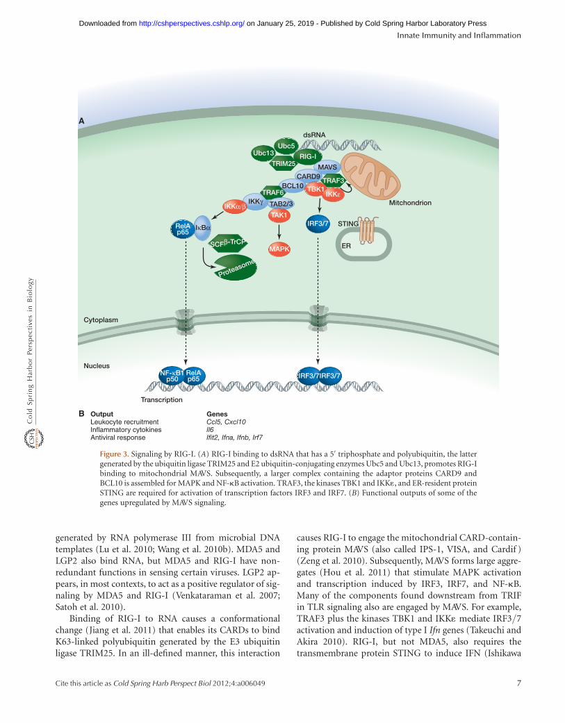
causes RIG-I to engage the mitochondrial CARD-contain-ing protein MAVS (also called IPS-1, VISA, and Cardif )(Zeng et al. 2010). Subsequently, MAVS forms large aggre-gates (Hou et al. 2011) that stimulate MAPK activationand transcription induced by IRF3, IRF7, and NF-kB.Many of the components found downstream from TRIFin TLR signaling also are engaged by MAVS. For example,TRAF3 plus the kinases TBK1 and IKK1 mediate IRF3/7activation and induction of type I Ifn genes (Takeuchi andAkira 2010). RIG-I, but not MDA5, also requires thetransmembrane protein STING to induce IFN (Ishikawa
IKKγ Mitchondrion
RIG-ITRIM25
dsRNA
MAVS
ER
STING
CARD9BCL10
TRAF6
TAB2/3IKKα/βTAK1
RelAp65
SCFβ-TrCP
Cytoplasm
Transcription
NucleusRelAp65
NF-κB1p50 IRF3/7IRF3/7
Proteasome
IκBα
MAPK
TRAF3TBK1
IRF3/7
IKKε
Ubc5Ubc13
A
B GenesCcl5, Cxcl10Il6Ifit2, Ifna, Ifnb, Irf7
OutputLeukocyte recruitmentInflammatory cytokinesAntiviral response
Figure 3. Signaling by RIG-I. (A) RIG-I binding to dsRNA that has a 5′ triphosphate and polyubiquitin, the lattergenerated by the ubiquitin ligase TRIM25 and E2 ubiquitin-conjugating enzymes Ubc5 and Ubc13, promotes RIG-Ibinding to mitochondrial MAVS. Subsequently, a larger complex containing the adaptor proteins CARD9 andBCL10 is assembled for MAPK and NF-kB activation. TRAF3, the kinases TBK1 and IKK1, and ER-resident proteinSTING are required for activation of transcription factors IRF3 and IRF7. (B) Functional outputs of some of thegenes upregulated by MAVS signaling.
Innate Immunity and Inflammation
Cite this article as Cold Spring Harb Perspect Biol 2012;4:a006049 7
on January 25, 2019 - Published by Cold Spring Harbor Laboratory Press http://cshperspectives.cshlp.org/Downloaded from

and Barber 2008). STING is located in the endoplasmicreticulum (ER), but its precise role in IFN induction bydsRNA requires further study. Experiments with fibro-blasts from gene-targeted mice also implicate TRAF6,IKKg, and the DD-containing proteins TRADD, RIP1,and FADD in IFN induction (Balachandran et al. 2004;Zhao et al. 2007; Michallet et al. 2008; Yoshida et al.2008). Note that loss of TRADD, RIP1, or FADD producesa defect less severe than does MAVS or TBK1 deficiency.The extent of IRF3 phosphorylation and dimerizationhas not been determined in cells lacking TRADD, RIP1,or FADD; it remains possible that these proteins, likeTRAF6, contribute to type I Ifn gene expression by activat-ing NF-kB (Wang et al. 2010a). In DCs, MAVS engages theCARD-containing adaptors CARD9 and BCL10 to activateNF-kB (Poeck et al. 2010). BCL10 engages TRAF6 in lym-phocytes to activate NF-kB (Sun et al. 2004b), and a similarpathway may operate downstream from MAVS. A role forFADD, TRADD, and RIP1 in MAVS signaling by DCs hasnot been examined.
5 NOD-LIKE RECEPTORS (NLRs)
Members of the Nod-like receptor (NLR) family of cytosolicPRRs are best known for their ability to signal NF-kB acti-vation (NOD1 and NOD2) or secretion of the pro-inflam-matory cytokines IL1b and IL18 (NLRP1/NALP1, NLRP3/NALP3/cryopyrin, and NLRC4/Ipaf ) (Fig. 4). These pro-teins typically contain a CARD or pyrin domain at the ami-no terminus, a central nucleotide-binding oligomerizationNACHT domain, and carboxy-terminal LRRs. NOD2 andNLRP3 have received considerable attention because theirmutation is linked to inflammatory disease. NOD2 muta-tions are associated with Crohn’s inflammatory bowel diseaseand Blau syndrome, whereas mutations in the CIAS1 geneencoding NLRP3 are associated with familial cold autoin-flammatory syndrome, Muckle–Wells syndrome, and neo-natal-onset multisystem inflammatory disease (Table 2).
NOD1 and NOD2 are sensors of different bacterialpeptidoglycan components, but they both interact withthe CARD-containing kinase RIP2 to activate MAPK andNF-kB signaling (Park et al. 2007). cIAPs are proposed tobind and ubiquitylate RIP2, and K63-linked polyubiquity-lation of RIP2 recruits TAK1 for IKK and MAPK activation(Yang et al. 2007; Hitotsumatsu et al. 2008; Bertrand et al.2009). NOD1 and NOD2 also have been shown to stimulateautophagy2 independently of RIP2 (Travassos et al. 2010).
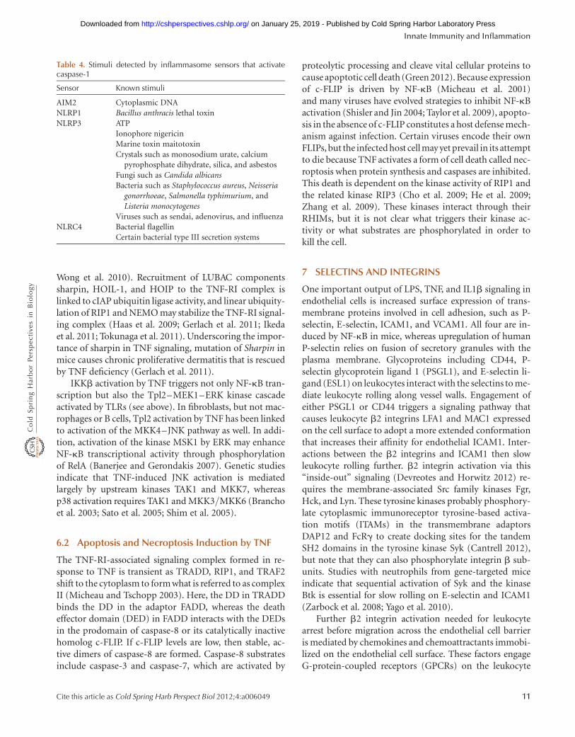
Distinct PAMPS and DAMPs trigger NLRP1, NLRP3,and NLRC4 to nucleate signaling complexes termed “in-flammasomes” (Table 4). The CARD- and PYRIN-do-main-containing adaptor ASC is a critical inflammasomecomponent, binding the CARD in the zymogen form ofthe aspartate-specific cysteine protease caspase-1 (Maria-thasan et al. 2004). The proximity of caspase-1 zymogenswithin the inflammasome complex is believed to facilitatetheir autocatalytic activation. Caspase-1 substrates includepro-IL18 and pro-IL1b, the latter being upregulated tran-scriptionally by MyD88-dependent TLR signaling. Inflam-masome activation also results in an extremely rapid formof cell death termed “pyroptosis.” The suicide of infectedmacrophages by pyroptosis is important for bacterial clear-ance (Miao et al. 2010), but the critical substrates of cas-pase-1 in this process still have to be determined.
Intriguingly, immunofluorescence microscopy of en-dogenous inflammasome components in mouse macro-phages infected with Salmonella typhimurium suggeststhat inflammasome assembly occurs at a single focus with-in a cell (Broz et al. 2010). One question that continues tovex the field is how NLRP1, NLRP3, and NLRC4 sensePAMPS and DAMPs, because direct binding has not beenshown. The diversity of entities that trigger NLRP3-de-pendent caspase-1 activation suggests that NLRP3 mightrespond to a particular stress-activated signaling pathway.Both potassium efflux and the generation of reactive oxy-gen species (ROS) have been proposed as critical events up-stream of NLRP3 activation, but the precise nature of NLRactivation remains obscure.
ASC-dependent caspase-1 activation is also triggered inresponse to cytoplasmic DNA that appears during an infec-tion or after tissue injury. The responsible PRR is not anNLR but the IFN-induced protein AIM2, which has aHIN200 domain for DNA binding and a pyrin domainto engage ASC. Cytoplasmic DNA also triggers type IIFN production, but this requires neither AIM2 norTLRs. STING is a critical signaling component in this path-way, but whether any one DNA receptor is essential for itsactivation is unclear (Hornung and Latz 2010).
6 THE PRO-INFLAMMATORY CYTOKINE TUMORNECROSIS FACTOR (TNF)
Induction of the cytokines IL1b and TNF by PRRs serves toamplify the inflammatory response because they too pro-mote NF-kB and MAPK activation. Binding of IL1b toIL1R triggers MyD88-dependent signaling (Muzio et al.1997), whereas TNF mediates most of its pro-inflamma-tory effects by binding to TNF receptor I (TNF-RI) (Pes-chon et al. 1998).
2Autophagy is the process by which cytoplasmic components, including or-ganelles and invading bacteria, are sequestered inside double-membranevesicles and then delivered to the lysosome for degradation.
K. Newton and V.M. Dixit
8 Cite this article as Cold Spring Harb Perspect Biol 2012;4:a006049
on January 25, 2019 - Published by Cold Spring Harbor Laboratory Press http://cshperspectives.cshlp.org/Downloaded from

6.1 NF-kB and MAPK Activation by TNF
TNF-RI (also called TNFRSF1A) is a type I transmembraneprotein that has cysteine-rich extracellular domains (CRDs)for TNF binding. Its cytoplasmic tail contains a DD thatrecruits the DD-containing adaptor TRADD and kinaseRIP1 (Fig. 5). TRADD facilitates binding of RIP1 to TNF-R1 and recruits TRAF2, which is an adaptor for the ubiq-uitin ligases cIAP1 and cIAP2 (Chen et al. 2008; Ermolaevaet al. 2008; Pobezinskaya et al. 2008). Analyses of cells
lacking cIAPs, RIP1, or TRAF2 indicate that all three con-tribute to NF-kB and MAPK activation, but the details ofhow they do so continue to be unraveled. Ubiquitylationof RIP1 by the cIAPs and E2 UbcH5 is believed to be im-portant for recruitment of NEMO and TAK1, and subse-quently for IKK activation (Varfolomeev et al. 2008; Xuet al. 2009), although TRAF2 ubiquitin ligase activity hasbeen invoked recently as well (Alvarez et al. 2010). In addi-tion, in some cell types, ubiquitylation of TRAF2 or cIAPsrather than RIP1 may support IKK activation (Li et al. 2009;
Cytoplasm
Nucleus
NLRP1NLRP3NLRC4
AS
C
Caspase-1
AIM2
AS
C
Caspase-1
DNA
PAMPs/DAMPs
?
ProIL-1β
ProIL-18
IL18
IL1β
NO
D1/
2
IKKγ TAB2/3IKKα/βTAK1
MAPK
RIP2
RelAp65
SCFβ-TrCP
Transcription
RelAp65
NF-κB1p50
Proteasome
IκBα
cIAP1/2
Bacterial peptidoglycan
ATG16L1Autophagy
?
Pyroptosis
A
B OutputInflammatory cytokines
GenesIl6, Tnf
Figure 4. Signaling by NLRs. (A) NLRP1, NLRP3, and NLRC4 respond to diverse PAMPS and DAMPs by engagingthe caspase-1 adaptor protein ASC, whereas AIM2 binds ASC in response to cytoplasmic dsDNA. Activation ofcaspase-1 within each inflammasome complex results in processing of pro-IL1b and pro-IL18 and secretion of theirbiologically active forms. Caspase-1 activation also triggers a rapid form of cell death termed pyroptosis. NOD1 andNOD2 sense different components of bacterial peptidoglycan and stimulate either the autophagy machinery or genetranscription via NF-kB and MAPK activation. The latter outcome requires interaction of NOD1 or NOD2 with thekinase RIP2, which may be ubiquitylated by cIAPs in order to recruit TAB2/3 and IKKg for TAK1 and IKK activa-tion. (B) Functional outputs of some of the genes upregulated by NOD1 or NOD2 signaling.
Innate Immunity and Inflammation
Cite this article as Cold Spring Harb Perspect Biol 2012;4:a006049 9
on January 25, 2019 - Published by Cold Spring Harbor Laboratory Press http://cshperspectives.cshlp.org/Downloaded from

Tpl2
TRAF2
NF-κBp105
LUBACcIAP1/2
RelAp65
MEK1
ERK1/2
Cytoplasm
Transcription
Nucleus
MSK1/2
SCFβ-TrCP
CREBATF
Proteasome
TAB2/3
TAK1
MKK3/6 MKK4/7
JNK1/2p38αRelAp65
NF-κB1p50
RelAp65
NF-κB1p50
IκBα
JNK1/2p38αERK1/2
AP1c/EBPβ
TRADD
TNF
TNF-R1
RIP1IKKγIKKα/β TRAF2
TRADD
RIP1RIP3
Caspase-3
Caspase-7
Apoptosis
FLIPFADD
Caspase-8
Necroptosis
?
A
B OutputLeukocyte recruitmentLeukocyte activationSurface receptorsInflammatory cytokinesIntracellular signaling (negative)Intracellular signaling (positive)Transcription factorsRemodeling of extracellular matrixVascular effectsPRRsCell adhesionSynthesis of inflammatory mediators
GenesCcl2, Ccl5, Ccl20, Cxcl1, Cxcl2, Cxcl3, Cxcl5, Cxcl10, Cx3cl1Csf1, Csf2Fas, Il18r1, Jag1Il1b, Il6, Il15, Lif, TnfBcl3, Nfkbia, Socs3, Tnfaip3, Traf1Ifi47Fos, Jun, JunBMmp3, Mmp9, Mmp14Edn1, VegfcNod2Icam1, Sele, Vcam1Ptgs2
Figure 5. Signaling by TNF-R1. (A) Binding of TNF to TNF-R1 causes the cytoplasmic death domain (DD) inTNF-R1 to bind the DD-containing proteins TRADD and RIP1. TRADD also binds TRAF2, which serves as anadaptor for the ubiquitin ligases cIAP1 and cIAP2. Ubiquitylation of RIP1, and potentially other components ofthe complex, recruits IKKg and TAK1 for NF-kB and MAPK activation. Recruitment of LUBAC for linear ubiqui-tylation of IKKg may stabilize the signaling complex. Translocation of TRADD, TRAF2, and RIP1 to the cytoplasmnucleates a second complex that contains the adaptor protein FADD and caspase-8. If c-FLIP levels are low, activa-tion of caspase-8 and its substrates caspase-3 and caspase-7 causes apoptotic cell death. Inhibition of protein syn-thesis and caspases, as might occur in a virus-infected cell, promotes necroptotic cell death that is dependent on thekinase activities of RIP1 and RIP3. (B) Functional outputs of some of the genes upregulated by TNF signaling.
K. Newton and V.M. Dixit
10 Cite this article as Cold Spring Harb Perspect Biol 2012;4:a006049
on January 25, 2019 - Published by Cold Spring Harbor Laboratory Press http://cshperspectives.cshlp.org/Downloaded from
Wong et al. 2010). Recruitment of LUBAC componentssharpin, HOIL-1, and HOIP to the TNF-RI complex islinked to cIAP ubiquitin ligase activity, and linear ubiquity-lation of RIP1 and NEMO may stabilize the TNF-RI signal-ing complex (Haas et al. 2009; Gerlach et al. 2011; Ikedaet al. 2011; Tokunaga et al. 2011). Underscoring the impor-tance of sharpin in TNF signaling, mutation of Sharpin inmice causes chronic proliferative dermatitis that is rescuedby TNF deficiency (Gerlach et al. 2011).
IKKb activation by TNF triggers not only NF-kB tran-scription but also the Tpl2–MEK1–ERK kinase cascadeactivated by TLRs (see above). In fibroblasts, but not mac-rophages or B cells, Tpl2 activation by TNF has been linkedto activation of the MKK4–JNK pathway as well. In addi-tion, activation of the kinase MSK1 by ERK may enhanceNF-kB transcriptional activity through phosphorylationof RelA (Banerjee and Gerondakis 2007). Genetic studiesindicate that TNF-induced JNK activation is mediatedlargely by upstream kinases TAK1 and MKK7, whereasp38 activation requires TAK1 and MKK3/MKK6 (Branchoet al. 2003; Sato et al. 2005; Shim et al. 2005).
6.2 Apoptosis and Necroptosis Induction by TNF
The TNF-RI-associated signaling complex formed in re-sponse to TNF is transient as TRADD, RIP1, and TRAF2shift to the cytoplasm to form what is referred to as complexII (Micheau and Tschopp 2003). Here, the DD in TRADDbinds the DD in the adaptor FADD, whereas the deatheffector domain (DED) in FADD interacts with the DEDsin the prodomain of caspase-8 or its catalytically inactivehomolog c-FLIP. If c-FLIP levels are low, then stable, ac-tive dimers of caspase-8 are formed. Caspase-8 substratesinclude caspase-3 and caspase-7, which are activated by
proteolytic processing and cleave vital cellular proteins tocause apoptotic cell death (Green 2012). Because expressionof c-FLIP is driven by NF-kB (Micheau et al. 2001)and many viruses have evolved strategies to inhibit NF-kBactivation (Shisler and Jin 2004; Taylor et al. 2009), apopto-sis in the absence of c-FLIP constitutes a host defense mech-anism against infection. Certain viruses encode their ownFLIPs, but the infected host cell may yet prevail in its attemptto die because TNF activates a form of cell death called nec-roptosis when protein synthesis and caspases are inhibited.This death is dependent on the kinase activity of RIP1 andthe related kinase RIP3 (Cho et al. 2009; He et al. 2009;Zhang et al. 2009). These kinases interact through theirRHIMs, but it is not clear what triggers their kinase ac-tivity or what substrates are phosphorylated in order tokill the cell.
7 SELECTINS AND INTEGRINS
One important output of LPS, TNF, and IL1b signaling inendothelial cells is increased surface expression of trans-membrane proteins involved in cell adhesion, such as P-selectin, E-selectin, ICAM1, and VCAM1. All four are in-duced by NF-kB in mice, whereas upregulation of humanP-selectin relies on fusion of secretory granules with theplasma membrane. Glycoproteins including CD44, P-selectin glycoprotein ligand 1 (PSGL1), and E-selectin li-gand (ESL1) on leukocytes interact with the selectins to me-diate leukocyte rolling along vessel walls. Engagement ofeither PSGL1 or CD44 triggers a signaling pathway thatcauses leukocyte b2 integrins LFA1 and MAC1 expressedon the cell surface to adopt a more extended conformationthat increases their affinity for endothelial ICAM1. Inter-actions between the b2 integrins and ICAM1 then slowleukocyte rolling further. b2 integrin activation via this“inside-out” signaling (Devreotes and Horwitz 2012) re-quires the membrane-associated Src family kinases Fgr,Hck, and Lyn. These tyrosine kinases probably phosphory-late cytoplasmic immunoreceptor tyrosine-based activa-tion motifs (ITAMs) in the transmembrane adaptorsDAP12 and FcRg to create docking sites for the tandemSH2 domains in the tyrosine kinase Syk (Cantrell 2012),but note that they can also phosphorylate integrin b sub-units. Studies with neutrophils from gene-targeted miceindicate that sequential activation of Syk and the kinaseBtk is essential for slow rolling on E-selectin and ICAM1(Zarbock et al. 2008; Yago et al. 2010).
Further b2 integrin activation needed for leukocytearrest before migration across the endothelial cell barrieris mediated by chemokines and chemoattractants immobi-lized on the endothelial cell surface. These factors engageG-protein-coupled receptors (GPCRs) on the leukocyte
Table 4. Stimuli detected by inflammasome sensors that activatecaspase-1
Sensor Known stimuli
AIM2 Cytoplasmic DNANLRP1 Bacillus anthracis lethal toxinNLRP3 ATP
Ionophore nigericinMarine toxin maitotoxinCrystals such as monosodium urate, calcium
pyrophosphate dihydrate, silica, and asbestosFungi such as Candida albicansBacteria such as Staphylococcus aureus, Neisseria
gonorrhoeae, Salmonella typhimurium, andListeria monocytogenes
Viruses such as sendai, adenovirus, and influenzaNLRC4 Bacterial flagellin
Certain bacterial type III secretion systems
Innate Immunity and Inflammation
Cite this article as Cold Spring Harb Perspect Biol 2012;4:a006049 11
on January 25, 2019 - Published by Cold Spring Harbor Laboratory Press http://cshperspectives.cshlp.org/Downloaded from

surface (see below). Note that integrin engagement alsoelicits “outside-in” signaling in leukocytes, and, similarlyto Fc receptor signaling (see below), this activates leukocyteeffector functions (Lowell 2011). Clustering of ICAM1 onendothelial cells also triggers signals that facilitate leukocytemigration across the endothelium into the surrounding tis-sue. Phosphorylation of VE-cadherin appears to loosen ad-herens junctions, whereas activation of myosin light chainkinase (MLCK) mediates endothelial cell contraction (Mul-ler 2011).
8 G-PROTEIN-COUPLED RECEPTORS (GPCRs)
Lipid-based inflammatory mediators such as prostaglan-dins, leukotrienes, and platelet-activating factor; vasoactiveamines such as histamine and serotonin; complement frag-ments C3b, C3a, and C5a; chemokines; proteases; and bac-terial or mitochondrial formylated peptides all activatesignaling by GPCRs linked to heterotrimeric G-proteinscomposed of a, b, and g subunits. Following ligand bind-ing, or cleavage in the case of protease-activated receptors(PARs), Ga and Gbg interact with ion channels or enzymessuch as adenylyl cyclase, phospholipase C (PLC), and phos-phoinositide 3-kinase (PI3K). In addition, active GPCRs
are phosphorylated by GPCR kinases (GRKs) to stimulatebinding of arrestins, adaptors that stimulate GPCR endocy-tosis as well as MAPK activation. To highlight some of thepathways engaged by GPCRs during inflammation, belowwe focus on signaling by the chemoattractants C5a andthe prototypical formylated peptide formyl-Met-Leu-Phe(fMLP) (Fig. 6).
8.1 C5a Receptor (C5aR) and Formyl PeptideReceptors (FPRs)
Complement protein C5a is produced by complementplasma proteases activated by IgM- and IgG-containingantibody complexes (the classical pathway), pathogenscoated with host mannose-binding lectin or C-reactiveprotein (the lectin pathway), or pathogens in isolation(the alternative pathway). C5a can also be generated bynon-complement proteases such as thrombin and kallik-rein, which are components of the clotting system activatedin response to endothelial cell injury. C5a and formylpeptides, the latter of bacterial or mitochondrial origin,stimulate leukocyte chemotaxis, degranulation, superoxideproduction for microbe killing, and, as mentioned above,activation of integrins for cell adhesion. Similarly to TNF,
ER
IP3R
GγGβGα
Ligand (e.g., C5a, fMLP)
GPCR
PLCβGγ
Gβ
CRAC
Ca2+
Ca2+
GγGβ
PIP2
IP3
STIM1
DAGPKC
Ras PI3KγPIP3
Prex1DOCK2
Rac
PAK1
Pixα
Cdc42 GIT2
WASP
F-actin polymerization
mDia1Cyfip1
Hem1
Abi ARP2/3Complex
WAVE
NA
DP
H
oxi
das
e
Rac
O2O2
–
Figure 6. Signaling by GPCRs activated by chemoattractants C5a and fMLP. C5a or fMLP binding to their respectiveGPCRs triggers dissociation of Gai-GTP from Gbg, the latter interacting with PLCb, the p101 regulatory subunit ofPI3Kg, and PAK1. Activated PLCb generates the second messengers IP3 and DAG to elevate intracellular calciumand activate PKC, respectively. These outcomes regulate JNK activation, vesicle exocytosis, and superoxide produc-tion by the NADPH oxidase. PIP3 generated by PI3Kg, whose activation also involves the GTPase Ras, stimulatesGEFs (DOCK2 and Prex1) that activate Rac GTPases. PAK1 interacts with the GEF PIXa for activation of anotherRho family GTPase called Cdc42. Rac1, Rac2, and Cdc42 together regulate chemotaxis by coordinating alterations tothe actin cytoskeleton via mDia1 and the ARP2/3 complex. Rac2 is also an essential component of the NADPH ox-idase. The signaling components regulating gene transcription are less defined.
K. Newton and V.M. Dixit
12 Cite this article as Cold Spring Harb Perspect Biol 2012;4:a006049
on January 25, 2019 - Published by Cold Spring Harbor Laboratory Press http://cshperspectives.cshlp.org/Downloaded from
C5a stimulates endothelial cells to increase expression ofcytokines, chemokines, and cell adhesion molecules suchas E-selectin, ICAM1, and VCAM1 (Albrecht et al. 2004).
C5a and fMLP activate predominantly pertussis tox-in-sensitive Gi proteins. The Gbg dimer that is releasedactivates several enzymes, including PLCb (Camps et al.1992). Hydrolysis of phosphatidylinositol 4,5-bisphos-phate in the plasma membrane by PLC yields the secondmessengers inositol 1,4,5-trisphosphate (IP3) and diacyl-glycerol (DAG) (Bootman 2012). Binding of IP3 to its re-ceptor causes depletion of calcium stores within the ERand relocation of the calcium-binding type I transmem-brane protein STIM1 from the ER to structures near theplasma membrane (Brechard et al. 2009). STIM1 then acti-vates the plasma membrane calcium-release-activated cal-cium channel (CRAC) to cause an influx of calcium intothe cell. Elevated intracellular calcium together with pro-tein kinase C (PKC) activation by DAG is important forvesicle exocytosis, superoxide production by the NADPHoxidase, and JNK activation (Li et al. 2000). Calcium stim-ulates lysosome exocytosis by activating the synaptotagminregulator of vesicle fusion SYT7 (Colvin et al. 2010).
GTP-bound Ras, activated by a mechanism that is un-clear, and Gbg dimers activate PI3Kg by binding to itsp101 regulatory subunit and p110g catalytic subunit, re-spectively. Phosphatidylinositol 3,4,5-trisphosphate (PIP3)produced by PI3Kg activates Rac guanine-nucleotide ex-change factors (GEFs), such as Prex1 and DOCK2 (Welchet al. 2002; Kunisaki et al. 2006), and contributes to super-oxide production and chemokinesis3 (Suire et al. 2006; Fer-guson et al. 2007; Nishio et al. 2007). The GTPase RhoG alsohas a role in superoxide production but is dispensable forneutrophil migration (Condliffe et al. 2006). The GTPaseRac2 appears to be essential for the assembly of filamentousactin (F-actin) and the NADPH oxidase, whereas Rac1localizes F-actin to the leading edge of the cell facing the che-moattractant (Sun et al. 2004a). This asymmetrical poly-merization of F-actin drives membrane protusions in thedirection of migration. Active Rac is thought to exert its ef-fect on the actin cytoskeleton by interacting with the adap-tor Cyfip1 (also called Sra1), which, in combination withseveral proteins, stimulates the ARP2/3 actin nucleationcomplex (Devreotes and Horwitz 2012).
The GEF PIXa also is required for F-actin assembly atthe leading edge in C5a-stimulated neutrophils. It is re-cruited to Gbg via the kinase PAK1 and appears to functionby interacting with the GTPase Cdc42 and the GTPase-ac-tivating (GAP) protein GIT2 (Li et al. 2003; Mazaki et al.
2006). Activated Cdc42 interacts with the adaptor WASPto engage the ARP2/3 actin nucleation complex. WASP ap-pears to work in concert with the actin-nucleating proteinmDia1, because neutrophils lacking both WASPand mDia1show a profound defect in chemotaxis (Shi et al. 2009).
9 Fc RECEPTORS
Repeated exposure to a polyvalent foreign substance canelicit an inflammatory response called a hypersensitivityreaction if the host makes antibodies against the substance.Immune complexes containing the antigen and IgG or IgMantibodies activate complement proteases, culminating inthe generation of C3a and C5a, which signal leukocyte re-cruitment and activation (see above); the opsonin C3b,which coats and promotes phagocytosis of bacteria; andthe membrane attack complex for bacterial cell lysis (C5b-9). In addition, complexes containing IgG or IgE antibod-ies engage Fc receptors on leukocytes. Members of theFc receptor family are type I transmembrane proteins(with the exception of human GPI-anchored FcgRIIIB)that produce activating (human FcgRI, FcgRIIA, FcgRIIC,FcgRIIIA, FcgRIIIB, and Fc1RI) or inhibitory (humanFcgRIIB) signals. Mast cells expressing the high-affinityreceptor for IgE, Fc1RI, play a central role in allergic reac-tions. Fc1RI engagement causes intracellular granules tofuse with the plasma membrane such that preformedinflammatory mediators including histamine, serotonin,and proteases are released into the extracellular environ-ment. Activated mast cells also secrete pro-inflammatoryprostaglandins, leukotrienes, and cytokines, but these aresynthesized de novo.
9.1 Fc1RI
Fc1RI is an abg2 heterotetramer. Its a-chain containsextracellular Ig-like domains for binding the heavy-chainconstant region of IgE, whereas the b-chain and a g-chainhomodimer transduce signals via cytoplasmic ITAMs(Fig. 7) (Cantrell 2012). IgE-induced clustering of Fc1RIpromotes activation of Src family kinases Lyn and Fyn.Lyn substrates include both positive and negative regulatorsof mast cell activation, which fine-tune the magnitude andduration of the response. Lyn stimulates activation byphosphorylating the FcRg ITAM, which recruits the SH2domains in Syk. Subsequent Syk-dependent phosphoryla-tion of the transmembrane adaptors LAT1 and LAT2 re-cruits additional SH2-containing signaling components,such as PLCg and the adaptors Grb2 and Gads. SH3 do-mains in Grb2 and Gads bind proline-rich regions in addi-tional proteins such as the adaptors SLP76 and Gab2. SLP76interacts with the Rho/Rac GEF VAV1, which contributes to
3Chemokinesis refers to random cell migration, whereas chemotaxis is di-rected cell migration along a chemical gradient.
Innate Immunity and Inflammation
Cite this article as Cold Spring Harb Perspect Biol 2012;4:a006049 13
on January 25, 2019 - Published by Cold Spring Harbor Laboratory Press http://cshperspectives.cshlp.org/Downloaded from

RelAp65
Cytoplasm
Transcription
Nucleus
Proteasome
SLP76Btk
JNK1/2
NF-κB1p50
IκBα
JNK1/2
AP1
βα
FcεR1
IgE-containingimmune complex
γ
SykLynFyn
LAT
ER
IP3R
CRAC
Ca2+
Ca2+
PIP2
STIM1
DAGPKCPLCγ
IP3
Gads
NFAT
Calcineurin
NFAT
cPLA2
Grb2Gab
2PIP3PI3Kδ
VAV1
IKKγ
BCL10
TRAF6
MALT1
IKKα/β
RelAp65
ArachidonicacidCOX1/2
ProstaglandinsThromboxane
Ca2+
cPLA2
Synthases
Cysteinyl-LTs
FLAP5-LO
Ca2+
LTC4synthase
LTA4 hydrolase
LTB4
5-LO
5-LO
A
B OutputLeukocyte recruitmentLeukocyte activationCell survivalInflammatory cytokinesCell adhesionRegulators of adaptive immune response
GenesCcl1, Ccl2, Ccl3, Ccl4Csf2, Il3Bcl2a1Il1a, Il1b, Il6, Lif, Tnf Itga2, Icam1Il11, Il25, Slamf1, Tnfrsf9
Figure 7. Signaling by Fc1RI. (A) Binding of the Fc region of antigen-bound IgE to Fc1RI activates the Src familykinases Lyn and Fyn. Tyrosine phosphorylation of the FcRg ITAM recruits the tyrosine kinase Syk, which is requiredfor phosphorylation of LAT transmembrane adaptor proteins. Phosphorylated LAT1 binds PLCg and the adaptorsGads and Grb2. Gads recruits the adaptor SLP76, which regulates activation of PLCg and the GEF Vav1. Grb2 bindsGab2, which is phosphorylated by Fyn and binds the p85 regulatory subunit of PI3Kd. PIP3 generated by PI3Kd re-tains signaling components such as Gab2, PLCg, and Btk at the plasma membrane. IP3 generated by PLCg depletes ERcalcium stores, which causes a STIM1-dependent influx of calcium that promotes mast cell degranulation. Elevatedintracellular calcium also activates the phosphatase calcineurin, stimulates NFAT-dependent gene expression, and trig-gers the translocation of cPLA2 and 5-lipoxygenase (5-LO) to the nuclear envelope, cytoplasmic lipid bodies, or ER.cPLA2 releases arachidonic acid from membrane phospholipids. COX enzymes and downstream synthases metabolizearachidonic acid into prostaglandins and thromboxane, whereas leukotriene (LT) synthesis from arachidonic acid in-volves five-lipoxygenase-activating protein (FLAP), 5-LO, and downstream LTC4 synthase or LTA4 hydrolase. DAGgenerated by PLCg activates PKC, which is important for IKK activation via MALT1, BCl10, and TRAF6, as wellas subsequent NF-kB-dependent gene transcription. IKKb has also been implicated in mast cell degranulation inde-pendent of NF-kB activation. (B) Functional outputs of some of the genes upregulated by Fc1RI signaling.
K. Newton and V.M. Dixit
14 Cite this article as Cold Spring Harb Perspect Biol 2012;4:a006049
on January 25, 2019 - Published by Cold Spring Harbor Laboratory Press http://cshperspectives.cshlp.org/Downloaded from
PLCg and JNK activation. Fyn-dependent phosphorylationof Gab2 recruits the SH2-containing p85 regulatory subunitof PI3Kd. PIP3 produced by PI3Kd retains proteins contain-ing plextrin homology (PH) domains at the plasma mem-brane, such as PLCg, Gab2, Akt, and Btk. The kinase Btkphosphorylates and enhances the activity of PLCg (Alva-rez-Errico et al. 2009).
PLC-g signaling triggers STIM1-dependent calcium in-flux (Bootman 2012), which is essential for normal mastcell degranulation, leukotriene synthesis, and activationof NFAT transcription factors via the calcium-dependentphosphatase calcineurin (Baba et al. 2008; Vig et al. 2008).NFAT promotes expression of the cytokines TNF andIL13. Eicosanoids including leukotrienes and prostaglan-dins are derived from arachidonic acid, which is liberatedfrom phospholipids by cytosolic phospholipase A2 in re-sponse to elevated intracellular calcium and MAPK activa-tion (Fujishima et al. 1999).
PKC activation by DAG is required for degranulationand activation of NF-kB, the latter contributing to the in-duction of TNF and IL6. BCL10, MALT1 (also called para-caspase), and TRAF6 regulate NF-kB activation but aredispensable for degranulation (Klemm et al. 2006; Chenet al. 2007; Yang et al. 2008), whereas IKKb is requiredfor both functions (Suzuki and Verma 2008). The mecha-nism by which IKKb is activated for degranulation remainsunclear, but, once activated, IKKb appears to promote exo-cytosis by phosphorylating the SNARE receptor SNAP23.
9.2 Fcg Receptors
Activating Fcg receptors, in common with Fc1RI, containcytoplasmic ITAMs and stimulate Src family kinases plusSyk. The downstream signaling events that promote phago-cytosis, degranulation, cytokine production, and super-oxide production are less well defined but probably involvemany of the components engaged by Fc1RI. Superoxideproduction by the NADPH oxidase that assembles onphagosomal membranes requires Vav-mediated activationof Rac GTPases, the putative Rac adaptor CAPRI, andPLCg. Depending on the cell type and context, CAPRI,VAV, and Rac also contribute to remodeling of the actin cy-toskeleton for phagocytosis, along with PI3K and its adap-tor Gab2 (Gu et al. 2003; Zhang et al. 2005; Hall et al. 2006;Utomo et al. 2006; Jakus et al. 2009). FcgRIIB is uniqueamongst Fc receptors in that it suppresses ITAM signal-ing through an immunoreceptor tyrosine-based inhibitorymotif (ITIM). Lyn-dependent ITIM phosphorylation re-cruits the SH2-containing inositol 5′-phosphatase (SHIP),which hydrolyzes PIP3 and thereby limits recruitment ofPH-domain-containing proteins such as PLC-g, Btk, andVAV (Lowell 2011).
10 INFLAMMATION AS A RISK FACTORFOR CANCER
Bacteria and viruses that establish persistent infectionsare linked to certain cancers. For example, Helicobacter py-lori bacteria increase the risk of gastric cancer and MALTlymphoma, whereas Hepatitis B and Hepatitis C virusesincrease the risk of hepatocellular carcinoma. Similarly,pancreatitis is a risk factor for pancreatic ductal adenocar-cinoma, and this has been modeled successfully in miceexpressing oncogenic K-Ras in adult pancreatic acinar cells(Guerra et al. 2011). Studies with gene-targeted mice haveconfirmed that inflammatory signaling pathways promotetumor development. For example, deletion of FcRg sup-presses squamous cell carcinoma driven by keratinocyte-specific expression of the human papilloma virus oncogeneHPV16 (Andreu et al. 2010). Similarly, IL6 deficiency inhematopoietic cells suppresses colon tumors that developin response to procarcinogen azoxymethane plus colitis-in-ducing dextran sulfate sodium (DSS) (Grivennikov et al.2009). IL6 enhances proliferation and survival of intestinalepithelial cells through activation of the transcription fac-tor STAT3.
In another mouse model, the ability of tobacco smoketo promote lung tumors driven by oncogenic K-Ras is re-duced by IKKb deletion in myeloid cells (Takahashi et al.2010). Finally, IL6 or TNF-R1 deficiency protects obesemice from hepatocellular carcinomas that form in responseto the pro-carcinogen diethylnitrosamine by limiting lipidaccumulation and inflammatory infiltrates in the liver(Park et al. 2010). Note, however, that in all of these tumormodels, inflammation alone is insufficient for tumor de-velopment, which implies that carcinogen-induced muta-tions are needed.
11 CONCLUDING REMARKS
Many of the major players in inflammatory signaling havebeen identified, but the importance and complexity ofposttranslational modifications such as ubiquitylation inthese pathways continue to be unraveled. Binding of TLRs,TNF and IL1 receptors, GPCRs, integrins, selectins, andFc receptors to their ligands triggers the formation ofmulti-subunit signaling complexes, but it remains to beseen how diverse inflammatory stimuli can activate intra-cellular PRRs such as NLRP3 and NLRC4. An attractive hy-pothesis is that posttranslational modifications to NLRfamily members are key to their activation. Once activated,both surface and intracellular PRRs stimulate transcriptionof inflammatory genes; TLRs, RLRs, and some NLRs (e.g.,NOD1 and NOD2) engage common downstream signalingpathways to stimulate transcription factors such as NF-kB,
Innate Immunity and Inflammation
Cite this article as Cold Spring Harb Perspect Biol 2012;4:a006049 15
on January 25, 2019 - Published by Cold Spring Harbor Laboratory Press http://cshperspectives.cshlp.org/Downloaded from

AP1, CREB, and c/EBPb, whereas caspase-1-activatingPRRs (e.g., AIM2, NLRP3, and NLRC4) stimulate similarpathways indirectly via the secretion of IL1b and IL18.Going forward, it will be important to understand how in-nate immune cells exposed to multiple inflammatory me-diators and stimuli in vivo integrate signals from diversereceptors, because this will offer insight into what criticalcomponents might be targeted for therapeutic benefit ininflammatory disorders.
REFERENCES∗Reference is also in this collection.
Albrecht EA, Chinnaiyan AM, Varambally S, Kumar-Sinha C, BarretteTR, Sarma JV, Ward PA. 2004. C5a-induced gene expression in humanumbilical vein endothelial cells. Am J Pathol 164: 849–859.
Alvarez SE, Harikumar KB, Hait NC, Allegood J, Strub GM, Kim EY, Ma-ceyka M, Jiang H, Luo C, Kordula T, et al. 2010. Sphingosine-1-phosphate is a missing cofactor for the E3 ubiquitin ligase TRAF2. Na-ture 465: 1084–1088.
Alvarez-Errico D, Lessmann E, Rivera J. 2009. Adapters in the organiza-tion of mast cell signaling. Immunol Rev 232: 195–217.
Andreu P, Johansson M, Affara NI, Pucci F, Tan T, Junankar S, Korets L,Lam J, Tawfik D, DeNardo DG, et al. 2010. FcRg activation regulatesinflammation-associated squamous carcinogenesis. Cancer Cell 17:121–134.
Baba Y, Nishida K, Fujii Y, Hirano T, Hikida M, Kurosaki T. 2008. Essen-tial function for the calcium sensor STIM1 in mast cell activation andanaphylactic responses. Nat Immunol 9: 81–88.
Balachandran S, Thomas E, Barber GN. 2004. A FADD-dependent innateimmune mechanism in mammalian cells. Nature 432: 401–405.
Banerjee A, Gerondakis S. 2007. Coordinating TLR-activated signalingpathways in cells of the immune system. Immunol Cell Biol 85: 420–424.
Bertrand MJ, Doiron K, Labbe K, Korneluk RG, Barker PA, Saleh M.2009. Cellular inhibitors of apoptosis cIAP1 and cIAP2 are requiredfor innate immunity signaling by the pattern recognition receptorsNOD1 and NOD2. Immunity 30: 789–801.
∗ Bootman M. 2012. Calcium signaling. Cold Spring Harb Perspect Biol doi:10.1101/cshperspect.a011171.
Brancho D, Tanaka N, Jaeschke A, Ventura JJ, Kelkar N, Tanaka Y, Kyuu-ma M, Takeshita T, Flavell RA, Davis RJ. 2003. Mechanism of p38 MAPkinase activation in vivo. Genes Dev 17: 1969–1978.
Brechard S, Plancon S, Melchior C, Tschirhart EJ. 2009. STIM1 but notSTIM2 is an essential regulator of Ca2+ influx-mediated NADPH ox-idase activity in neutrophil-like HL-60 cells. Biochem Pharmacol 78:504–513.
Broz P, Newton K, Lamkanfi M, Mariathasan S, Dixit VM, Monack DM.2010. Redundant roles for inflammasome receptors NLRP3 andNLRC4 in host defense against Salmonella. J Exp Med 207: 1745–1755.
Camps M, Carozzi A, Schnabel P, Scheer A, Parker PJ, Gierschik P. 1992.Isozyme-selective stimulation of phospholipase C-b2 by G protein b
g-subunits. Nature 360: 684–686.∗ Cantrell D. 2012. Lymphocyte activation and behavior. Cold Spring Harb
Perspect Biol doi: 10.1101/cshperspect.a006056.Chang M, Jin W, Sun SC. 2009. Peli1 facilitates TRIF-dependent Toll-like
receptor signaling and proinflammatory cytokine production. NatImmunol 10: 1089–1095.
Chen Y, Pappu BP, Zeng H, Xue L, Morris SW, Lin X, Wen R, Wang D.2007. B cell lymphoma 10 is essential for Fc1R-mediated degranula-tion and IL-6 production in mast cells. J Immunol 178: 49–57.
Chen NJ, Chio II, Lin WJ, Duncan G, Chau H, Katz D, Huang HL, PikeKA, Hao Z, Su YW, et al. 2008. Beyond tumor necrosis factor
receptor: TRADD signaling in toll-like receptors. Proc Natl Acad Sci105: 12429–12434.
Cho YS, Challa S, Moquin D, Genga R, Ray TD, Guildford M, Chan FK.2009. Phosphorylation-driven assembly of the RIP1–RIP3 complexregulates programmed necrosis and virus-induced inflammation.Cell 137: 1112–1123.
Colvin RA, Means TK, Diefenbach TJ, Moita LF, Friday RP, Sever S, Cam-panella GS, Abrazinski T, Manice LA, Moita C, et al. 2010. Synaptotag-min-mediated vesicle fusion regulates cell migration. Nat Immunol 11:495–502.
Condliffe AM, Webb LM, Ferguson GJ, Davidson K, Turner M, VigoritoE, Manifava M, Chilvers ER, Stephens LR, Hawkins PT. 2006. RhoGregulates the neutrophil NADPH oxidase. J Immunol 176: 5314–5320.
Cusson-Hermance N, Khurana S, Lee TH, Fitzgerald KA, Kelliher MA.2005. Rip1 mediates the Trif-dependent toll-like receptor 3- and 4-induced NF-kB activation but does not contribute to interferon regu-latory factor 3 activation. J Biol Chem 280: 36560–36566.
Das M, Sabio G, Jiang F, Rincon M, Flavell RA, Davis RJ. 2009. Inductionof hepatitis by JNK-mediated expression of TNF-a. Cell 136: 249–260.
∗ Devreotes P, Horwitz R. 2012. Cell migration and chemotaxis. Cell death.Cold Spring Harb Perspect Biol doi: 10.1101/cshperspect.a005959.
Doffinger R, Smahi A, Bessia C, Geissmann F, Feinberg J, Durandy A,Bodemer C, Kenwrick S, Dupuis-Girod S, Blanche S, et al. 2001.X-linked anhidrotic ectodermal dysplasia with immunodeficiency iscaused by impaired NF-kB signaling. Nat Genet 27: 277–285.
Ermolaeva MA, Michallet MC, Papadopoulou N, Utermohlen O, Krani-dioti K, Kollias G, Tschopp J, Pasparakis M. 2008. Function of TRADDin tumor necrosis factor receptor 1 signaling and in TRIF-dependentinflammatory responses. Nat Immunol 9: 1037–1046.
Ferguson GJ, Milne L, Kulkarni S, Sasaki T, Walker S, Andrews S, CrabbeT, Finan P, Jones G, Jackson S, et al. 2007. PI(3)Kg has an importantcontext-dependent role in neutrophil chemokinesis. Nat Cell Biol 9:86–91.
Fujishima H, Sanchez Mejia RO, Bingham CO III, Lam BK, Sapirstein A,Bonventre JV, Austen KF, Arm JP. 1999. Cytosolic phospholipase A2 isessential for both the immediate and the delayed phases of eicosanoidgeneration in mouse bone marrow-derived mast cells. Proc Natl AcadSci 96: 4803–4807.
Gerlach B, Cordier SM, Schmukle AC, Emmerich CH, Rieser E, Haas TL,Webb AI, Rickard JA, Anderton H, Wong WW, et al. 2011. Linear ubiq-uitination prevents inflammation and regulates immune signalling.Nature 471: 591–596.
∗ Green D. 2012. Cell death. Cold Spring Harb Perspect Biol doi: 10.1101/cshperspect.a006080.
Grivennikov S, Karin E, Terzic J, Mucida D, Yu GY, Vallabhapurapu S,Scheller J, Rose-John S, Cheroutre H, Eckmann L, et al. 2009. IL-6and Stat3 are required for survival of intestinal epithelial cells and de-velopment of colitis-associated cancer. Cancer Cell 15: 103–113.
Gu H, Botelho RJ, Yu M, Grinstein S, Neel BG. 2003. Critical role for scaf-folding adapter Gab2 in FcgR-mediated phagocytosis. J Cell Biol 161:1151–1161.
Guerra C, Collado M, Navas C, Schuhmacher AJ, Hernandez-Porras I,Canamero M, Rodriguez-Justo M, Serrano M, Barbacid M. 2011.Pancreatitis-induced inflammation contributes to pancreatic cancerby inhibiting oncogene-induced senescence. Cancer Cell 19: 728–739.
Haas TL, Emmerich CH, Gerlach B, Schmukle AC, Cordier SM, Rieser E,Feltham R, Vince J, Warnken U, Wenger T, et al. 2009. Recruitment ofthe linear ubiquitin chain assembly complex stabilizes the TNF-R1 sig-naling complex and is required for TNF-mediated gene induction. MolCell 36: 831–844.
Hall AB, Gakidis MA, Glogauer M, Wilsbacher JL, Gao S, Swat W, BruggeJS. 2006. Requirements for Vav guanine nucleotide exchange factorsand Rho GTPases in FcgR- and complement-mediated phagocytosis.Immunity 24: 305–316.
He S, Wang L, Miao L, Wang T, Du F, Zhao L, Wang X. 2009. Receptorinteracting protein kinase-3 determines cellular necrotic response toTNF-a. Cell 137: 1100–1111.
K. Newton and V.M. Dixit
16 Cite this article as Cold Spring Harb Perspect Biol 2012;4:a006049
on January 25, 2019 - Published by Cold Spring Harbor Laboratory Press http://cshperspectives.cshlp.org/Downloaded from
Hitotsumatsu O, Ahmad RC, Tavares R, Wang M, Philpott D, TurerEE, Lee BL, Advincula R, Malynn BA, Werts C, et al. 2008. Theubiquitin-editing enzyme A20 restricts nucleotide-binding oligomeri-zation domain containing 2-triggered signals. Immunity 28: 381–390.
Hornung V, Latz E. 2010. Intracellular DNA recognition. Nat Rev Immu-nol 10: 123–130.
Hoshino K, Sasaki I, Sugiyama T, Yano T, Yamazaki C, Yasui T, KikutaniH, Kaisho T. 2010. Critical role of IkB kinase a in TLR7/9-inducedtype I IFN production by conventional dendritic cells. J Immunol184: 3341–3345.
Hou F, Sun L, Zheng H, Skaug B, Jiang Q, Chen ZJ. 2011. MAVS formsfunctional prion-like aggregates to activate and propagate antiviral in-nate immune response. Cell 146: 448–461.
Ikeda F, Deribe YL, Skanland SS, Stieglitz B, Grabbe C, Franz-Wachtel M,van Wijk SJ, Goswami P, Nagy V, Terzic J, et al. 2011. SHARPIN forms alinear ubiquitin ligase complex regulating NF-kB activity and apopto-sis. Nature 471: 637–641.
Ishikawa H, Barber GN. 2008. STING is an endoplasmic reticulum adap-tor that facilitates innate immune signalling. Nature 455: 674–678.
Israel A. 2010. The IKK complex, a central regulator of NF-kB activation.Cold Spring Harb Perspect Biol 2: a000158.
Jakus Z, Simon E, Frommhold D, Sperandio M, Mocsai A. 2009. Criticalrole of phospholipase Cg2 in integrin and Fc receptor-mediated neu-trophil functions and the effector phase of autoimmune arthritis. J ExpMed 206: 577–593.
Jiang F, Ramanathan A, Miller MT, Tang CQ, Gale M, Patel SS, Marcotri-giano J. 2011. Structural basis of RNA recognition and activation byinnate immune receptor RIG-I. Nature 479: 423–427.
Kanarek N, London N, Schueler-Furman O, Ben-Neriah Y. 2010. Ubiq-uitination and degradation of the inhibitors of NF-kB. Cold SpringHarb Perspect Biol 2: a000166.
Kang YJ, Chen J, Otsuka M, Mols J, Ren S, Wang Y, Han J. 2008. Macro-phage deletion of p38a partially impairs lipopolysaccharide-inducedcellular activation. J Immunol 180: 5075–5082.
Kim C, Sano Y, Todorova K, Carlson BA, Arpa L, Celada A, Lawrence T,Otsu K, Brissette JL, Arthur JS, et al. 2008. The kinase p38a serves celltype-specific inflammatory functions in skin injury and coordinatespro- and anti-inflammatory gene expression. Nat Immunol 9: 1019–1027.
Klemm S, Gutermuth J, Hultner L, Sparwasser T, Behrendt H, Peschel C,Mak TW, Jakob T, Ruland J. 2006. The Bcl10–Malt1 complex segre-gates Fc1RI-mediated nuclear factor kB activation and cytokine pro-duction from mast cell degranulation. J Exp Med 203: 337–347.
Komander D, Reyes-Turcu F, Licchesi JD, Odenwaelder P, Wilkinson KD,Barford D. 2009. Molecular discrimination of structurally equivalentLys 63-linked and linear polyubiquitin chains. EMBO Rep 10: 466–473.
Kunisaki Y, Nishikimi A, Tanaka Y, Takii R, Noda M, Inayoshi A, Wata-nabe K, Sanematsu F, Sasazuki T, Sasaki T, et al. 2006. DOCK2 is aRac activator that regulates motility and polarity during neutrophilchemotaxis. J Cell Biol 174: 647–652.
Laplantine E, Fontan E, Chiaravalli J, Lopez T, Lakisic G, Veron M, AgouF, Israel A. 2009. NEMO specifically recognizes K63-linked poly-ubiquitin chains through a new bipartite ubiquitin-binding domain.EMBO J 28: 2885–2895.
Li S, Wang L, Dorf ME. 2009. PKC phosphorylation of TRAF2 mediatesIKKa/b recruitment and K63-linked polyubiquitination. Mol Cell33: 30–42.
Li Z, Hannigan M, Mo Z, Liu B, Lu W, Wu Y, Smrcka AV, Wu G, Li L, Liu M,Huang CK, Wu D. 2003. Directional sensing requires Gb g-mediatedPAK1 and PIX a-dependent activation of Cdc42. Cell 114: 215–227.
Li Z, Jiang H, Xie W, Zhang Z, Smrcka AV, Wu D. 2000. Roles of PLC-b2and -b3 and PI3Kg in chemoattractant-mediated signal transduction.Science 287: 1046–1049.
Lin SC, Lo YC, Wu H. 2010. Helical assembly in the MyD88–IRAK4–IRAK2 complex in TLR/IL-1R signalling. Nature 465: 885–890.
Lowell CA. 2011. Src-family and Syk kinases in activating and inhibitorypathways in innate immune cells: Signaling cross talk. Cold SpringHarb Perspect Biol 3: a002352.
Lu C, Xu H, Ranjith-Kumar CT, Brooks MT, Hou TY, Hu F, Herr AB,Strong RK, Kao CC, Li P. 2010. The structural basis of 5′ triphosphatedouble-stranded RNA recognition by RIG-I C-terminal domain.Structure 18: 1032–1043.
Mariathasan S, Newton K, Monack DM, Vucic D, French DM, Lee WP,Roose-Girma M, Erickson S, Dixit VM. 2004. Differential activationof the inflammasome by caspase-1 adaptors ASC and Ipaf. Nature430: 213–218.
Mazaki Y, Hashimoto S, Tsujimura T, Morishige M, Hashimoto A, Ari-take K, Yamada A, Nam JM, Kiyonari H, Nakao K, et al. 2006. Neutro-phil direction sensing and superoxide production linked by theGTPase-activating protein GIT2. Nat Immunol 7: 724–731.
Miao EA, Leaf IA, Treuting PM, Mao DP, Dors M, Sarkar A, Warren SE,Wewers MD, Aderem A. 2010. Caspase-1-induced pyroptosis is an in-nate immune effector mechanism against intracellular bacteria. NatImmunol 11: 1136–1142.
Michallet MC, Meylan E, Ermolaeva MA, Vazquez J, Rebsamen M, Cur-ran J, Poeck H, Bscheider M, Hartmann G, Konig M, et al. 2008.TRADD protein is an essential component of the RIG-like helicaseantiviral pathway. Immunity 28: 651–661.
Micheau O, Tschopp J. 2003. Induction of TNF receptor I-mediatedapoptosis via two sequential signaling complexes. Cell 114: 181–190.
Micheau O, Lens S, Gaide O, Alevizopoulos K, Tschopp J. 2001. NF-kBsignals induce the expression of c-FLIP. Mol Cell Biol 21: 5299–5305.
Mukhopadhyay S, Pluddemann A, Hoe JC, Williams KJ, Varin A, Make-peace K, Aknin M, Bowdish DME, Smale ST, Barclay AN, et al. 2010.Immune inhibitory ligand CD200 induction by TLRs and NLRs limitsmacrophage activation to protect the host from meningococcal septi-cemia. Cell Host Microbe 16: 236–247.
Muller WA. 2011. Mechanisms of leukocyte transendothelial migration.Annu Rev Pathol 28: 323–344.
Muzio M, Ni J, Feng P, Dixit VM. 1997. IRAK (Pelle) family memberIRAK-2 and MyD88 as proximal mediators of IL-1 signaling. Science278: 1612–1615.
Newton K, Matsumoto ML, Wertz IE, Kirkpatrick DS, Lill JR, Tan J,Dugger D, Gordon N, Sidhu SS, Fellouse FA, et al. 2008. Ubiquitinchain editing revealed by polyubiquitin linkage-specific antibodies.Cell 134: 668–678.
Nishio M, Watanabe K, Sasaki J, Taya C, Takasuga S, Iizuka R, Balla T, Ya-mazaki M, Watanabe H, Itoh R, et al. 2007. Control of cell polarity andmotility by the PtdIns(3,4,5)P3 phosphatase SHIP1. Nat Cell Biol 9:36–44.
Park JH, Kim YG, McDonald C, Kanneganti TD, Hasegawa M, Body-Malapel M, Inohara N, Nunez G. 2007. RICK/RIP2 mediates innateimmune responses induced through Nod1 and Nod2 but not TLRs.J Immunol 178: 2380–2386.
Park EJ, Lee JH, Yu GY, He G, Ali SR, Holzer RG, Osterreicher CH,Takahashi H, Karin M. 2010. Dietary and genetic obesity promote liverinflammation and tumorigenesis by enhancing IL-6 and TNF expres-sion. Cell 140: 197–208.
Peschon JJ, Torrance DS, Stocking KL, Glaccum MB, Otten C, Willis CR,Charrier K, Morrissey PJ, Ware CB, Mohler KM. 1998. TNF receptor-deficient mice reveal divergent roles for p55 and p75 in several modelsof inflammation. J Immunol 160: 943–952.
Pobezinskaya YL, Kim YS, Choksi S, Morgan MJ, Li T, Liu C, Liu Z. 2008.The function of TRADD in signaling through tumor necrosis factorreceptor 1 and TRIF-dependent Toll-like receptors. Nat Immunol 9:1047–1054.
Poeck H, Bscheider M, Gross O, Finger K, Roth S, Rebsamen M, Han-nesschlager N, Schlee M, Rothenfusser S, Barchet W, et al. 2010. Rec-ognition of RNA virus by RIG-I results in activation of CARD9 andinflammasome signaling for interleukin 1b production. Nat Immunol11: 63–69.
Innate Immunity and Inflammation
Cite this article as Cold Spring Harb Perspect Biol 2012;4:a006049 17
on January 25, 2019 - Published by Cold Spring Harbor Laboratory Press http://cshperspectives.cshlp.org/Downloaded from

Rahighi S, Ikeda F, Kawasaki M, Akutsu M, Suzuki N, Kato R, Kensche T,Uejima T, Bloor S, Komander D, et al. 2009. Specific recognition of lin-ear ubiquitin chains by NEMO is important for NF-kB activation. Cell136: 1098–1109.
Reiley W, Zhang M, Wu X, Granger E, Sun SC. 2005. Regulation of thedeubiquitinating enzyme CYLD by IkB kinase g-dependent phos-phorylation. Mol Cell Biol 25: 3886–3895.
Rothlin CV, Ghosh S, Zuniga EI, Oldstone MB, Lemke G. 2007. TAM re-ceptors are pleiotropic inhibitors of the innate immune response. Cell131: 1124–1136.
Sato S, Sanjo H, Takeda K, Ninomiya-Tsuji J, Yamamoto M, Kawai T,Matsumoto K, Takeuchi O, Akira S. 2005. Essential function for thekinase TAK1 in innate and adaptive immune responses. Nat Immunol6: 1087–1095.
Satoh T, Kato H, Kumagai Y, Yoneyama M, Sato S, Matsushita K, Tsuji-mura T, Fujita T, Akira S, Takeuchi O. 2010. LGP2 is a positive regulatorof RIG-I- and MDA5-mediated antiviral responses. Proc Natl Acad Sci107: 1512–1517.
Shembade N, Ma A, Harhaj EW. 2010. Inhibition of NF-kB signaling byA20 through disruption of ubiquitin enzyme complexes. Science 327:1135–1139.
Shi Y, Zhang J, Mullin M, Dong B, Alberts AS, Siminovitch KA. 2009. ThemDial formin is required for neutrophil polarization, migration, andactivation of the LARG/RhoA/ROCK signaling axis during chemo-taxis. J Immunol 182: 3837–3845.
Shim JH, Xiao C, Paschal AE, Bailey ST, Rao P, Hayden MS, Lee KY, Bus-sey C, Steckel M, Tanaka N, et al. 2005. TAK1, but not TAB1 or TAB2,plays an essential role in multiple signaling pathways in vivo. GenesDev 19: 2668–2681.
Shisler JL, Jin XL. 2004. The vaccinia virus K1L gene product inhibitshost NF-kB activation by preventing IkBa degradation. J Virol 78:3553–3560.
Skaug B, Jiang X, Chen ZJ. 2009. The role of ubiquitin in NF-kB regula-tory pathways. Annu Rev Biochem 78: 769–796.
Suire S, Condliffe AM, Ferguson GJ, Ellson CD, Guillou H, Davidson K,Welch H, Coadwell J, Turner M, Chilvers ER, et al. 2006. Gbgs and theRas binding domain of p110g are both important regulators ofPI(3)Kg signalling in neutrophils. Nat Cell Biol 8: 1303–1309.
Sun CX, Downey GP, Zhu F, Koh AL, Thang H, Glogauer M. 2004a. Rac1is the small GTPase responsible for regulating the neutrophil chemo-taxis compass. Blood 104: 3758–3765.
Sun L, Deng L, Ea CK, Xia ZP, Chen ZJ. 2004b. The TRAF6 ubiquitin li-gase and TAK1 kinase mediate IKK activation by BCL10 and MALT1 inT lymphocytes. Mol Cell 14: 289–301.
Suzuki K, Verma IM. 2008. Phosphorylation of SNAP-23 by IkB kinase 2regulates mast cell degranulation. Cell 134: 485–495.
Takahashi H, Ogata H, Nishigaki R, Broide DH, Karin M. 2010. Tobaccosmoke promotes lung tumorigenesis by triggering IKKb- and JNK1-dependent inflammation. Cancer Cell 17: 89–97.
Takeuchi O, Akira S. 2010. Pattern recognition receptors and inflamma-tion. Cell 140: 805–820.
Taylor SL, Frias-Staheli N, Garcia-Sastre A, Schmaljohn CS. 2009. Han-taan virus nucleocapsid protein binds to importin a proteins and in-hibits tumor necrosis factora-induced activation of nuclear factor kB.J Virol 83: 1271–1279.
Tokunaga F, Sakata S, Saeki Y, Satomi Y, Kirisako T, Kamei K, Nakagawa T,Kato M, Murata S, Yamaoka S, et al. 2009. Involvement of linear poly-ubiquitylation of NEMO in NF-kB activation. Nat Cell Biol 11: 123–132.
Tokunaga F, Nakagawa T, Nakahara M, Saeki Y, Taniguchi M, Sakata S,Tanaka K, Nakano H, Iwai K. 2011. SHARPIN is a component ofthe NF-kB-activating linear ubiquitin chain assembly complex. Na-ture 471: 633–636.
Travassos LH, Carneiro LA, Ramjeet M, Hussey S, Kim YG, MagalhaesJG, Yuan L, Soares F, Chea E, Le Bourhis L, et al. 2010. Nod1 andNod2 direct autophagy by recruiting ATG16L1 to the plasma mem-brane at the site of bacterial entry. Nat Immunol 11: 55–62.
Tseng PH, Matsuzawa A, Zhang W, Mino T, Vignali DA, Karin M. 2010.Different modes of ubiquitination of the adaptor TRAF3 selectivelyactivate the expression of type I interferons and proinflammatory cy-tokines. Nat Immunol 11: 70–75.
Utomo A, Cullere X, Glogauer M, Swat W, Mayadas TN. 2006. Vav pro-teins in neutrophils are required for FcgR-mediated signaling to RacGTPases and nicotinamide adenine dinucleotide phosphate oxidasecomponent p40(phox). J Immunol 177: 6388–6397.
Varfolomeev E, Goncharov T, Fedorova AV, Dynek JN, Zobel K, DeshayesK, Fairbrother WJ, Vucic D. 2008. c-IAP1 and c-IAP2 are critical me-diators of tumor necrosis factora (TNFa)-induced NF-kB activation.J Biol Chem 283: 24295–24299.
Venkataraman T, Valdes M, Elsby R, Kakuta S, Caceres G, Saijo S, IwakuraY, Barber GN. 2007. Loss of DExD/H box RNA helicase LGP2 mani-fests disparate antiviral responses. J Immunol 178: 6444–6455.
Vereecke L, Beyaert R, van Loo G. 2011. Genetic relationships betweenA20/TNFAIP3, chronic inflammation, and autoimmune disease. Bio-chem Soc Trans 39: 1086–1091.
Vig M, DeHaven WI, Bird GS, Billingsley JM, Wang H, Rao PE, Hutch-ings AB, Jouvin MH, Putney JW, Kinet JP. 2008. Defective mast cell ef-fector functions in mice lacking the CRACM1 pore subunit ofstore-operated calcium release-activated calcium channels. Nat Im-munol 9: 89–96.
Wan Y, Xiao H, Affolter J, Kim TW, Bulek K, Chaudhuri S, Carlson D,Hamilton T, Mazumder B, Stark GR, et al. 2009. Interleukin-1 recep-tor-associated kinase 2 is critical for lipopolysaccharide-mediatedpost-transcriptional control. J Biol Chem 284: 10367–10375.
Wang J, Basagoudanavar SH, Wang X, Hopewell E, Albrecht R, Garcia-Sastre A, Balachandran S, Beg AA. 2010a. NF-kB RelA subunit is cru-cial for early IFN-b expression and resistance to RNAvirus replication.J Immunol 185: 1720–1729.
Wang Y, Ludwig J, Schuberth C, Goldeck M, Schlee M, Li H, Juranek S,Sheng G, Micura R, Tuschl T, et al. 2010b. Structural and functionalinsights into 5′-ppp RNA pattern recognition by the innate immunereceptor RIG-I. Nat Struct Mol Biol 17: 781–787.
Welch HC, Coadwell WJ, Ellson CD, Ferguson GJ, Andrews SR,Erdjument-Bromage H, Tempst P, Hawkins PT, Stephens LR. 2002.P-Rex1, a PtdIns(3,4,5)P3- and Gbg-regulated guanine-nucleotideexchange factor for Rac. Cell 108: 809–821.
Wong WW, Gentle IE, Nachbur U, Anderton H, Vaux DL, Silke J. 2010.RIPK1 is not essential for TNFR1-induced activation of NF-kB. CellDeath Differ 17: 482–487.
Xia ZP, Sun L, Chen X, Pineda G, Jiang X, Adhikari A, Zeng W, Chen ZJ.2009. Direct activation of protein kinases by unanchored polyubiqui-tin chains. Nature 461: 114–119.
Xu M, Skaug B, Zeng W, Chen ZJ. 2009. A ubiquitin replacement strategyin human cells reveals distinct mechanisms of IKK activation by TNFaand IL-1b. Mol Cell 36: 302–314.
Yago T, Shao B, Miner JJ, Yao L, Klopocki AG, Maeda K, Coggeshall KM,McEver RP. 2010. E-selectin engages PSGL-1 and CD44 through acommon signaling pathway to induce integrin aLb2-mediated slowleukocyte rolling. Blood 116: 485–494.
Yamamoto M, Okamoto T, Takeda K, Sato S, Sanjo H, Uematsu S, SaitohT, Yamamoto N, Sakurai H, Ishii KJ, et al. 2006. Key function for theUbc13 E2 ubiquitin-conjugating enzyme in immune receptor signal-ing. Nat Immunol 7: 962–970.
Yang Y, Yin C, Pandey A, Abbott D, Sassetti C, Kelliher MA. 2007. NOD2pathway activation by MDP or Mycobacterium tuberculosis infectioninvolves the stable polyubiquitination of Rip2. J Biol Chem 282:36223–36229.
Yang YJ, Chen W, Carrigan SO, Chen WM, Roth K, Akiyama T, Inoue J,Marshall JS, Berman JN, Lin TJ. 2008. TRAF6 specifically contributesto Fc1RI-mediated cytokine production but not mast cell degranula-tion. J Biol Chem 283: 32110–32118.
Yoshida R, Takaesu G, Yoshida H, Okamoto F, Yoshioka T, Choi Y, Akira S,Kawai T, Yoshimura A, Kobayashi T. 2008. TRAF6 and MEKK1 play a
K. Newton and V.M. Dixit
18 Cite this article as Cold Spring Harb Perspect Biol 2012;4:a006049
on January 25, 2019 - Published by Cold Spring Harbor Laboratory Press http://cshperspectives.cshlp.org/Downloaded from
pivotal role in the RIG-I-like helicase antiviral pathway. J Biol Chem283: 36211–36220.
Zarbock A, Abram CL, Hundt M, Altman A, Lowell CA, Ley K. 2008.PSGL-1 engagement by E-selectin signals through Src kinase Fgrand ITAM adapters DAP12 and FcRg to induce slow leukocyte rolling.J Exp Med 205: 2339–2347.
Zeng W, Sun L, Jiang X, Chen X, Hou F, Adhikari A, Xu M, Chen ZJ.2010. Reconstitution of the RIG-I pathway reveals a signaling role of un-anchored polyubiquitin chains in innate immunity. Cell 141: 315–330.
Zhang J, Guo J, Dzhagalov I, He YW. 2005. An essential function for thecalcium-promoted Ras inactivator in Fcg receptor-mediated phago-cytosis. Nat Immunol 6: 911–919.
Zhang DW, Shao J, Lin J, Zhang N, Lu BJ, Lin SC, Dong MQ, Han J.2009. RIP3, an energy metabolism regulator that switches TNF-induced cell death from apoptosis to necrosis. Science 325: 332–336.
Zhao T, Yang L, Sun Q, Arguello M, Ballard DW, Hiscott J, Lin R. 2007.The NEMO adaptor bridges the nuclear factor-kB and interferon reg-ulatory factor signaling pathways. Nat Immunol 8: 592–600.
Innate Immunity and Inflammation
Cite this article as Cold Spring Harb Perspect Biol 2012;4:a006049 19
on January 25, 2019 - Published by Cold Spring Harbor Laboratory Press http://cshperspectives.cshlp.org/Downloaded from

January 31, 20122012; doi: 10.1101/cshperspect.a006049 originally published onlineCold Spring Harb Perspect Biol
Kim Newton and Vishva M. Dixit Signaling in Innate Immunity and Inflammation
Subject Collection Signal Transduction
Cell Signaling and Stress ResponsesGökhan S. Hotamisligil and Roger J. Davis
Second Messengers
D. ScottAlexandra C. Newton, Martin D. Bootman and John
Protein Regulation in Signal TransductionMichael J. Lee and Michael B. Yaffe
Signals and ReceptorsCarl-Henrik Heldin, Benson Lu, Ron Evans, et al.
Synaptic Signaling in Learning and MemoryMary B. Kennedy
Cell Death SignalingDouglas R. Green and Fabien Llambi
Vertebrate ReproductionSally Kornbluth and Rafael Fissore
Signaling Networks that Regulate Cell MigrationPeter Devreotes and Alan Rick Horwitz
Signaling in Lymphocyte ActivationDoreen Cantrell Computation, and Decision Making
Signaling Networks: Information Flow,
Evren U. Azeloglu and Ravi IyengarSignaling in Muscle Contraction
Ivana Y. Kuo and Barbara E. Ehrlich Post-Genomic EraSignal Transduction: From the Atomic Age to the
al.Jeremy Thorner, Tony Hunter, Lewis C. Cantley, et
Toll-Like Receptor SignalingKian-Huat Lim and Louis M. Staudt
SuperfamilyβSignaling by the TGFJeffrey L. Wrana
Signaling Pathways that Regulate Cell DivisionNicholas Rhind and Paul Russell
Subversion of Cell Signaling by PathogensNeal M. Alto and Kim Orth
http://cshperspectives.cshlp.org/cgi/collection/ For additional articles in this collection, see
Copyright © 2012 Cold Spring Harbor Laboratory Press; all rights reserved
on January 25, 2019 - Published by Cold Spring Harbor Laboratory Press http://cshperspectives.cshlp.org/Downloaded from