Signal Perception by the Secretion Stress-Responsive CssRS ...
15
Signal Perception by the Secretion Stress-Responsive CssRS Two- Component System in Bacillus subtilis David Noone, Eric Botella, Clodagh Butler, Annette Hansen,* Inga Jende, and Kevin M. Devine Smurfit Institute of Genetics, Trinity College Dublin, Dublin, Ireland The CssRS two-component system responds to heat and secretion stresses in Bacillus subtilis by controlling expression of HtrA and HtrB chaperone-type proteases and positively autoregulating its own expression. Here we report on the features of the CssS extracellular loop domain that are involved in signal perception and on CssS subcellular localization. Individual regions of the CssS extracellular loop domain contribute differently to signal perception and activation. The conserved hydrophilic 26-amino- acid segment juxtaposed to transmembrane helix 1 is involved in the switch between the deactivated and activated states, while the conserved 19-amino-acid hydrophobic segment juxtaposed to transmembrane 2 is required for signal perception and/or transduction. Perturbing the size of the extracellular loop domain increases CssS kinase activity and makes it unresponsive to secretion stress. CssS is localized primarily at the septum but is also found in a punctate pattern with lower intensity throughout the cell cylinder. Moreover, the CssRS-controlled HtrA and HtrB proteases are randomly distributed in foci throughout the cell surface, with more HtrB than HtrA foci in unstressed cells. T wo-component signal transduction systems are the predomi- nant mechanism by which bacteria sense and respond to pre- vailing conditions. The prototypical system consists of two pro- teins, a sensor kinase and a response regulator, that are usually encoded by genes within the same operon (for reviews, see refer- ence 19). In response to a specific signal(s), the sensor kinase au- tophosphorylates a histidine residue and activates its cognate response regulator by transfer of the phosphoryl group to a con- served aspartate residue. For the transcription factor class of re- sponse regulators (the majority), phosphorylation usually in- creases their binding affinity for specific DNA sequences, thereby directing a characteristic spectrum of transcriptional changes within the cell. Two-component systems (TCS) function as cog- nate pairs ensuring that the elicited cellular response is appropri- ate to the stimulus perceived (for reviews, see references 14 and 27). Our understanding of signal perception by sensor kinases lags significantly behind understanding of other aspects of two-com- ponent-system function. While the stimulus to which an individ- ual two-component system responds is often known (e.g., phos- phate limitation or altered osmolarity), the signal perceived by the histidine kinase is usually unknown. Identifying these signals is a formidable challenge because of the multitude of stimuli that can be detected and the highly variable nature of sensing domains. While classification of histidine kinases based on the organization of their sensing domains has provided some insight into the cel- lular compartment from which a signal emanates, the nature of the signal and the mechanism of signal perception are known for only a very few TCS (27, 31). Perhaps the best characterized are the CitA and DcuS sensor kinases that detect citrate and other C4- dicarboxylates. Sensor kinase activation is achieved by direct li- gand binding to PAS domain-like motifs in the periplasmically located sensing domains (15, 25, 41). The sensing domains of the BvgS and EvgS sensor kinases display similarity to those of high- affinity periplasmic solute binding proteins, suggesting a direct interaction with an as-yet-unidentified ligand (4, 8). Other sensor kinases with identified signals include FixL, whose activity is con- trolled by reversible oxygen binding to a heme cofactor bound to a PAS domain, ArcB, whose activity is controlled by reversible disulfide bond formation, and DesK, whose activity is controlled by membrane fluidity (for a review, see reference 27). The poten- tial complexity of signal perception is indicated by the WalRK (YycFG, VicKR, and MicAB) two-component system that coordi- nates cell wall metabolism and cell division in Bacillus subtilis (5, 11, 20). In addition to a PAS domain in the extracellular loop, WalK has a second cytoplasmically located PAS domain that me- diates WalK translocation to the septum to make specific interac- tions with the divisome and two auxiliary proteins, YycH and YycI, that modulate WalK activation through intramembrane in- teractions (12, 13, 46, 47, 49). Thus, the level of WalK kinase activity is a function of the integration of activating and inhibiting signals potentially emanating from three cellular compartments. However, the accepted view of extracytoplasmic domains func- tioning in signal perception is challenged by the finding that an Escherichia coli envZ null mutant can be complemented by a ho- mologous EnvZ from Xenorhabdus nematophilus lacking a periplasmic-sensing domain (29). The CssRS two-component system is one of the mechanisms by which B. subtilis detects and responds to cell envelope stress (9, 22, 23, 51). CssS is a typical sensing kinase with two transmem- brane domains flanking an extracellular loop of 137 amino acids and is induced in response to high-level production of homolo- gous or heterologous proteins and by heat stress (35, 36, 51). Translocation of secreted proteins is required for induction, al- though the intensity of the response is not directly proportional to the level of heterologous proteins produced (51). The CssRS regu- Received 8 July 2011 Accepted 25 January 2012 Published ahead of print 3 February 2012 Address correspondence to David Noone, [email protected]. * Present address: Novozymes, Bagsvaerd, Denmark. D.N., E.B., and C.B. contributed equally to this article. Copyright © 2012, American Society for Microbiology. All Rights Reserved. doi:10.1128/JB.05767-11 1800 jb.asm.org 0021-9193/12/$12.00 Journal of Bacteriology p. 1800 –1814
Transcript of Signal Perception by the Secretion Stress-Responsive CssRS ...

Signal Perception by the Secretion Stress-Responsive CssRS Two-Component System in Bacillus subtilis
David Noone, Eric Botella, Clodagh Butler, Annette Hansen,* Inga Jende, and Kevin M. Devine
Smurfit Institute of Genetics, Trinity College Dublin, Dublin, Ireland
The CssRS two-component system responds to heat and secretion stresses in Bacillus subtilis by controlling expression of HtrAand HtrB chaperone-type proteases and positively autoregulating its own expression. Here we report on the features of the CssSextracellular loop domain that are involved in signal perception and on CssS subcellular localization. Individual regions of theCssS extracellular loop domain contribute differently to signal perception and activation. The conserved hydrophilic 26-amino-acid segment juxtaposed to transmembrane helix 1 is involved in the switch between the deactivated and activated states, whilethe conserved 19-amino-acid hydrophobic segment juxtaposed to transmembrane 2 is required for signal perception and/ortransduction. Perturbing the size of the extracellular loop domain increases CssS kinase activity and makes it unresponsive tosecretion stress. CssS is localized primarily at the septum but is also found in a punctate pattern with lower intensity throughoutthe cell cylinder. Moreover, the CssRS-controlled HtrA and HtrB proteases are randomly distributed in foci throughout the cellsurface, with more HtrB than HtrA foci in unstressed cells.
Two-component signal transduction systems are the predomi-nant mechanism by which bacteria sense and respond to pre-
vailing conditions. The prototypical system consists of two pro-teins, a sensor kinase and a response regulator, that are usuallyencoded by genes within the same operon (for reviews, see refer-ence 19). In response to a specific signal(s), the sensor kinase au-tophosphorylates a histidine residue and activates its cognateresponse regulator by transfer of the phosphoryl group to a con-served aspartate residue. For the transcription factor class of re-sponse regulators (the majority), phosphorylation usually in-creases their binding affinity for specific DNA sequences, therebydirecting a characteristic spectrum of transcriptional changeswithin the cell. Two-component systems (TCS) function as cog-nate pairs ensuring that the elicited cellular response is appropri-ate to the stimulus perceived (for reviews, see references 14and 27).
Our understanding of signal perception by sensor kinases lagssignificantly behind understanding of other aspects of two-com-ponent-system function. While the stimulus to which an individ-ual two-component system responds is often known (e.g., phos-phate limitation or altered osmolarity), the signal perceived by thehistidine kinase is usually unknown. Identifying these signals is aformidable challenge because of the multitude of stimuli that canbe detected and the highly variable nature of sensing domains.While classification of histidine kinases based on the organizationof their sensing domains has provided some insight into the cel-lular compartment from which a signal emanates, the nature ofthe signal and the mechanism of signal perception are known foronly a very few TCS (27, 31). Perhaps the best characterized are theCitA and DcuS sensor kinases that detect citrate and other C4-dicarboxylates. Sensor kinase activation is achieved by direct li-gand binding to PAS domain-like motifs in the periplasmicallylocated sensing domains (15, 25, 41). The sensing domains of theBvgS and EvgS sensor kinases display similarity to those of high-affinity periplasmic solute binding proteins, suggesting a directinteraction with an as-yet-unidentified ligand (4, 8). Other sensorkinases with identified signals include FixL, whose activity is con-trolled by reversible oxygen binding to a heme cofactor bound to
a PAS domain, ArcB, whose activity is controlled by reversibledisulfide bond formation, and DesK, whose activity is controlledby membrane fluidity (for a review, see reference 27). The poten-tial complexity of signal perception is indicated by the WalRK(YycFG, VicKR, and MicAB) two-component system that coordi-nates cell wall metabolism and cell division in Bacillus subtilis (5,11, 20). In addition to a PAS domain in the extracellular loop,WalK has a second cytoplasmically located PAS domain that me-diates WalK translocation to the septum to make specific interac-tions with the divisome and two auxiliary proteins, YycH andYycI, that modulate WalK activation through intramembrane in-teractions (12, 13, 46, 47, 49). Thus, the level of WalK kinaseactivity is a function of the integration of activating and inhibitingsignals potentially emanating from three cellular compartments.However, the accepted view of extracytoplasmic domains func-tioning in signal perception is challenged by the finding that anEscherichia coli envZ null mutant can be complemented by a ho-mologous EnvZ from Xenorhabdus nematophilus lacking aperiplasmic-sensing domain (29).
The CssRS two-component system is one of the mechanismsby which B. subtilis detects and responds to cell envelope stress (9,22, 23, 51). CssS is a typical sensing kinase with two transmem-brane domains flanking an extracellular loop of 137 amino acidsand is induced in response to high-level production of homolo-gous or heterologous proteins and by heat stress (35, 36, 51).Translocation of secreted proteins is required for induction, al-though the intensity of the response is not directly proportional tothe level of heterologous proteins produced (51). The CssRS regu-
Received 8 July 2011 Accepted 25 January 2012
Published ahead of print 3 February 2012
Address correspondence to David Noone, [email protected].
* Present address: Novozymes, Bagsvaerd, Denmark.
D.N., E.B., and C.B. contributed equally to this article.
Copyright © 2012, American Society for Microbiology. All Rights Reserved.
doi:10.1128/JB.05767-11
1800 jb.asm.org 0021-9193/12/$12.00 Journal of Bacteriology p. 1800–1814
lon is small: transcriptome analysis shows it to contain only thecssRS, htrA, and htrB operons (23). A subsequent study suggestedthat citM, ylxF, yloA, and ykoJ expression is also regulated in aCssRS-dependent manner, although the putative CssR bindingsequence is not evident in the promoters of these operons (30).The response to the activating signal is amplified by positive au-toregulation, leading to increased CssRS expression and expres-sion of the HtrA and HtrB chaperones-proteases that refold ordegrade misfolded proteins within the cell envelope. Both pro-teases have single transmembrane domains and are probably lo-cated at the outer surface of the plasma membrane, although HtrAalso accumulates in the culture medium in a truncated form (2).CssRS is also implicated in the mechanism by which peptidogly-can recognition proteins (PGLYRPs) kill bacterial cells and in thecellular response to rhamnolipoid biosurfactants (24, 50). TheCssRS system of B. subtilis has many similarities to the Cpx systemof Escherichia coli, to the extent that they might be consideredfunctional homologues (39, 43). The Cpx system comprises a two-component system (CpxA kinase and CpxR response regulator)and CpxP, a small periplasmically located protein that negativelyregulates CpxA activity (39, 43). The Cpx system is induced byenvelope stress instigated by stimuli such as alkaline stress or mis-folding of some pilin proteins (43). Genetic analysis shows thecognate stimulus to be sensed by the CpxA periplasmic loop do-main: gain-of-function mutations cluster in locations adjacent totransmembrane helices 1 and 2 and to regions within the loopinterior (38). Mechanistically, the evidence suggests that detectionof the cognate stimulus alters the autokinase/phosphatase ratio ofCpxA: under activating conditions, the kinase activity predomi-nates, whereas the phosphatase activity predominates in the ab-sence of a stimulus (38).
We chose to investigate signal perception by the CssRS two-component system of B. subtilis, reasoning that the nature of thesignal would be amenable to genetic analysis similar to that per-formed on the Cpx system (38). Overexpression of heterologousproteins is an activating stimulus for CssS, suggesting that thesignal detected by the extracellular loop domain may emanatefrom some aspect of the secretion apparatus or process or theaccumulation of misfolded proteins. Therefore, we sought toidentify the features of the CssS extracellular loop domain that areinvolved in signal perception and to establish the CssS cellularlocation. Our results show that two regions of the extracellularloop domain have distinct roles in signal perception: the regionadjacent to transmembrane helix 1 functions in the switch be-tween the deactivated and activated states, while the region adja-cent to transmembrane helix 2 is required for signal perceptionand/or transduction. Moreover, the CssS kinase is localized at theseptum and is distributed throughout the cell cylinder in a punc-tate manner.
MATERIALS AND METHODSBacterial growth conditions. Bacterial strains and plasmids used in thisstudy are listed in Table 1. E. coli strain TG1 and B. subtilis strains wereroutinely maintained and propagated on Luria Bertani (LB) supple-mented with agar (Becton Dickinson, Cockeysville, MD) (1.5% wt/vol) asappropriate and grown at 37°C (33). E. coli and B. subtilis transformationswere performed as described previously (1, 44). Additions to growth me-dia were at the following concentrations: X-Gal (5-bromo-4-chloro-3-indolyl-ß-D-galactopyranoside), 100 �g/ml; ampicillin, 100 �g/ml; chlor-amphenicol, 5 �g/ml; erythromycin, 1.0 �g/ml; spectinomycin, 100 �g/ml; kanamycin, 10 �g/ml; starch, 1%.
DNA manipulation and transformation. Standard procedures forDNA manipulation and analysis were used (44). E. coli strain TG1 (16)was used as the host for all plasmid constructions. Oligonucleotides usedin this study are listed in Table 2 and were purchased from Eurofins MWG(Ebersberg, Germany). Restriction enzymes, DNA ligase, and DNA poly-merases were purchased from New England BioLabs (Beverly, MA) andfrom Boehringer (Mannheim, Germany). Chromosomal and plasmidDNA was purified using columns purchased from Genomed Inc. (St.Louis, MO) and Qiagen Ltd. (Crawley, United Kingdom). Verification ofstrains and plasmid constructs was performed by sequencing, Southernanalysis, and diagnostic PCR as appropriate.
Strain and plasmid construction. Strain CB1 (�cssS), which has thecssS gene precisely replaced with the spectinomycin resistance gene, wasconstructed by integration of plasmid pCB101 into the chromosome ofstrain 168 by a double-crossover event, selecting for spectinomycin resis-tance. Plasmid pCB101 was constructed by cloning a C-terminal fragment(283 bp, including the stop codon) of cssR (generated by PCR using oligo-nucleotides FRS1 and RRS2) and a fragment (296 bp) of the yuxN pro-moter (generated by PCR using oligonucleotides FRS3 and RRS4) into theEcoRI and HincII sites, respectively, of plasmid pDG1726 flanking thespectinomycin gene. Plasmid pCB90 was constructed as follows: a 788-bpfragment encoding the cssS transmitter domain (including the down-stream terminator) was amplified using primers FRS5 and RRS6 and wascloned into the XbaI site of a pDG641 derivative (the HindIII site outsidethe multiple cloning site is inactivated) 5= to the erm gene. A secondfragment (296 bp, encompassing the yuxN promoter region describedabove) was generated using primers FRS3 and RRS4 and cloned into theNdeI site of the pDG641 derivative 3= to the erm gene. The resultingplasmid, pCB90, was used to introduce all mutations into the cssS gene.Plasmid pCBrs2 (encoding a cssS gene with transmembrane helix 1 de-leted) was constructed by generating two PCR fragments by PCR usingprimer pair FRS1 and RRS9 and primer pair FRS10 and RRS11B. The firstfragment encodes the cssR C terminus and the region up to the first cssStransmembrane domain. The second fragment encodes the region fromthe end of the first transmembrane domain to the beginning of the cssStransmitter domain. These two fragments were annealed, and a singlefragment was generated by overlapping PCR using primer pair FRS1 andRRS11. The resulting fragment was digested with AgeI and HindIII andcloned into similarly digested pCB90, generating plasmid pCBrs2. StrainCB2 was generated by transformation with linearized pCBrs2 selecting forerythromycin resistance and spectinomycin sensitivity. A similar strategywas adopted to generate plasmid pCBrs3 (truncated extracellular loop) bythe use of primer pair FRS1 and RRS13 and primer pair FRS12 andRRS11B. Plasmid pCBrs4 (deletion of sensing domain) was constructedusing primer pair FRS1 and RRS14 and primer pair FRS15 and RRS6. Thefirst fragment spanned the cssR C terminus up to the beginning of the firstcssS transmembrane domain, while the second fragment extended fromthe end of the second transmembrane domain up to the end of the cssSgene, including the stop codon and terminator. The two fragments werecombined by overlapping PCR and cloned into the XbaI site of pCB90H3.The promoter region of the downstream yuxN gene (amplified by FRS3and RRS4) was then cloned into the same vector using NdeI. PlasmidpCBrsWT was similarly constructed using primer pair FRS1 and RRS6 togenerate the cssR cssS fragment and primer pair FRS3 and RRS4 to gener-ate the yuxN promoter fragment. Strains CB3, CB4, and CB5 were gener-ated by transforming strain CB1 with linearized plasmids pCBrs3,pCBrs4, and pCBrsWT, respectively, and integration by double-crossoverevents into the chromosome. A PhtrA-lacZ transcriptional fusion encodedon plasmid pCH9 was introduced into strains CB1, CB2, CB3, CB4, CB5,and 168 (linearized pCH9; integration by double-crossover events intothe amyE locus), generating strains CB6, CB8, CB9, CB10, CB11, andCB7, respectively. Plasmid pKTH10 (encoding amyQ from B. amylolique-faciens) was transformed into strains CB7, CB11, and CB9 to generatestrains CB12, CB13, and CB14, respectively.
The extracellular loop region of CssS was randomly mutated using the
Signal Perception by CssRS System in B. subtilis
April 2012 Volume 194 Number 7 jb.asm.org 1801

TABLE 1 Strains and plasmids used in this study
Strain orplasmid Relevant properties Reference
E. coli strainTG-1 supE thi-1 �(lac-proAB) �(mcrB-hsdSM) [F= traD36 proAB lacIqZ�M15] 44
B. subtilis strains168 trpC2 28CB1 �cssS spc replacement of cssS with spectinomycin resistance gene This workCB2 cssS1 erm CssS with transmembrane 1 (TM1) deleted This workCB3 cssS2 erm CssSTEL with truncated extracellular loop This workCB4 cssS3 erm CssS with entire sensing domain deleted This workCB5 cssS4 erm wild-type CssS regenerated using pCBrsWT This workCB6 CB1 amyE::pCH9 containing PhtrA-lacZ transcriptional fusion at amyE locus This workCB7 amyE::pCH9 wild-type 168 with PhtrA-lacZ transcriptional fusion at amyE locus This workCB8 CB2 amyE::pCH9 containing PhtrA-lacZ transcriptional fusion at amyE locus This workCB9 CB3 amyE::pCH9 containing PhtrA-lacZ transcriptional fusion at amyE locus This workCB10 CB4 amyE::pCH9 containing PhtrA-lacZ transcriptional fusion at amyE locus This workCB11 CB5 amyE::pCH9 containing PhtrA-lacZ transcriptional fusion at amyE locus This workCB12 CB7 transformed with pKTH10 overexpressing AmyQ This workCB13 CB11 transformed with pKTH10 overexpressing AmyQ This workCB14 CB9 transformed with pKTH10 overexpressing AmyQ This workHA58 cssS5 erm CssS with amino acids 40–60 deleted from extracellular loop region This workHA64 cssS6 erm CssS with amino acids 85–105 deleted from extracellular loop region This workHA60 cssS7 erm CssS with amino acids 130–150 deleted from extracellular loop region This workHA70 HA58 containing pKTH10 overexpressing AmyQ This workHA73 HA64 containing pKTH10 overexpressing AmyQ This workHA71 HA60 containing pKTH10 overexpressing AmyQ This workHA54 cssS8 erm CssS with L33Q substitution This workHA62 cssS9 erm CssS with N47Y substitution This workHA56 cssS10 erm CssS with Q49H substitution This workHA68 HA54 containing pKTH10 overexpressing AmyQ This workHA72 HA62 containing pKTH10 overexpressing AmyQ This workHA69 HA56 containing pKTH10 overexpressing AmyQ This workEB66 Wild-type strain 168 carrying a CssS-3�-C-Myc tag at the cssS locus This workDN1980 CssS D153A amyE::PhtrA-lacZ This workDN1980Q CssS D153A � pKTH10 amyE::PhtrA-lacZ This workDN1981 CssS S154A amyE::PhtrA-lacZ This workDN1981Q CssS S154A � pKTH10 amyE::PhtrA-lacZ This workDN1982 CssS Y155A amyE::PhtrA-lacZ This workDN1982Q CssS Y155A � pKTH10 amyE::PhtrA-lacZ This workDN1983 CssS R156A amyE::PhtrA-lacZ This workDN1983Q CssS R156A � pKTH10 amyE::PhtrA-lacZZ This workDN1984 CssS D157A amyE::PhtrA-lacZ This workDN1984Q CssS D157A � pKTH10 amyE::PhtrA-lacZ This workDN1985 CssS D158A amyE::PhtrA-lacZ This workDN1985Q CssS D158A � pKTH10 amyE::PhtrA-lacZ This workDN1986 CssS with amino acids �153–159 deleted amyE::PhtrA-lacZ This workDN1986Q CssS with amino acids �153–159 deleted � pKTH10 amyE::PhtrA-lacZ This workDN1987 CssS L33Q R156A amyE::PhtrA-lacZPhtrA-lacZ This workDN1987Q CssS L33Q R156A � pKTH10 amyE::PhtrA-lacZ This workDN1988 CssS with amino acids 59-80 replaced by a 3� FLAG amyE::PhtrA-lacZ This workDN1988Q CssS with amino acids 59-80 replaced by a 3� FLAG � pKTH10 amyE::PhtrA-lacZ This workDN1990 CssS with amino acids 62-80 replaced by a 3� FLAG amyE::PhtrA-lacZ This workDN1990Q CssS �62-80::3� FLAG �pKTH10 amyE::PhtrA-lacZ This work168D 168 cssS-spec amyE::PhtrA-lacZ This work168DQ 168 cssS-spec amyE::PhtrA-lacZ pKTH10 This work168E 168 Pspac-cssRS amyE::PhtrA-lacZ This work168EQ 168 Pspac-cssRS amyE::PhtrA-lacZ pKTH10 This workHA54C HA54 Pspac-cssRS �Erm::spec This workHA54CQ HA54C Pspac-cssRS pKTH10 This workHA62C HA62 Pspac-cssRS �Erm::spec This workHA62CQ HA62C Pspac-cssRS pKTH10 This work
(Continued on following page)
Noone et al.
1802 jb.asm.org Journal of Bacteriology

error-prone DNA polymerase Mutazyme II (Stratagene, United King-dom) as follows: a DNA fragment extending from the cssR gene to the endof the first CssS transmembrane helix domain was generated by PCR usingoligonucleotides FRS1 and RVmut and Phusion high-fidelity polymerase(Finnzyme, Vantaa, Finland). A second fragment was amplified from thefirst transmembrane domain to the beginning of the region of cssS that iscontained in pCB90 by the use of FWmut and RRS11B and the MutazymeII error-prone polymerase (pooled and used in 6 separate reactions).These two fragments were joined by overlapping PCR, eluted using a gel,cloned into the AgeI and HindIII sites of pCB90, and transformed into E.coli Tg1. Plasmid was prepared from pooled E. coli transformants, linear-ized by the use of ClaI, and transformed into CB6 (�cssS amyE::PhtrA-lacZ) as previously described. Colonies were screened for �-galactosidaseactivity on X-Gal-containing agar plates. The loop regions of cssS genesencoded in the blue transformants were sequenced (MWG-Biotech AG,Ebersberg, Germany). This strategy identified amino acids important forCssS function. To ensure that the amino acid changes were the only ones
present in the CssS protein, strains with these amino acid changes werereconstructed using site-directed mutagenesis. Strains HA54 (CssSL33Q), HA62 (CssS N47Y), and HA56 (CssS Q49H) were constructed aspreviously described using pCB90, and two DNA fragments were gener-ated from each strain by the use of primer pair FRS1 and cssSR6 andprimer pair cssSF5 and RVmut (HA54), primer pair FRS1 and cssSR4 andprimer pair cssSF3 and RVmut (HA62), and primer pair FRS1 and cssSR8and primer pair cssSF7 and RVmut (HA54). Each pair of fragments wasjoined by overlapping PCR using the outside primers (FRS1 and RVmut)and cloned into pCB90. Integration of the resultant pHA54, pHA62, andpHA56 plasmids into the chromosome of B. subtilis 168 yielded strainsHA54, HA62, and HA56, respectively. Plasmid pKTH10 was then trans-formed into strains HA54, HA62, and HA56 to generate strains HA68,HA72, and HA69, respectively. The pCB90-based strategy outlined abovewas also used to generate deletions in subregions of the CssS extracellularloop as follows: to make a deletion of amino acids 40 to 60, two DNAfragments were generated using primer pair FRS1 and cssSR10 and primer
TABLE 1 (Continued)
Strain orplasmid Relevant properties Reference
DN1980C DN1980 Pspac-cssRS �Erm::spec This workDN1980CQ DN1980C Pspac-cssRS pKTH10 This workDN1984C DN1984 Pspac-cssRS �Erm::spec This workDN1987C DN1987 Pspac-cssRS �Erm::spec This workDN1987CQ DN1987C Pspac-cssRS pKTH10 This workCB9H CB9 CssSH250A This workCB9HQ CB9H pKTH10 This work
PlasmidspDG641 pJRD184 derivative encoding ampicillin and erythromycin resistance 18pCB90H3 pDG641 with the HindIII site outside the MCS inactivated This workpDG1726 pSB119 derivative 18pKTH10 Contains amyQ gene from B. amyloliquefaciens 37pM4xZ pMUTin4 derivative lacking lacZ gene B. Jester, unpublished datapCB101 pDG1726 derivative with cssR and yuxN fragments flanking spc gene This workpCB90 pDG641 derivative with DNA fragments encoding the cssS transmitter domain
and the yuxN promoter regions flanking the erm geneThis work
pCBrs2 pCB90 derivative with cssS1 gene (CssS with Tm1 deleted) This workpCBrs3 pCB90 derivative with cssS2 gene (CssSTEL with truncated loop) This workpCBrs4 pCB90 derivative with cssS3 gene (CssS with sensing domain deleted) This workpCBrsWT pCB90 derivative with wild-type cssS gene (wild-type CssS) This workpCH9 contains PhtrA-lacZ fusion C. Hough, unpublished
datapHA54 pCB90 derivative with cssS8 encoding CssS with L33Q substitution This workpHA62 pCB90 derivative with cssS encoding CssS with N47Y substitution This workpHA56 pCB90 derivative with cssS10 encoding CssS with Q49H substitution This workpHA58 pCB90 derivative with cssS5 encoding CssS with amino acids 40–60 deleted This workpHA60 pCB90 derivative with cssS7 encoding CssS with amino acids 130–150 deleted This workpHA64 pCB90 derivative with cssS6 encoding CssS with amino acids 85–105 deleted This workpDN1980 pCB90 derivative with cssS encoding CssS with D153A substitution This workpDN1981 pCB90 derivative with cssS encoding CssS with S154A substitution This workpDN1982 pCB90 derivative with cssS encoding CssS with Y155A substitution This workpDN1983 pCB90 derivative with cssS encoding CssS with R156A substitution This workpDN1984 pCB90 derivative with cssS encoding CssS with D157A substitution This workpDN1985 pCB90 derivative with cssS encoding CssS with D158A substitution This workpDN1986 pCB90 derivative with cssS encoding CssS with amino acids 153–159 deleted This workpDN1987 pCB90 derivative with cssS encoding CssS with L33Q and R156A substitutions This workpDN1988 pCB90 derivative with CssS amino acids 59–80 replaced by 3� FLAG motif This workpDN1990 pCB90 derivative with CssS amino acids 62–80 replaced by 3� FLAG motif This workpDNH250A pCBrs3 containing the mutation CssSH250A This workpDN3020 pM4xZ with 5= 300 bp of cssR, including ribosome binding site This workpEB34 pMyc2 derivative with cssS cloned to produce a CssS-3� c-Myc-tagged protein This work
Signal Perception by CssRS System in B. subtilis
April 2012 Volume 194 Number 7 jb.asm.org 1803

TABLE 2 Oligonucleotides used in this study
Name Sequence (5=–3=)FRS1 CGGAATTCTAGACCGGTCGCGAGGTCTATGACGAAAACRRS2 CGGAATTCTAGACCGGTTCATGATGACATCATCCTGFRS3 CGGGATCCATATGTTTGAGCGGCTGTTTGCATTGRRS4 CGGGATCCATATGGAGGCCTGCTAGGATATGFRS5 GCTCTAGACCGGTCGAGACGTTGATCGGCAAGCTTGGCCATACRRS6 GCTCTAGACAGAGATTTTAACGGCTATTCRRS7 CGTTTTTAAATCAGCAGAGATATFRS8 ATATCTCTGCTGATTTAAAAACGRRS9 CGTGTTTGAAAATAACACCTGAAACGCGAGCGGCTTFRS10 GTGTTATTTTCAAACACGRRS11B GCACAAGCTTTTGGCGCAFRS12 GACGATTTGGCCTATACCRRS13 GGTATAGGCCAAATCGTCATTAGTGAAAAAATCTCGRRS14 AGGCCTTGATAAATACTTGTTTTTCATGATGACATCFRS15 AAGTATTTATCAAGGCCTRVmut ATCTCGCAGCGTGTTTGAcssSF3 CGATTGAATATGAGCAGCATGcssSF5 CACGCAGCGAGATTTTTTCcssSF7 GAAAATGAGCACCATGTTCTGcssSR4 CATGCTGCTCATATTCAATCGcssSR6 GAAAAAATCTCGCTGCGTGcssSR8 CAGAACATGGTGCTCATTTTCcssSF9 CTGCGAGATTTTTTCACTAATATTGAAAGGCGCTATTACAGcssSR10 ATTAGTGAAAAAATCTCGCAGcssSF11 GTACAGCACGTGCTCCTTAAGGTGTACAAGCTGGCTGcssSR12 AAGGAGCACGTGCTGTACcssSF13 CGACGTCAATGGAGAGAAAGCGCTTGATTCTTATCGGGcssSR14 TTTCTCTCCATTGACGTCGRVmut ATCTCGCAGCGTGTTTGAFWmut TCAAACACGCTGCGAGATCssR5=pET GGAATTCCATATGTTGTCATACACCATTTATCTAGCssR3=pET CCGCTCGAGTGATGACATCATCCTGTAGCCCssS5=pET GGAATTCCATATGAGGCCTCTTGTATCATTTGCss3=SpET CCGCTCGAGTTTTGGCACTGCTATGCGGOE148 CCCAAGCTTCGACATGTTCAGTATTGTGGAOE149 CGGGATCCTTTTGGCACTGCTATGCGGTAOE151 CTCGCACCGCTCTAGAAAGCOE152 AAAACTGCAGGCTCGTCCTGAATGATATGCcssSD153AFwd CGCGCTTGCTTCTTATCGGGACGATTTGcssSD153ARev CAAATCGTCCCGATAAGAAGCAAGCGCcssSS154AFwd CGCGCTTGATGCTTATCGGGACGATTTGcssSS154ARev CAAATCGTCCCGATAAGCATCAAGCGCGcssSY155AFwd CGCGCTTGATTCTGCTCGGGACGATTTGcssSY155ARev CAAATCGTCCCGAGCAGAATCAAGCGCGcssSR156AFwd CTTATGCGGACGATTTGGCCTATACCcssSR156ARev GGTATAGGCCAAATCGTCCGCATAAGcssSD157AFwd CTTATCGGGCCGATTTGGCCTATACCcssSD157ARev GGTATAGGCCAAATCGGCCCGATAAGcssSD158AFwd CTTATCGGGACGCTTTGGCCTATACCcssSD158ARev GGTATAGGCCAAAGCGTCCCGATAAGcssS�7Fwd GCCTATACCTTGTTCAAACAGCcssS�7Rev CAAGGTATAGGCAAGCGCGTAAGAGAGCcssSHA54combinF GGTGTACAAGCTGGCTGATAAcssSHA54combinR TTATCAGCCAGCTTGTACACCcssS�22F GACTACAAAGACCATGACGGTGATTATAAAGcssS�22R CGTCATGGTCTTTGTAGTCTGGCAGGCGGTACTCTGTCAGcssSFLAGFwd GATTATAAAGATCATGACATCGACTACAAGGATGACGATGACAAGCACGTGCTCCTTCCTGAAAATcssSFLAGRev CATGATCTTTATAATCACCGTCATGGTCTTTGTAGTCAATCGAACCTGGCAGGCGGTACcssSdelERmUPfc CTTGTATCATTTGAAAAACACGTCSpecdelErmUPR CGTTACGTTATTAGCGAGCCAGTCATATCCGAAGCAGACCTTGTCAGAGSpecdelEmDWNF CAATAAACCCTTGCCCTCGCTACGGAAAGGGTGGAACCATTGAAGGcssSdelErmDWNRc AAAGGCGTAATAGGAAGAACGGCspec-fwd GACTGGCTCGCTAATAACGTAACGTGACTGGCAAGAGspec-rev CGTAGCGAGGGCAAGGGTTTATTGTTTTCTAAAATCTGcssRSpM4F GCCCAAGCTTTGACGTTGAAAGGATGTGAAGAGCcssRSpM4R CGGGATCCTGTAGTCATTGCTGCCAAGCTCTAAGCcssSH250AF GCTGATTTAAAAACGCCGGTCATGGTCcssSH250AR AGAGATATTTTGCAATAGAGTTCTTTCTG
Noone et al.
1804 jb.asm.org Journal of Bacteriology

pair cssSF9 and RVmut; to delete amino acids 85 to 105, two DNA frag-ments were generated using primer pair FRS1 and CssSR12 and primerpair cssSF11 and cssSR12; and to delete amino acids 130 to 150, two DNAfragments were generated using primer pair FRS1 and cssSR14 and primerpair cssSF13 and RVmut. Each pair of DNA fragments was joined byoverlapping PCR, digested with AgeI and HindIII, and cloned into simi-larly digested pCB90 plasmids. This generated plasmids pHA58 (deletionof amino acids 40 to 60), pHA64 (deletion of amino acids 85 to 105), andpHA60 (deletion of amino acids 130 to 150). These plasmids were trans-formed into CB6 to generate strains HA58, HA64, and HA60, respectively.Strains HA58, HA64, and HA60 were then transformed with plasmidpKTH10 to generate strains HA70, HA73, and HA71, respectively.
Strains DN1980 through DN1986 and strain DN1990 were con-structed using pCB90 as previously described. Each cssS allele was gener-ated by overlapping PCR using two DNA fragments each that were gen-erated by the following primer pairs: for strain DN1980, primer pair FRS1and cssS_D153ARev and primer pair cssS_D153AFwd and RRS11b; forstrain DN1981, primer pair FRS1 and cssS_S154ARev and primer paircssS_S154AFwd and RRS11b; for strain DN1982, primer pair FRS1 andcssS_Y155ARev and primer pair cssS_Y155AFwd and RRS11b; for strainDN1983, primer pair FRS1 and cssS_R156ARev and primer paircssS_R156AFwd and RRS11b; for strain DN1984, primer pair FRS1 andcssS_D157ARev and primer pair cssS_D157AFwd and RRS11b; for strainDN1985, primer pair FRS1 and cssS_D158ARev and primer paircssS_D158AFwd and RRS11b; for strain DN1986, primer pair FRS1and cssS�7Rev and primer pair cssS�7Fwd and RRS11b; and forDN1990, primer pair FRS1 and cssS3xFLAGRev and primer paircssS3xFLAGFwd and RRS11B. Each fragment pair was joined using theoutside primers (FRS1 and RRS11B) and cloned into pCB90. Integra-tion of the resultant pDN1980, pDN1981, pDN1982, pDN1983,pDN1984, pDN1985, pDN1986, and pDN1990 plasmids into the chro-mosome of B. subtilis strain CB6 yielded strains DN1980, DN1981,DN1982, DN1983, DN1984, DN1985, DN1986, and DN1990, respec-tively. To generate a secretion stress, plasmid pKTH10 was trans-formed into each of these strains to generate strains DN1980Q,DN1981Q, DN1982Q, DN1983Q, DN1984Q, DN1985Q, DN1986Q,and DN1990Q. Plasmid pDN1987 was constructed as described aboveusing primer pair FRS1 and cssSHA54combinRev and primer paircssSHA54combinF and RRS11b and plasmids pHA54 and pDN1983 astemplates for the respective upstream and downstream fragments.Plasmid pDN1988 was constructed as described above using primerpair FRS1 and cssS�22R and primer pair cssS�22F and RRS11b andplasmid pDN1990 as the template. Integration of the resultantpDN1987 and pDN1988 plasmids into the chromosome of B. subtilisCB6 yielded strains DN1987 and DN1988. Plasmid pKTH10 was thentransformed into strains DN1987 and DN1988 to generate strainsDN1987Q and DN1988Q.
Strain EB66 containing c-Myc-tagged CssS was constructed by trans-forming wild-type strain 168 with plasmid pEB34. Plasmid pEB34 wasconstructed by amplifying a DNA fragment encoding the 3= end of cssSwith primers OE148 and OE149 and cloning the fragment into the Hin-dIII-BamHI sites of plasmid pMyc2 (laboratory stock), which encodes apolylinker followed by a 3� c-Myc tag. The translational cssS-3�-C-Mycfusion generated in pMyc2 was then amplified using primers OE151 andOE152 and cloned into the XbaI-PstI sites of pDG780 (17, 18) to obtainpEB34. All plasmids were verified by sequencing.
To express cssS alleles under the control of the IPTG (isopropyl-�-D-thiogalactopyranoside)-inducible Pspac promoter (thereby eliminatingthe positive-feedback loop), strains were constructed in which the eryth-romycin resistance gene (erm) downstream of the cssS terminator wasreplaced by a spectinomycin resistance gene (spc). This was achieved bylong flanking homology PCR (LFH-PCR) using upstream (UP) anddownstream (DWN) fragments of �800 bp of homologous DNA flankingthe erm resistance gene as previously described (32). The 5= end of theupstream fragment did not extend to the mutated region of the cssS gene
in these strains. Upstream and downstream homologous DNA fragmentswere amplified using primer pair cssSdelERmUPfc and SpecdelErmUPRand primer pair SpecdelEmDWNF and cssSdelErmDWNRc, respectively.These fragments were joined to a DNA fragment carrying the spc resis-tance gene previously amplified using primers spec-fwd and spec-revfrom pDG1726. Successful exchange of antibiotic cassettes was confirmedby loss of erythromycin resistance and gain of spectinomycin resistance intransformed strains. Expression of the cssRS operon in the relevant strainswas then placed under the control of the IPTG-inducible Pspac promoterby transforming each strain with plasmid pDN3020, with insertion occur-ring by a Campbell-type event. Plasmid pDN3020 was constructed bycloning a 320-bp fragment, amplified using primers cssRSpM4F andcssRSpM4R, into HindIII-BamHI-digested plasmid pM4xZ, a version ofpMUTIN4 that has the lacZ gene deleted. The cssS gene on the chromo-some of all strains was sequenced to confirm that it contained the desiredmutation(s).
The active-site histidine residue of CssS in strain CB9 was changed toalanine (strain CB9H H250A) by the use of site-directed mutagenesis ofplasmid pCBrs3 and the 5= phosphorylated primer pair cssSH250AF andcssSH250AR following the Phusion site-directed mutagenesis protocol(Fermentas GMBH, Germany). The mutated pDNH250A plasmid wassequenced to confirm that only the desired mutation was obtained. Trans-formation of strain CB6 with linearized plasmid pDNH250A generatedstrain CB9H (CssSH250A).
Secretion stress was induced by transforming plasmid pKTH10 into allrelevant strains selecting for kanamycin resistance. Increased �-amylaseproduction was confirmed by plating transformants on LB agar platescontaining starch.
Antibody production, protein expression, and purification. DNAfragments encoding CssR and the intracellular domains of CssS were gen-erated by PCR using primer pair CssR5=pET and CssR3=pET and primerpair CssS5=pET and CssS3=pET, respectively, digested with NdeI andXhoI, and cloned into similarly digested pET21b (Novagen), and the re-sultant plasmids were established in expression strain BL21(DE3). Ex-pression of the proteins was induced by addition of 1 mM IPTG when agrowing culture reached an optical density at 600 nm (OD600) of 0.6 to0.8. Cells were harvested by centrifugation at 3 h postinduction, resus-pended in ice-cold lysis buffer (500 mM NaH2PO4, 300 mM NaCl, 20 mMimidazole, 0.1% [vol/vol] Tween), and lysed by treatment with lysozyme(1 mg/ml)–1% [vol/vol] protease inhibitor and sonicated. Cell debris wasremoved by centrifugation at 12,000 rpm for 30 min at 4°C. The superna-tant was subjected to affinity chromatography on nickel2�-nitrilotriaceticacid (Ni2�-NTA) His·Bind resins (Novagen). Each protein was bound for2 h and washed four times in buffer (50 mM NaH2PO4, 300 mM NaCl, 40mM imidazole) containing 0.1% (vol/vol) Tween and four times withoutTween. Proteins were eluted in buffer (50 mM NaH2PO4, 300 mM NaCl,250 mM imidazole) containing 1% (vol/vol) protease inhibitor. Eluateswere dialyzed overnight (20 mM Tris-HCl [pH 8.0], 300 mM NaCl, 50%[vol/vol] glycerol) and stored at �20°C. The purified CssR and =CssSproteins were emulsified in Freund’s complete adjuvant and injected intoNew Zealand White rabbits. Antibodies were immunoaffinity purifiedfrom the sera by the use of purified protein coupled to CNBr-activatedSepharose 4 FastFlow (Pharmacia) following the recommendations of themanufacturer. Antibodies were eluted using 0.2 M glycine (pH 2.2) intotubes containing 2 M Tris (pH 8.8) and desalted, and the buffer wasexchanged using MicroSep concentration columns (Pall) into 1� phos-phate-buffered saline (PBS) containing 40% glycerol, bovine serum albu-min (BSA; 1 mg/ml), and 0.1% sodium azide, aliquoted, and stored at�20°C.
Western analysis. Western analysis was carried out as described pre-viously (21).
Enzymatic assays. The specific activity of �-galactosidase was deter-mined as previously described: 1 unit of activity is defined as the nano-moles of o-nitrophenyl-�-D-galactopyranoside (ONPG) hydrolyzed perminute per milligram of protein (35).
Signal Perception by CssRS System in B. subtilis
April 2012 Volume 194 Number 7 jb.asm.org 1805

Immunofluorescence microscopy. Immunofluorescence micros-copy was performed essentially as described previously (48). In brief, B.subtilis strains 168, HA54, and EB66 were grown in LB to an OD600 of 0.6and fixed in 2.6% paraformaldehyde for 20 min at room temperaturefollowed by 30 min on ice. Bacteria were washed three times in PBS bufferand resuspended at an OD600 of 0.4. Aliquots (20 �l) of the cell suspensionwere individually applied to a multiwell microscope slide that was pre-treated with 0.1% (wt/vol) poly-L-lysine. The excess suspension was re-moved after 30 min, each well was washed three times with 20 �l of PBS,and 20 �l of GTE buffer (50 mM glucose, 20 mM Tris-HCl [pH 7.5], 10mM EDTA) was then added to each well for 10 min. The adherent bacteriawere then incubated with lysozyme (2 mg/ml) dissolved in GTE buffer for2 min and washed 10 times with 20 �l of PBS buffer. Slides were immersedin ice-cold methanol for 5 min and allowed to dry completely. Sampleswere then rehydrated with 20 �l of PBS for 5 min and blocked with asolution of PBS supplemented with 2% BSA (PBS-BSA) for 20 min atroom temperature. Wells were individually incubated with a 1:1,000 di-lution (in PBS containing 2% BSA) of polyclonal HtrA and HtrB antibod-ies and with an 1:500 anti-c-myc monoclonal antibody (Invitrogen) for 1h. Wells were then washed 10 times with 20 �l of PBS-BSA and incubatedwith anti-rabbit IgG conjugated to fluorescein (Sigma) at a 1:400 dilutionin PBS-BSA for 1 h in the dark and washed again as previously described.An antifade solution containing 4=,6=diamidino-2-phenylindole (DAPI)(SlowFade Gold antifade; Invitrogen) was then applied. Samples wereobserved using an Olympus BX61 microscope, and images were collectedand treated using CellP software version 3.3 (Olympus).
RESULTSDevelopment of a system to investigate the CssS extracellularloop domain in vivo. To accurately assess the physiological effectof mutating the CssS extracellular loop, we developed a system forroutine mutation of this domain in vivo without otherwise per-turbing the location or organization of the cssRS operon (Fig. 1A).In strain CB6, the cssS gene has been precisely replaced with thespectinomycin resistance gene (spc) and there is a PhtrAlacZ tran-scriptional fusion positioned at the amyE locus (not shown in Fig.1A). Plasmid pCB90 contains the cssS transmitter domain (blackarrow) and the cssRS operon terminator, downstream of whichthe erythromycin resistance (erm) gene and a region of the yuxNpromoter are located (Fig. 1A). Several DNA fragments, each en-coding the 3= end of cssR (wild type) and a mutated cssS-sensingdomain (box with asterisks), were generated by overlapping PCRand cloned into pCB90, thereby reconstituting a mutated cssSgene on the plasmids (pCBR2, -3, and -4 in Fig. 1A). The wild-typecssS gene was similarly reconstructed on plasmid pCB90(pCBrsWT)—this is referred to as reconstituted wild-type CssS inthis report to distinguish it from CssS in wild-type strain 168.Integration of each plasmid into the CB6 chromosome by a dou-ble-crossover event (selecting for erythromycin resistance andscreening for spectinomycin sensitivity) transfers these (mutated)
FIG 1 Schematic representation of the in vivo system developed to mutate the CssS extracellular loop domain without otherwise affecting the cssRS operon. (A)In strain CB6, the cssS gene is precisely replaced with the spectimomycin resistance gene (spc). Derivatives of plasmid pCB90 (pCBR2/3/4/WT), encoding mutatedcssS genes (indicated by asterisks within the cssS arrow), were linearized and transformed into strain CB6. Integration into the chromosome occurs by adouble-crossover event (indicated by pairs of large crosses) through homologous recombination between the chromosomal and plasmid-encoded cssR and yuxNpromoter regions. The cssRS operon in the resulting strains (CB8, -9, -10, and -11) is precisely reconstituted and contains the designed cssS mutation in anotherwise unaltered operon. Horizontal arrows (individually filled) represent the genes at the cssRS locus. Bent arrows represent promoters, while lollipopsrepresent terminators. Straight lines at the extremities represent chromosomes, whereas bent lines represent plasmids. (B) Schematic representation of thepostulated structures of CssS with various regions deleted. The proteins are shown embedded in the lipid bilayer. Thick lines spanning the membrane representtransmembrane helices. Thick lines in the cytoplasm represent the HATPase domain, while H represents the active histidine site.
Noone et al.
1806 jb.asm.org Journal of Bacteriology

cssS genes into the chromosome and restores the normal cssRSoperon configuration. The only change to the cssRS operon inthese strains is the desired mutation in the region encoding thecssS extracellular loop domain (note that the erm gene is locateddownstream of the cssRS operon terminator). The effect of eachmutation on CssS function was determined by monitoring expres-sion of the PhtrA-lacZ transcriptional fusion at the amyE locus.
Partial deletion of the extracellular loop domain results in aconstitutively active CssS kinase. Four strains were initially con-structed using the system described for Fig. 1A. The wild-type cssSgene was reconstituted (strain CB11) to verify that the systemfunctions as predicted (construct A in Fig. 1B). The cssS gene instrain CB8 encodes a protein with the N-terminal region and thefirst transmembrane domain deleted (construct B in Fig. 1B). ThecssS gene in strain CB9 encodes a protein with an extracellularloop domain of 20 amino acids (amino acids 30 to 39 and 157 to166 from the amino- and carboxy-terminal regions, respectively,of the extracellular loop) (construct C in Fig. 1B). We call thisprotein CssS1 and the encoding gene cssS1. The cssS gene in strainCB10 encodes a protein with the entire sensing domain deletedand is predicted to be cytoplasmically located (construct D in Fig.1B). The activity of each CssS protein was determined under non-stress conditions and during a secretion stress induced by high-level expression of the heterologous AmyQ �-amylase (the amyQgene is encoded on the multicopy pKTH10 plasmid). Representa-tive results are shown in Table 3. Strains CB11 and CB13 express areconstituted wild-type CssS protein. There is an �28-fold in-crease in PhtrA-lacZ expression in response to a secretion stress(compare fold induction data for strains CB11 and CB13), aninduction level comparable to that observed when wild-type strain168 was similarly stressed. Thus, we conclude that the system de-veloped to mutate CssS (Fig. 1A) operates as expected. The accu-mulation of �-galactosidase in the strains expressing CssS withtransmembrane 1 (strain CB8; construct B in Fig. 1B) or the entiresensing domain (strain CB10; construct D in Fig. 1B) deleted wasonly �2-fold higher than that observed in the strain expressingthe reconstituted wild-type CssS protein (strain CB11) in the ab-sence of a secretion stress (Table 3). However, accumulation of�-galactosidase in strain CB9 expressing CssS1 (with a 20-amino-acid extracellular loop domain; construct C in Fig. 1B) was more
than 320-fold higher than that observed with the strain CB11 ex-pressing the reconstituted wild-type CssS protein (Table 3).Moreover, this highly elevated level of �-galactosidase in strainCB9 was observed throughout the growth cycle (data not shown)and was not further increased upon exposure to a secretion stress(compare CB9 and CB14 data in Table 3). Importantly, mutatingthe CssS1 active-site histidine to an alanine residue (strain CB9HH250A; Table 3) completely abolished the high level of �-galacto-sidase accumulation and secretion stress induction (CB9HQH250A and pKTH10; Table 3). Western analysis shows the CssSH250A protein to be present in strain CB9H at a level comparableto that of wild-type CssS in strain 168, confirming the loss ofincreased CssS activity (data not shown). Thus, we infer that theCssS1 kinase protein is constitutively active. These data show thatthe extracellular loop domain maintains CssS kinase activity at alow level under nonstress conditions and that partial deletion ofthis domain results in a constitutively active kinase that is unre-sponsive to secretion stress. Furthermore, it is evident that thesecretion stress generated in this study (AmyQ �-amylase over-production) induced CssS to only �10% of its maximal (consti-tutive) activity.
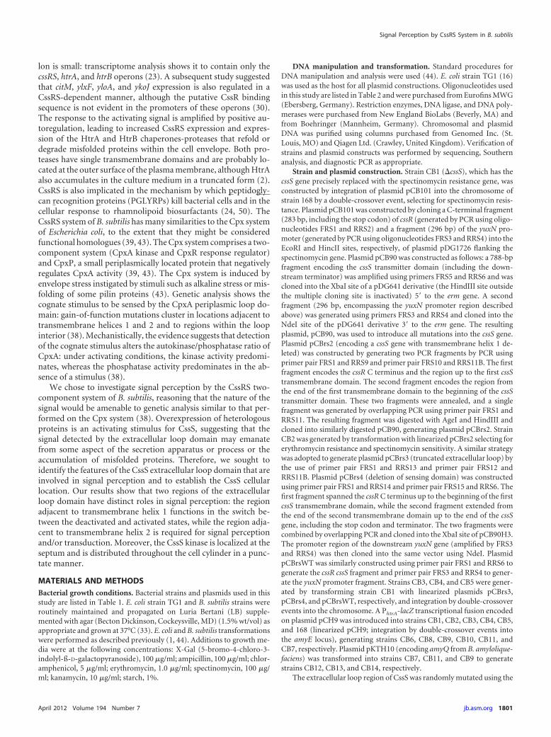
CssS1, CssR, HtrA, and HtrB protein levels are highly ele-vated in strain CB3 encoding CssS1. The activated CssRS two-component system positively autoregulates its own expressionand increases expression of the HtrA and HtrB serine proteases(9). Therefore, HtrA, HtrB, CssR, and CssS1 (truncated by 117amino acids) protein levels should be highly elevated in strain CB3expressing CssS1. The levels of these four proteins in strain CB3(expressing CssS1) were compared with those in strain CB5 (ex-pressing the reconstituted wild-type CssS) by Western analysisusing specific polyclonal antibodies. The results are shown in Fig.2. A band with the predicted size of wild-type CssS protein (toppanel, arrow, Mr [54.2]) is present in strain CB5, and its intensityincreased between the exponential-growth and stationary phases(OD600 of 1, 2, and 4). These mobility and expression profiles ofreconstituted CssS in strain CB5 are similar to those of wild-typeCssS in strain 168 (data not shown), further supporting the viewthat the CssS mutation system that was developed (Fig. 1A) func-tions as predicted. A smaller band of Mr (�25), present in lysatesof CB3 harvested at an OD600 of 4.0, is likely to be a processed formof CssS. CssS protein was not detected by Western analysis instrain CB2 (CssS construct B in Fig. 1B) or in strain CB4 (CssSconstruct D in Fig. 1B), indicating that these mutated proteins areprobably unstable and degraded (data not shown). However, theCssS1 protein accumulates to a very high level in strain CB3 (Fig.2, top panel, arrowhead). As expected, the CssS1 protein (strainCB3) is smaller than the full-length CssS protein (strain CB5) dueto the 117-amino-acid deletion within the extracellular loop do-main (Fig. 2). Notably, the level of CssS1 protein in strain CB3 iselevated throughout the growth cycle, consistent with the consti-tutive expression of PhtrA-lacZ observed in strain CB13 (Table 3).Similarly, the cellular levels of CssR (Fig. 2, second panel), HtrA(Fig. 2, third panel), and HtrB (Fig. 2, bottom panel) were all verysignificantly elevated throughout the growth cycle in strain CB3(expressing the CssS1 protein) compared to the level observed instrain CB5 (expressing the reconstituted wild-type CssS protein).The faster-migrating bands observed only in strain CB3 may in-dicate that significant CssRS, HtrA, and HtrB protein degradationoccurs due to their elevated cellular levels. We have previouslyreported that a processed form of HtrA accumulates in the me-
TABLE 3 Activity of CssS sensor kinases measured by expression of thePhtrA-lacZ fusion
Strain CssS proteina
Fold increase in�-galactosidaseaccumulationb
CB11 Reconstituted wild type 1CB13 Reconstituted wild type � pKTH10 28CB6 cssS gene deleted 1CB8 CssS TM1 domain deleted (construct B) 2CB10 Sensing domain deleted (construct D) 2CB9 Loop domain of 20-aa length (construct C) 325CB14 Loop domain of 20-aa length (construct C) �
pKTH10321
CB9H Loop domain of 20 aa with H250A mutation 1CB9HQ Loop domain of 20 aa with H250A mutation �
pKTH101
a Constructs B to D are shown in Fig. 1B. aa, amino acid.b Normalized to the �-galactosidase accumulation in strain CB11 containing areconstituted CssS protein generated using the system described for Fig. 1.
Signal Perception by CssRS System in B. subtilis
April 2012 Volume 194 Number 7 jb.asm.org 1807

dium during growth of wild-type strain 168 (2). We found a 200-fold increase in the level of processed HtrA in the culture super-natant of strain CB3 compared to the level found in wild-typestrain 168 (data not shown). Clearly, the cellular level of all CssRS
regulon proteins is significantly elevated in strains expressing theCssS1 kinase (20-amino-acid extracellular loop domain). We pro-pose that these greatly increased protein levels are due to the for-mation of an unregulated-feedback expression loop caused by theconstitutive CssS1 kinase acting within a positively acting autoreg-ulatory circuit.
Functional analysis of the CssS extracellular loop domain.An alignment of the extracellular loop domains of CssS ortho-logues from B. subtilis, B. licheniformis, B pumilis, and B. amyloliq-uefaciens is shown in Fig. 3. The amino acids positioned adjacentto the transmembrane helices (26 amino acids at the amino-ter-minal end and 19 amino acids at the carboxy-terminal end of theextracellular loop) are highly conserved, with some additionalconserved amino acid clusters scattered throughout the remain-der of the domain. The truncation of the loop domain in the CssS1protein (the loop is composed of amino acids 30 to 39 followed byamino acids 157 to 166, shown shaded in gray in Fig. 3) removes asignificant portion of both conserved regions. Therefore, to refineour analysis of CssS extracellular loop domain function, three ap-proaches were adopted, informed by the conservation profile seenin Fig. 3: (i) deletion of small regions of the loop; (ii) randommutation of the loop by PCR mutagenesis; and (iii) site-directedmutagenesis.
(i) Small deletions within the CssS extracellular loop do-main. Three deletions, each of 21 amino acids, were separatelymade in the N-terminal (amino acids 41 to 61), central (aminoacids 86 to 106), and C-terminal (amino acids 131 to 151) regionsof the CssS extracellular loop domain (marked in bold in Fig. 3).CssS activity was monitored in the resultant strains (by measuring�-galactosidase accumulation from the PhrA-lacZ fusion) in theabsence (HA58, HA64, and HA60) and presence (HA70, HA73,and HA71) of secretion stress. CssS activity was measured con-temporaneously in control strains CB11, CB12, CB9, and CB14.Representative results are presented in Table 4. Subjecting wild-type cells to a secretion stress increased PhtrA-lacZ expression�32-fold as expected (compare data showing �-galactosidase ac-cumulation in strains CB11 and CB12). Moreover, expression of
FIG 2 Western analysis of the CssRS operon proteins within strain CB3. Sam-ples were harvested at OD600 of �1, 2, and 4, separated by sodium dodecylsulfate-polyacrylamide gel electrophoresis (SDS-PAGE), and transferred tomembrane and probed with polyclonal antibodies (Ab) directed against CssS,CssR, HtrA, and HtrB. The migration position of each full-length protein isindicated by an arrow, while the migration position of truncated proteins isindicated by an arrowhead. The CssS protein in CB5 is full length and in CB3is truncated by 117 amino acids, the deletion in the extracellular loop region.The positive-control =CssS protein is also truncated and contains only thecytoplasmic domain (C). CssR and HtrB are full length in both strains, whileHtrA undergoes a processing event that results in a cell-associated full-lengthand truncated protein. The migration positions of standard proteins are indi-cated on the right side of the figure with their Mr values.
FIG 3 Alignment of the CssS proteins from B. subtlis strain 168, B. amyloliquefaciens strain FZB42, B. licheniformis strain ATCC 14580, and B. pumilis SAFR-032.Asterisks signify amino acid identity and colons conservative changes at each position. The amino acids that form the CssS1 (truncated) loop domain are shadedin gray. The 21-amino-acid deletions made in strains HA58, HA64, and HA60 are shown in bold. The positions at which amino acid substitutions L33Q, N47Y,Q49H, D153A, S154A, Y155A, R156A, D157A, and D158A were made are underlined. The sequence replaced by the 3� FLAG tag epitope is shown in red, whilethe 7-amino-acid deletion (DSYRDDL) is shown in orange. The 20 amino acids preceding (amino acids 10 to 29) and the 20 amino acids succeeding (amino acids167 to 186) these sequences in B. subtilis form transmembrane helices 1 and 2, respectively. Protein identity codes are as follows: B. subtilis, NP_391182.2; B.amyloliquefaciens, YP_001422575.1; B. licheniformis, YP_080579.1; and B. pumilis, YP_001488168.1.
Noone et al.
1808 jb.asm.org Journal of Bacteriology

PhtrA-lacZ in strains CB9 and CB14 was �340-fold higher thanthat observed in nonstressed wild-type cells, consistent with pre-vious results. Each 21-amino-acid deletion within the loopdomain affected CssS activity differently. Deletion of the amino-terminal 21-amino-acid segment (strain HA58) increased PhtrA-lacZ expression �320-fold, similar to that observed in strain CB9carrying the constitutively active CssS1 (Table 4). Deletion of thecentral 21-amino-acid segment (strain HA64) increased PhtrA-lacZ expression �98-fold (approximately 1/3 of the level seen instrain CB9), while deletion of the carboxy-terminal 21-amino-acid segment increased PhtrA-lacZ expression �27-fold (approxi-mately 1/13 of the level seen in strain CB9). Interestingly, intro-duction of a secretion stress (strains HA70, HA73, and HA71) didnot further increase PhtrA-lacZ expression relative to that observedin nonstressed strains (strains HA58, HA64, and HA60, respec-tively; Table 4). The results suggest that these mutated CssS pro-teins are insensitive to secretion stress. However, an importantcaveat is that AmyQ overexpression may not generate a secretionstress in cells with elevated HtrA and HtrB protein levels.
(ii) Random mutation of the CssS loop domain. Deletionanalysis affects both the amino acid composition and the size ofthe extracellular loop domain. To distinguish between these twoeffects, the system described in Fig. 1A was used to randomlyintroduce amino acid changes specifically into the CssS extra-cellular loop domain, screening for colonies with increased �-ga-
lactosidase expression on X-Gal-containing agar plates. Threestrains showed highly elevated �-galactosidase expression, andeach had a single amino acid substitution within the CssS loopdomain. To ensure that only this single amino acid was changed ineach protein, cssS alleles encoding these amino acid changes werereconstructed by site-directed mutagenesis (as outlined in Fig.1A) and inserted into the chromosome to generate strains HA54(CssS L33Q), HA62 (CssS N47Y), and HA56 (CssS Q49H) (Table4). �-Galactosidase production increased between 271- and 339-fold in these three strains, a level similar to that observed in strainCB9 expressing CssS1 (117 amino acids deleted from the extracel-lular loop domain). These results are consistent with and extendthe deletion analysis reported in the previous section: all threeamino acid substitutions are located within the conserved regionof the CssS loop domain juxtaposed to transmembrane helix 1,and two of the three substitutions (N47Y and Q49H) are locatedwithin the loop region that is deleted in strain HA58 (amino acids41 to 61 deleted; Fig. 3). Subjecting each of these strains to secre-tion stress (by introducing pKTH10 into strains HA54, HA62, andHA56 to generate strains HA68, HA72, and HA69) did not furtherincrease PhtrA-lacZ expression (Table 4). Thus, each of the threenonconservative amino acid substitutions within this conservedloop region juxtaposed to transmembrane helix 1 was sufficient togenerate a constitutively active CssS kinase, showing that this re-gion plays a crucial role in maintaining CssS with very low kinaseactivity in the absence of secretion stress and in the switch tohigher activity upon induction.
(iii) Site-directed mutation of the CssS loop domain. The re-gion of the CssS loop domain flanking transmembrane helix 2 isalso highly conserved (Fig. 3). However, mutations were notfound in this region in the collection of mutagenized strains. Sinceonly gain-of-function CssS mutants (i.e., increased �-galactosi-dase production) are detected in our screen, we posited that thisregion of the CssS loop domain might contribute differently tosignal perception and/or transduction. Therefore, an alanine-scanning mutagenesis of the conserved DSYRDD motif was per-formed and, in addition, a strain expressing CssS with a 7-amino-acid (DSYRDDL) deletion was generated. CssS activity wasdetermined in the resultant strains by measuring expression of thePhtrA-lacZ fusion in the absence and presence of secretion stress.Representative results are presented in Table 4. Mutation of five ofthe six amino acids within this conserved CssS region does notresult in increased �-galactosidase accumulation. The exceptionalcase is DN1982 (expressing CssS Y155A), where a small increase(�13-fold) in accumulation was observed that, curiously, was re-duced in response to secretion stress. Importantly, CssS kinaseactivity was not induced by a secretion stress in any of thesestrains, signifying that they are signal blind (Table 4). Althoughdeletion of 7 amino acids from this region (strain DN1986;deletion of DSYRDDL) resulted in a 16-fold increase in �-galac-tosidase accumulation under nonstress conditions, there was onlymarginal induction (1.5-fold) in response to a secretion stress(Table 4). These results show that mutating the conserved regionflanking transmembrane helix 2 of the extracellular loop domainabolishes CssS induction by a secretion stress and that it is re-quired for signal perception and/or transduction. Clearly, theconserved regions of the CssS extracellular loop domain flankingthe transmembrane helices function differently in signal percep-tion and transduction.
To further investigate the different functions of the conserved
TABLE 4 Fold induction of �-galactosidase in B. subtilis strainsexpressing mutated CssS proteins from the natural promoter
Strains (�/�pKTH10)a CssS mutation
Fold increase in �-galactosidaseexpression overwild-type CssSinduced by amylaseb Fold induction
of CssS �-galactosidaseby amylasec
NopKTH10
�pKTH10
CB11/CB12 Wild type 1 32 32CB9/CB14 �ECL 343 349 1
HA58/HA70 �40–60 AAd 327 313 1HA64/HA73 �85–105 AA 98 88 1HA60/HA71 �130–150 AA 27 22 1
HA54/HA68 L33Q 339 325 1HA62/HA72 N47Y 330 325 1HA56/HA69 Q49H 271 307 1.1
DN1980/DN1980Q D153A �1 1 1DN1981/DN1981Q S154A �1 1 1DN1982/DN1982Q Y155A 13 1.5 0.1DN1983/DN1983Q R156A 1 1 1DN1984/DN1984Q D157A 1 1 1DN1985/DN1985Q D158A 2 1 1DN1986/DN1986Q Deletion of DSYRDD 16 24 1.5
DN1987/DN1987Q L33Q R156A 68 77 1
DN1988/DN1988Q CssS�58–79::3�FLAG 76 82 1DN1991/DN1991Q CssS�61–79::3�FLAG
(� 3AA)326 354 1
a �, strain with plasmid; �, strain without plasmid.b Values represent the extent to which CssS variants are active relative to wild-typeCssS.c Values represent the extent to which each CssS variant is induced by amylase(pKTH10).d AA, amino acids.
Signal Perception by CssRS System in B. subtilis
April 2012 Volume 194 Number 7 jb.asm.org 1809

loop regions juxtaposed to the transmembrane helices, we con-structed a strain expressing CssS that combined the L33Q gain-of-function mutation (confers constitutive noninducible kinase ac-tivity) and the R156A loss-of-function mutation (confers lownoninducible kinase activity). We found that �-galactosidase ac-cumulation increased 68-fold and 77-fold in the absence(DN1987) and presence (DN1987Q) of secretion stress, respec-tively, compared to the noninduced CB11 (reconstituted wild-type CssS) strain (Table 4). However, Western analysis showedthat this CssS variant was unstable: combining the R156A andL33Q mutations reduced the very high level of protein observed incells expressing CssS L33Q to approximately the level of CssS ob-served in noninduced wild-type cells. We posit that the increasedPhtrA-lacZ expression in strains DN1987 and DN1987Q may havebeen due to a low steady-state level of constitutively active kinase,the residue of the competing processes of high-level expressionand protein degradation.
(iv) Replacement of a nonconserved region of the CssS loopdomain with the heterologous 3� FLAG epitope. To assess theimportance of amino acid identity within the relatively noncon-served region of the CssS loop domain, we replaced a 22-amino-acid segment (amino acids 59 to 80; shown in red in Fig. 3) with aheterologous 3� FLAG epitope of identical length and monitoredCssS activity in the absence and presence of secretion stress. Asshown in Table 4, this substitution resulted in a partially consti-tutively active CssS kinase (76- to 82-fold elevated activity) that isunresponsive to secretion stress. Interestingly, replacing the sameamino acids with 3� FLAG and increasing the size of the loopdomain by 3 amino acids (amino acids GSI; indicated in red boldin Fig. 3) generated a constitutively active CssS kinase (326- to354-fold elevated activity).
In summary, these studies show that individual regions of theCssS extracellular loop domain function differently in signal per-ception and transduction. The conserved region adjacent to trans-membrane helix 1 is likely to be involved in maintaining the CssSkinase in a low activity state under nonstress conditions and in theswitch to increased activity in response to secretion stress. Theconserved region adjacent to transmembrane helix 2 is requiredfor signal sensing and/or signal transduction, since all mutationsin this region render CssS signal blind. The size of the extracellularloop was also important, since all perturbations led to an increase(27- to 343-fold; Table 4) in CssS activity.
Mutation of the CssS loop domain in strains lacking the pos-itive autoregulatory loop of cssRS expression. The positive auto-regulation of cssRS expression amplified the effects of gain-of-function mutations (e.g., L33Q), resulting in very high cellularCssS levels, while loss-of-function mutations (e.g., D153A) re-sulted in low cellular CssS levels. Such a disparity in protein levelscomplicates a comparison of the activities of different CssS vari-ants. To address this issue, we placed cssRS expression under thecontrol of the IPTG-inducible Pspac promoter (thereby removingthe autoregulatory loop), allowing us to compare the effects ofselected mutations in the loop domain in strains with comparablecellular CssS protein levels. All such strains were grown with ad-dition of 100 �M IPTG, a level of inducer that significantly in-creased the cellular level of CssS protein (Fig. 4; compare strain168D with 168E and strain 168DQ with 168EQ). CssS proteinlevels were very similar in all strains (except DN1987) expressingcssRS from the Pspac promoter, regardless of the presence or ab-sence of pKTH10 (compare strains in Fig. 4A with those in Fig.
4B), showing that the autoregulatory loop had been removed andthat expression was no longer inducible by secretion stress. Impor-tantly, the level of CssS protein in cells expressing gain-of-function(Fig. 4 [HA54C, HA54CQ, HA62C, and HA62CQ]) and loss-of-function (Fig. 4 [DN1980C, DN1980CQ, and DN1984CQ]) vari-ants was very similar to that in cells expressing wild-type protein(168E and 168EQ; Fig. 4), showing that neither type of mutationdestabilized CssS protein and allowing direct comparison of theiractivities (Table 5). Increasing the CssS protein level in wild-typestrain 168E �20-fold (Fig. 4) increased the noninduced level ofPhtrA-lacZ expression only �3-fold (compare strains 168D and168E; Table 5). However, applying a secretion stress (strain 168EQ) in this background increased PhtrA-lacZ expression only 19-fold, very similar to the 16-fold-increased expression observed inthe wild-type background (strain 168DQ), showing that CssRSlevels are not limiting in wild-type cells (Table 5). Expression ofPhtrA-lacZ in strains with gain-of-function CssS variants (HA54C,HA54CQ, HA62C, and HA62C) was elevated more than 100-fold,confirming increased CssS kinase activity and showing that thepositive autoregulatory loop makes an approximately 3-fold con-tribution (e.g., compare HA54C and HA62C in Table 5 with HA54and HA62, respectively, in Table 4) to expression. However, ex-pression of PhtrA-lacZ in strains with loss-of-function CssS vari-ants (DN1980C, DN1980CQ, and DN1984C) was similar to thatin a strain with wild-type CssS (168E) and was not induced bysecretion stress, signifying that mutations in this region of the loopdomain made the protein signal blind and/or incapable of trans-ducing the signal (Table 5). We were unable to establish plasmidpKTH10 in strain DN1984C, indicating that the cell is unable tomount a secretion stress response. Generally, we found that strainsexpressing gain-of-function CssS variants (e.g., HA54, HA62)were more readily transformed with pKTH10 than wild-type cellswhereas strains expressing loss-of-function CssS variants that are
FIG 4 Western blot analysis of CssS protein levels in strains expressing CssSunder the control of the IPTG-inducible Pspac promoter. Cultures were grownto an OD600 of �2.0 and harvested, and lysates were prepared for SDS-PAGEas described in the text. Lysate samples containing 10 �g for protein wereloaded onto each lane, and CssS protein was detected as described in the text.(A) Strains in the absence of a secretion stress (no pKTH10 plasmid). (B)Strains with secretion stress (containing pKTH10 plasmid). Strains CB6 andCB6Q are the negative controls, having the cssS gene deleted. The filter wasexposed to film for 30 s. Protein sizes are shown at the side of the image.
Noone et al.
1810 jb.asm.org Journal of Bacteriology

unresponsive to secretion stress (e.g., CB6, DN1980C, DN1984C)were very poorly transformable, if at all, with pKTH10. Theseanalyses also confirm that combining the L33Q gain-of-functionmutation with the loss-of-function R156A, neither of which af-fects protein stability individually, destabilized CssS, with only alow level of full-length protein detectable by Western analysis(DN1987C and DN1987CQ; Fig. 4). In summary, these data con-firm that the conserved regions of the CssS loop domain adjacentto the transmembrane helices function differently in signal per-ception, activation, and transduction.
Localization of the CssS, HtrA, and HtrB proteins in B. sub-tilis. We established the cellular localization of CssS, HtrA, andHtrB by immunofluorescence in exponentially growing B. subtiliscells (Fig. 5). CssS is primarily located at the septa of dividing cellsand at the poles of many cells (Fig. 5A to C). Two separate butjuxtaposed CssS bands positioned at the septa and poles of cellsthat are dividing or have just divided are shown in Fig. 5A. Somecells also showed a punctate distribution of CssS along the cellcylinder but with lower intensity (Fig. 5C). A minority of cellsshowed no staining (see, e.g., Fig. 5B): it is unclear whether thisresult represents cells not expressing CssRS or whether there wasinsufficient antibody penetration for visualization. We concludefrom these and many additional images that CssS is localized at thesepta of dividing cells and in a punctate manner along the cellcylinder.
HtrA was located at only one or a few foci in exponentiallygrowing wild-type 168 cells but was not detected in all cells (Fig.5D). This is consistent with our demonstration that HtrA is ex-pressed at low levels in nonstressed cells and that it is processedand shed into the medium during growth (2, 34, 35). The numberof HtrA foci increased very significantly in cells expressing veryhigh HtrA levels (strain HA54, expressing the constitutively activeCssS L33Q), and the foci were distributed along the length of thecell cylinder in an apparently random manner (Fig. 5E). In addi-tion, a lower level of continuous fluorescence covers the entiresurface of cells of strain HA54, suggesting that the very high levelof HtrA saturated all potential focal binding sites and that surplusprotein became evenly distributed over the cell surface. HtrB wasalso located in foci distributed throughout the length of wild-type168 cells in an apparently random manner (Fig. 5F). Importantly,
TABLE 5 Fold induction of �-galactosidase in B. subtilis strains expressing mutated CssS proteins under the control of the Pspac promoter
Strains (�/� pKTH10)a CssS mutation
Fold increase in expression overwild-type CssS induced by amylaseb
Fold induction of CssSby amylasecNo pKTH10 � pKTH10
168D/168DQ Wild type 1 16 16168E/168EQ Wild-type CssS under Pspac control 3 19 6
HA54C/HA54CQ CssS L33Q under Pspac control 117 100 0.85HA62C/HA62CQ CssS B47Y under Pspac control 129 106 0.82
DN1980C/DN1980CQ CssS D153A under Pspac control 3.3 3.8 1.1DN1984C/SNO CssS D157A under Pspac control 3.1 ND
DN1987C/DN1987CQ CssS L33Q R156A under Pspac control 63 49 0.8a �, strain with plasmid; �, strain without plasmid; SNO, strain not obtained.b Values represent the extent to which CssS variants are active relative to wild-type CssS. ND, not determined.c Values represent the extent to which each CssS variant is induced by amylase (pKTH10).
FIG 5 Localization of the CssS, HtrA, and HtrB proteins in B. subtilis. TheCssS, HtrA, and HtrB proteins were visualized by differential interferencecontrast microscopy (DIC) and by immunostaining (immunofluores-cence) microscopy in nonstressed wild-type strain 168 and in strain HA54(CssS L33Q) cells that express a constitutive CssS kinase. CssS (A, B, and C)was visualized by staining with an anti-myc-tag antibody, while HtrA (Dand E) and HtrB (F and G) were stained with specific affinity-purifiedpolyclonal antisera. Visualization of primary antibodies was performed bystaining with a fluorescein isothiocyanate (FITC)-labeled secondary anti-body. Bar, 4 �m for all images.
Signal Perception by CssRS System in B. subtilis
April 2012 Volume 194 Number 7 jb.asm.org 1811

HtrB foci were observed in the vast majority of cells, while thenumber of HtrB foci per cell was also significantly higher than thenumber of HtrA foci. The number of HtrB foci was elevated instrain HA54 (expressing constitutively active CssS L33Q) com-pared with wild-type strain 168 (Fig. 5G). Like HtrA, the entiresurface of HA54 cells was covered with a continuous lower level offluorescence, again suggesting that all potential focal binding siteswere saturated and the HtrB surplus protein became evenly dis-tributed over the entire cell surface (Fig. 5G). These data show thatHtrA and HtrB were confined to discrete foci on the surface ofnonstressed cells. HtrB was present at most of the potential bind-ing site foci in nonstressed cells, whereas HtrA was largely absentfrom the cell surface under this condition, probably because it wasprocessed and shed into the medium (2). However, increasingHtrA expression showed many additional foci to which it couldbind. Moreover, when highly expressed, both proteins were alsoevenly distributed at a lower level throughout the cell length. Inview of the fact that CssS localizes primarily to cell septa and poles,it is interesting that neither HtrA nor HtrB showed septal or polarlocalization.
DISCUSSION
The CssS kinase is a member of the EnvZ/OmpR family of kinasesthat has two transmembrane helices flanking a 137-amino-acidextracellular loop proposed to encode a PAS domain (6, 31).While CssS responds to heat and secretion stress stimuli, the iden-tity of the signal, how it is perceived by CssS, and how perceptionactivates kinase activity are largely unknown. Here we have iden-tified several features of the extracytoplasmic loop domain that areimportant for signaling. The extracytoplasmic loop domain func-tions to maintain the activity of CssS kinase at a very low level inthe absence of signal. Deletion of 117 amino acids from the extra-cytoplasmic loop domain (CssS1) increased kinase activity morethan 300-fold even in the absence of secretion stress. CssS1 ap-pears to be unresponsive to stress, although it must be remem-bered for all strains showing increased CssS kinase activity thatAmyQ overexpression may not generate a secretion stress in cellshaving highly elevated levels of HtrA and HtrB proteases. Thus,the nonstressed (PhtrA-lacZ expression similar to that in cssS nullstrains) and CssS1 (PhtrA-lacZ expression increased 300-fold) pro-tein states appear to be the extremities of CssS kinase activity,representing the “off” and “on” conditions, respectively, in thecell. It is interesting that the heat and secretion stresses (PhtrA-lacZexpression increased 6- and 25-fold, respectively [reference 35 andthis study]) induced CssS kinase activity to a level at the lower endof its capability. The CpxRA two-component system also re-sponds to cell envelope and secretion stresses in E. coli (10, 43). Ina manner similar to that seen with CssS, an in-frame deletion of 32amino acids in the central region of the CpxA periplasmic sensingdomain (CpxA24) results in a gain of function, renders it insensi-tive to secretion stress, and results in a significantly elevated levelof CpxRA protein in the cell due to positive autoregulation of thecpxRA operon (38, 40). Thus, the negative role of the periplasmicsensing domain and amplification of the signal through positiveautoregulation are themes common to the CssRS (of B. subtilis)and CpxRA (of E. coli) two-component systems in the detectionand response to secretion stresses. However, this is not universalamong two-component systems, since deletion of the extracellu-lar loop (encoding a PAS domain) of PhoR in B. subtilis does notresult in constitutive activity or render it insensitive to detecting
phosphate limitation (E. Botella and K. M. Devine, unpublishedresults).
The two regions of the CssS extracellular loop domain that arejuxtaposed to the transmembrane helices are highly conservedamong CssS orthologues of closely related bacilli (Fig. 3) and havedistinct functions. The 26-amino-acid region that is adjacent totransmembrane helix 1 functions in the activity switch that occursupon cognate stimulus detection. In the absence of signal, thisregion functions to maintain the CssS kinase in a low activity state.However, single nonconservative amino acid substitutions(L33Q, N47Y, Q49H) in this region generate CssS variants that areconstitutively active. Interestingly, Raivio and Silhavy (38) alsoreported a gain-of-function mutation (cpxA104) localized to thisregion of the CpxA loop domain. The amino acid composition ofthis CssS region is very hydrophilic, with 9 hydroxyl groups (Ser,Thr, and Tyr), 5 carboxyl groups (Asp and Glu), 2 positive charges(Arg and His), and 4 amide groups (Gln and Asn). Among theremaining 6 hydrophobic amino acids, there is an unusual pair ofadjacent Phe residues. The L33Q mutation is especially interest-ing, since the hydrophobic Leu is directly adjacent to the trans-membrane helix (amino acids 10 to 29), while the mutation placesthe more hydrophilic glutamine at this position. It is tempting tospeculate that this conserved hydrophilic domain interacts withthe outer leaftlet or face of the cytoplasmic membrane to maintainCssS in a state of low kinase or high phosphatase activity. Signalperception (or mutation, as in this study) may disrupt these inter-actions, thereby facilitating activation of CssS by increasing kinaseor decreasing phosphatase activity.
The role of the conserved 19-amino-acid region adjacent totransmembrane helix 2 in signal perception is distinct from that ofthe conserved region adjacent to transmembrane helix 1. CssSvariants mutated in this region are signal blind and have activitiessimilar to those of the noninduced wild-type CssS protein. Thus,this region is required for either signal perception and/or trans-duction. CssS differs from CpxA in that all point mutations in thisCssS region are inactive and signal blind whereas the CpxA103(R164P) variant mutated in this region has increased activity, al-though it is also signal blind. The strain expressing the CssS L33QR156A double-mutant variant (DN1987) is especially interesting.Expression of each CssS single-mutant variant shows that bothproteins are stable. However, the CssS variant with both muta-tions (DN1987C and DN1987CQ) is highly unstable, with only alow level of full-length CssS visible and extensive protein degra-dation being observed. The size of the loop domain and featureslocated between these two conserved membrane-associated do-mains are also important for signal perception. All size perturba-tions of the loop domain increase CssS activity. Moreover, chang-ing the amino acid composition of part of the loop domain (byreplacement of 22 amino acids with a 22-amino-acid 3� FLAGtag) increased CssS activity 76-fold. When this replacement wascombined with a 3-amino-acid increase in the size of the loopdomain, CssS activity increased 326-fold, similar to that observedwhen most of the loop domain was deleted.
It is not possible to ascertain the effect of these perturbationson the various enzymatic activities of the CssS kinase because ofdifficulties in purifying integral membrane proteins and becausesuch activities probably depend on proper membrane localiza-tion. Kinases such as CssS and CpxA have autophosphorylation,phosphotransfer, and phosphatase enzymatic activities. Thus,while the mutated CssS proteins in this study had altered activities,
Noone et al.
1812 jb.asm.org Journal of Bacteriology
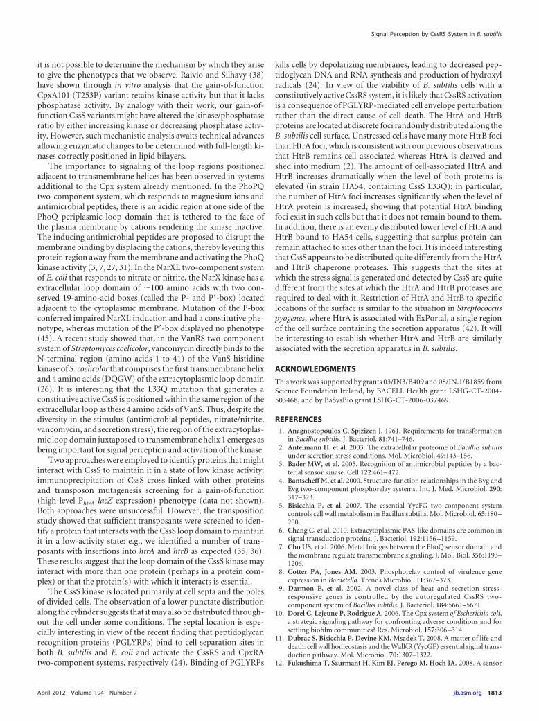
it is not possible to determine the mechanism by which they ariseto give the phenotypes that we observe. Raivio and Silhavy (38)have shown through in vitro analysis that the gain-of-functionCpxA101 (T253P) variant retains kinase activity but that it lacksphosphatase activity. By analogy with their work, our gain-of-function CssS variants might have altered the kinase/phosphataseratio by either increasing kinase or decreasing phosphatase activ-ity. However, such mechanistic analysis awaits technical advancesallowing enzymatic changes to be determined with full-length ki-nases correctly positioned in lipid bilayers.
The importance to signaling of the loop regions positionedadjacent to transmembrane helices has been observed in systemsadditional to the Cpx system already mentioned. In the PhoPQtwo-component system, which responds to magnesium ions andantimicrobial peptides, there is an acidic region at one side of thePhoQ periplasmic loop domain that is tethered to the face ofthe plasma membrane by cations rendering the kinase inactive.The inducing antimicrobial peptides are proposed to disrupt themembrane binding by displacing the cations, thereby levering thisprotein region away from the membrane and activating the PhoQkinase activity (3, 7, 27, 31). In the NarXL two-component systemof E. coli that responds to nitrate or nitrite, the NarX kinase has aextracellular loop domain of �100 amino acids with two con-served 19-amino-acid boxes (called the P- and P=-box) locatedadjacent to the cytoplasmic membrane. Mutation of the P-boxconferred impaired NarXL induction and had a constitutive phe-notype, whereas mutation of the P=-box displayed no phenotype(45). A recent study showed that, in the VanRS two-componentsystem of Streptomyces coelicolor, vancomycin directly binds to theN-terminal region (amino acids 1 to 41) of the VanS histidinekinase of S. coelicolor that comprises the first transmembrane helixand 4 amino acids (DQGW) of the extracytoplasmic loop domain(26). It is interesting that the L33Q mutation that generates aconstitutive active CssS is positioned within the same region of theextracellular loop as these 4 amino acids of VanS. Thus, despite thediversity in the stimulus (antimicrobial peptides, nitrate/nitrite,vancomycin, and secretion stress), the region of the extracytoplas-mic loop domain juxtaposed to transmembrane helix 1 emerges asbeing important for signal perception and activation of the kinase.
Two approaches were employed to identify proteins that mightinteract with CssS to maintain it in a state of low kinase activity:immunoprecipitation of CssS cross-linked with other proteinsand transposon mutagenesis screening for a gain-of-function(high-level PhtrA-lacZ expression) phenotype (data not shown).Both approaches were unsuccessful. However, the transpositionstudy showed that sufficient transposants were screened to iden-tify a protein that interacts with the CssS loop domain to maintainit in a low-activity state: e.g., we identified a number of trans-posants with insertions into htrA and htrB as expected (35, 36).These results suggest that the loop domain of the CssS kinase mayinteract with more than one protein (perhaps in a protein com-plex) or that the protein(s) with which it interacts is essential.
The CssS kinase is located primarily at cell septa and the polesof divided cells. The observation of a lower punctate distributionalong the cylinder suggests that it may also be distributed through-out the cell under some conditions. The septal location is espe-cially interesting in view of the recent finding that peptidoglycanrecognition proteins (PGLYRPs) bind to cell separation sites inboth B. subtilis and E. coli and activate the CssRS and CpxRAtwo-component systems, respectively (24). Binding of PGLYRPs
kills cells by depolarizing membranes, leading to decreased pep-tidoglycan DNA and RNA synthesis and production of hydroxylradicals (24). In view of the viability of B. subtilis cells with aconstitutively active CssRS system, it is likely that CssRS activationis a consequence of PGLYRP-mediated cell envelope perturbationrather than the direct cause of cell death. The HtrA and HtrBproteins are located at discrete foci randomly distributed along theB. subtilis cell surface. Unstressed cells have many more HtrB focithan HtrA foci, which is consistent with our previous observationsthat HtrB remains cell associated whereas HtrA is cleaved andshed into medium (2). The amount of cell-associated HtrA andHtrB increases dramatically when the level of both proteins iselevated (in strain HA54, containing CssS L33Q): in particular,the number of HtrA foci increases significantly when the level ofHtrA protein is increased, showing that potential HtrA bindingfoci exist in such cells but that it does not remain bound to them.In addition, there is an evenly distributed lower level of HtrA andHtrB bound to HA54 cells, suggesting that surplus protein canremain attached to sites other than the foci. It is indeed interestingthat CssS appears to be distributed quite differently from the HtrAand HtrB chaperone proteases. This suggests that the sites atwhich the stress signal is generated and detected by CssS are quitedifferent from the sites at which the HtrA and HtrB proteases arerequired to deal with it. Restriction of HtrA and HtrB to specificlocations of the surface is similar to the situation in Streptococcuspyogenes, where HtrA is associated with ExPortal, a single regionof the cell surface containing the secretion apparatus (42). It willbe interesting to establish whether HtrA and HtrB are similarlyassociated with the secretion apparatus in B. subtilis.
ACKNOWLEDGMENTS
This work was supported by grants 03/IN3/B409 and 08/IN.1/B1859 fromScience Foundation Ireland, by BACELL Health grant LSHG-CT-2004-503468, and by BaSysBio grant LSHG-CT-2006-037469.
REFERENCES1. Anagnostopoulos C, Spizizen J. 1961. Requirements for transformation
in Bacillus subtilis. J. Bacteriol. 81:741–746.2. Antelmann H, et al. 2003. The extracellular proteome of Bacillus subtilis
under secretion stress conditions. Mol. Microbiol. 49:143–156.3. Bader MW, et al. 2005. Recognition of antimicrobial peptides by a bac-
terial sensor kinase. Cell 122:461– 472.4. Bantscheff M, et al. 2000. Structure-function relationships in the Bvg and
Evg two-component phosphorelay systems. Int. J. Med. Microbiol. 290:317–323.
5. Bisicchia P, et al. 2007. The essential YycFG two-component systemcontrols cell wall metabolism in Bacillus subtilis. Mol. Microbiol. 65:180 –200.
6. Chang C, et al. 2010. Extracytoplasmic PAS-like domains are common insignal transduction proteins. J. Bacteriol. 192:1156 –1159.
7. Cho US, et al. 2006. Metal bridges between the PhoQ sensor domain andthe membrane regulate transmembrane signaling. J. Mol. Biol. 356:1193–1206.
8. Cotter PA, Jones AM. 2003. Phosphorelay control of virulence geneexpression in Bordetella. Trends Microbiol. 11:367–373.
9. Darmon E, et al. 2002. A novel class of heat and secretion stress-responsive genes is controlled by the autoregulated CssRS two-component system of Bacillus subtilis. J. Bacteriol. 184:5661–5671.
10. Dorel C, Lejeune P, Rodrigue A. 2006. The Cpx system of Escherichia coli,a strategic signaling pathway for confronting adverse conditions and forsettling biofilm communities? Res. Microbiol. 157:306 –314.
11. Dubrac S, Bisicchia P, Devine KM, Msadek T. 2008. A matter of life anddeath: cell wall homeostasis and the WalKR (YycGF) essential signal trans-duction pathway. Mol. Microbiol. 70:1307–1322.
12. Fukushima T, Szurmant H, Kim EJ, Perego M, Hoch JA. 2008. A sensor
Signal Perception by CssRS System in B. subtilis
April 2012 Volume 194 Number 7 jb.asm.org 1813

histidine kinase co-ordinates cell wall architecture with cell division inBacillus subtilis. Mol. Microbiol. 69:621– 632.
13. Fukushima T, et al. 2011. A role for the essential YycG sensor histidinekinase in sensing cell division. Mol. Microbiol. 79:503–522.
14. Gao R, Stock AM. 2009. Biological insights from structures of two-component proteins. Annu. Rev. Microbiol. 63:133–154.
15. Gerharz T, Reinelt S, Kaspar S, Scapozza L, Bott M. 2003. Identificationof basic amino acid residues important for citrate binding by the periplas-mic receptor domain of the sensor kinase CitA. Biochemistry 42:5917–5924.
16. Gibson TJ. 1984. Studies on the Epstein-Barr virus genome. University ofCambridge, Cambridge, United Kingdom.
17. Glaser P, et al. 1993. Bacillus subtilis genome project: cloning and se-quencing of the 97 kb region from 325 degrees to 333 degrees. Mol. Mi-crobiol. 10:371–384.
18. Guérout-Fleury AM, Shazand K, Frandsen N, Stragier P. 1995. Antibi-otic-resistance cassettes for Bacillus subtilis. Gene 167:335–336.
19. Hoch JA, Silhavy TJ (ed). 1995. Two-component signal transduction.ASM Press, Washington, DC.
20. Howell A, et al. 2003. Genes controlled by the essential YycG/YycF two-component system of Bacillus subtilis revealed through a novel hybridregulator approach. Mol. Microbiol. 49:1639 –1655.
21. Howell A, Dubrac S, Noone D, Varughese KI, Devine KM. 2006.Interactions between the YycFG and PhoPR two-component systems inBacillus subtilis: the PhoR kinase phosphorylates the non-cognate YycFresponse regulator upon phosphate limitation. Mol. Microbiol. 59:1199 –1215.
22. Hyyryläinen HL, et al. 2001. A novel two-component regulatory systemin Bacillus subtilis for the survival of severe secretion stress. Mol. Micro-biol. 41:1159 –1172.
23. Hyyryläinen HL, Sarvas M, Kontinen VP. 2005. Transcriptome analysisof the secretion stress response of Bacillus subtilis. Appl. Microbiol. Bio-technol. 67:389 –396.
24. Kashyap DR, et al. 2011. Peptidoglycan recognition proteins kill bacteriaby activating protein-sensing two component systems. Nat. Med. 17:676 –683.
25. Kaspar S, et al. 1999. The periplasmic domain of the histidine autokinaseCitA functions as a highly specific citrate receptor. Mol. Microbiol. 33:858 – 872.
26. Koteva K, et al. 2010. A vancomycin photoprobe identifies the histidinekinase VanSsc as a vancomycin receptor. Nat. Chem. Biol. 6:327–329.
27. Krell T, et al. 2010. Bacterial sensor kinases: diversity in the recognition ofenvironmental signals. Annu. Rev. Microbiol. 64:539 –559.
28. Kunst F, et al. 1997. The complete genome sequence of the gram-positivebacterium Bacillus subtilis. Nature 390:249 –256.
29. Leonardo MR, Forst S. 1996. Re-examination of the role of the periplas-mic domain of EnvZ in sensing of osmolarity signals in Escherichia coli.Mol. Microbiol. 22:405– 413.
30. Lulko AT, et al. 2007. Production and secretion stress caused by overex-pression of heterologous alpha-amylase leads to inhibition of sporulationand a prolonged motile phase in Bacillus subtilis. Appl. Environ. Micro-biol. 73:5354 –5362.
31. Mascher T, Helmann JD, Unden G. 2006. Stimulus perception in bac-terial signal-transducing histidine kinases. Microbiol. Mol. Biol. Rev. 70:910 –938.
32. Mascher T, Margulis NG, Wang T, Ye RW, Helmann JD. 2003. Cell wallstress responses in Bacillus subtilis: the regulatory network of the bacitra-cin stimulon. Mol. Microbiol. 50:1591–1604.
33. Miller JH. 1972. Experiments in molecular genetics, p 431– 432. ColdSpring Harbor Laboratory Press, Cold Spring Harbor, NY.
34. Möglich A, Ayers RA, Moffat K. 2009. Structure and signaling mecha-nism of Per-ARNT-Sim domains. Structure 17:1282–1294.
35. Noone D, Howell A, Devine KM. 2000. Expression of ykdA, encoding aBacillus subtilis homologue of HtrA, is heat shock inducible and negativelyautoregulated. J. Bacteriol. 182:1592–1599.
36. Noone D, Howell A, Collery R, Devine KM. 2001. YkdA and YvtA,HtrA-like serine proteases in Bacillus subtilis, engage in negative autoreg-ulation and reciprocal cross-regulation of ykdA and yvtA gene expression.J. Bacteriol. 183:654 – 663.
37. Palva I. 1982. Molecular cloning of the alpha-amylase gene from Bacillusamyloliquefaciens and its expression in Bacillus subtilis. Gene 19:81– 87.
38. Raivio TL, Silhavy TJ. 1997. Transduction of envelope stress in Esche-richia coli by the Cpx two-component system. J. Bacteriol. 179:7724 –7733.
39. Raivio TL, Silhavy TJ. 1999. The �E and Cpx regulatory pathways: over-lapping but distinct stress responses. Curr. Opin. Microbiol. 2:159 –165.
40. Raivio TL, Popkin DL, Silhavy TJ. 1999. The Cpx envelope stress re-sponse is controlled by amplification and feedback inhibition. J. Bacteriol.181:5263–5272.
41. Reinelt S, Hofmann E, Gerharz T, Bott M, Madden DR. 2003. Thestructure of the periplasmic ligand-binding domain of the sensor kinaseCitA reveals the first extracellular PAS domain. J. Biol. Chem. 278:39189 –39196.
42. Rosch JW, Caparon MG. 2005. The ExPortal: an organelle dedicated tothe biogenesis of secreted proteins in Streptococcus pyogenes. Mol. Micro-biol. 58:959 –968.
43. Ruiz N, Silhavy TJ. 2005. Sensing external stress: watchdogs of the Esch-erichia coli cell envelope. Curr. Opin. Microbiol. 8:122–126.
44. Sambrook J, Fritsch EF, Maniatis T. 1989. Molecular cloning: a labora-tory manual, 2nd ed. Cold Spring Harbor Laboratory Press, Cold SpringHarbor, NY.
45. Stewart V. 2003. Nitrate- and nitrite-responsive sensors NarX and NarQof proteobacteria. Biochem. Soc. Trans. 31:1–10.
46. Szurmant H, Mohan MA, Imus PM, Hoch JA. 2007. YycH and YycIinteract to regulate the essential YycFG two-component system in Bacillussubtilis. J. Bacteriol. 189:3280 –3289.
47. Szurmant H, White RA, Hoch JA. 2007b. Sensor complexes regulatingtwo-component signal transduction. Curr. Opin. Struct. Biol. 17:706 –715.
48. Szurmant H, Fukushima T, Hoch JA. 2007. The essential YycFG two-component system of Bacillus subtilis. Methods Enzymol. 422:396 – 417.
49. Szurmant H, Bu L, Brooks CL III, Hoch JA. 2008. An essential sensorhistidine kinase controlled by transmembrane helix interactions with itsauxiliary proteins. Proc. Natl. Acad. Sci. U. S. A. 105:5891–5896.
50. Wecke T, Bauer T, Harth H, Mäder U, Mascher T. 2011. The rhamno-lipid stress response of Bacillus subtilis. FEMS Microbiol. Lett. 323:113–123.
51. Westers H, et al. 2006. The CssRS two-component regulatory systemcontrols a general secretion stress response in Bacillus subtilis. FEBS J.273:3816 –3827.
Noone et al.
1814 jb.asm.org Journal of Bacteriology