Sigma xipresentation
17
The Histological Characterizations of Dendritic Cell and T-lymphocyte Interactions within the context of Pancreatic Adenocarcinoma Anjlee Panjwani Dr. George Miller and Rocky Barilla New York University Medical Center
-
Upload
anjleepanjwani -
Category
Science
-
view
126 -
download
2
Transcript of Sigma xipresentation

The Histological Characterizations of Dendritic Cell and T-lymphocyte Interactions within the context of
Pancreatic AdenocarcinomaAnjlee Panjwani
Dr. George Miller and Rocky BarillaNew York University Medical Center
2
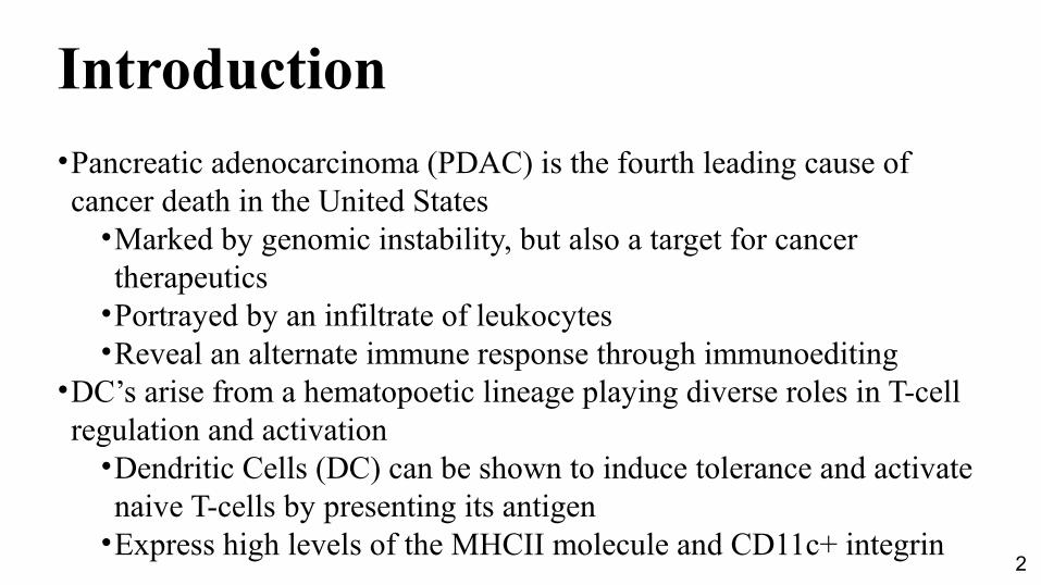
Introduction•Pancreatic adenocarcinoma (PDAC) is the fourth leading cause of cancer death in the United States
•Marked by genomic instability, but also a target for cancer therapeutics
•Portrayed by an infiltrate of leukocytes•Reveal an alternate immune response through immunoediting
•DC’s arise from a hematopoetic lineage playing diverse roles in T-cell regulation and activation
•Dendritic Cells (DC) can be shown to induce tolerance and activate naive T-cells by presenting its antigen
•Express high levels of the MHCII molecule and CD11c+ integrin

Function of Dendritic Cells: T-cell Activation
DC naive T-cell
Th1 cell
Th2 cell
T regulatory cell
On the figure to the right,the DC’s present their antigento the naive T-cells in order toactivate them. As it can be seen, the DC’s can favor a different response by the T-cells they activate, whether it is a Th1 responseor a Th2 response.

4
Question
How can we reveal the behavior of dendritic cells interacting with other leukocytes and other types of cells in the context of pancreatic adenocarcinoma?

5
Goals•To make observations regarding the locations of dendritic cells relative to T-lymphocytes and other leukocytes that are present in lymphoid tissue
•Through these observations, there can be many suggestions regarding the dynamics of DC interactions and the type of effector responses that may be elicited by the DC. T
•To make conclusions regarding morphology (shapes and sizes of cells) and quantity of DC
•Suggest much about the cell’s activation state and possibly elucidate the current boundaries to DC-T cell mediated therapies for pancreatic cancer.

6
Materials and Methods
FC1242 cells were injected into the pancreas of the mouse. After 8-10 weeks, the spleen was harvested, fixed, and flash-frozen in a liquid nitrogen bath after being inserted in OCT media. Tissue were then sentto the CryoStat for frozen slides

Materials and Methods: Immunohistochemistry
An immunohistochemistry was conducted on the frozen spleen tissue. Antigen retrieval was done with Proteinase K, and the slides were blocked using 10% goat serum, H202, and avidin/streptavidin blocking kit. Slides were washed in Tris Buffered Saline with .1% tween.

8
Figure 1. There was an elevated number of CD11c+ dendritic cells in the pancreatic
cancer model

Figure 1a. There is an elevated number of CD11c+ dendritic cells in the spleen of FC1242-pancreatic cancer-bearing mice as compared to the spleen of non-tumor bearing mice.

Figure 1b. Elevated number of CD11c+ DC were observed in the peri-follicular region
• Elevated number of CD11c+ DC in the spleen tissue overall
• Normally, CD11c+ are seen around areas in which the T-cells congregate
• DCs were observed around the follicular (B-cell) regions of the tissue

11
Results•Cells expressing the antigen CD11c+ were counted in 5-10 40x high power fields (HPF) on each biological replicate/specimen, and averaged (Figure 1B).
•There were a greater amount of cells (avg= 369.67) compared to the non-tumor bearing spleen model (avg=221.1) (Figure 1B)
•Observed CD11c+ cells were expressed in larger quantity in marginal zones and around the B cell follicles (Figure 1A). Cells in the non-tumor bearing spleen (sham- control) were seen to have a smaller average of dendritic cells in both extra-follicular (avg=174.6) and perifollicular regions (avg= 46.5).
•In the tumor-bearing spleen, there was an average of 279.67 in the perifollicular regions and an average of 90 extra-follicular regions(Figure 1C).

Figure 2. Spleen-infiltrating DC of tumor-bearing mice congregate with invading pancreatic tumor cells

Figure 2a. Immunofluorescence on spleen tissue expressing CD11c (green) and CK19 (red).

Figure 2b. Invasive cancer IHC with CD11c (Magenta) and CK19 (brown).

15
Results•Invading cancer cells, represented by CK19 staining, as well as DC were observed in contact or close proximity to one another,
•Especially around the arterioles of the spleen where afferent blood flow, naive T cells, and activated DC normally enter the spleen.
•The cancer cells began to form ductal structures, which is typical of PDAC cells, and also known as a more “differentiated” state (Figure 2B);
•there was also many mesenchymal tumor cells (Figure 2B, arrow) that were not in ductal structures but had enlarged nuclei that were misshapen, indicating genomic instability.
•“macro-metastases” seem to intermingle with dendritic cells entering the spleen (Figure 2A)
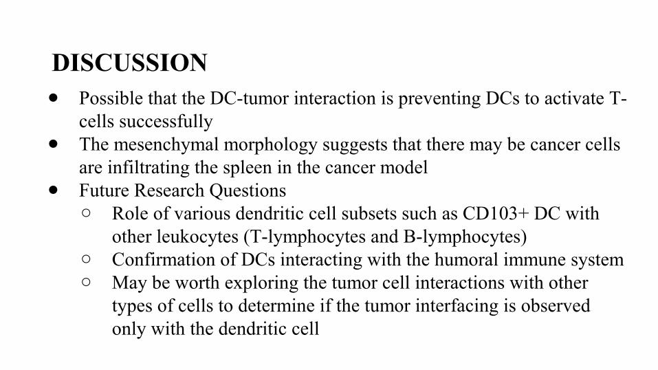
DISCUSSION● Possible that the DC-tumor interaction is preventing DCs to activate T-
cells successfully● The mesenchymal morphology suggests that there may be cancer cells
are infiltrating the spleen in the cancer model● Future Research Questions
○ Role of various dendritic cell subsets such as CD103+ DC with other leukocytes (T-lymphocytes and B-lymphocytes)
○ Confirmation of DCs interacting with the humoral immune system○ May be worth exploring the tumor cell interactions with other
types of cells to determine if the tumor interfacing is observed only with the dendritic cell

17
Acknowledgments
Rocky Barilla and Dr. George Miller, MD, Ph.DResearch Fellow and Principle Investigator
NYU Langone Medical Center Dept. of Surgery and Cell Biology
Dr. JoAnn GensertSenior Research Biology InstructorThe Bronx High School of Science