Siebel Stem Cell Institute - First Virtual Group Stem Cell Institute 2015 Annual Report... ·...
39
a UNIVERSITY OF CALIFORNIA, BERKELEY and STANFORD UNIVERSITY collaboration Siebel Stem Cell Institute ANNUAL REPORT 2015
Transcript of Siebel Stem Cell Institute - First Virtual Group Stem Cell Institute 2015 Annual Report... ·...

a U N I V E R S I T Y O F C A L I F O R N I A , B E R K E L E Y
a n d S T A N F O R D U N I V E R S I T Y c o l l a b o r a t i o n
Siebel Stem Cell InstituteA N N U A L R E P O R T
2 0 1 5


C O N T E N T S
I n t r o d u c t i o n . . . . . . . . . . . . . . . . . . . . . . . . . . . . . . . . . . . . . . . 1
S i e b e l S t e m C e l l I n s t i t u t e a t S t a n f o r d . . . . . . . . . . . . . . . . . . . . 2
S e e d G r a n t s B u i l d L o n g - T e r m C o l l a b o r a t i o n s . . . . . . . . . . . . . . 1 6
S i e b e l S t e m C e l l I n s t i t u t e a t B e r k e l e y . . . . . . . . . . . . . . . . . . . 1 8
S i e b e l S t e m C e l l I n s t i t u t e S y m p o s i u m . . . . . . . . . . . . . . . . . . . 2 9
C o n c l u s i o n . . . . . . . . . . . . . . . . . . . . . . . . . . . . . . . . . . . . . . . 3 0
A p p e n d i x A : S i e b e l S t e m C e l l I n s t i t u t e S y m p o s i u m A g e n d a a n d S p e a k e r s . . . . . . . . . . . . . . . . . . . . . . . . . . . . . . . . 3 2
A p p e n d i x B : M a k e n a C a p i t a l M a n a g e m e n t Q 4 F u n d R e p o r t . . . . . 3 4
A p p e n d i x C : F u n d A c c o u n t a n d A c t i v i t y S u m m a r y O c t o b e r 1 , 2 0 1 4 – S e p t e m b e r 3 0 , 2 0 1 5 . . . . . . . . . . . . . . . . . . . 3 5

S I E B E L S T E M C E L L I N S T I T U T E
1
I N T R O D U C T I O N
With deep gratitude to the Thomas and Stacey Siebel Foundation, we are pleased to present the eighth annual report on the Siebel Stem Cell Institute . The Institute facilitates a generative cross-pollination of ideas and resources among stem cell science investigators at the University of California, Berkeley and Stanford University . By establishing the Siebel Stem Cell Institute, the Thomas and Stacey Siebel Foundation has made it possible for leading investigators to collaborate with their peers and with early-career scientists to work toward unleashing the power of stem cells for therapeutic purposes . Since its inception in 2008, the Siebel Stem Cell Institute has brought together 83 established and emerging investigators from across the U .S . and ten foreign countries to cultivate the extraordinary healing potential of stem cells . The Siebel Stem Cell Institute enables this vital exchange of resources at a critical moment in the life and biomedical sciences . Recent technological advances have made it possible to analyze cellular behaviors in real time and to shape genetic material in ways that were inconceivable in the recent past . With the generous support of the Thomas and Stacey Siebel Foundation, researchers at Berkeley and Stanford are getting a better look at our bodies’ fundamental building blocks in real time, elucidating the subtleties of gene expression, developing predictive models that will learn from data to advance the pace of discovery, improving and advancing groundbreaking genome editing technologies, and making key contributions to the creation of new therapies for intractable illnesses . The Siebel Stem Cell Institute is composed of the Siebel Visiting Scholars and Postdoctoral Fellows, innovative research partnerships funded by Siebel Stem Cell Institute Seed Grants, and, most recently, Siebel Faculty Fellows . The flexibility and responsive nature of the Siebel Stem Cell Institute advances our long-term research goals by aiding the recruitment and cultivation of core faculty who drive this work forward in collaboration with students and colleagues at both institutions . As this remarkable research enterprise evolves, we remain dedicated to the mission of the Siebel Stem Cell Institute: to foster creativity and ingenuity at the forefront of the biomedical sciences . In this report, we are pleased to share updates on the activities of the Siebel Stem Cell Institute in 2015 .

2
S I E B E L S T E M C E L L I N S T I T U T E
S I E B E L S T E M C E L L I N S T I T U T E A T S T A N F O R D
Under the oversight of Irving L . Weissman, MD, the Virginia and D .K . Ludwig Professor for Clinical Investigation in Cancer Research and the director of the Siebel Stem Cell Institute, the Stanford Institute for Stem Cell Biology and Regenerative Medicine, and the Ludwig Center for Cancer Stem Cell Research and Medicine, eleven Siebel Scholars furthered their research in blood-forming stem cells, embryonic stem cells, and cancer stem cells . Sharing a common goal to target the root causes of today’s most devastating diseases and translate discoveries into new therapies, these scholars made important progress in their research during 2015 . To follow, please find updates on these extraordinary scholars:
Maddalena Adorno, PhD, Instructor, Institute for Stem Cell Biology and Regenerative Medicine(Lab: Michael Clarke, MD, Karel H . and Avice N . Beekhuis Professor in Cancer Biology)
Michelle Ho, PhD, Bioengineering(Lab: Chris Garcia, PhD, Professor of Molecular and Cellular Physiology and of Structural Biology)
Wan-Jin Lu, PhD, Postdoctoral Research Fellow, Institute for Stem Cell Biology and Regenerative Medicine(Lab: Philip Beachy, PhD, Ernest and Amelia Gallo Professor and Professor of Biochemistry and of Developmental Biology)
Siddhartha S. Mitra, PhD, Senior Scientist, Institute for Stem Cell Biology and Regenerative Medicine(Lab: Samuel Cheshier, MD, PhD, Assistant Professor of Neurosurgery and, by courtesy, of Neurology at Lucile Packard Children’s Hospital)
Aaron Newman, PhD, Postdoctoral Research Fellow, Institute for Stem Cell Biology and Regenerative Medicine(Lab: Ash Alizadeh, MD, PhD, Assistant Professor of Oncology)
Vittorio Sebastiano, PhD, Assistant Professor, OB/GYN and Institute for Stem Cell Biology and Regenerative Medicine(Lab: Vittorio Sebastiano, PhD)
Navdar Sever, PhD, Research Scientist, Institute for Stem Cell Biology and Regenerative Medicine
3
S I E B E L S T E M C E L L I N S T I T U T E
(Lab: Philip Beachy, PhD, Ernest and Amelia Gallo Professor and Professor of Biochemistry and of Developmental Biology)
Mark Wossidlo, PhD, Postdoctoral Research Fellow, Institute for Stem Cell Biology and Regenerative Medicine(Lab: Joanna Wysocka, PhD, Professor of Chemical and Systems Biology and of Developmental Biology)
Ryo Yamamoto, MD, PhD, Postdoctoral Research Fellow, Institute for Stem Cell Biology and Regenerative Medicine(Lab: Hiromitsu Nakauchi, MD, PhD, Professor of Genetics)
Nan Yang, PhD, Postdoctoral Research Fellow, Institute for Stem Cell Biology and Regenerative Medicine(Lab: Marius Wernig, MD, Associate Professor of Pathology and, by courtesy, of Chemical and Systems Biology)
Fangfang Zhu, PhD, Postdoctoral Research Fellow, Institute for Stem Cell Biology and Regenerative Medicine(Lab: Irving Weissman, MD, Virginia and D . K . Ludwig Professor for Clinical Investigation in Cancer Research)
4
S I E B E L S T E M C E L L I N S T I T U T E
S I E B E L S C H O L A R S :
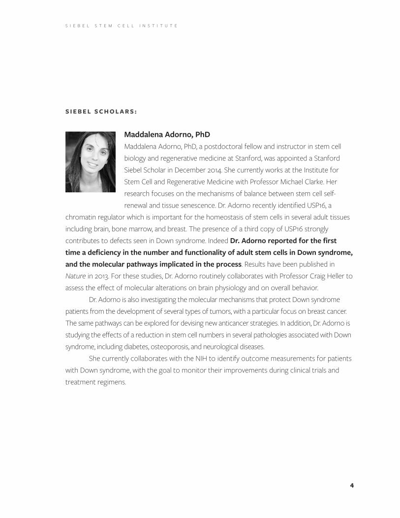
Maddalena Adorno, PhD Maddalena Adorno, PhD, a postdoctoral fellow and instructor in stem cell biology and regenerative medicine at Stanford, was appointed a Stanford Siebel Scholar in December 2014 . She currently works at the Institute for Stem Cell and Regenerative Medicine with Professor Michael Clarke . Her research focuses on the mechanisms of balance between stem cell self-renewal and tissue senescence . Dr . Adorno recently identified USP16, a
chromatin regulator which is important for the homeostasis of stem cells in several adult tissues including brain, bone marrow, and breast . The presence of a third copy of USP16 strongly contributes to defects seen in Down syndrome . Indeed Dr. Adorno reported for the first time a deficiency in the number and functionality of adult stem cells in Down syndrome, and the molecular pathways implicated in the process . Results have been published in Nature in 2013 . For these studies, Dr . Adorno routinely collaborates with Professor Craig Heller to assess the effect of molecular alterations on brain physiology and on overall behavior .
Dr . Adorno is also investigating the molecular mechanisms that protect Down syndrome patients from the development of several types of tumors, with a particular focus on breast cancer . The same pathways can be explored for devising new anticancer strategies . In addition, Dr . Adorno is studying the effects of a reduction in stem cell numbers in several pathologies associated with Down syndrome, including diabetes, osteoporosis, and neurological diseases .
She currently collaborates with the NIH to identify outcome measurements for patients with Down syndrome, with the goal to monitor their improvements during clinical trials and treatment regimens .

5
S I E B E L S T E M C E L L I N S T I T U T E
Michelle Ho, PhD Michelle Ho, PhD, has been a Siebel Scholar since September 2012 . In 2015, she was a fifth-year graduate student in bioengineering at Stanford, jointly advised by Dr . Weissman and Dr . Chris Garcia . Broadly, she is interested in using protein engineering and biochemical engineering approaches to develop potentially therapeutic immunomodulatory molecules . One of the targets she has been working on is the “don’t eat me” signal,
which has been shown to regulate phagocytosis, an essential process in the body to clear pathogens, dead cells, and cancer cells . The “don’t eat me” signal consists of a pair of proteins: CD47, which is expressed by essentially all cell types in the body, but at abnormally high levels on most of the cancer cell types in order to evade immune surveillance; and SIRPα, which is expressed mostly by professional phagocytes, or white blood cells dedicated to phagocytosis .
Dr . Ho has made a significant contribution to the development of a CD47-targeting molecule that instructs the macrophages to better recognize and eliminate cancer cells, and an article describing this work appeared in the journal Science . CD47-targeting molecules can target essentially all cells in the body, raising concerns with low bioavailability and off-target effects that can lead to toxicity . Therefore, Dr. Ho continued to improve potential therapeutics on the “don’t eat me” axis by using a more targeted approach — namely to engineer an equivalent SIRPα-targeting molecule that promotes tumor clearance by macrophages . This work was recently published in Journal of Biological Chemistry .
In addition, she is targeting the KIT/stem cell factor (SCF) interaction for applications in radiation/chemotherapy for the treatment of cancer, as well as the preparatory regime for bone marrow transplant . These treatments destroy the bloodforming system and render the patient anemic and susceptible to infections and other complications . It has been shown that SCF can mitigate these symptoms, speed up the recovery of the immune system, and increase survival . It does so by engaging the receptor KIT on the hematopoietic/blood-forming stem cells . Activation of KIT initiates a complex network of signals that leads to growth and survival of the stem cell . However, SCF also activates mast cells and induces allergic responses . In a Phase I clinical trial, SCF has exhibited significant toxicity related to mast cell activation in human patients . Using protein engineering approaches, Dr. Ho and her colleagues have created a

6
S I E B E L S T E M C E L L I N S T I T U T E
version of SCF that retains the ability to stimulate stem cell growth and survival, and that exerts minimal effects on mast cells to reduce toxicity . By thus lowering mortality and morbidity rates of radiation/chemotherapy and conditioned bone marrow transplant, Dr . Ho seeks to decrease posttreatment mortality from infection, thereby speeding patient recovery and lowering healthcare costs . Dr . Ho is currently preparing the manuscript for publication .
Wan-Jin Lu, PhD Wan-Jin Lu, PhD, is a postdoctoral fellow and became a Siebel Scholar in December 2015 . She has worked with Dr . Philip Beachy at Stanford University’s Institute of Stem Cell Biology and Regenerative Medicine since 2011 . Dr . Lu has extensive experience and expertise in the field of genetics and cancer biology . Her current research focuses on the role of Hedgehog
signaling in maintenance and regeneration of cells responsible for taste . Taste sensation becomes a significant clinical issue in cancer patients treated with a Hedgehog (Hh) pathway antagonist who are forced or choose to discontinue therapy due to loss of taste, a specific biological effect caused by this class of mechanism-based cancer therapeutic agents . Alluded to by several clinical reports in 2012, Dr . Lu established a pharmacological blockade of Hh signaling in mice, and found that Hh antagonism leads to dramatic loss of taste receptor cells (TRCs), indicating a central role of Hh signaling in TRC maintenance . She also found that regeneration of TRCs occurs upon removal of Hh blockade, due to the Hh signaling elicited from the innervating neurons .
Dr . Lu’s research aims to provide new insights on tissue regeneration, a process that not only involves proliferation of the stem/progenitor cells but also their differentiation to the appropriate cell fates for restoring their original functions . Dr . Lu will continue her research as a Siebel Scholar to investigate how neurons can guide stem/progenitor cell activities for regeneration by identifying signals that could facilitate the process . Her advances will expand our biological understanding of homeostasis and regeneration of sensory organs generally, and will improve our ability to prevent loss of taste or facilitate its recovery in patients undergoing cancer therapy.
7
S I E B E L S T E M C E L L I N S T I T U T E
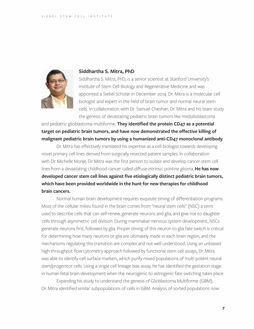
Siddhartha S. Mitra, PhDSiddhartha S . Mitra, PhD, is a senior scientist at Stanford University’s Institute of Stem Cell Biology and Regenerative Medicine and was appointed a Siebel Scholar in December 2014 . Dr . Mitra is a molecular cell biologist and expert in the field of brain tumor and normal neural stem cells . In collaboration with Dr . Samuel Cheshier, Dr . Mitra and his team study the genesis of devastating pediatric brain tumors like medulloblastoma
and pediatric glioblastoma multiforme . They identified the protein CD47 as a potential target on pediatric brain tumors, and have now demonstrated the effective killing of malignant pediatric brain tumors by using a humanized anti-CD47 monoclonal antibody .
Dr . Mitra has effectively translated his expertise as a cell biologist towards developing novel primary cell lines derived from surgically resected patient samples . In collaboration with Dr . Michelle Monje, Dr . Mitra was the first person to isolate and develop cancer stem cell lines from a devastating childhood cancer called diffuse intrinsic pontine glioma . He has now developed cancer stem cell lines against five etiologically distinct pediatric brain tumors, which have been provided worldwide in the hunt for new therapies for childhood brain cancers.
Normal human brain development requires exquisite timing of differentiation programs . Most of the cellular milieu found in the brain comes from “neural stem cells” (NSC) a term used to describe cells that can self-renew, generate neurons and glia, and give rise to daughter cells through asymmetric cell division . During mammalian nervous system development, NSCs generate neurons first, followed by glia . Proper timing of this neuron to glia fate switch is critical for determining how many neurons or glia are ultimately made in each brain region, and the mechanisms regulating this transition are complex and not well understood . Using an unbiased high throughput flow cytometry approach followed by functional stem cell assays, Dr . Mitra was able to identify cell surface markers, which purify mixed populations of multi potent neural stem/progenitor cells . Using a single cell lineage bias assay, he has identified the gestation stage in human fetal brain development when the neurogenic to astrogenic fate switching takes place .
Expanding his study to understand the genesis of Glioblastoma Multiforme (GBM), Dr . Mitra identified similar subpopulations of cells in GBM . Analysis of sorted populations now

8
S I E B E L S T E M C E L L I N S T I T U T E
suggests that the gene signature of the molecular subtype classification in GBM is not evident in the glioma stem cell, but rather the epigenetic and somatic mutation signature is . Dr . Mitra’s research as a Siebel Scholar focused on understanding normal and glioma stem/progenitor lineage hierarchy . He has identified key signaling pathways such as the JAK-STAT and Notch1 signaling pathways which are upregulated during gliomagenesis which can now be targeted to treat this devastating disease.
Aaron Newman, PhD Aaron Newman, PhD, is an instructor at Stanford and became a Siebel Scholar in August 2011 . Since then, he has worked in the laboratories of Drs . Alizadeh and Weissman to develop novel genome informatics methods to (1) improve the detection and monitoring of cancer, and (2) identify and characterize tumor-associated cell subsets that can be leveraged to define improved clinical biomarkers and therapeutic targets .
With Dr . Alizadeh, Dr. Newman developed CIBERSORT, a novel data analysis method for enumerating different cell types, such as immune cells and stem cells, in gene expression profiles of bulk tissues . Unlike methods that require living cells as input, this approach can be used to analyze both fresh and archival clinical specimens with relative ease and high-throughput, and has utility for the discovery of novel cellular biomarkers . When applied to gene expression profiles from 25 tumor types, CIBERSORT revealed complex associations between 22 distinct immune cell types and cancer survival . Papers describing the CIBERSORT method and its application to tumor-infiltrating immune cells across cancers were published in Nature Methods and Nature Medicine in 2015 .
Dr . Newman has also created new computational tools for noninvasive detection and monitoring of cancer patients . Working with Drs . Alizadeh and Diehn, Dr . Newman co-developed CAPP-Seq, a new approach for ultrasensitive detection of blood-based circulating tumor DNA . A manuscript describing this work was published in Nature Medicine in 2014 . Dr . Newman is continuing to refine this method to improve its detection limit, extend it to diverse cancer types, and perform biopsy-free screening and tumor genotyping . Several new manuscripts describing these efforts are currently in preparation and under review .
9
S I E B E L S T E M C E L L I N S T I T U T E
Finally, with Dr . Weissman and colleagues at the Hopkins Marine Station in Pacific Grove, CA, Dr . Newman has studied the genome and histocompatibility locus of Botryllus schlosseri, a marine colonial organism that engages in a natural transplantation reaction . When two colonies physically engage, they either fuse to become one colony or reject one another in a reaction reminiscent of organ and tissue rejection in humans . Dr . Newman developed new software tools that led to the discovery of a single gene whose sequence variants reliably predict fusion/rejection outcomes . This research has potentially significant implications for transplantation medicine, including allogeneic stem cell transplants, and was published in Science in 2013 .
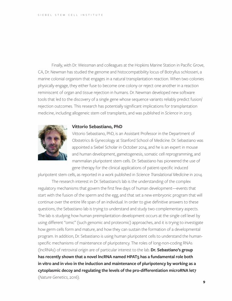
Vittorio Sebastiano, PhD Vittorio Sebastiano, PhD, is an Assistant Professor in the Department of Obstetrics & Gynecology at Stanford School of Medicine . Dr . Sebastiano was appointed a Siebel Scholar in October 2014, and he is an expert in mouse and human development, gametogenesis, somatic cell reprogramming, and mammalian pluripotent stem cells . Dr . Sebastiano has pioneered the use of gene therapy for the clinical applications of patient-specific induced
pluripotent stem cells, as reported in a work published in Science Translational Medicine in 2014 .The research interest in Dr . Sebastiano’s lab is the understanding of the complex
regulatory mechanisms that govern the first few days of human development—events that start with the fusion of the sperm and the egg, and that set a new embryonic program that will continue over the entire life span of an individual . In order to give definitive answers to these questions, the Sebastiano lab is trying to understand and study two complementary aspects . The lab is studying how human preimplantation development occurs at the single cell level by using different “omic” (such genomic and proteomic) approaches, and it is trying to investigate how germ cells form and mature, and how they can sustain the formation of a developmental program . In addition, Dr . Sebastiano is using human pluripotent cells to understand the human-specific mechanisms of maintenance of pluripotency . The roles of long-non-coding RNAs (lncRNAs) of retroviral origin are of particular interest to the lab . Dr. Sebastiano’s group has recently shown that a novel lncRNA named HPAT5 has a fundamental role both in vitro and in vivo in the induction and maintenance of pluripotency by working as a cytoplasmic decoy and regulating the levels of the pro-differentiation microRNA let7 (Nature Genetics, 2016) .
10
S I E B E L S T E M C E L L I N S T I T U T E
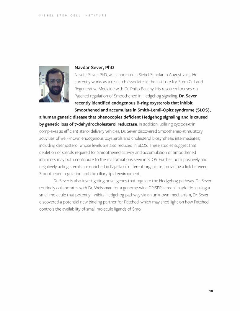
Navdar Sever, PhD Navdar Sever, PhD, was appointed a Siebel Scholar in August 2015 . He currently works as a research associate at the Institute for Stem Cell and Regenerative Medicine with Dr . Philip Beachy . His research focuses on Patched regulation of Smoothened in Hedgehog signaling . Dr. Sever recently identified endogenous B-ring oxysterols that inhibit Smoothened and accumulate in Smith-Lemli-Opitz syndrome (SLOS),
a human genetic disease that phenocopies deficient Hedgehog signaling and is caused by genetic loss of 7-dehydrocholesterol reductase . In addition, utilizing cyclodextrin complexes as efficient sterol delivery vehicles, Dr . Sever discovered Smoothened-stimulatory activities of well-known endogenous oxysterols and cholesterol biosynthesis intermediates, including desmosterol whose levels are also reduced in SLOS . These studies suggest that depletion of sterols required for Smoothened activity and accumulation of Smoothened inhibitors may both contribute to the malformations seen in SLOS . Further, both positively and negatively acting sterols are enriched in flagella of different organisms, providing a link between Smoothened regulation and the ciliary lipid environment .
Dr . Sever is also investigating novel genes that regulate the Hedgehog pathway . Dr . Sever routinely collaborates with Dr . Weissman for a genome-wide CRISPR screen . In addition, using a small molecule that potently inhibits Hedgehog pathway via an unknown mechanism, Dr . Sever discovered a potential new binding partner for Patched, which may shed light on how Patched controls the availability of small molecule ligands of Smo .

11
S I E B E L S T E M C E L L I N S T I T U T E
Mark Wossidlo, PhD Mark Wossidlo, PhD, is a postdoctoral research fellow at Stanford and was appointed a Siebel Scholar in November 2014 . He came to Stanford in 2012 as a postdoctoral fellow in the laboratory of Dr . Renee Reijo Pera and subsequently joined the laboratory of Dr . Joanna Wysocka in 2014 . Dr . Wossidlo has deep and extensive research experience in epigenetics (the study of the inheritable chemical modifications of the mammalian genome
associated with gene expression) and early mammalian embryogenesis . He has contributed greatly to the understanding of DNA methylation reprogramming by identifying epigenetic key players . In this intrinsic process during early embryogenesis, Dr . Wossidlo discovered the involvement of the sixth base of the genome, 5-hydroxymethyl-cytosine, in active demethylation of the zygotic genome and identified Tet3 as the responsible enzyme . While investigating potential DNA repair pathways in the process of zygotic reprogramming, he also shed new light on mechanisms of DNA methylation reprogramming .
His current research focus at Stanford is to understand the impact of epigenetic reprogramming on the development of mammalian embryos, and in particular human preimplantation embryos . The analysis of epigenetic reprogramming in mammals has mainly focused on the mouse model and little is known about human embryonic development . Human embryogenesis is remarkably inefficient: only 30-50% of fertilized oocytes develop to the blastocyst stage at day five in human embryogenesis . This is in stark contrast to the mouse model, where 80-90% of zygotes develop to term . In his current research, Dr . Wossidlo discovered an essential role for Tet enzymes in early mouse and human embryogenesis . His work demonstrates for the first time that DNA methylation reprogramming is a key component of the clock that regulates timing and extent of embryonic genome activation in mammalian embryos . Furthermore, by using novel sequencing technologies to analyze single cell gene expression patterns in early human embryos, Dr . Wossidlo created the first in silico three-dimensional model of a human blastocyst at day five and day six of human embryogenesis . This model identified epigenetic key factors, which are able to generate “naïve” human pluripotent stem cells in vitro and are the starting point to generate improved pluripotent human stem cells for regenerative medicine .

12
S I E B E L S T E M C E L L I N S T I T U T E
In addition, Dr . Wossidlo is collaborating with several groups in Stanford and outside to investigate the fundamentals of early human embryogenesis and the characteristics of true totipotency and pluripotency in vivo . The long-term objective of his studies is focused on the embryo intrinsic epigenetic reprogramming that underlies early cell fate decisions and cellular potency . Understanding these fundamental processes will ultimately translate into the generation of improved toti- and pluripotent stem cells and the treatment of infertility, particularly through improved outcomes of human-assisted reproduction and induced reprogramming of somatic cells for regenerative medicine .
Ryo Yamamoto, MD, PhD Ryo Yamamoto, MD, PhD, is a research associate at the Institute of Stem Cell Biology and Regenerative Medicine and was appointed a Siebel Scholar in December 2015 .
Dr, Yamamoto joined Dr . Hiromitsu Nakauchi’s lab and started research on hematopoietic stem cells (HSCs) at the University of Tokyo after years of clinical training in hematology . During this time, he has
demonstrated heterogeneity of HSCs extensively with single cell transplantation assay . He has analyzed over 500 single HSC transplanted mice in great detail . Subsequently, he discovered a novel type of lineage-restricted hematopoietic progenitor cells with long-term repopulating capacity such as megakaryocytic repopulating progenitors (MRPs), common myeloid repopulating progenitors (CMRPs) and megakryo-erythroid repopulating progenitors (MERPs) . He showed that these repopulating progenitors are able to differentiate directly from HSCs rather than from multipotent progenitors by using a paired-daughter cell analysis and in vivo single cell transplantation . His research provided new insights into the biology of HSCs and was published in the journal CELL .
Now relocated to Stanford University, he is working on single cell transplantation of aged HSCs . He has already discovered that aged HSCs show atypical kinetics in vivo at the single cell level . To determine differences in molecular signatures between young and aged HSCs, he performed RNAseq of single cells of young and aged HSCs in collaboration with the Weissman lab .
13
S I E B E L S T E M C E L L I N S T I T U T E
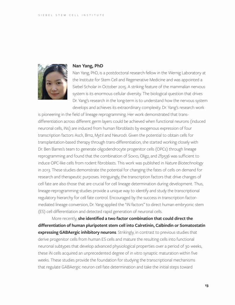
Nan Yang, PhD Nan Yang, PhD, is a postdoctoral research fellow in the Wernig Laboratory at the Institute for Stem Cell and Regenerative Medicine and was appointed a Siebel Scholar in October 2015 . A striking feature of the mammalian nervous system is its enormous cellular diversity . The biological question that drives Dr . Yang’s research in the long-term is to understand how the nervous system develops and achieves its extraordinary complexity . Dr . Yang’s research work
is pioneering in the field of lineage reprogramming . Her work demonstrated that trans-differentiation across different germ layers could be achieved when functional neurons (induced neuronal cells, iNs) are induced from human fibroblasts by exogenous expression of four transcription factors Ascl1, Brn2, Myt1l and Neurod1 . Given the potential to obtain cells for transplantation-based therapy through trans-differentiation, she started working closely with Dr . Ben Barres’s team to generate oligodendrocyte progenitor cells (OPCs) through lineage reprogramming and found that the combination of Sox10, Olig2, and Zfp536 was sufficient to induce OPC-like cells from rodent fibroblasts . This work was published in Nature Biotechnology in 2013 . These studies demonstrate the potential for changing the fates of cells on demand for research and therapeutic purposes . Intriguingly, the transcription factors that drive changes of cell fate are also those that are crucial for cell lineage determination during development . Thus, lineage-reprogramming studies provide a unique way to identify and study the transcriptional regulatory hierarchy for cell fate control . Encouraged by the success in transcription factor-mediated lineage conversion, Dr . Yang applied the “iN factors” to direct human embryonic stem (ES) cell differentiation and detected rapid generation of neuronal cells .
More recently, she identified a two factor combination that could direct the differentiation of human pluripotent stem cell into Calretinin, Calbindin or Somatostatin expressing GABAergic inhibitory neurons . Strikingly, in contrast to previous studies that derive progenitor cells from human ES cells and mature the resulting cells into functional neuronal subtypes that develop advanced physiological properties over a period of 30 weeks, these iN cells acquired an unprecedented degree of in vitro synaptic maturation within five weeks . These studies provide the foundation for studying the transcriptional mechanisms that regulate GABAergic neuron cell fate determination and take the initial steps toward

14
S I E B E L S T E M C E L L I N S T I T U T E
studying inhibitory synaptic transmission in vitro . She is now leveraging her expertise in lineage reprogramming, stem cell biology, and neurobiology to develop reprogramming-based human cell culture models and to study the fundamental processes underlying the development and function of the human nervous system under normal and pathological conditions .
Fangfang Zhu, PhD Fangfang Zhu, PhD, was appointed a Siebel Scholar in September 2014 . Interested in the study of human hematopoietic hierarchy, Dr. Zhu designed a strategy for the identification of regulators for hematopoietic development . Hematopoietic stem cells can first differentiate into hematopoietic progenitors, such as common myeloid progenitor cells, then into more restricted lineage progenitors . One of
these progenitors is Megakaryocyte-erythroid progenitors (MEPs) that could further differentiate into either megakaryocyte progenitors (MkPs) that give rise to megakaryocytes that generate platelets, or erythroid progenitors (EPs) that give rise to erythrocytes . During this process, transcription factors (TFs) are activated/inactivated and play a critical regulatory role on the cell fate decision . The identification of megakaryopoiesis regulators will enable the in vitro generation of megakaryocytes from HSCs (or other cell types), which is important for the treatment of genetic blood diseases, cancer, and other pathologies affecting platelet production . Dr . Zhu is working on the project of a CRISPR-mediated gene knockout screen to determine the TFs that regulate human megakaryopoiesis .
In addition, the “don’t eat me” signal, CD47/SIRPα, may play an important role in the development of hematopoietic stem cells . Dr . Zhu studies the role of CD47 during embryo development by overexpressing the mouse CD47 in the hematopoietic system . She designed a TALEN-based targeting strategy for the ectopic expression of mouse CD47 in a specific locus of mouse genome, and received the CD47-overexpressing mouse ESCs . Collaborating with colleagues, Dr . Zhu injected mouse ESCs into blastocysts to test the establishment of germline-transmitting CD47 mice . She continues to investigate the effect of CD47-overexpression during mouse embryo development, especially in the hematopoietic system .

S I E B E L S T E M C E L L I N S T I T U T E
15
F U T U R E P R O G R A M S : S I E B E L FA C U LT Y F E L LO W S P R O G R A M
We have decided to extend the Siebel Stem Cell Institute funding to include the support of a small group of Siebel Faculty Fellows: individuals recently awarded PhDs who have shown sufficient innovation and independence that they are able to design and carry out their immediate postdoctoral research themselves . Siebel Faculty Fellows will act as principal investigators on grants and choose with whom they would like to collaborate . They will do research largely independently, with senior institute faculty serving as advisors rather than as scientific leaders . In an institute that has filled its junior faculty slots, this decision will provide a continuing infusion of new scientists whose careers we can facilitate and who can conduct high-level science within the institute in return . The Siebel Institute funding will provide a package for each Siebel Faculty Fellow to establish a small independent lab .
The Siebel Faculty Fellows program is currently being set up along lines similar to other prestigious research fellowships, such as the Whitehead Institute Fellows Program or the UCSF Sandler Fellows Program . Siebel Faculty Fellows will be funded for a three-year term, which can be extended to five years if needed .
16
S I E B E L S T E M C E L L I N S T I T U T E
The Siebel Stem Cell Institute encourages investigators at Berkeley and Stanford to work together on innovative research projects that may be considered too speculative for other sources of funding . By providing seed grants for collaborative endeavors, the Institute enables investigators to share equipment and bring new perspectives to one another’s work . Since the inception of the Siebel Stem Cell Institute in 2008, seed grants have supported 33 researchers working on 14 innovative projects, several of which were deemed successful enough to warrant multiple years of support . The flexibility of the Institute enables collaborations among researchers at Berkeley and Stanford to develop over time . Recipients of seed grants have:
• made important advancements in instrumentation, creating new technologies for more efficient and accurate analysis of cellular proteins and for the imaging of living cells;
• published the results of their work in highly regarded journals; and
• garnered additional support that has enabled them to continue to develop their work .
The Institute-supported collaboration between the Tjian laboratory at Berkeley and the Steven Quake laboratory at Stanford is one such example . Initiated in 2010, this project sought to apply single cell gene expression profiling methods developed in the Quake laboratory to studies of motor neuron differentiation from human embryonic stem cells . For many years, “basal” or “general” core promoter factors (regulatory proteins) such as TAF9B were considered necessary for gene expression, but were thought not to vary among different cell types . Core factors are known to work in combination with various other proteins to switch genes on or off in specific cell types; however, the specific core promoter factors and partner proteins that guide a cell into becoming a neuron have not been well characterized .
In 2014, funding from the Institute supported the completion and publication of a study that resulted in the unexpected finding that the TBP-associated basal transcription factor TAF9B regulates gene expression during motor neuron development . These findings might, in the
S E E D G R A N T S B U I L D L O N G - T E R M C O L L A B O R A T I O N S
S I E B E L S T E M C E L L I N S T I T U T E
future, improve our ability to guide stem cells into forming neurons, or to reprogram other types of specialized cells into becoming motor neurons . This new information could also prove useful for researchers interested in better understanding neuronal development and might aid in the design of therapies to treat neuronal injuries or diseases, such as motor neuron disease . The study was published in eLife in 2014 .
With an additional seed grant in 2015, this study was extended to evaluate the role of TAF9B in the adult brain, in collaboration with the laboratories of Daniela Kaufer (Berkeley) and Michelle Monje (Stanford) . TAF9B was known to be present at elevated levels in the brain and spinal cord relative to other tissues of adult mice, but it was assumed that this expression was uniform in all cell types of the brain . By querying newly available databases for TAF9B expression in different neuronal cell types, a five-fold enrichment was found in oligodendrocyte progenitors and newly formed oligodendrocytes compared to neurons — a pattern similar to enhancer-binding transcription factors that regulate oligodendrocyte differentiation .
To confirm this experimentally, embryonic stem cells from normal and TAF9B mutant mice were grown in culture and differentiated into oligodendrocytes . Preliminary data suggest that differentiation of embryonic stem cells from TAF9B mutant mice generates many fewer neural progenitor cells, oligodendrocyte progenitors, and mature oligodendrocytes, and instead shows increased expression of astrocyte-specific markers, suggesting a skewed differentiation potential toward astrocyte generation . These studies are being followed up with in vivo experiments in normal and TAF9B mutant mice to confirm the in vitro observations and to investigate the role of TAF9B in developmental and adaptive myelination processes .
17
18
S I E B E L S T E M C E L L I N S T I T U T E
Under the leadership of Dr . David Schaffer, the Siebel Stem Cell Institute makes vital contributions to Berkeley’s research enterprise, drawing top scientific talent to campus to work toward unleashing the power of stem cells to support human health .
From postdoctoral researchers and early-career faculty to established leaders breaking new ground in this dynamic field, members of the Siebel Stem Cell Institute community at Berkeley are creating advanced instrumentation to image live cells at new levels of refinement, mapping the molecular behaviors that lead to cancer and that determine the way that genetic information is transmitted, and changing our assumptions about aging . Ultimately, these investigations lay the groundwork for the development of treatments for a wide array of health challenges, from cancer and Parkinson’s disease to obesity and the healing of chronic wounds .
Over the past year, the Siebel Stem Cell Institute’s impact at Berkeley continued in several significant ways, supporting collaborative opportunities for leading scientists and driving scientific advances . In 2015, the Institute at Berkeley:
• welcomed Siebel Visiting Scholars Dana Carroll, PhD, and Christophe Zimmer, PhD;
• continued to support the work of Siebel Senior Faculty Fellow and Assistant Professor of Genetics, Genomics and Development Xavier Darzacq;
• expanded the collaboration that originated between the Robert Tjian laboratory at Berkeley and the Steven Quake laboratory at Stanford, with the support of a Siebel Seed Grant, to include the laboratories of Daniela Kaufer at Berkeley and Michelle Monje at Stanford; and
• provided critical resources for Andrew Dillin, Thomas and Stacey Siebel Distinguished Chair in Stem Cell Biology and Professor of Genetics, Genomics and Development, and Dirk Hockemeyer, Assistant Professor of Cell and Developmental Biology .
Support of these scientists and collaborations yielded important scientific advances, including:• development of state-of-the-art microscopes for visualizing live cells in vivo,
expanding our understanding of fundamental cellular processes;
S I E B E L S T E M C E L L I N S T I T U T E A T B E R K E L E Y
19
S I E B E L S T E M C E L L I N S T I T U T E
• applications of the CRISPR/Cas9 genome editing technology, including work toward a possible genetic treatment for sickle cell disease;
• algorithms and predictive machine learning models advancing regenerative medicine through new understanding of gene regulation and tissue differentiation;
• explorations into how stem cells maintain their identity as stem cells; and
• contributions to understanding the reactivation of the restorative enzyme telomerase in human tumors and its effect on tumor progression .
About the Director: Dr . David Schaffer is a Professor of Chemical and Biomolecular Engineering, Bioengineering, and Neurobiology at Berkeley . He applies engineering principles to enhance stem cell and gene therapy approaches for neuroregeneration, work that includes novel approaches for molecular engineering and evolution of new viral vectors as well as new technologies to investigate and control stem cell fate decisions . Dr . Schaffer is also a co-founder of 4D Molecular Therapeutics (4DMT), a gene therapy product research and development endeavor formed to unlock the full potential of gene therapy for patients with genetic diseases . The company’s robust discovery platform empowers the creation of customized gene delivery vehicles (novel adeno-associated virus vectors) to deliver genes to any tissue or organ in the body — such as the delivery of normal genes to tissues with defective genes (as occurs in genetic diseases) . This discovery technology was originally developed by Dr . Schaffer over a period of 15 years at Berkeley .

20
S I E B E L S T E M C E L L I N S T I T U T E
In 2014, Berkeley welcomed Xavier Darzacq to a permanent position as Assistant Professor of Genetics, Genomics, and Development, and as the inaugural Siebel Stem Cell Institute Faculty Fellow . Dr . Darzacq’s first engagement with Berkeley was as a Siebel Stem Cell Institute Visiting Scholar in 2012 . Dr . Darzacq, who was recruited from the Ecole Normale Superieure in Paris, is an expert in the study of living cells using super-resolution microscopy, and his work in this area led to a collaboration with the Tjian lab
at Berkeley . Dr . Darzacq was recruited to Berkeley with the help of a $4 .3 million California Institute for Regenerative Medicine Research (CIRM) Research Leadership Award, whose goal is to enable institutions to recruit outstanding young stem cell research innovators to pursue their work in California . The Siebel Stem Cell Institute was a crucial source of the institutional matching funds required to enable Berkeley’s eligibility for this award .
Dr . Darzacq is a member of a community of microscopists who design and modify super-resolution imaging and data analysis platforms . During his second year as a Siebel Stem Cell Senior Faculty Fellow, Dr. Darzacq and his laboratory have built and brought online two state-of-the-art imaging systems: a lattice light sheet microscope and a multi-focus microscope . In addition to providing a foundation for the work of the Darzacq and Tjian labs, these systems are available for use by investigators throughout Berkeley and the Siebel Stem Cell Institute .
Imaging of live cells enables a valuable expanded understanding of the cell, but such direct observation can involve challenges or tradeoffs such as loss of resolution as the cell moves through space over time, and the possibility that light exposure may cause damage to the cell . Successfully surmounting these significant challenges, the lattice light sheet microscope allows high-speed 3D imaging of groups of living cells over long periods of time, with exquisite signal-to-noise ratio and minimal specimen damage . Additionally, Siebel Visiting Scholar Christophe Zimmer, working with the Darzacq laboratory, is developing a compressive sensing illumination and detection strategy to further reduce the sample exposure times and to increase the speed of image reconstruction with this system . (More information on Dr . Zimmer’s work follows .)
S E N I O R F A C U L T Y F E L L O W
21
S I E B E L S T E M C E L L I N S T I T U T E
The multi-focus microscope provides simultaneous imaging of multiple focal planes in a sample, and is ideal for tracking the 3D movements of single molecules within individual cells . The Darzacq lab is using these systems to study the organization in space of chromosomal DNA and the movements of individual gene regulatory proteins in human embryonic stem cells, as well as in more specialized cells derived from the embryonic cells . Through collaborations with other Berkeley labs, these systems are also being used to investigate cellular differentiation in model organisms — such as fruit flies and worms — that have been widely studied and have much available data .
Dr . Claire Dugast-Darzacq, Assistant Research Scientist, was a Siebel Visiting Scholar in 2012-13 and currently heads the Darzacq laboratory’s studies of skin regeneration during wound healing . Chronic skin ulcers are a pervasive and dangerous public health problem . Normal wound healing is partly mediated by the mobilization of skin fibroblasts — particular cells within the second-most layer of skin which generate connective tissue — and their differentiation into contractile myofibroblasts to effect wound closure . The Darzacq laboratory has demonstrated that the transcriptional regulatory protein MED12L is largely down-regulated upon differentiation of primary human dermal fibroblasts into myofibroblasts . The laboratory is using super-resolution microscopy and genome editing to test the hypothesis that MED12L influences cell fate by locking in instructive interactions between proteins bound to DNA regulatory elements .
22
S I E B E L S T E M C E L L I N S T I T U T E
Through the Siebel Visiting Scholar program, national and international scholars of distinction are invited to become scholars-in-residence at Berkeley or Stanford . These scholars fully integrate with local faculty and students, leveraging the substantial capabilities and infrastructure of the two institutions to initiate increasingly innovative research collaborations .
2 0 1 5 S I E B E L V I S I T I N G S C H O L A R S AT B E R K E L E Y : D A N A C A R R O L L A N D
C H R I S T O P H E Z I M M E R
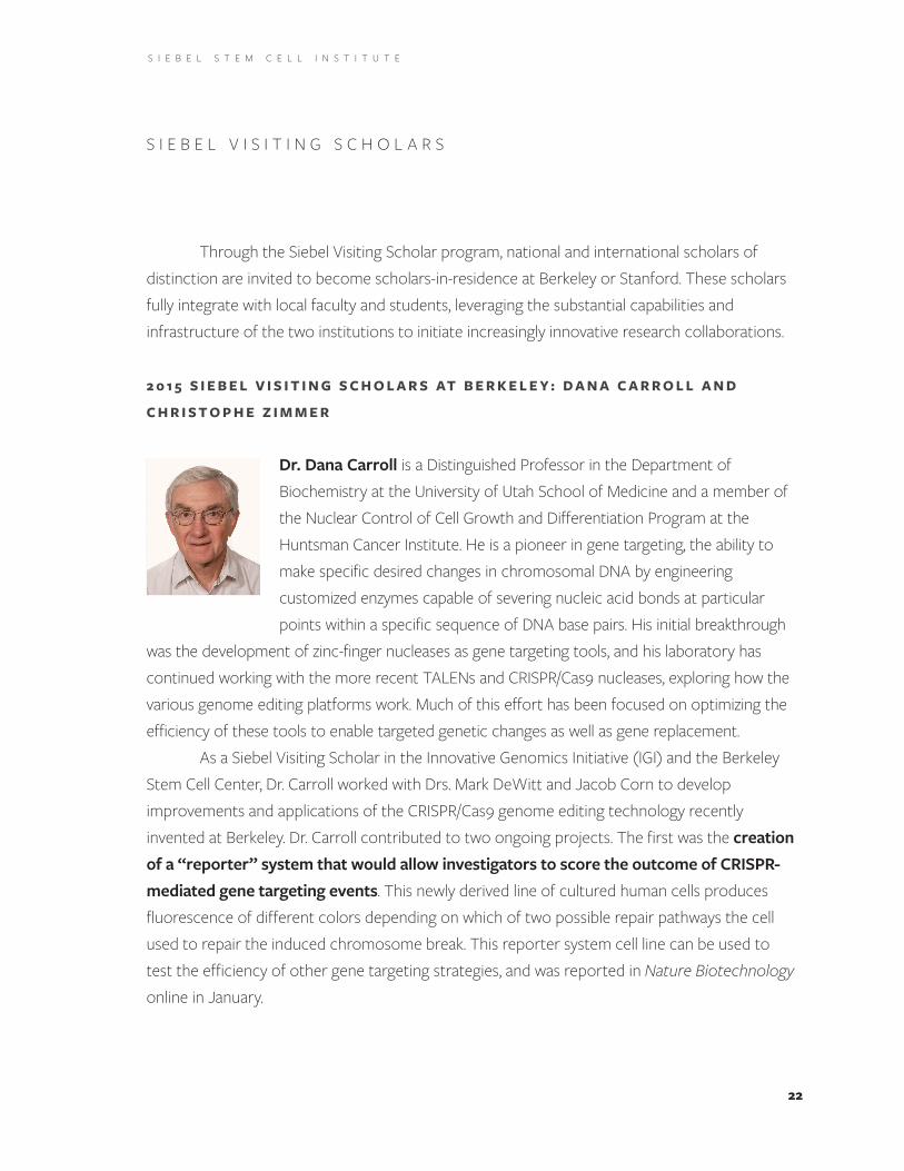
Dr. Dana Carroll is a Distinguished Professor in the Department of Biochemistry at the University of Utah School of Medicine and a member of the Nuclear Control of Cell Growth and Differentiation Program at the Huntsman Cancer Institute . He is a pioneer in gene targeting, the ability to make specific desired changes in chromosomal DNA by engineering customized enzymes capable of severing nucleic acid bonds at particular points within a specific sequence of DNA base pairs . His initial breakthrough
was the development of zinc-finger nucleases as gene targeting tools, and his laboratory has continued working with the more recent TALENs and CRISPR/Cas9 nucleases, exploring how the various genome editing platforms work . Much of this effort has been focused on optimizing the efficiency of these tools to enable targeted genetic changes as well as gene replacement .
As a Siebel Visiting Scholar in the Innovative Genomics Initiative (IGI) and the Berkeley Stem Cell Center, Dr . Carroll worked with Drs . Mark DeWitt and Jacob Corn to develop improvements and applications of the CRISPR/Cas9 genome editing technology recently invented at Berkeley . Dr . Carroll contributed to two ongoing projects . The first was the creation of a “reporter” system that would allow investigators to score the outcome of CRISPR-mediated gene targeting events . This newly derived line of cultured human cells produces fluorescence of different colors depending on which of two possible repair pathways the cell used to repair the induced chromosome break . This reporter system cell line can be used to test the efficiency of other gene targeting strategies, and was reported in Nature Biotechnology online in January .
S I E B E L V I S I T I N G S C H O L A R S
23
S I E B E L S T E M C E L L I N S T I T U T E
The second project, in collaboration with the Children’s Hospital of Oakland Research Institute (CHORI), is working toward the development of a genetic treatment for sickle cell disease . Sickle cell disease is the result of a single base-pair mutation — making it an attractive target for genetic correction — in the beta-globin gene that interferes with the usual process of transporting oxygen from the lungs to the rest of the body via the red blood cells . Drs . Carroll, DeWitt, and Corn tested a number of guide RNA sequences for targeting the beta-globin gene and identified guide sequences that were particularly effective — first in cultured human erythroleukemia cells, and then in the type of human stem cells that give rise to all the other blood cells . These gene-corrected human hematopoietic stem cells were transplanted into immune-deficient mice, where they persisted for 16 weeks and continued to produce a potentially curative proportion of circulating red blood cells . The initial studies have been submitted for publication, and the project is continuing as a collaboration between the IGI and CHORI .

24
S I E B E L S T E M C E L L I N S T I T U T E
Dr. Christophe Zimmer, Laboratory Head at the Institut Pasteur, is a physicist and engineer . After his initial work in astrophysics — where he contributed to the discovery of oceans below the surfaces of Europa and Callisto, two of Jupiter’s four largest moons — he has applied computational and physics-based approaches to biological research . Dr . Zimmer’s lab builds complex mathematical and statistical models of chromosome architecture and develops high-resolution and high-throughput imaging approaches . As a
Siebel Visiting Scholar at Berkeley, Dr . Zimmer collaborated with the Darzacq and Tjian labs on two projects: developing machine learning models to predict how genes are turned on by enhancers; and new algorithms to enable faster acquisition of data for lattice light sheet microscopy .
Machine learning project: Enhancers are regions of DNA that control and regulate gene expression, even across vast genetic distances; enhancers can be located millions of base pairs away from the promoter and protein coding sequence that they regulate . While enhancers control gene expression, promoters are regions that initiate the first step of gene expression . The specific ways and rules by which enhancers and promoters interact to drive gene expression are still mysterious .
Dr. Zimmer is developing a machine learning approach to discover the underlying rules behind these interactions, creating processes that will enable computers to learn from known data relationships to make predictions on other data sets . In Dr . Zimmer’s approach, many pairs of enhancers and promoters are provided, together with their known protein occupancy and whether they interact or not . These elements serve as data to “train” a network to predict whether two arbitrary enhancers and promoters interact or not . During his time as a Siebel Visiting Scholar, Dr . Zimmer developed a gene regulation algorithm whose accuracy he demonstrated using simulated data . His next step will be to refine the network using public data sets and/or high-throughput experiments . Ultimately, this project should yield new insights into the rules that govern how enhancers turn on specific genes in stem cell differentiation, development, and disease.
Compressive light sheet microscopy project: The Darzacq lab at Berkeley, noted earlier, has finalized the assembly of a lattice light sheet microscope, enabling researchers to

25
S I E B E L S T E M C E L L I N S T I T U T E
obtain 3D images of living systems over time with excellent spatial resolution and minimal light exposure damage to cells . However, the current system requires illumination in sequence of many focal planes in order to reconstruct a comprehensive 3D volume, which limits the resolution of the system as well as the ability to study fast cellular processes .
Dr. Zimmer worked to develop a “compressive sensing” image analysis program, which leverages the redundancy of natural signals to reconstruct them from a smaller number of measurements . Using this method, 2D projections will be recorded from many focal planes at the same time and then will be used to generate 3D images . If successful, this project will allow faster imaging of embryos and molecules as complete volumes, with applications not only in stem cell biology but for development and neurobiology as well .
26
S I E B E L S T E M C E L L I N S T I T U T E
S I E B E L F A C U L T Y S U P P O R T
Over the past year, the Siebel Stem Cell Institute was again a key source of support for the laboratories of two leading Berkeley stem cell researchers, Andrew Dillin and Dirk Hockemeyer, and their substantial contributions to the advancement of the stem cell research landscape .
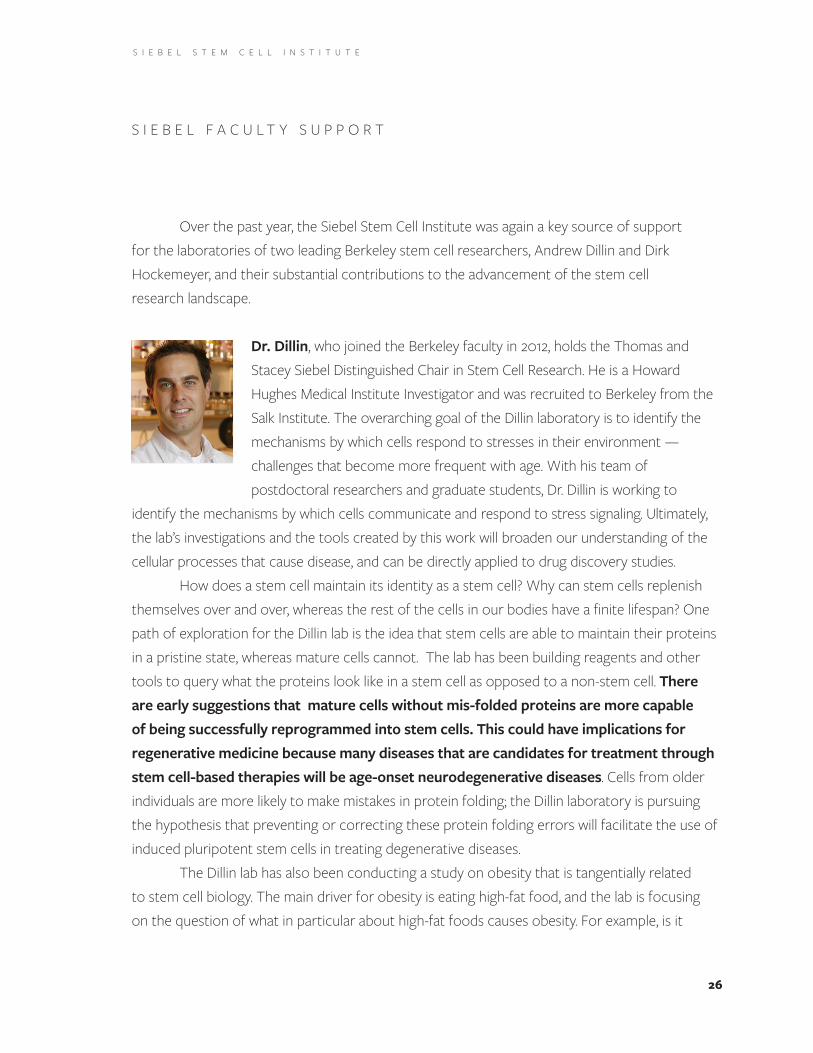
Dr. Dillin, who joined the Berkeley faculty in 2012, holds the Thomas and Stacey Siebel Distinguished Chair in Stem Cell Research . He is a Howard Hughes Medical Institute Investigator and was recruited to Berkeley from the Salk Institute . The overarching goal of the Dillin laboratory is to identify the mechanisms by which cells respond to stresses in their environment — challenges that become more frequent with age . With his team of postdoctoral researchers and graduate students, Dr . Dillin is working to
identify the mechanisms by which cells communicate and respond to stress signaling . Ultimately, the lab’s investigations and the tools created by this work will broaden our understanding of the cellular processes that cause disease, and can be directly applied to drug discovery studies .
How does a stem cell maintain its identity as a stem cell? Why can stem cells replenish themselves over and over, whereas the rest of the cells in our bodies have a finite lifespan? One path of exploration for the Dillin lab is the idea that stem cells are able to maintain their proteins in a pristine state, whereas mature cells cannot . The lab has been building reagents and other tools to query what the proteins look like in a stem cell as opposed to a non-stem cell . There are early suggestions that mature cells without mis-folded proteins are more capable of being successfully reprogrammed into stem cells. This could have implications for regenerative medicine because many diseases that are candidates for treatment through stem cell-based therapies will be age-onset neurodegenerative diseases . Cells from older individuals are more likely to make mistakes in protein folding; the Dillin laboratory is pursuing the hypothesis that preventing or correcting these protein folding errors will facilitate the use of induced pluripotent stem cells in treating degenerative diseases .
The Dillin lab has also been conducting a study on obesity that is tangentially related to stem cell biology . The main driver for obesity is eating high-fat food, and the lab is focusing on the question of what in particular about high-fat foods causes obesity . For example, is it

27
S I E B E L S T E M C E L L I N S T I T U T E
merely the assimilation of actual calories, or does sensory perception contribute? Will an animal still gain weight when it eats high-fat food if its sense of smell is diminished? By temporarily damaging the neurons in mice that pertain to sense of smell, the lab has found that animals without the ability to smell do not gain weight, despite eating as much of a high-fat diet as untreated mice who retain their sense of smell and gain weight on the same diet . Thus the lab is gaining a different point of view on obesity: it may not only be about assimilating calories, but about perceiving those calories as well . This has potential to play a role medically as a treatment for morbid obesity . The lab is currently working on preparing these results for publication .
In 2014, the Dillin lab was recognized with a prestigious award from the Glenn Foundation, which supports research on the biological processes of aging . Subsequently, the Dillin lab and a team of faculty colleagues established the new Paul F . Glenn Center for Aging Research in collaboration with UC San Francisco . Now in its second year, the Glenn Center has hired several new faculty who have started labs and leveraged Glenn Foundation support to raise further funds . We are pleased to share this news as it demonstrates the reach of the Siebel Foundation’s support and its capacity to leverage additional support — in this case, from the Glenn Foundation — which, in turn, draws further support to the university and the groundbreaking research of its faculty .
Dr. Dirk Hockemeyer received his PhD from Rockefeller University and was a postdoctoral fellow in the laboratory of Rudolf Jaenisch at MIT’s Whitehead Institute for Biomedical Research . Since joining the Berkeley faculty in 2013, he has been awarded the New Scholar in Aging of the Ellison Medical Foundation award and the H .W . Mossman Award in Developmental Biology of the American Association of Anatomists . The focus of the Hockemeyer lab is to develop primary human cell systems to illuminate and
explain the early events in the formation of a cancer, whereby normal cells are transformed into cancer cell, and to investigate the mechanisms of telomerase regulation during stem cell maintenance, differentiation, and immortalization .
28
S I E B E L S T E M C E L L I N S T I T U T E
The bulk of the DNA in the human genome is divided between 23 pairs of chromosomes, and the ends of these chromosomes contain a repetitive stretch of DNA known as a telomere . Every time a cell divides, a portion of the telomere is lost and can be restored by an enzyme called telomerase . Telomerase participates in both healthy cellular regeneration and in the development of cancerous cells . In the majority of human cells — with the exception of stem cells — there is no telomerase activity, due to the down-regulation of the active protein component (called TERT) after birth . Therefore, the telomeres in these cells shorten after each cell division . However, 90% of human cancers have very high TERT activity, which enables them to divide continuously to drive tumor growth .
While mouse models of cancer have provided important insights into basic mechanisms, key differences in tumor biology between mice and humans have hindered the translation of these findings into successful therapeutics . In the last year, the Hockemeyer lab made substantial contributions to the understanding of how telomerase activity can be reactivated in human tumors and the effect of this increased telomerase activity on tumor progression .
Previous studies of human tumor DNA had identified mutations in the regulatory DNA controlling telomerase gene expression, including the emergence of potentially regulatory “ETS” sequences . The Hockemeyer lab hypothesized that these sequences could prevent normal silencing of telomerase gene expression, and used gene editing to introduce mutations commonly found in human tumors into normal human embryonic stem cells . Unexpectedly, these changes did not increase the activity of the telomerase enzyme in these cells, nor did they increase the length of the telomeres . Next, the lab caused the genetically engineered stem cells to develop into more specialized cell types, such as nerve cells . This process of differentiation would normally silence the gene that encodes TERT, but the mutations prevented the gene from being silenced . This led to abnormally high levels of telomerase activity and long telomeres . Cells containing the mutations were injected into mice, and the cells formed a mass of tumors that contained very long telomeres . These results together suggest that cancer-causing mutations in the regulatory regions controlling TERT gene expression stop this gene from being properly silenced in more specialized cells, and that this, on its own, can promote the formation of tumors. These findings are likely to underpin future efforts to treat cancers by targeting the expression and activity of the telomerase enzyme . The findings are in press in eLife .
S I E B E L S T E M C E L L I N S T I T U T E
29
The 6th annual joint workshop of the Berkeley and Stanford Siebel Stem Cell Institutes was held at Berkeley on March 23, 2015 in the Li Ka Shing Center .
The workshop featured 15 talks by Stanford and Berkeley affiliates, including Drs . Irv Weissman, Steven Artandi, Julie Baker, Maria Barna, Michael Clarke, Kyle Loh, Michael Longaker, Michelle Monje, Aaron Newman, and Thomas Rando from Stanford, and David Schaffer, Xavier Darzacq, Jennifer Doudna, Dirk Hockemeyer, and Michael Rape from Berkeley . Speakers were Seibel Stem Cell Institute affiliates from both institutions, as well as additional Stanford faculty members nominated by Berkeley Stem Cell Center affiliates as prospective collaborators . Attendance at the talks was open to the entire campus community, which took full advantage of this outstanding opportunity to hear some of the most prominent stem cell researchers in California share their research . More than 200 faculty members, postdoctoral fellows, students, and staff attended the symposium, and a coffee break and reception provided opportunities for attendees to connect individually with the symposium speakers . Please see the appendix for the symposium agenda and list of speakers .
An integral part of the workshop was a private speakers’ lunch, at which each Stanford faculty participant was paired with a Berkeley faculty host for extended scientific discussion . Graduate student and postdoctoral Siebel Fellows from both institutions were similarly seated together . The collaboration between the Tjian, Kaufer, and Monje laboratories described in this annual report was initiated following discussions at this lunch .
S I E B E L S T E M C E L L I N S T I T U T E S Y M P O S I U M
30
S I E B E L S T E M C E L L I N S T I T U T E
C O N C L U S I O N
Since its inception, the Siebel Stem Cell Institute has served as an important incubator for ideas and collaborations among biomedical researchers at all stages of their careers . With the crucial support of the Thomas and Stacey Siebel Foundation, stem cell biologists at Berkeley and Stanford are deciphering the mysteries of aging, elucidating the cellular mechanisms that make the difference between healthy tissues and malevolent growth, and finding new ways to direct and accelerate the human body’s own healing potential . The Siebel Stem Cell Institute fosters creativity and collaboration among scientists who are shaping the future of human health — a future that is brighter because of the Thomas and Stacey Siebel Foundation . Thank you!

Siebel Stem Cell Institute
2 0 1 5 A N N U A L R E P O R T
A P P E N D I C E S

32
S I E B E L S T E M C E L L I N S T I T U T E A p p e n d i x A
Siebel Stem Cell Institutes UCB-Stanford Workshop | Monday March 23, 2015245 Li Ka Shing Center, UC Berkeley
Session 1, Session Chair - Richard Harland10:00 David Schaffer, Welcome
10:05–10:30 Irv Weissman (Stanford) “Cancer Stem Cells and Anti-CD47 Cancer Therapies”
10:30-10:55 Michael Longaker (Stanford) “Identifying the Mouse Skeletal Stem Cell”
10:55-11:20 Kyle Loh (Stanford) “A Roadmap from Human Pluripotency to Bone, Muscle, Heart and Other Mesoderm Cell Types”
11:20-11:45 Jennifer Doudna (UC Berkeley) “CRISPR Biology: Origins and Applications of Transformative Technology for Genome Engineering”
11:45-12:10 Maria Barna (Stanford) “Specialized Ribosomes: a New Frontier in Gene Regulation, Cell Specification, and Organismal Development”
12:10-12:35 Michael Rape (UC Berkeley) “Building a Face, One Ubiquitin at a Time”
Lunch break 12:35-1:25
Session 2, Session Chair - Irina Conboy
1:30-1:55 Michael Clarke (Stanford) “Molecular Regulators of Normal and Diseased Stem Cells”
1:55-2:20 Steven Artandi (Stanford) “Encoding Immortality: Telomerase Regulation in the Germline”
2:20-2:45 Dirk Hockemeyer (UC Berkeley) “Determining the Impact of Telomerase Dysregulation on Tumorigenesis Using Genetically-Defined Human Stem Cell Models”
2:45–3:10 Thomas Rando (Stanford) “Molecular Regulation of Stem Cell Quiescence”
3:10-3:35 Julie Baker (Stanford) “Endogenous Retroviral Elements and Trophoblast Stem Cells”
33
S I E B E L S T E M C E L L I N S T I T U T E A p p e n d i x A
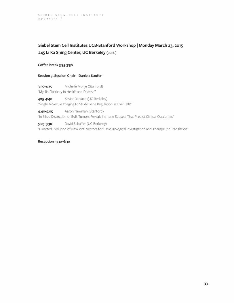
Coffee break 3:35-3:50
Session 3, Session Chair - Daniela Kaufer
3:50-4:15 Michelle Monje (Stanford) “Myelin Plasticity in Health and Disease”
4:15-4:40 Xavier Darzacq (UC Berkeley) “Single Molecule Imaging to Study Gene Regulation in Live Cells”
4:40-5:05 Aaron Newman (Stanford) “In Silico Dissection of Bulk Tumors Reveals Immune Subsets That Predict Clinical Outcomes”
5:05-5:30 David Schaffer (UC Berkeley) “Directed Evolution of New Viral Vectors for Basic Biological Investigation and Therapeutic Translation”
Reception 5:30-6:30
Siebel Stem Cell Institutes UCB-Stanford Workshop | Monday March 23, 2015245 Li Ka Shing Center, UC Berkeley (cont.)
34
S I E B E L S T E M C E L L I N S T I T U T E A p p e n d i x B
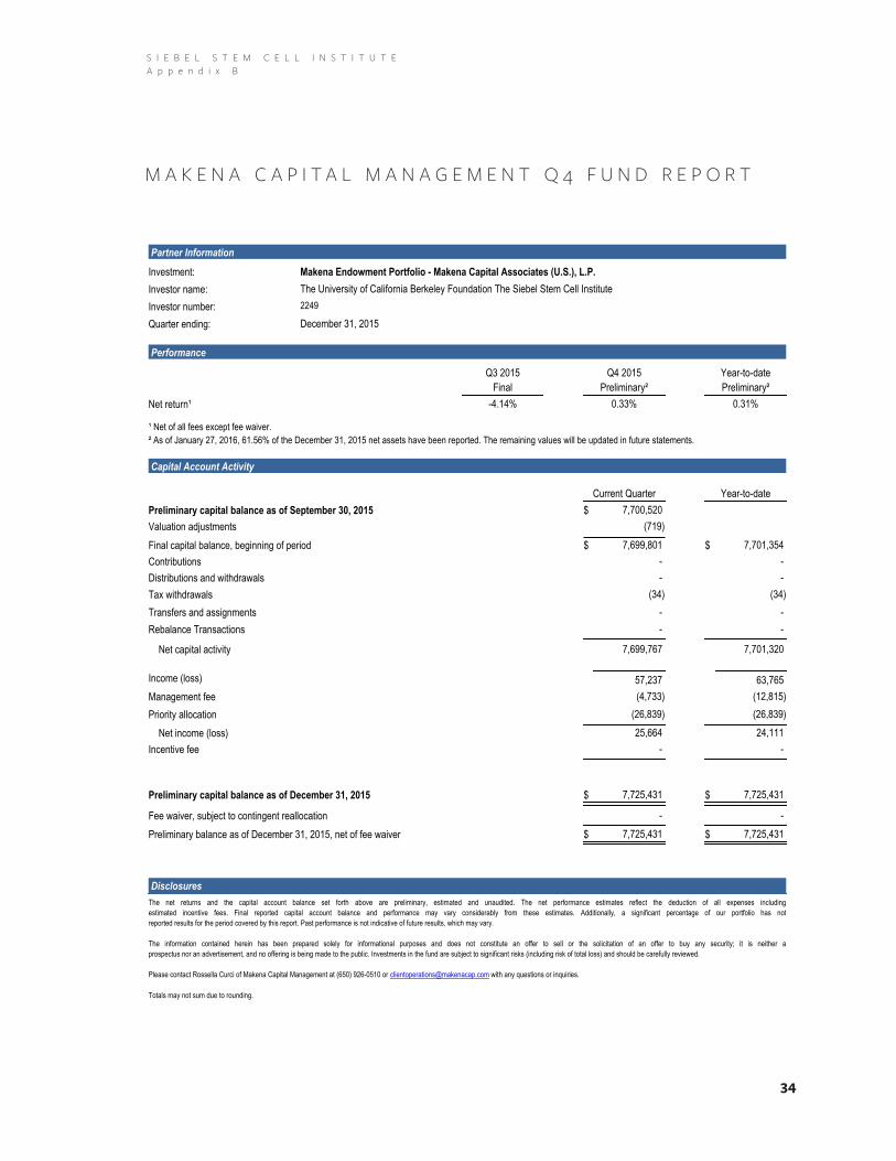
1000046528PARENT INVESTOR ID: 2249 | GROUP ID: 2249 | REPORT TYPE: Detail | FUND ID: MCAMP | FUND DESC: Makena Capital Associates (U.S.), L.P. - Main P
Partner Information
Investment: Makena Endowment Portfolio - Makena Capital Associates (U.S.), L.P.
Investor name: The University of California Berkeley Foundation The Siebel Stem Cell Institute
Investor number: 2249
Quarter ending: December 31, 2015
Performance
Q3 2015
Final
Year-to-date
Preliminary²
Q4 2015
Preliminary²
Net return¹ -4.14% 0.33% 0.31%
¹ Net of all fees except fee waiver.
² As of January 27, 2016, 61.56% of the December 31, 2015 net assets have been reported. The remaining values will be updated in future statements.
Capital Account Activity
Current Quarter Year-to-date
$Preliminary capital balance as of September 30, 2015 7,700,520
Valuation adjustments (719)
Final capital balance, beginning of period 7,699,801 7,701,354 $$
- - Contributions
- - Distributions and withdrawals
Tax withdrawals (34)(34)
Transfers and assignments - -
Rebalance Transactions - -
7,701,320 7,699,767 Net capital activity
Income (loss) 57,237 63,765
(4,733) (12,815)Management fee
Priority allocation (26,839)(26,839)
Net income (loss) 24,111 25,664
- - Incentive fee
7,725,431 Preliminary capital balance as of December 31, 2015 7,725,431 $ $
Fee waiver, subject to contingent reallocation - -
Preliminary balance as of December 31, 2015, net of fee waiver 7,725,431 $ $ 7,725,431
Disclosures
The net returns and the capital account balance set forth above are preliminary, estimated and unaudited. The net performance estimates reflect the deduction of all expenses including
estimated incentive fees. Final reported capital account balance and performance may vary considerably from these estimates. Additionally, a significant percentage of our portfolio has not
reported results for the period covered by this report. Past performance is not indicative of future results, which may vary.
The information contained herein has been prepared solely for informational purposes and does not constitute an offer to sell or the solicitation of an offer to buy any security; it is neither a
prospectus nor an advertisement, and no offering is being made to the public. Investments in the fund are subject to significant risks (including risk of total loss) and should be carefully reviewed.
Please contact Rossella Curci of Makena Capital Management at (650) 926-0510 or [email protected] with any questions or inquiries.
Totals may not sum due to rounding.
M A K E N A C A P I T A L M A N A G E M E N T Q 4 F U N D R E P O R T

35
S I E B E L S T E M C E L L I N S T I T U T E
F U N D A C C O U N T A N D A C T I V I T Y S U M M A R Y October 1, 2014 – September 30, 2015
BERKELEY STANFORD
Account Balance History
Beginning Balance (October 1, 2014) $735,503 $891,918
Fund Distribution (November 30, 2014) $1,000 $1,000
Fund Distribution (February 28, 2015) $450,000 $450,000
Fund Distribution (August 31, 2015) $50,000 $50,000
Expenditures
Scholar Support $104,718 $348,273
Seed Grant Collaborations, Equipment and supplies $407,401 $198,438
Operational Support $51,079 $46,727
Total Expenditures $563,198 $593,438
Ending Balance (September 30, 2015) $673,305 $799,480
S I E B E L S T E M C E L L I N S T I T U T E A p p e n d i x C