Shoulder Mobility Deficits
13
Shoulder Mobility Deficits ICD-9-CM codes: 726.0 Adhesive capsulitis of the shoulder ICF codes: Activities and Participation Domain codes: d4452 Reaching (Using the hands and arms to extend outwards and touch and grasp something, such as when reaching across a table or desk for a book.) Body Structure code: s7201 Joints of shoulder region Body Functions code: b7100 Mobility of a single joint Common Historical Findings Lateral shoulder pain - worsens with positions or activities which put stretch on the glenohumeral joint Progresses to stiffness Gradual, usually insidious, onset of symptoms Common Impairment Findings - Related to the Reported Activity Limitation or Participation Restrictions: ROM limitations - external rotation and abduction are most limited, flexion and internal rotation are least limited Pain at end ranges--some motions are more painful than others (external rotation with abduction is typically the most painful) Limited glenohumeral accessory movements Physical Examination Procedures: Glenohumeral External Rotation ROM Measurement Performance Cues: Remember that glenohumeral ROM is different than shoulder ROM (shoulder ROM is the sum of glenohumeral and scapular ROM) Stand in patient’s axillary region Stabilize scapula with forearm Be precise with stabilization of humeral abduction (to 90 degrees if possible) and horizontal abduction (maintain 0 degrees) Joe Godges, DPT, MA, OCS KP So Cal Ortho PT Residency 1
Transcript of Shoulder Mobility Deficits
Shoulder Mobility Deficits ICD-9-CM codes: 726.0 Adhesive capsulitis of the shoulder ICF codes: Activities and Participation Domain codes:
d4452 Reaching (Using the hands and arms to extend outwards and touch and grasp something, such as when reaching across a table or desk for a book.)
Body Structure code: s7201 Joints of shoulder region Body Functions code: b7100 Mobility of a single joint
Common Historical Findings
Lateral shoulder pain - worsens with positions or activities which put stretch on the glenohumeral joint
Progresses to stiffness Gradual, usually insidious, onset of symptoms
Common Impairment Findings - Related to the Reported Activity Limitation or Participation Restrictions:
ROM limitations - external rotation and abduction are most limited, flexion and internal rotation are least limited
Pain at end ranges--some motions are more painful than others (external rotation with abduction is typically the most painful)
Limited glenohumeral accessory movements Physical Examination Procedures:
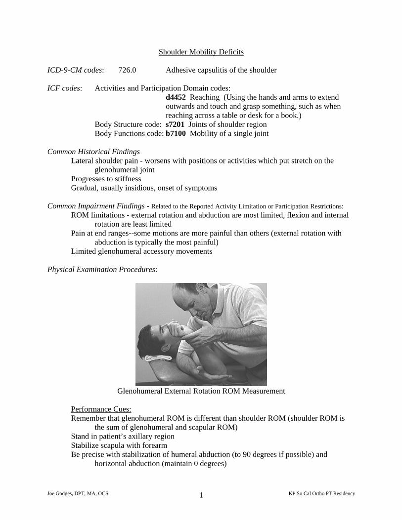
Glenohumeral External Rotation ROM Measurement
Performance Cues:Remember that glenohumeral ROM is different than shoulder ROM (shoulder ROM is
the sum of glenohumeral and scapular ROM) Stand in patient’s axillary region Stabilize scapula with forearm Be precise with stabilization of humeral abduction (to 90 degrees if possible) and
horizontal abduction (maintain 0 degrees)
Joe Godges, DPT, MA, OCS KP So Cal Ortho PT Residency 1
If glenohumeral external rotation ROM is greater at 90 degrees of abduction than at 45 degrees of abduction, suspect a muscle flexibility deficit of the subscapularis
Normal glenohumeral external rotation ROM is 90 degrees
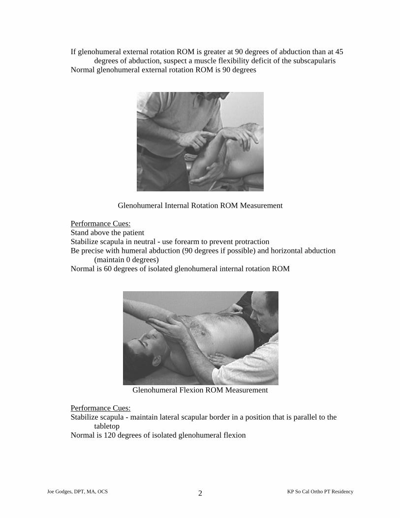
Glenohumeral Internal Rotation ROM Measurement Performance Cues: Stand above the patient Stabilize scapula in neutral - use forearm to prevent protraction Be precise with humeral abduction (90 degrees if possible) and horizontal abduction
(maintain 0 degrees) Normal is 60 degrees of isolated glenohumeral internal rotation ROM
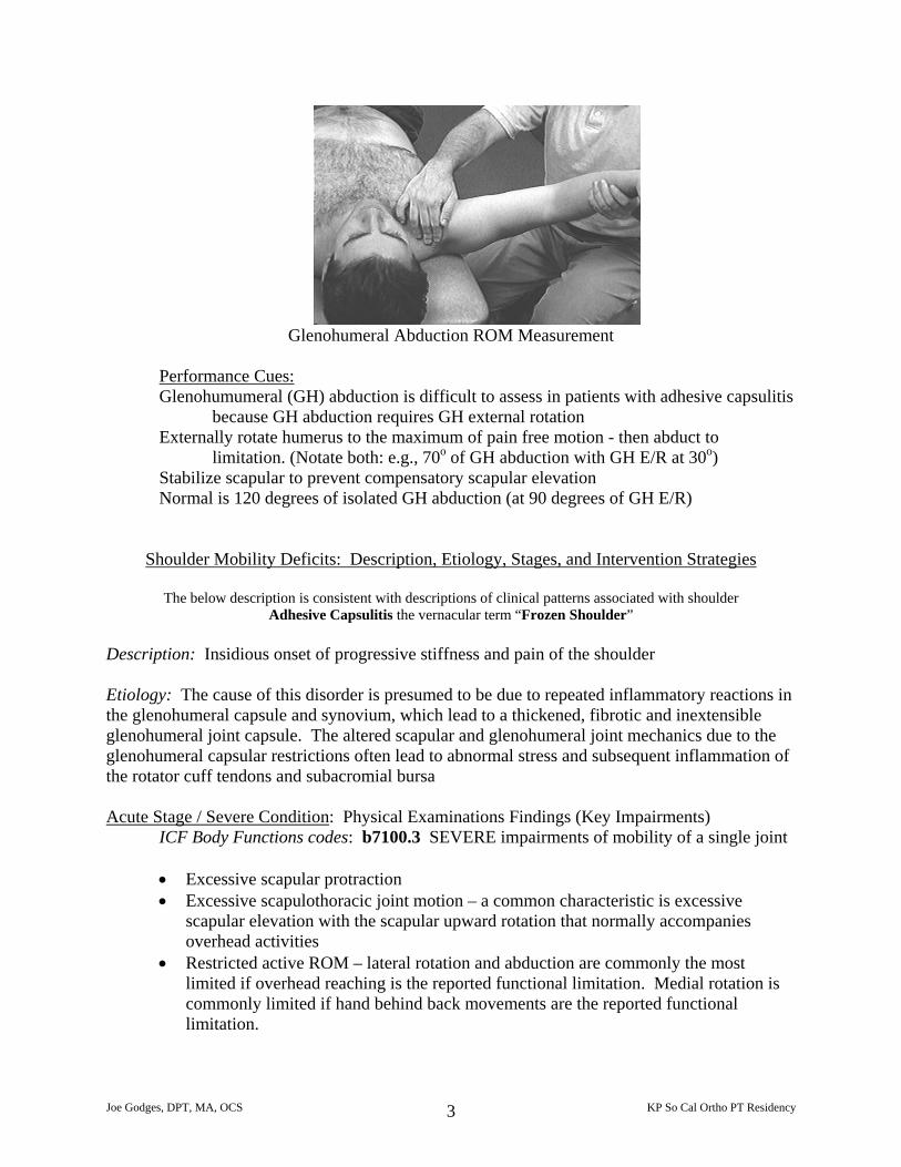
Glenohumeral Flexion ROM Measurement
Performance Cues: Stabilize scapula - maintain lateral scapular border in a position that is parallel to the
tabletop Normal is 120 degrees of isolated glenohumeral flexion
Joe Godges, DPT, MA, OCS KP So Cal Ortho PT Residency 2
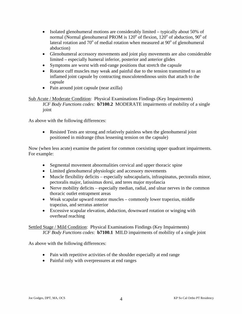
Glenohumeral Abduction ROM Measurement
Performance Cues: Glenohumumeral (GH) abduction is difficult to assess in patients with adhesive capsulitis
because GH abduction requires GH external rotation Externally rotate humerus to the maximum of pain free motion - then abduct to
limitation. (Notate both: e.g., 70o of GH abduction with GH E/R at 30o) Stabilize scapular to prevent compensatory scapular elevation Normal is 120 degrees of isolated GH abduction (at 90 degrees of GH E/R)
Shoulder Mobility Deficits: Description, Etiology, Stages, and Intervention Strategies
The below description is consistent with descriptions of clinical patterns associated with shoulder Adhesive Capsulitis the vernacular term “Frozen Shoulder”
Description: Insidious onset of progressive stiffness and pain of the shoulder Etiology: The cause of this disorder is presumed to be due to repeated inflammatory reactions in the glenohumeral capsule and synovium, which lead to a thickened, fibrotic and inextensible glenohumeral joint capsule. The altered scapular and glenohumeral joint mechanics due to the glenohumeral capsular restrictions often lead to abnormal stress and subsequent inflammation of the rotator cuff tendons and subacromial bursa Acute Stage / Severe Condition: Physical Examinations Findings (Key Impairments)
ICF Body Functions codes: b7100.3 SEVERE impairments of mobility of a single joint
• Excessive scapular protraction • Excessive scapulothoracic joint motion – a common characteristic is excessive
scapular elevation with the scapular upward rotation that normally accompanies overhead activities
• Restricted active ROM – lateral rotation and abduction are commonly the most limited if overhead reaching is the reported functional limitation. Medial rotation is commonly limited if hand behind back movements are the reported functional limitation.
Joe Godges, DPT, MA, OCS KP So Cal Ortho PT Residency 3
• Isolated glenohumeral motions are considerably limited – typically about 50% of normal (Normal glenohumeral PROM is 120o of flexion, 120o of abduction, 90o of lateral rotation and 70o of medial rotation when measured at 90o of glenohumeral abduction)
• Glenohumeral accessory movements and joint play movements are also considerable limited – especially humeral inferior, posterior and anterior glides
• Symptoms are worst with end-range positions that stretch the capsule • Rotator cuff muscles may weak and painful due to the tension transmitted to an
inflamed joint capsule by contracting musculotendinous units that attach to the capsule
• Pain around joint capsule (near axilla) Sub Acute / Moderate Condition: Physical Examinations Findings (Key Impairments)
ICF Body Functions codes: b7100.2 MODERATE impairments of mobility of a single joint
As above with the following differences:
• Resisted Tests are strong and relatively painless when the glenohumeral joint positioned in midrange (thus lessening tension on the capsule)
Now (when less acute) examine the patient for common coexisting upper quadrant impairments. For example:
• Segmental movement abnormalities cervical and upper thoracic spine • Limited glenohumeral physiologic and accessory movements • Muscle flexibility deficits – especially subscapularis, infraspinatus, pectoralis minor,
pectoralis major, latissimus dorsi, and teres major myofascia • Nerve mobility deficits – especially median, radial, and ulnar nerves in the common
thoracic outlet entrapment areas • Weak scapular upward rotator muscles – commonly lower trapezius, middle
trapezius, and serratus anterior • Excessive scapular elevation, abduction, downward rotation or winging with
overhead reaching Settled Stage / Mild Condition: Physical Examinations Findings (Key Impairments)
ICF Body Functions codes: b7100.1 MILD impairments of mobility of a single joint As above with the following differences:
• Pain with repetitive activities of the shoulder especially at end range • Painful only with overpressures at end ranges
Joe Godges, DPT, MA, OCS KP So Cal Ortho PT Residency 4
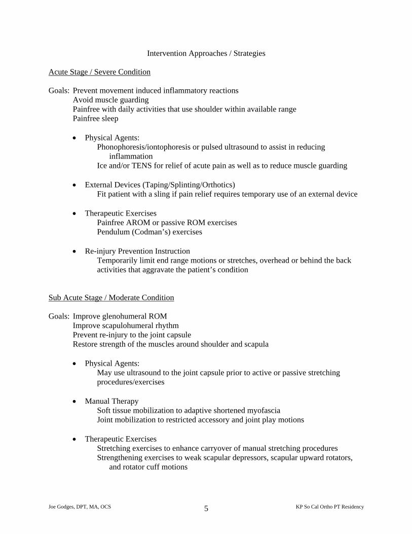
Intervention Approaches / Strategies Acute Stage / Severe Condition Goals: Prevent movement induced inflammatory reactions
Avoid muscle guarding Painfree with daily activities that use shoulder within available range Painfree sleep
• Physical Agents:
Phonophoresis/iontophoresis or pulsed ultrasound to assist in reducing inflammation
Ice and/or TENS for relief of acute pain as well as to reduce muscle guarding
• External Devices (Taping/Splinting/Orthotics) Fit patient with a sling if pain relief requires temporary use of an external device
• Therapeutic Exercises
Painfree AROM or passive ROM exercises Pendulum (Codman’s) exercises
• Re-injury Prevention Instruction Temporarily limit end range motions or stretches, overhead or behind the back activities that aggravate the patient’s condition
Sub Acute Stage / Moderate Condition Goals: Improve glenohumeral ROM
Improve scapulohumeral rhythm Prevent re-injury to the joint capsule Restore strength of the muscles around shoulder and scapula
• Physical Agents:
May use ultrasound to the joint capsule prior to active or passive stretching procedures/exercises
• Manual Therapy
Soft tissue mobilization to adaptive shortened myofascia Joint mobilization to restricted accessory and joint play motions
• Therapeutic Exercises
Stretching exercises to enhance carryover of manual stretching procedures Strengthening exercises to weak scapular depressors, scapular upward rotators,
and rotator cuff motions
Joe Godges, DPT, MA, OCS KP So Cal Ortho PT Residency 5
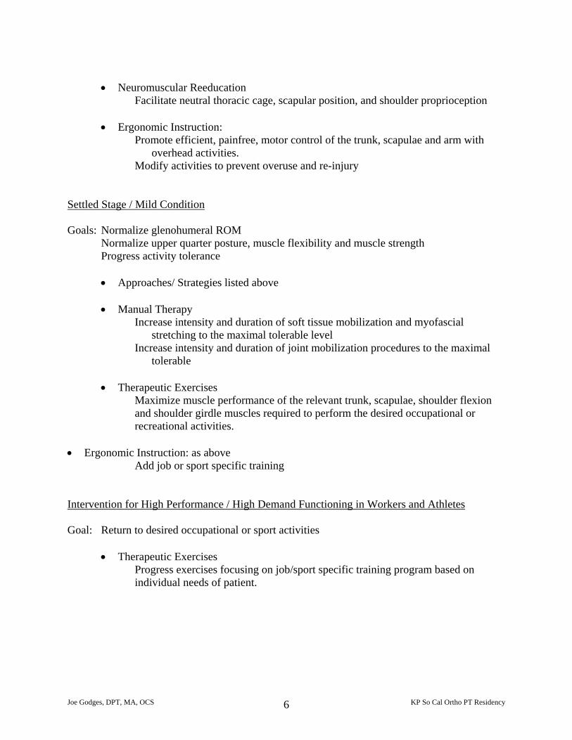
• Neuromuscular Reeducation
Facilitate neutral thoracic cage, scapular position, and shoulder proprioception
• Ergonomic Instruction: Promote efficient, painfree, motor control of the trunk, scapulae and arm with
overhead activities. Modify activities to prevent overuse and re-injury
Settled Stage / Mild Condition Goals: Normalize glenohumeral ROM
Normalize upper quarter posture, muscle flexibility and muscle strength Progress activity tolerance
• Approaches/ Strategies listed above
• Manual Therapy
Increase intensity and duration of soft tissue mobilization and myofascial stretching to the maximal tolerable level
Increase intensity and duration of joint mobilization procedures to the maximal tolerable
• Therapeutic Exercises
Maximize muscle performance of the relevant trunk, scapulae, shoulder flexion and shoulder girdle muscles required to perform the desired occupational or recreational activities.
• Ergonomic Instruction: as above
Add job or sport specific training Intervention for High Performance / High Demand Functioning in Workers and Athletes Goal: Return to desired occupational or sport activities
• Therapeutic Exercises Progress exercises focusing on job/sport specific training program based on individual needs of patient.
Joe Godges, DPT, MA, OCS KP So Cal Ortho PT Residency 6
Selected References Deyle GD, Bang MD. Examination and treatment of the shoulder. Orthopaedic Physical Therapy Clinics of North America. 1999;8:83-115. Gross J, Fetto J, Rosen E. Musculoskeletal Examination. Blackwell Science, 1996. Hannifan JA, Chiaia TA. Adhesive capsulitis: a treatment approach. Clinical Orthop Rel Res. 2000; 372:95-109. Loyd JA. Adhesive capsulitis of the shoulder, diagnosis and treatment. South Medical Journal. 1993;76:879-883. Neviaser JS. Adhesive capsulitis and the stiff and painful shoulder. Orthop Clin of North Am. 1980;11:327-333. Nicholson GG. The effects of passive joint mobilization on pain and hypomobility associated with adhesive capsulitis of the shoulder. J Orthop Sports Phys Ther. 1985; 6(4): 238-246. Placzek J, Roubal P, Freeman C, et al. Long term effectiveness of translational manipulation for adhesive capsulitis. Clin Orthop and Rel Res. 1998;356:181-191. Rizk TE, Christopher RP, Pinals RS, et al. Adhesive capsulitis (frozen shoulder): a new approach to its management. Arch Phys Med Rehabil. 1983;64:29-33. Roubal PJ, Dobitt D, Placzek JD. Glenohumeral gliding manipulation following interscalene brachial plexus block in patients with adhesive capsulitis. J Orthop Sports Phys Ther. 1996;24:66-77. Tomberlin J, Saunders D. Evaluation, Treatment, and Prevention of Musculoskeletal Disorders, 3rd ed., Vol. 2, (Extremities). Educational Opportunities, 1994. Vermeulen HM, Oberman WR, et. al. End-range mobilization techniques in adhesive capsulitis of the shoulder joint: a multiple subject case report. Phys Ther. 2000;80:1204-1213. Wadsworth T. Frozen shoulder. Phys Ther. 1986;66:1878-83.
Joe Godges, DPT, MA, OCS KP So Cal Ortho PT Residency 7
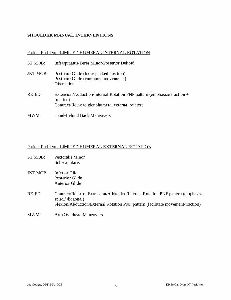
SHOULDER MANUAL INTERVENTIONS
Patient Problem: LIMITED HUMERAL INTERNAL ROTATION ST MOB: Infraspinatus/Teres Minor/Posterior Deltoid JNT MOB: Posterior Glide (loose packed position)
Posterior Glide (combined movements) Distraction
RE-ED: Extension/Adduction/Internal Rotation PNF pattern (emphasize traction +
rotation) Contract/Relax to glenohumeral external rotators
MWM: Hand-Behind Back Maneuvers Patient Problem: LIMITED HUMERAL EXTERNAL ROTATION ST MOB: Pectoralis Minor
Subscapularis JNT MOB: Inferior Glide
Posterior Glide Anterior Glide
RE-ED: Contract/Relax of Extension/Adduction/Internal Rotation PNF pattern (emphasize
spiral/ diagonal) Flexion/Abduction/External Rotation PNF pattern (facilitate movement/traction)
MWM: Arm Overhead Maneuvers
Joe Godges, DPT, MA, OCS KP So Cal Ortho PT Residency 8
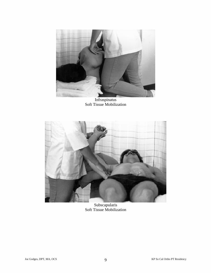
Infraspinatus
Soft Tissue Mobilization
Subscapularis
Soft Tissue Mobilization
Joe Godges, DPT, MA, OCS KP So Cal Ortho PT Residency 9
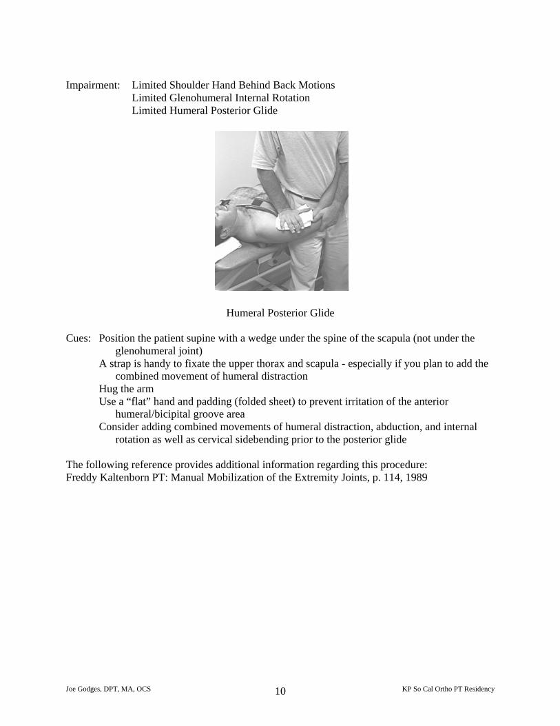
Impairment: Limited Shoulder Hand Behind Back Motions
Limited Glenohumeral Internal Rotation Limited Humeral Posterior Glide
Humeral Posterior Glide Cues: Position the patient supine with a wedge under the spine of the scapula (not under the
glenohumeral joint) A strap is handy to fixate the upper thorax and scapula - especially if you plan to add the
combined movement of humeral distraction Hug the arm Use a “flat” hand and padding (folded sheet) to prevent irritation of the anterior
humeral/bicipital groove area Consider adding combined movements of humeral distraction, abduction, and internal
rotation as well as cervical sidebending prior to the posterior glide
The following reference provides additional information regarding this procedure: Freddy Kaltenborn PT: Manual Mobilization of the Extremity Joints, p. 114, 1989
Joe Godges, DPT, MA, OCS KP So Cal Ortho PT Residency 10
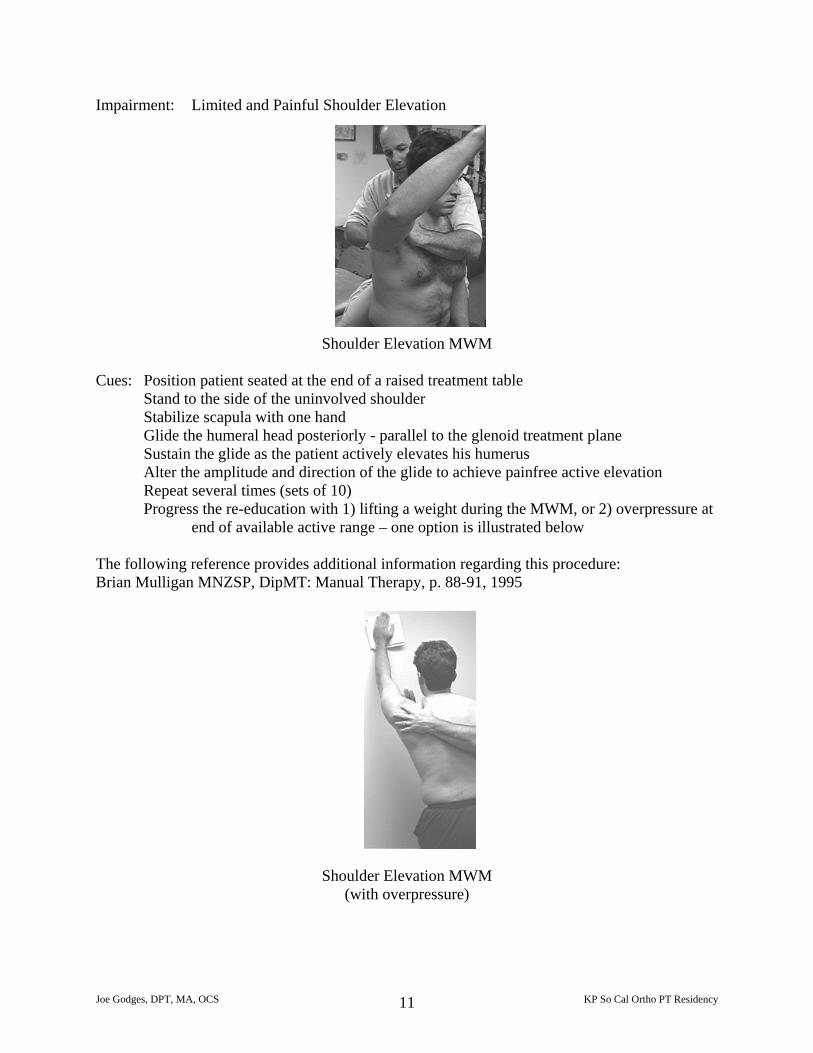
Impairment: Limited and Painful Shoulder Elevation
Shoulder Elevation MWM Cues: Position patient seated at the end of a raised treatment table
Stand to the side of the uninvolved shoulder Stabilize scapula with one hand Glide the humeral head posteriorly - parallel to the glenoid treatment plane Sustain the glide as the patient actively elevates his humerus Alter the amplitude and direction of the glide to achieve painfree active elevation Repeat several times (sets of 10) Progress the re-education with 1) lifting a weight during the MWM, or 2) overpressure at
end of available active range – one option is illustrated below
The following reference provides additional information regarding this procedure: Brian Mulligan MNZSP, DipMT: Manual Therapy, p. 88-91, 1995
Shoulder Elevation MWM (with overpressure)
Joe Godges, DPT, MA, OCS KP So Cal Ortho PT Residency 11
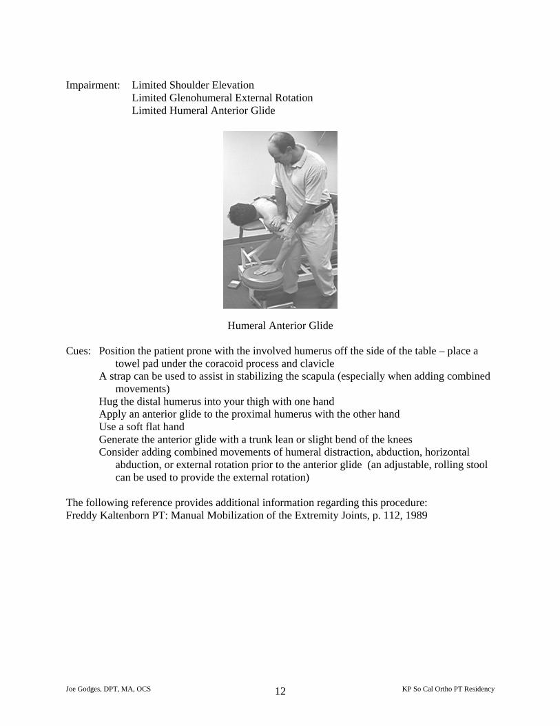
Impairment: Limited Shoulder Elevation
Limited Glenohumeral External Rotation Limited Humeral Anterior Glide
Humeral Anterior Glide Cues: Position the patient prone with the involved humerus off the side of the table – place a
towel pad under the coracoid process and clavicle A strap can be used to assist in stabilizing the scapula (especially when adding combined
movements) Hug the distal humerus into your thigh with one hand Apply an anterior glide to the proximal humerus with the other hand Use a soft flat hand Generate the anterior glide with a trunk lean or slight bend of the knees Consider adding combined movements of humeral distraction, abduction, horizontal
abduction, or external rotation prior to the anterior glide (an adjustable, rolling stool can be used to provide the external rotation)
The following reference provides additional information regarding this procedure: Freddy Kaltenborn PT: Manual Mobilization of the Extremity Joints, p. 112, 1989
Joe Godges, DPT, MA, OCS KP So Cal Ortho PT Residency 12
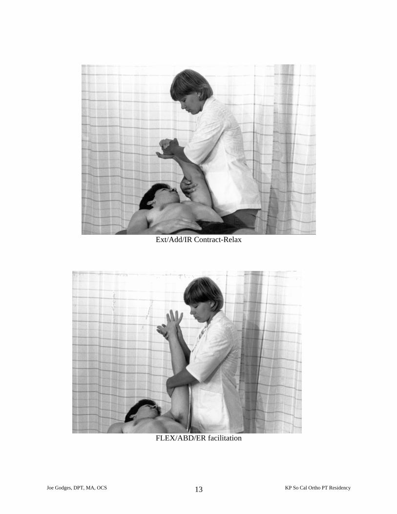
Ext/Add/IR Contract-Relax
FLEX/ABD/ER facilitation
Joe Godges, DPT, MA, OCS KP So Cal Ortho PT Residency 13