Shotgun mass mapping of Lactobacillus species and subspecies from caries related isolates by...
10
RESEARCH ARTICLE Shotgun mass mapping of Lactobacillus species and subspecies from caries related isolates by MALDI-MS Frank Schmidt 1, 2 , Thomas Fiege 3 , Hanne K. Hustoft 1 , Susanne Kneist 3 and Bernd Thiede 1 1 The Biotechnology Centre of Oslo, University of Oslo, Oslo, Norway 2 Interfaculty Institute for Genetics and Functional Genomics, Ernst-Moritz-Arndt University of Greifswald, Greifswald, Germany 3 Biological Laboratory, Centre of Dentistry, Friedrich Schiller University of Jena, Jena, Germany A taxonomical study of 90 isolates of lactobacilli isolated from soft and hard carious dentine of 70 deciduous molars is presented. The Lactobacillus strains were determined by shotgun mass mapping (SMM). This method based on MALDI-MS analysis of Lactobacillus isolates treated with trypsin followed by database comparison against a library of mass spectra derived from 20 refer- ence strains. The SMM method allowed to discriminate different Lactobacillus subspecies. The method was used to analyse Lactobacillus isolates of unknown identity derived from carious dentine. Application of the SMM method to isolates from hard carious dentine revealed a nearly similar distribution of L. paracasei ss paracasei (29%), L. paracasei ss tolerans (32%) and L. casei ss rhamnosus (23%) as dominant subspecies. On the other hand, samples derived from soft carious dentine showed a clear bias only to L. paracasei ss paracasei (60%), whereas L. paracasei ss tolerans (14%) and L. casei ss rhamnosus (12%) were clear minorities. Compared to existent methods, SMM has unique potential for the analysis of Lactobacillus strains on subspecies level. Received: November 7, 2007 Revised: October 27, 2008 Accepted: November 4, 2008 Keywords: Caries / Lactobacillus / MALDI-MS / Shotgun mass mapping / Subspecies 1994 Proteomics 2009, 9, 1994–2003 1 Introduction Dental caries is still a major health problem in most indus- trialized countries, affecting 60–90% of school children and most adults [1]. The level of healthcare spending in Germany per capita in 2000 amounted to $2741, corresponding to 10.6% on total healthcare spending. The specific targeting of persons at increased risk of caries (“high-risk groups”) is thus without question one of the major social-medicine challenges facing dentistry [2]. Lactobacilli are involved in the caries etiopathogenesis. Lactobacilli are nonpathogenic, Gram-positive, nonspore- forming, aciduric and acidophilic rod-shaped bacteria. More than 50 different species were partly sequenced so far. Lac- tobacilli have a positive influence on intestinal balance [3] and an antimutagenic effect was proven [4]. They seemed to be mostly transient and up to 5 years of age Lactobacillus flora is established. In adults, lactobacilli usually comprise less than 1% of the cultivable microflora of the plaque. Their proportions and prevalence increase at advanced caries lesions both in saliva and of the enamel [5–12]. After caries removal and placement of restoration lactobacilli drop down in saliva [13–15]. Methods for rapid and reliable Lactobacillus subspecies identification have to be developed to investigate if subspecies are responsible for caries development. Most studies still identify lactobacilli or Lactobacillus spp. on Rogosa medium as total counts [16–18]. Few studies of oral Lactobacillus species in association with dental caries have been performed using biochemical or reliable genetic-based methods. Thereby, strains identified as Lactobacillus casei, L. Correspondence: Dr. Bernd Thiede, The Biotechnology Centre of Oslo, University of Oslo, Gaustadalleen 21, P.O. Box 1125, Blin- dern, 0317 Oslo, Norway E-mail: [email protected] Fax: 147-22840501 Abbreviations: cfu, colony forming units; DSMZ, German type culture collection and cell cultures; SMM, shotgun mass map- ping DOI 10.1002/pmic.200701028 © 2009 WILEY-VCH Verlag GmbH & Co. KGaA, Weinheim www.proteomics-journal.com
-
Upload
frank-schmidt -
Category
Documents
-
view
212 -
download
0
Transcript of Shotgun mass mapping of Lactobacillus species and subspecies from caries related isolates by...

RESEARCH ARTICLE
Shotgun mass mapping of Lactobacillus species and
subspecies from caries related isolates by MALDI-MS
Frank Schmidt1, 2, Thomas Fiege3, Hanne K. Hustoft1, Susanne Kneist3 and Bernd Thiede1
1 The Biotechnology Centre of Oslo, University of Oslo, Oslo, Norway2 Interfaculty Institute for Genetics and Functional Genomics, Ernst-Moritz-Arndt University of Greifswald,
Greifswald, Germany3 Biological Laboratory, Centre of Dentistry, Friedrich Schiller University of Jena, Jena, Germany
A taxonomical study of 90 isolates of lactobacilli isolated from soft and hard carious dentine of 70deciduous molars is presented. The Lactobacillus strains were determined by shotgun massmapping (SMM). This method based on MALDI-MS analysis of Lactobacillus isolates treated withtrypsin followed by database comparison against a library of mass spectra derived from 20 refer-ence strains. The SMM method allowed to discriminate different Lactobacillus subspecies. Themethod was used to analyse Lactobacillus isolates of unknown identity derived from cariousdentine. Application of the SMM method to isolates from hard carious dentine revealed a nearlysimilar distribution of L. paracasei ss paracasei (29%), L. paracasei ss tolerans (32%) and L. casei ssrhamnosus (23%) as dominant subspecies. On the other hand, samples derived from soft cariousdentine showed a clear bias only to L. paracasei ss paracasei (60%), whereas L. paracasei ss tolerans(14%) and L. casei ss rhamnosus (12%) were clear minorities. Compared to existent methods,SMM has unique potential for the analysis of Lactobacillus strains on subspecies level.
Received: November 7, 2007Revised: October 27, 2008
Accepted: November 4, 2008
Keywords:
Caries / Lactobacillus / MALDI-MS / Shotgun mass mapping / Subspecies
1994 Proteomics 2009, 9, 1994–2003
1 Introduction
Dental caries is still a major health problem in most indus-trialized countries, affecting 60–90% of school children andmost adults [1]. The level of healthcare spending in Germanyper capita in 2000 amounted to $2741, corresponding to10.6% on total healthcare spending. The specific targeting ofpersons at increased risk of caries (“high-risk groups”) isthus without question one of the major social-medicinechallenges facing dentistry [2].
Lactobacilli are involved in the caries etiopathogenesis.Lactobacilli are nonpathogenic, Gram-positive, nonspore-forming, aciduric and acidophilic rod-shaped bacteria. Morethan 50 different species were partly sequenced so far. Lac-tobacilli have a positive influence on intestinal balance [3]and an antimutagenic effect was proven [4]. They seemed tobe mostly transient and up to 5 years of age Lactobacillus florais established. In adults, lactobacilli usually comprise lessthan 1% of the cultivable microflora of the plaque. Theirproportions and prevalence increase at advanced carieslesions both in saliva and of the enamel [5–12]. After cariesremoval and placement of restoration lactobacilli drop downin saliva [13–15]. Methods for rapid and reliable Lactobacillussubspecies identification have to be developed to investigateif subspecies are responsible for caries development. Moststudies still identify lactobacilli or Lactobacillus spp. onRogosa medium as total counts [16–18]. Few studies of oralLactobacillus species in association with dental caries havebeen performed using biochemical or reliable genetic-basedmethods. Thereby, strains identified as Lactobacillus casei, L.
Correspondence: Dr. Bernd Thiede, The Biotechnology Centre ofOslo, University of Oslo, Gaustadalleen 21, P.O. Box 1125, Blin-dern, 0317 Oslo, NorwayE-mail: [email protected]: 147-22840501
Abbreviations: cfu, colony forming units; DSMZ, German typeculture collection and cell cultures; SMM, shotgun mass map-ping
DOI 10.1002/pmic.200701028
© 2009 WILEY-VCH Verlag GmbH & Co. KGaA, Weinheim www.proteomics-journal.com

Proteomics 2009, 9, 1994–2003 1995
casei ss casei, L. casei ss rhamnosus, L. fermentum, L. plan-tarum, L. salivarius, L. acidophilus and L. rhamnosus have beenfrequently considered to be related with caries [9–12, 19–22].However, the development of the PCR in the early 1980senabled genotyping by PCR fingerprinting to determineLactobacillus species [10, 23, 24]. Recently, genotyping ofribosomes was shown as an approach to distinguish bacteriaon subspecies level [25, 26]. However, incomplete or erro-neous genetic data are still major drawbacks of genotypingmethods [27]. 2-DE and MS-based quantitative techniquescan also be used to distinguish bacteria [28, 29]. However, adetailed proteome analysis is time-consuming and ineffi-cient to compare large sets of bacteria samples. Thus, whole-cell protein profiles separated by PAGE and capillary gelelectrophoresis proved to be useful to distinguish Lactoba-cillus species on the protein level [30, 31].
Direct analysis of bacteria by MS was already used in1975 for the identification of bacteria using characteristicphospholipids and ubiquinones [32]. The development ofESI-MS and MALDI-MS enabled the fast and accurate iden-tification of bacteria [33–35]. Proteins, nucleic acids, lipids,phospholipids, lipopolysaccharides, oligosaccharides may beused as strain-specific markers for the identification andtaxonomic characterization of bacteria. Currently, two majormethods were established to analyse proteins of bacteria byMS [35–37]. The intact protein profiling (IPP) (or top-down)method determines molecular masses of intact proteinswhereas peptide mixtures resulting from enzymatic digestedproteins are analysed by shotgun mass mapping (SMM) (orbottom-up). In addition, sequence analysis by MS/MS can beapplied to enzymatic derived peptides [38, 39]. However, thisapproach often suffers due to incomplete or missing geno-mic data to determine bacterial subspecies.
The aim of this study was to differentiate 90 isolates oflactobacilli from carious dentine [40, 41] of 70 deciduousmolars by MALDI-MS. For this purpose, 20 relevant refer-ence strains from the German type culture collection andcell cultures (DSMZ; Braunschweig, Germany) were cho-sen to build up a MALDI-MS reference mass spectralibrary. The selected strains are known to be a part of thegerm population within the mouth of humans and couldplay a role in caries progression. References and wild-typeisolates were analysed by SMM of tryptic digests andresulting mass spectra were compared using the programMS-Screener which was already employed to compare largesets of mass spectra [42–44]. In addition, the mass spectrawere clustered to verify the results from SMM and to clas-sify the Lactobacillus isolates. Using the SMM method incombination with cluster analysis, we determined 90% ofthe Lactobacillus isolates from carious dentine down to thesubspecies level. Moreover, the classification of the identi-fied Lactobacillus subspecies showed a clear bias for L.paracasei ss paracasei in carious soft dentine, whereas inhard dentine the subspecies L. paracasei ss paracasei, L.paracasei ss tolerans and L. casei ss rhamnosus were found tobe equally common.
2 Materials and methods
2.1 Growing conditions of Lactobacillus references
The strains were grown from frozen stocks in MRS broth(Difco). After overnight growth under anaerobic conditionsat 35 6 27C (VT 5042 EK, Heraeus), 10 mL MRS broth wereinoculated with each strain and incubated as described abovefor 48 6 0.5 h. The purity of the cultures at the end of the latelog-growth-phase was controlled by Gram-stain and har-vested by centrifugation at 4000 U/min for 5 min. (Megafuge1.0 Sepatech, Heraeus). The pellet was resuspended inphosphate buffer (5 mL, pH 7.0) and washed three timeswith the same buffer. After centrifugation the supernatantwas rejected and the pellet was used for SMM analysis byMALDI-MS.
2.2 Lactobacillus wild-type assembling and growing
conditions
In a controlled clinical trial, 70 primary lower second molarsfrom 70 children with the age of 6–7 years with comparabledeep carious lesions were paired. Pretreatment radiographswere used to classify lesion size and to exclude apical patho-genesis. None of the lesions had irreversible signs of pulpitis(i.e. no information on spontaneous or provoked pulpalpain). Only pairs of molars without clinical signs of inflam-mation or pathological radicular pulp changes were includedin the study. The molars of 35 pairs were treated by indirectpulp cupping procedure, and after matching, half of themwere mechanically excavated in one sitting (one-step, O-group) and half of them were subjected to step-wise (S-group) excavating [40]. The microflora was determined aftercaries excavation. The teeth were extracted after 16 months ofmicrobiological control to determine the etiopathogenic roleof germs for carious progression in dentine histologically.Sixty-seven percent of the primary molars were free frompulpal inflammations. Soft carious dentine was significantlyhigher infected than the clinically acceptable hard dentine.The microorganisms were only eliminated in 40% of thecavity floors. In the infected teeth acidogenic streptococciand lactobacilli were involved in pulpal inflammations.Results indicate that the latter genera of microorganisms areof etiologic significance for carious progression in dentine[40].
The cavities were isolated with rubber dam for microbialcontrol. The first step in the step-wise excavation (S-group)procedure included removal of the central cariogenic bio-mass and the superficial parts of the necrotic and deminer-alized dentine, and the complete excavation of the peripheraldemineralized dentine. A calcium hydroxide-containing basematerial and a temporary filling were applied. After a treat-ment interval of 11 months, the temporary filling wasremoved with a sharp excavator. The bottom of the cavity wassealed with a calcium hydroxide base material, and the toothwas finally restored. Carious dentine was excavated up to the
© 2009 WILEY-VCH Verlag GmbH & Co. KGaA, Weinheim www.proteomics-journal.com

1996 F. Schmidt et al. Proteomics 2009, 9, 1994–2003
hard cavity floor by excavation in one sitting (O-group). Thebottom of the cavity was sealed with a calcium hydroxidebase material, and the tooth was finally restored (Fig. 1).
For microbiological procedures, samples of soft and harddentine were obtained with a sterile excavator damped in sa-line before application of the calcium hydroxide base mate-rial and the temporary or finally filling were applied (Fig. 1).
Soft and hard dentine samples were transferred to 1 mL ofsaline, vortex mixed for 30 s and serially diluted ten-foldbefore 0.1 mL were plated three-fold on brain heart agar(Difco) with 5% human bank blood and incubated parallel for2 and 7 days at anaerobic conditions and 2 days at aerobicconditions at 377C. After incubation, the total number of col-ony forming units (cfu) was counted on agar plates yielded50–300 colonies. Colonies with different morphology, usuallydifferent species or subspecies, were isolated by using a ste-reomicroscope. Bacterial counts were expressed as log10 cfuper sample. Of these, mean and standard errors were calcu-lated and, furthermore, the percentages of lactobacilli in themicrobial population were determined after identification.Statistical analyses of bacterial counts conducted using pairedt-test; p,0.05 were considered statistically significant.
Gram-positive rods growing onto the Rogosa medium(Merck AG, Darmstadt, Germany) were designated Lactoba-cillus and tested for fermentation of amygdalin, cellobiose,mannitol, melezitose, raffinose, rhamnose and sorbitol, pro-duction of CO2 from glucose and hydrolysis of arginine andesculin. A total of 90 isolates were confirmed to be lactobacilli.The strains were lyophilized and stored frozen in micro-banks (Mast Diagnostica, Reinfeld, Germany) at 2187C [40],and subjected to profiling by MS.
Figure 1. Overview of deep carious lesions treatment. Deep car-ious lesions were treated by one-step (O) and step-wise (S)excavation of carious dentine. Microbiological examination wasperformed before final filling after one-step (O1: Ca(OH)2, phos-phate cement), before temporary filling (S1: zinc oxide-brenzca-techine (ZOB)) and final filling (S2: Ca(OH)2, phosphate cement)after step-wise excavation and after clinical observation at 11months (O2, S3).
2.3 SMM by MALDI MS
One vial of trypsin (20 mg) was diluted in 80 mL trypsinresuspension buffer (Promega, Madison, WI, USA) to obtaina final concentration of 0.25 mg/mL. One mL trypsin of thissolution was added to 4 mL of 25 mM ammonium bicarbo-nate (pH 7.8) and mixed with 1 mL entire Lactobacillus pellet.The suspensions were incubated for 2 h at 377C with gentleshaking followed by centrifugation at 15 7006g for 10 min(Centrifuge 5415D, Eppendorf, Hamburg, Germany). After-wards, 1 mL of the supernatants were mixed with 1 mL 2,5-dihydroxy benzoic acid (DHB; Bruker Daltonics, Bremen,Germany) matrix solution and spotted onto a MALDI plate.The tryptic peptides were measured in reflector mode in themass range of 900–4000 Da after external calibration with astandard peptide mixture (Kemptide, bradykinin, substanceP, glu-fibrinopeptide B and dynorphin A 2–17). Each samplewas prepared once and mass spectra were acquired manuallyfrom various positions of each analyte until intensitieshigher than 10 000 counts were achieved. Basic settings ofthe MALDI-TOF/TOF instrument (Ultraflex II, Bruker Dal-tonics) were as follows: ion source 1, 25 kV; ion source 2,21.85 kV; lens, 9.60 kV; reflector, 26.3 kV; reflector 2,13.85 kV; deflector mode, polarity positive. The mass spectrawere internally calculated from the raw mass spectra usingthe sophisticated numerical annotation procedure (SNAP)algorithms of FlexAnalysis 2.4, using minimal S/N ratio of 5,relative intensity threshold of 0, maximum number of 200peaks, quality factor threshold of 20 and averagine buildingblock as peak filter settings.
2.4 Decontamination, database comparison and
clustering using MS-screener
The mass spectra were standardized by labelling up to 80peaks with highest intensity. Afterwards, the generated. xmlpeak lists files were exported and postprocessed in the MS-Screener program (Version 1.0.1) which was developed byFrank Schmidt and Axel Rack (can be downloaded at http://www.mpiib-berlin.mpg.de/2D-PAGE/) [42]. The programwas used to detect common mass peaks and to eliminatecontaminant masses derived from human keratins, trypsinautoproteolysis and the MALDI matrix. Moreover, the halfdecimal place rule (HDPR) was applied to consider that theaverage mass value of a peptide after the decimal place can beapproximately calculated by dividing the molecular masswith 2 (up to m/z 2000) in order to check the external cali-bration of the spectra. Three independent mass spectra of thereference strains were stored in the MS-Screener databasetab and compared with mass spectra derived from clinicalsamples using the similarity search function. For the simi-larity search, only the presence or absence of peaks was takeninto account using a mass tolerance of 60.25 Da. A similar-ity of at least 20% (corresponding to 16 matched queriesexcept for L. buchneri and L. delbruekii ss delbruekii) wasrequired to accept a first hit with SMM. The second and third
© 2009 WILEY-VCH Verlag GmbH & Co. KGaA, Weinheim www.proteomics-journal.com

Proteomics 2009, 9, 1994–2003 1997
hits to the database were 8% and 12% lower on average,which was equivalent to 7 and 12 mass peaks, respectively.Please note that at less than 20% similarity (16 matchedqueries), the matched queries to the second hit can be notsignificant anymore. Referring to PMF, identified proteinscan be revised as a rule of thumb by multiplication ofsequence coverage (% value) and matched masses. A proteinis considered to be identified by a value .300 in humans[45]. Here, we transferred this rule of thumb with at least 16matching peptides with a similarity of 20%.
In addition, MS-Screener was used to generate peak listsmatrices (peak alignment) as a prerequisite for a clusteranalysis. For this purpose, mass spectra were intervalizedwith an accuracy of 60.25 Da and subsequently binaryencoded. Intervalization means that peaks detected in one ofthe spectra were used to generate intervals within a toleranceof 60.25 Da. Identical peaks of the spectra were assignedinto existing ones, whereas new peaks were used to open anadditional interval. Considering binary encoding, only thepresence or absence (L/O) of peaks was taken into account.Single linkage as well as the Sørensen coefficient (Dice),which supports present and absent metrics, were applied forhierarchical clustering. This similarity calculation considersmore on joint occurrences than on missed matches. Inaddition, a 1000 iterations comprising bootstrapping wasapplied to reference replicates to evaluate the robustness ofeach cluster. Moreover, principal component analysis (PCA)was employed to confirm the cluster results by a secondmethod. All calculations were realized using the free ofcharge program Past (http://folk.uio.no/ohammer/past/download.html).
3 Results
3.1 SMM of Lactobacillus references by MALDI-MS
The SMM method was based on mass analysis of completeLactobacillus germs after enzymatic digestion. Therefore,lactobacilli pellets were incubated for 2 h with trypsin. Scan-ning electron microscopy images of L. paracasei ss paracasei20008 and L. paracasei ss tolerans prior to and after addition oftrypsin confirmed that the cell membranes were cracked(Supporting Information Fig. 1). Mass spectra of all 20-reference strains could be recorded by SMM (Table 1). Thenumber of mass peaks varied between 24 (L. buchneri) and196 (L. delbrueckii ss bulgaricus) with 151 peaks in averageusing an S/N ratio of 5. Mass spectra obtained from differentLactobacillus species were strikingly dissimilar using thismethod. On the other hand, the SMMs of six L. casei/para-casei subspecies revealed related but not identical massspectra as shown in Fig. 2. In addition, hierarchical cluster-ing of the 20 references including all three replicates wasapplied to confirm the discrimination of each of the speciespatterns (Fig. 3A). First, mass labelling was limited up to 80peaks with highest intensity to standardize the mass spectra
Figure 2. Comparison of six L. casei related reference subspecieswith a Lactobacillus isolate derived from caries region S1. Themass spectra obtained from six L. casei subspecies (L. paracaseiss paracasei 20008, L. paracasei ss tolerans, L. paracasei ss para-casei 5457, L. casei ss rhamnosus 20178, L. casei ss rhamnosus20022 and L. casei ss casei) were noticeably related to each otherbut also showed apparent discriminations. The mass spectradisplayed in the range from m/z 900 to 3000 of the isolate resultedin a similarity of 52% to L. casei ss rhamnosus 20022, 28% to L.paracasei ss paracasei and 25% to L. casei ss casei. The remain-ing subspecies yielded a percentage of similar peaks lower than20%. All three most intense peaks of the isolate were clearlydetected only in L. casei ss rhamnosus 20022 as shown by dottedlines.
© 2009 WILEY-VCH Verlag GmbH & Co. KGaA, Weinheim www.proteomics-journal.com

1998 F. Schmidt et al. Proteomics 2009, 9, 1994–2003
Figure 3. Cluster dendrogram,bootstrap analysis and principlecomponent analysis of refer-ence mass spectra obtained bythe SMM method. The clusterdendrogram obtained frommass spectra from three repli-cates of 20 references is given(A). The cluster dendrogramcontained one main groupwhich accumulates the massspectra from L. casei and para-casei, related subspecies con-sidering a similarity higher than0.3. The remaining Lactobacillussubspecies were obviously dis-similar to each other and formedown branches. The PCAobtained from three replicatesof 20 references was shown in(B). Numbering was accordingto Table 1.
because a higher number of labelled peaks resulted in incor-rect assignments for mass peaks with low intensity. Thedendrogram obtained from 60 mass spectra of the 20 differ-ent Lactobacillus strains displayed the assignment of thethree replicates of all subspecies. In addition, nonparametricbootstrap values based on 1000 pseudo-replicates were usedto evaluate the branches. As a result, most of the replicateswere estimated to be .90 except for L. paracasei ss tolerans(73), L. casei ss rhamnosus 20178 (58), L. casei ss rhamnosus20022 (74) and L. alimentarius (89), and L. buchneri (36)
(Fig. 3A). The similarity coefficients between replicates ofeach species varied between 0.98 (L. paracasei ss paracasei5457) and 0.68 (e.g. L. coryniformis ss coryniformis) in contrastto the subspecies with a variation of similarity between 0.68(L. casei ss rhamnosus 20022 and L. casei ss casei) and 0.05 (L.delbrueckii ss delbrueckii). Considering the similarity at thespecies level, it varied between 0.4 (e.g. L. delbrueckii ss lactisand L. alimentarius) and 0.05, except for L. paracasei ss para-casei 5457 (0.76) and L. plantarum (0.76). The high similaritybetween the replicates (0.98–0.68) and the lower similarities
© 2009 WILEY-VCH Verlag GmbH & Co. KGaA, Weinheim www.proteomics-journal.com

Proteomics 2009, 9, 1994–2003 1999
Table 1. List of investigated Lactobacillus subspecies derivedfrom the German type culture collection and cell cul-tures (DSMZ)
Lactobacillus taxon DSMZstrain
SMM900–4000 Da(no. peaks)
L. acidophilus 20079 186L. alimentarius 20181 149L. brevis 20054 196L. buchneri 20057 24L. casei ss casei 20011 148
L. casei ss rhamnosus 20022 117
L. casei ss rhamnosus (zea) 20178 169
L. coryniformis ss coryniformis 20001 137L. coryniformis ss torquens 20004 161L. delbrueckii ss bulgaricus 20080 196L. delbrueckii ss delbrueckii 20074 44L. delbrueckii ss lactis 20072 177L. gasseri 20077 142L. paracasei ss paracasei 5457 172
L. paracasei ss tolerans 20012 180
L. paracasei ss paracasei 20008 172
L. plantarum 2601 155L. reuteri 20016 174L. salivarius ss salivarius 20555 152L. viridescens 20410 172
Strains presented in Figure 2 are shown in bold.References were measured by SMM and analysed by nano LC-MALDI-MS/MS. Phenotypic appearances such as colours andviscosities were determined for each of the subspecies.
between subspecies (0.68–0.05) allowed to discriminate sub-species using SMM in combination with cluster analysis.Moreover, PCA was applied to confirm the results of thecluster analysis. The score plot showed that the replicatesgrouped according to their respective subspecies. L. casei orL. paracasei subspecies were grouped in the first quarter,except L. paracasei ss paracasei 5457. The remaining sub-species were distributed over all four quarters (Fig. 3B). Incontrast to the cluster analysis, subspecies of several speciesgroups (e.g. L. salivarius ss salivarius and L. acidophilus) wereclosely arranged in quarter 3 (bottom right) and a clear dis-crimination was not obtained, even if the similarity of iden-tical peaks was low. Closely related subspecies were betterseparated by PCA than by cluster analysis. By contrast, sub-species with lower similarity were better separated by clusteranalysis. Furthermore, the reproducibility of referencesspectra recorded by SMM was examined. For this purpose,three independent sample preparations and mass measure-ments have been carried out in order to calculate the per-centage of identical peaks. Automatic labelling was used toobtain coincident normalization and the accordance of masspeaks within three corresponding spectra was on average72% with a minimum of 60% (L. casei ss rhamnosus 20178)and a maximum of 87% (L. salivarius ss salivarius) (Fig. 4).
Figure 4. MALDI-TOF mass spectra of three replicates of L. sali-varius ss salivarius. The SMM of the three independent prepara-tions revealed an overall similarity of 87%.
3.2 Similarity based identification of Lactobacillus
isolates from carious dentine in comparison to
references by SMM
The SMM approach was applied to 90 isolates derived fromyoung patients with caries and yielded to mass spectra for allof them. The similarity of identical peaks of the first hits toreference spectra varied between 0 and 60% and showed anaverage of 41%. In summary, 44 of the spectra matched asfirst hit with at least 20% similarity (corresponding to 16matched queries, except for L. buchneri and L. delbruekii ssdelbruekii) to the reference strain of L. paracasei ss paracasei,19 spectra to the reference of L. paracasei ss tolerance, 12 to L.casei ss rhamnosus 20022 and 1 to L. casei ss rhamnosus 20178.Furthermore, one strain was identified as L. delbrueckii sslactis, and three as L. gasseri (Supporting Information Table1). The second and third hits to the database were 8 and 12%lower on average, which was equivalent to 7 and 12 masspeaks, respectively. Seventy-eight of the 82 identified isolatesrevealed different subspecies from the same species group asthe next two hits. As an example, the mass spectrum of anisolate showed highest similarity to L. casei ss rhamnosus20022 and less to the remaining L. casei subspecies (Fig. 2). Adetailed report of all identifications is given in SupportingInformation Table 1.
© 2009 WILEY-VCH Verlag GmbH & Co. KGaA, Weinheim www.proteomics-journal.com

2000 F. Schmidt et al. Proteomics 2009, 9, 1994–2003
3.3 Species classification of all Lactobacillus isolates
from carious dentine by SMM
The molars of 35 pairs were treated by the indirect pulpcupping procedure. After matching, half of them weremechanically excavated in one sitting, named one-step (O),and half of them were subjected to step-wise excavating (S) asdepicted in Fig. 1. Soft carious dentine harboured a mean of3.6 6 106105 cfu. After carious excavation the microorgan-isms were drastically reduced. Independent of one-step orstep-wise excavation, the total counts on the hard cavity floorwas very low with 44.1 6 1976103 cfu (O1) and3.6 6 13.76103 cfu (S2), respectively. After one-step (O2)and step-wise (S3) caries removal, the residual microflorawas equal, with 1.8 6 6.36103 cfu/sample (O2) and0.1 6 2.86103 cfu/sample (S3) (paired t-test; significancelevel p,0.05; p = 0.114) [40, 41].
Taxonomical examination of carious dentine by SMMwas performed with samples isolated before one-step finalfilling (O1), temporary filling after step-wise excavation (S1),final filling after step-wise excavation (S2), one-step final fill-ing (O2) and final filling after step-wise excavation after clin-ical observation at 11 months (S3) (Fig. 1). The identificationon the species level by SMM revealed a clear dominance(84%) of species from L. casei/paracasei (Fig. 5).
Figure 5. Distribution of Lactobacillus species of carious dentinedetermined by SMM.
3.4 Subspecies classification of Lactobacillus isolates
from carious dentine identified by SMM and
cluster analysis
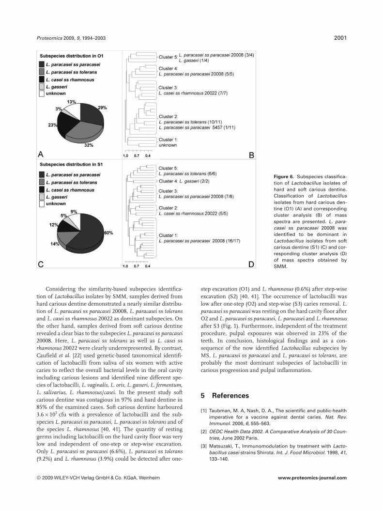
The distribution of Lactobacillus subspecies was comparedfor samples derived from O1 (hard carious dentine, 31strains) and S1 (soft carious dentine, 42 strains) by SMM.Samples obtained from hard carious dentine (O1) resulted ina nearly similar distribution of L. paracasei ss paracasei 20008(29%), L. paracasei ss tolerans (32%) and L. casei ss rhamnosus20022 (23%). Furthermore, 3% were assigned to L. gasseriand 13% of the mass spectra revealed no similarities with thereferences (Fig. 6A). A similar distribution of five main clus-ters was obtained by cluster analysis (Fig. 6B). Here, L. para-casei ss tolerans dominated as the major component cluster 2(10 of 11 spectra were identified as the first hit by SMM).Moreover, cluster 3 was assigned to L. casei ss rhamnosus20022, clusters 4 and 5 to L. paracasei ss paracasei 20008.Clusters 3 and 4 were also connected to each other, since themass spectra displayed a high similarity which was already
shown in the cluster analysis of reference mass spectra(Fig. 3A). In contrast, samples derived from soft cariousdentine (S1) displayed a clear bias to the subspecies L. para-casei ss paracasei 20008 (60%) whereas L. paracasei ss tolerans(14%) and L. casei ss rhamnosus 20022 (12%) were only foundas clear minorities (Fig. 6C). A similar distribution wasobtained by cluster analysis resulting in two main clusters(Fig. 6D). The main cluster could be further divided intothree subclusters (clusters 1–3) which mainly contained L.paracasei ss paracasei 20008. As already shown in the clusteranalysis of group O1, the L. casei ss rhamnosus 20022 cluster(cluster 2) was also related to the L. paracasei ss paracasei20008 clusters (Fig. 6D).
4 Discussion
Lactobacillus subspecies are acidophilic Gram-positive rods,commonly recognized by their growth on Rogosa medium.Further biochemical tests often give an imprecise taxonomicalspecification. Lactobacillus subspecies are often referred to aslactobacilli due to uncertain identifications. Many clinicalstudies on dental caries also failed to identify Lactobacillussubspecies using conventional methods. However, isolatedLactobacillus subspecies from carious dentine were identifiedby combined PCR-RFLP and PAGE [31]. Nevertheless, the per-sample cost using PCR-RFLP is high, time-consuming andcan only discriminate between a few subspecies, whereasPAGE suffers from a complex banding pattern.
Tryptic digestion of bacteria was first applied to a familyof small acid-soluble proteins (SAPs) [38]. Identification ofBacillus spores was accomplished by peptide sequencingcombined with genome-based database searches. However,sequence information of tryptic peptides from Lactobacillusstrains was useful to distinguish species, but failed to differ-entiate at the subspecies level due to the lack of knownsequences in the database (unpublished data). Therefore, theaim of this study was to establish a method to identify andclassify clinical isolates from carious dentine of primarymolars. At first, mass spectra of 20 reference strains wererecorded after tryptic digestion and resulted in suitable massspectra. Subsequently, this approach was applied to well-characterized clinical isolates from caries patients. Thus, themajority (.90%) of 90 clinical isolates of lactobacilli could beidentified by SMM down to the subspecies level. The SMMmethod was easy to perform and resulted in unique massspectra for each of the Lactobacillus subspecies and isolateswith high discrimination rates.
Interestingly, our investigations clearly indicated a biasfor L. casei and L. paracasei species in dental caries (84%).These findings correlated with results from the study byByun et al. [10], which identified the most prevalent lactoba-cilli (68%) as the L. casei group using 16S rDNA ampliconsequences and real-time PCR. However, since the analysiswas based on approximately 400 bp, it provided only anindication of the subspecies.
© 2009 WILEY-VCH Verlag GmbH & Co. KGaA, Weinheim www.proteomics-journal.com
Proteomics 2009, 9, 1994–2003 2001
Figure 6. Subspecies classifica-tion of Lactobacillus isolates ofhard and soft carious dentine.Classification of Lactobacillusisolates from hard carious den-tine (O1) (A) and correspondingcluster analysis (B) of massspectra are presented. L. para-casei ss paracasei 20008 wasidentified to be dominant inLactobacillus isolates from softcarious dentine (S1) (C) and cor-responding cluster analysis (D)of mass spectra obtained bySMM.
Considering the similarity-based subspecies identifica-tion of Lactobacillus isolates by SMM, samples derived fromhard carious dentine demonstrated a nearly similar distribu-tion of L. paracasei ss paracasei 20008, L. paracasei ss toleransand L. casei ss rhamnosus 20022 as dominant subspecies. Onthe other hand, samples derived from soft carious dentinerevealed a clear bias to the subspecies L. paracasei ss paracasei20008. Here, L. paracasei ss tolerans as well as L. casei ssrhamnosus 20022 were clearly underrepresented. By contrast,Caufield et al. [22] used genetic-based taxonomical identifi-cation of lactobacilli from saliva of six women with activecaries to reflect the overall bacterial levels in the oral cavityincluding carious lesions and identified nine different spe-cies of lactobacilli, L. vaginalis, L. oris, L. gasseri, L. fermentum,L. salivarius, L. rhamnosus/casei. In the present study softcarious dentine was contagious in 97% and hard dentine in85% of the examined cases. Soft carious dentine harboured3.66105 cfu with a prevalence of lactobacilli and the sub-species L. paracasei ss paracasei, L. paracasei ss tolerans and ofthe species L. rhamnosus [40, 41]. The quantity of restinggerms including lactobacilli on the hard cavity floor was verylow and independent of one-step or step-wise excavation.Only L. paracasei ss paracasei (6.6%), L. paracasei ss tolerans(9.2%) and L. rhamnosus (3.9%) could be detected after one-
step excavation (O1) and L. rhamnosus (0.6%) after step-wiseexcavation (S2) [40, 41]. The occurrence of lactobacilli waslow after one-step (O2) and step-wise (S3) caries removal. L.paracasei ss paracasei was resting on the hard cavity floor afterO2 and L. paracasei ss paracasei, L. paracasei and L. rhamnosusafter S3 (Fig. 1). Furthermore, independent of the treatmentprocedure, pulpal exposures was observed in 23% of theteeth. In conclusion, histological findings and as a con-sequence of the now identified Lactobacillus subspecies byMS, L. paracasei ss paracasei and L. paracasei ss tolerans, areprobably the most dominant subspecies of lactobacilli incarious progression and pulpal inflammation.
5 References
[1] Taubman, M. A, Nash, D. A., The scientific and public-healthimperative for a vaccine against dental caries. Nat. Rev.Immunol. 2006, 6, 555–563.
[2] OEDC Health Data 2002. A Comparative Analysis of 30 Coun-tries, June 2002 Paris.
[3] Matsuzaki, T., Immunomodulation by treatment with Lacto-bacillus casei strains Shirota. Int. J. Food Microbiol. 1998, 41,133–140.
© 2009 WILEY-VCH Verlag GmbH & Co. KGaA, Weinheim www.proteomics-journal.com
2002 F. Schmidt et al. Proteomics 2009, 9, 1994–2003
[4] Wollowski, I., Rechkemmer, G., Pool-Zobel, B. L., Protectiverole of probiotics and prebiotics in colon cancer. Am. J. Clin.Nutr. 2001, 73, 451S–455S.
[5] Loesche, W. J., Syed, S. A., The predominant cultivable floraof carious plaque and carious dentine. Caries Res. 1973, 7,201–216.
[6] Edwardsson, S., Bacteriological studies on deep areas ofcarious dentine. Odontol Rev. 1974, 35, 1–143.
[7] Van Houte, J., Aasenden, R., Peebles, T. C., Lactobacilli inhuman dental plaque and saliva. J. Dent. Res. 1981, 60, 2–5.
[8] Marchant, S., Brailsford, S. R., Twomey, A. C., Roberts, G. J.,Beighton, D., The predominant microflora of nursing carieslesions. Caries Res. 2001, 35, 397–406.
[9] Becker, M. R., Paster, B. J., Leys, E. J., Moeschberger, M. L. etal., Molecular analysis of bacterial species associated withchildhood caries. J. Clin. Microbiol. 2002, 40, 1001–1009.
[10] Byun, R., Nadkarni, M. A., Chhour, K. L., Martin, F. E. et al.,Quantitative analysis of diverse Lactobacillus species pres-ent in advanced dental caries. J. Clin. Microbiol. 2004, 42,3128–3136.
[11] Munson, M. A., Banerjee, A., Watson, T. F., Wade, W. G.,Molecular analysis of the microflora associated with dentalcaries. J. Clin. Microbiol. 2004, 42, 3023–3029.
[12] Chhour, K. L., Nadkarni, M. A., Byun, R., Martin, F. E. et al.,Molecular analysis of microbial diversity in advanced caries.J. Clin. Microbiol. 2005, 43, 843–849.
[13] Wright, J. T., Cutter, G. R., Dasanyanake, A. P., Stiles, H. M.,Caufield, P. W., Effect of conventional dental restorativetreatment on bacteria in saliva. Community Dent. Oral Epi-demiol. 1992, 20, 138–143.
[14] Kneist, S., Side effects of microbial diagnosis of saliva.(German) Oralprophylaxe 1998, 20, 208–217.
[15] Wicht, M. J., Haak, R., Schutt-Gerowitt, H., Kneist, S., Noack,M. J., Suppression of caries-related microorganisms indentine lesions after shortterm chlorhexidine or antibiotictreatment. Caries Res. 2004, 38, 436–441.
[16] Rogosa, M., Mitchell, J. A., Wiseman, R. F., A selective me-dium for the isolation and enumeration of oral lactobacilli. J.Dent Res. 1951, 30, 682–689.
[17] Kidd, E. A., Joyston-Bechal, S., Beighton, D., Microbiologicalvalidation of assessments of caries activity during cavitypreparation. Caries Res. 1993, 27, 402–408.
[18] Bjørndal, L., Larsen, T., Thylstrup, A., A clinical and micro-biological study of deep carious lesions during stepwiseexcavation using long treatment intervals. Caries Res. 1997,31, 411–417.
[19] Bowden, G. H. W., In: Jonson, N. W. (ed.), Risk Markers forOral Diseases. Vol. 1 Dental Caries. Cambridge UniversityPress, Cambrigde 1991.
[20] Botha, S. J., Bot, F. S., Botha, F. S., Senekal, R., Lactobacillispecies associated with active caries lesions. J. Dent. Assoc.S. Afr. 1998, 53, 3–6.
[21] Bjørndal, L., Larsen, T., Changes in the cultivable flora indeep carious lesions following a stepwise excavation pro-cedure. Caries Res. 2000, 34, 502–508.
[22] Caufield, P. W., Li, Y., Dasanayake, A., Saxena, D., Diversity oflacobacilli in the oral cavities of young woman with dentalcaries. Caries Res. 2007, 41, 2–8.
[23] Welsh, J., McClelland, M., Fingerprinting genomes usingPCR with arbitrary primers. Nucleic Acids Res. 1990, 25,7213–7218.
[24] Acedo-Felix, E., Perez-Martinez, G., Significant differencesbetween Lactobacillus casei subsp. casei ATCC 393T and acommonly used plasmid-cured derivative revealed by apolyphasic study. Int. J. Syst. Evol. Microbiol. 2003, 53, 67–75.
[25] Sun, L., Teramoto, K., Sato, H., Torimura, M. et al., Charac-terization of ribosomal proteins as biomarkers for matrix-assisted laser desorption/ionization mass spectral identifi-cation of Lactobacillus plantarum. Rapid Commun. MassSpectrom. 2006, 20, 3789–3798.
[26] Pineda, F. J., Antoine, M. D., Demirev, P. A., Feldman, A. B. etal., Microorganism identification by matrix-assisted laser/desorption ionization MS and model-derived ribosomalprotein biomarkers. Anal. Chem. 2003, 75, 3817–3822.
[27] Vásquez, A., Molin, G., Pettersson, B., Antonsson, M.,Ahrné, S., DNA-based classification and sequence heroge-ities in the 16S rRNA genes of Lactobacillus casei/paracaseiand related species. Syst. Appl. Microbiol. 2005, 28, 430–441.
[28] Jungblut, P. R., Schaible, U. E., Mollenkopf, H. J., Zimny-Arndt, U. et al., Comparative proteome analysis of Myco-bacterium tuberculosis and Mycobacterium bovis BCGstrains: Towards functional genomics of microbial patho-gens. Mol. Microbiol. 1999, 33, 1103–1117.
[29] Schmidt, F., Donahoe, S., Hagens, K., Mattow, J. et al.,Complementary analysis of the Mycobacterium tubercu-losis proteome by two-dimensional electrophoresis andisotope-coded affinity tag technology. Mol. Cell. Proteomics2004, 3, 24–42.
[30] Chavez de Paz, L. E., Molander, A., Dahlen, G., Gram-positiverods prevailing in teeth with apical periodontitis undergoingroot canal treatment. Int. Endod. J. 2004, 37, 579–587.
[31] Gomez-Zavaglia, A., Abraham, A., Giorgieri, S., De Antoni,G., Application of polyacrylamide gel electrophoresis andcapillary gel electrophoresis to the analysis of Lactobacillusdelbrueckii whole-cell proteins. J. Dairy Sci. 1999, 82, 870–877.
[32] Anhalt, J. P, Fenselau, C., Identification of bacteria usingmass-spectrometry. Anal. Chem. 1975, 47, 219–225.
[33] Claydon, M. A., Davey, S. N., Edwards-Jones, V., Gordon, D.B., The rapid identification of intact microorganisms usingmass spectrometry. Nat. Biotechnol. 1996, 14, 1584–1586.
[34] Erhard, M., von Dohren, H., Jungblut, P., Rapid typing andelucidation of new secondary metabolites of intact cyano-bacteria using MALDI-TOF mass spectrometry. Nat. Bio-technol. 1997, 15, 906–909.
[35] Fenselau, C., Demirev, P. A., Characterization of intactmicroorganisms by MALDI mass spectrometry. Mass Spec-trom. Rev. 2001, 20, 157–171.
[36] VerBerkmoes, N. C., Connelly, H. M., Pan, C., Hettich, R. L.,Mass spectrometric approaches for characterizing bacterialproteomes. Expert Rev. Proteomics 2004, 1, 433–447.
[37] Dworzanski, J. P., Snyder, A. P., Classification and identifica-tion of bacteria using mass spectrometry-based proteomics.Expert Rev. Proteomics 2005, 2, 863–878.
[38] Warscheid, B., Fenselau, C., Characterization of Bacillusspore species and their mixtures using postsource decaywith a curved-field reflectron. Anal. Chem. 2003, 75, 5618–5627.
© 2009 WILEY-VCH Verlag GmbH & Co. KGaA, Weinheim www.proteomics-journal.com

Proteomics 2009, 9, 1994–2003 2003
[39] Warscheid, B., Fenselau, C., A targeted proteomics approachto the rapid identification of bacterial cell mixtures bymatrix-assisted laser desorption/ionization mass spectrom-etry. Proteomics 2004, 4, 2877–2892.
[40] Heinrich, R., Kneist, S., Künzel, W., A clinical controlled trialof indirect pulp cupping on deciduous molars. (German)Dtsch. Zahnärztl. Z. 1991, 46, 581–583.
[41] Heinrich, R., Künzel, W., Kneist, S., Microflora after one-stepand step-wise indirect pulp cupping. (German) Zahn MundKieferheilkd. 1985, 18, 216–220.
[42] Schmidt, F., Schmid, M., Mattow, J., Facius, A. et al., Iterativedata analysis is the key for exhaustive analysis of peptidemass fingerprints from proteins separated by two-dimen-
sional electrophoresis. J. Am. Soc. Mass Spectrom. 2003,14, 943–956.
[43] Krah, A., Schmidt, F., Becher, D., Schmid, M. et al., Analysisof automatically generated peptide mass fingerprints ofcellular proteins and antigens from Heliobacter pylori 26695separated by two-dimensional electrophoresis. Mol. Cell.Proteomics 2003, 2, 1271–1283.
[44] Mattow, J., Schmidt, F., Hohenwarter, W., Siejak, F. et al.,Protein identification and tracking in two-dimensional elec-trophoretic gels by minimal protein identifiers. Proteomics2004, 4, 2927–2941.
[45] Thiede, B., Höhenwarter, W., Krah, A., Mattow, J. et al., Pep-tide mass fingerprinting. Methods 2005, 35, 237–247.
© 2009 WILEY-VCH Verlag GmbH & Co. KGaA, Weinheim www.proteomics-journal.com