
G AN A LEF AND B ET L ITERACY C ENTERS Shoshana Freedman 2011-2012.


SHOSHANA LEBOVIC - FACULTY ADVISOR
FOREWORD
LAByrinth Volume II SPRING 2014
LABy
rinth
201
4: S
CIE
NC
E JO
UR
NAL
OF
MAN
HAT
TAN
HS
FOR
GIR
LS
Page 3
During our first Science Club meeting, I became immediately inspired by an amazing group of students who chose to spend a weekly lunch researching and discussing scientific literature. I could not have anticipated, however, the amount of curiousity, dedication, and pure joy of learning I would witness as the year progressed.
The articles you are about to read are the result of the hard work and dedication of eleven unique students, each of whom chose to independently research a topic of interest. Several students submitted their papers to various science competitions: The Dupont Challenge (Fried, Grossman, Laub, Liebling, Schuster, Seif), The DNA Day Essay Contest (Hershkovitz), and The Design a Brain Experiment (Huberfeld, Sokolow). A group of students also particpated in the Jerusalem Science Contest and we were very proud and excited when Esther Rothman became the only female winner of this year's competition.
I am sure you will notice that there are a wide range of topics in this issue of LAByrinth - from synthetic vocal chords to the forensic evidence from Kriyas Yam Suf. And I think you will come to realize - as I did - that all of the essays have a key commonality: Each author has probed, analyzed and then summarized current, cutting-edge scientific literature that surrounds her topic of choice. All of the students wrote with the awareness that in order for them to properly present a topic, they had to fundamentally understand where the research is "at now".
I would also like to mention that nothing in this journal could have been written, edited, or assembled without the constant guidance and presence of Mrs. From, whose name should also be written on the top of this page. On behalf of the Science Club, I would like to express my deep Hakaras Hatov to Mrs. From for all of the time and devotion she gave to the supervision of this project.
And, of course, I have much grattitude toward the students, who gave me the opportunity to learn and discover alongside them.
I know you will enjoy reading this publication as much as I did.

Page 4

The hills are alive with the sound of music; well they were alive anyway. Since her debut in Disney’s Mary Poppins in 1964, Julie Andrews has been the epitome of musical theater for stage and screen. However, it is less well known that Andrews no longer has her famous singing voice. During a routine 1997 surgery to remove non-cancerous throat nodules, Andrews’ doctor accidentally botched up her vocal cords, leaving Andrews with no singing voice, and leaving her doctor with a lawsuit of over twenty million dollars. In a way, however, Andrews got lucky, because although she can no longer sing, four following surgeries left her with the ability to still speak. The latter is not the case for countless other singers, public speakers, and sports announcers who have permanently lost even their speaking voices through surgery, injury, infection, cancer, or simple overuse. Recent breakthroughs in technology, though, may turn these people’s lives around – Synthetic Vocal Cords.
What are anatomical vocals cords? What do they look like? Most people who have never seen it, picture the voice box very different from its actual appearance. A person’s voice box, or larynx is located in the throat, below the base of the hyoid bone and the tongue. It is anterior to the inferior portion of the pharynx, and superior to the trachea. (See Figure 1)
The common misconception is that the larynx is more cord-like, when in actuality, these “cords” are more structurally akin to “folds”. The vocal folds are open during breathing and closed by the pivoting of certain cartilages for speech or singing. The production of sound is quite complex. Diaphragm action pushes air from the lungs through the vocal folds, producing a periodic train of air pulses. The process of converting the air pressure from the lungs into audible vibrations by passing through the vocal folds is called Phonation.
As a fluid speeds up, the pressure below the moving mass drops. This phenomenon, known as Bernoulli’s Effect is an important physical effect associated with
the voice. When air flows faster through the folds of the larynx, this induces a pressure drop which ultimately pulls the folds together. As the top of the folds is opening, the bottom is in the process of closing, and as soon as the top is closed, the pressure buildup begins to open the bottom. The vibration then acts like a wave, which travels from the bottom of the vocal folds to the top of them. Each vibration allows a brief puff of air to escape, producing an audible sound at the frequency of the opening; this is called Voicing. The folds can vibrate between one hundred to one thousand times per second, depending on the pitch of one’s voice. Damage to the vocal cords can cause the air to pass through the folds in a haphazard way, resulting in a scratchy voice, at best, or no voice at all, when extreme, severe damage is present.
Figure 1: The larynx is located in the throat, below the base of the hyoid bone and the tongue.
A NEWLY FOUND VOICE
By: Miri Fried* '15
LAByrinth Volume II SPRING 2014
Page 5*Miri received an honorable mention for her submission of this paper to The Dupont Challenge.

Page 6
For this reason, singers are constantly being faced with the challenge of not stretching their vocal cords and damaging them. The art of increasing one’s vocal range does not happen overnight, but rather, it may take years for a singer to discover their full range. When a singer is singing very low notes, the vocal cords are further apart from each other and the vibrations between them are very slow. Similarly, when a singer is singing very high notes, the vocal cords are much closer together and the vibrations between them are faster. If a singer desires to increase his or her high vocal range, essentially what they what to do is to decrease to amount of space between their vocal cords while still allowing air to pass through so that the desired sound can be produced. The less space there is between the cords, the higher the pitch of the produced sound. If a singer hits a very high note which they have never been able to hit before, that means that their vocal chords have the ability to remain that close together while still allowing air to pass through. It might take a while for one’s vocal cords to get used to that position, and therefore, constant warm ups and practice sessions are required. Once the muscles in the vocal cords are comfortable with that close position, the note produced at that point can become stronger and stronger until the singer is ready to attempt the next highest note and so on. Those who are too impatient to practice and to gradually increase their range try to push the air through, when their vocal cords are really not ready to take that position. Just like stretching or pulling any muscle with no gradual warming up or practice, the vocal cords will too become a source of pain, and after enough time of constantly being used in the wrong way, the vocal cords will not function properly due to serious damage.
PEG30 CAN HEAL DAMAGED VOCAL CHORDS
About six percent of the United States population has some sort of vocal disorder. These include Spasmodic Dysphonia, which is an alteration in control of laryngeal function, Laryngeal cancer, Laryngitis, Vocal fold cysts, or simple hoarseness. Therefore, a cure for vocal cord damage is in high demand. In 1997, Dr. Steven Zeitels, a professor of laryngeal surgery at Harvard Medical School, had already started working on a treatment. In 2002, he enlisted the help of Dr. Robert Langer, a professor in the department of chemical engineering at MIT. Dr. Langer and his lab considered two approaches when they joined the team: They could either develop a synthetic material that mimics vocal cord properties, or they could attempt to engineer artificial vocal cord tissue. Eventually,
LAByrinth Volume II SPRING 2014
synthetic approach because creating a completely natural vocal cord would take too much time. Additionally, instead of looking at the scarred vocal tissue as a roadblock, the team decided to remedy lack of vocal cord functioning, despite the presence of the scar tissue.
With their direction in place, the team chose Polyethylene Glycol (PEG) as a starting material, because PEG had previously been used in many FDA-approved drugs and medical devices, and because its molecular structure could be easily altered, allowing the researchers to control its elasticity and viscosity. Viscoelasticity is crucial for voice production because the material must be able to vibrate like normal vocal cords. PEG can either be a liquid, or a low-melting solid, depending on its molecular structure. After many trials, the researchers identified PEG30, a polyether compound with a viscoelasticity that is comparable to that of vocal cords (see Figure 2).
Figure 2: PEG30 is a polyether compound that can be infused into damaged vocal chords.
Rather than a surgically implanted prosthetic, the gel would be used as an injectable device, infused into the already damaged vocal cords. This means that instead of completely removing the impaired tissue, and inserting the new, artificial cords, the PEG would work together with the scarred tissue, “patching it up” so to speak. In a study recently published in the Annals of Otology, Rhinology & Laryngology, the researchers conducted a series of tests on dogs with healthy vocal cords, and determined after four months that the dogs had no visible damage to their voices, leading to the probability it would do no harm to humans. The team also concluded that once approved for
human use, a patient would have to receive an injection of the gel at least once every six months to keep the vocal cords functioning properly.
When exposed to air, PEG breakdown can take about ten days. One might question therefore why the patients only need ingestions once every six months, when there is a constant flow of oxygen and carbon dioxide running through the vocal cords. The reason why the injections are not so frequent is because water slows the disintegration of PEG, and since humans contain many different body fluids, some of which interact with the vocal cords, the PEG remains intact for a longer period of time.
Zeitels conceived and now directs the Voice Restoration Research Program, which is a collaborative effort of investigators at Harvard and MGH, as well as Robert Langer at MIT. The research group received the 2010 B r o y l e s M a l o n e y A w a r d o f t h e A m e r i c a n Bronchoesophagological Association for their efforts in restoring human voice loss. In addition, Julie Andrews herself is now an honorary chairwoman of the Voice Health Institute (VHI), formerly known as the Institute of laryngology and Voice Restoration (ILVR).
One defining feature of science is that if an innovation is waiting to be discovered, and the need exists, it will not have to åwait much longer. Dr. Zeitels saw the need for a solution to vocal cord damage, and he did not wait passively for someone else to discover a treatment. Rather, he proactively enlisted the help of Dr. Langer, put together a team, and through their collaboration, invented a creative and elegant solution – Synthetic Vocal Cords. The ingenuity of the human mind is awe inspiring. Perhaps there are other uses for PEG30 waiting around the bend. Who knows? Maybe the hills will soon be alive with the sound of music once more.
Page 7
Works Cited1. Hyperphysics. “Vocal Sound Production.”
Hyperphysics.edu. 2000. Web. 7 February 2014.2. Physics. “Voice.” Colorado.edu. 2001. Web. 7
February 2014. 3. Popular Science. “Harvard-MIT Team's New Synthetic
Vocal Cord Gel Gives Voice to the Voiceless.” Popsci.com. 2011. Web. 7 February 2014.
4. ACS Chemistry for Life. “A material to rejuvenate aging and diseased human vocal cords.” Acs.org. 2012. Web. 7 February 2014.
LAByrinth Volume II SPRING 2014

Page 8

And until now, medical science disagreed with me. Historically, there were two methods for dealing with end-stage renal disease (ESRD): dialysis and live or deceased kidney donation. But now, scientists in University of California San Francisco, University of Michigan, Vanderbilt University, and other research centers throughout the nation are collaborating to produce a new, revolutionary device that could change the way that Bobbi and her fellow dialysis patients--600,000 a year and increasing in number—deal with their disease: the bioartificial kidney.
The kidney, fist-sized, reddish-brown and bean-shaped, is essential to keeping our blood, and therefore our bodies, healthy and free of impurities. Most people are born with two, though we are capable of living perfectly healthy lives using only one (see Figure 2). Each kidney is staffed with millions of nephrons, filtering units each made up of two components- a glomerulus, which filters the blood to remove waste, and a tubule, which once more filters the blood and waste to retrieve chemicals and nutrients the body needs. The waste, or ultrafiltrate, is then excreted as urine. This filtration does far more than only removing waste. It also helps to maintain many of the body’s systems balances the body’s salt, potassium, acid and body fluid levels (5).
It had all started years ago, when they still lived in Brooklyn and my Bobbi’s doctor told her some of the results of her blood tests were worrisome. Bobbi was in her early sixties at the time, and did her best to stay positive. She celebrated her children’s weddings and grandchildren’s births without showing too many signs of her underlying anxiety. Seven years later, though, things had come to a head. She was weak and obviously ill, and while her kidneys seemed to be functioning normally, further testing showed that they were lying dormant, utterly ineffective. Her nephrologist put her on what would turn into a ten year (and counting) regimen of dialysis.
Bobbi is lucky, really. According to University of California San Francisco (UCSF), the five-year survival rate for dialysis patients is 34% and it decreases with every passing year (4). Despite those gloomy odds, dialysis is a remarkable innovation that has saved millions of lives since it was invented in the 1940s. It is a practical alternative to kidney transplantation, which is an expensive, risky and to some, an ethically murky process.
However, dialysis can be limiting to a person as well. The dialysis patient’s mobility can become constrained by her need to be consistently present for three-day-a-week, three-hour-a-pop dialysis sessions. In addition, since dialysis is not a perfect replica of usual kidney function, the patient must adhere to strict dietary restraints, such as limits on her sodium and potassium, as well as the consumption of an enormous array of medications. And really, dialysis is by no means a perfect process. Three hours of what can be best described as “medical vampirism”, involves manually removing and filtering the blood externally, to mimic the action of a working kidney. See figure 1. Whenever I see my Bobbi do this, it leaves me convinced that there must be an alternative to dialysis.
CONQUERING DIALYSIS
By: Chani Grossman '14
F i g u r e 2 : T h edifferences betweena healthy kidneyand a failed kidney-‐stonybrookmedicalcenter.org
LAByrinth Volume II SPRING 2014
Page 9

When a patient experiences renal (kidney) failure, the first stop is usually hemodialysis. In hemodialysis, the patient’s blood is removed from the body and passed through an artificial cleansing membrane. This membrane removes waste and impurities from the blood, but it doesn’t allow the body to reclaim the electrolytes, salts, glucose and water that are otherwise discarded with the waste (1). Some lucky patients are only treated temporarily, on the way to a kidney transplant; others are not so fortunate, stuck on a dead-end, debilitating path, with life maintained but of reduced quality. Only one function of the kidney is being replaced; the others, also vital to survival, are not replicated by dialysers. This means a painfully high mortality rate.
Scientists, in their search to find a better way to treat those with ESRD, realized that using real renal tubule cells in an artificial kidney could allow the device to perform both functions of a healthy kidney. The laboratory of Dr David Humes of the University of Michigan, working with labs at Cleveland Clinic, used this idea to develop a brand-new bioartificial kidney that can do both of the steps of normal kidney function using a two-pronged approach: the blood goes through both a regular dialysing hemofilter and a brand-new renal tubule assist device (RAD), which replaces the tubules and performs all of the vital functions an ordinary, functional kidney would do. This RAD is made up of artificial membranes carpeted with over one billion renal proximal tubule cells which both return the nutrients in the waste to the bloodstream and maintain the functioning of many of the body’s systems (see Figure 3) (3).
One incredible effect of this development has the opportunity to save huge numbers of people on dialysis: the adjustment of levels of cytokines, or immune system cell signaling molecules. One of the key causes of death in these patients is bacterial sepsis and Systemic Inflammatory Response Syndrome (SIRS), which in many cases can lead to Multiple System Organ Failure syndrome (MSOF). This infection and inflammatory failure has been linked to altered cytokine levels in the kidneys. With the new bioartificial kidney (see Figure 4), healthy cytokine levels are maintained and chances of SIRS, which causes a quarter of a million deaths a year, are reduced. This can mean the difference between life and death for ARF patients, who now, in dialysis, suffer far too often from infection and illness related to inflammation- which can lead to MSOF and, subsequently, death. Now, there’s new hope (3).
This device, however, is extracorporeal, as it is much too large to fit inside the body; up until now, it has only been used in the manner of a regular dialysis machine. A team of researchers from UCSF and Vanderbilt, though, are now working on a project to miniaturize the device, putting all of the prodigious power of Humes’s machine into a bioartificial kidney the size of a coffee cup. They hope to accomplish this by using not only biological research but also new developments in microelectromechanical systems (MEMS) and nanotechnology to create a hemofilter that can act just like a regular kidney’s glomerulus, with regular blood flow. Using this along with Humes’s RAD may mean that what scientists call the “holy grail” of nephrology, the implantable bioartificial kidney, may become a reality. (6)
RAD CAN REPLACE TUBULE FUNCTIONING
Figure 3: Bioarti>icial kidney with and without RAD.
Figure 4: Shuvo Roy, a scientist working on the bioarti>icial kidney,
holds a prototype-‐ sfgate.com
LAByrinth Volume II SPRING 2014
Page 10

The miniaturized, implantable bioartificial kidney is still in the preclinical stages; it won’t be entering the third stage of development, clinical trial, until about 2017. In the comments on an article on the Vanderbilt University website about Dr William Fissell, one of the scientists working on the bioartificial kidney, the overwhelming theme was of desperation and gratitude, with comments along the lines of “God bless you,” and “if you need research subjects, my daughter/sister/husband has been on dialysis for six years and rejected a kidney transplant” and “it’s a shame it won’t be ready until post 2017; my prognosis isn’t good and I wish I’d have the chance to have one.” When this is ready, it will change the way that millions of people all around the world deal with their disease, and it can’t come a moment too soon.
Bobbi is on the list for a kidney transplant, but she knows about the limited odds of receiving one. Whenever I’ve told one of her family members about this new device, the new potential for dealing with her disease, the response has been “halevai.” If only. A bioartificial kidney has long seemed impossible, a fruitless goal, but scientists are proving that it may, indeed, be nearly here.
Works Cited
1. "Dialysis Information." The National Kidney Foundation: A to Z Health Guide. N.p., n.d. Web. 3 Feb. 2014. <http://www.kidney.org/atoz/content/dialysisinfo.cfm>.
2. Govern, Paul. "Project seeks to create 'bioartificial' kidney | Research News @ Vanderbilt | Vanderbilt University."Vanderbilt Research. N.p., 11 July 2013. Web. 6 Feb. 2014. <http://news.vanderbilt.edu/2013/07/project-seeks-to- create-bioartificial-kidney/>.
3. Humes, David, William Fissell, and William Weitzel. "The bioartificial kidney in the treatment of acute renal failure."Nature.com. Nature Publishing Group, n.d. Web. 5 Feb. 2014. <http://www.nature.com/ki/journal/v61/n80s/full/4493038a.html>.
4. "Kidney Disease Statistics for the United States." Kidney Diseases Statistics for the United States. N.p., n.d. Web. 5 Feb. 2014. <http://kidney.niddk.nih.gov/kudiseases/pubs/kustats/>.
5. "National Kidney and Urologic Diseases Information Clearinghouse (NKUDIC)." The Kidneys and How They Work Page. N.p., n.d. Web. 5 Feb. 2014. <http://kidney.niddk.nih.gov/kudiseases/pubs/yourkidneys/index.aspx>.
6. "The Kidney Project - School of Pharmacy." The Kidney Project - School of Pharmacy. N.p., n.d. Web. 6 Feb. 2014. <http://pharmacy.ucsf.edu/kidney- project/>.
LAByrinth Volume II SPRING 2014
Page 11

Page 12

You know that frustrating moment when you recover from your daydream and realize that not only is your dirty plate disposed of, but your handwritten essay, which is due tomorrow, is buried beneath the garbage? Next, chaos ensues as you start screaming and rummaging through the garbage like a madman until the kitchen is left in shambles. This is kind of what happens to individuals who have Familial Dysautonomia (FD). Not to worry, in actuality there are no garbage or lost essays to be found inside people with FD, but they do have a mutation on their DNA which results in a similar outcome: “chaos” in their bodies. The mutation that FD patients have causes a sort of mix-up during a process known as protein splicing; important portions of protein information get cut out along with the unnecessary pieces. The outcome is incompetent protein production, and therefore the autoimmune system faces dire consequences.
Familial Dysautonomia, also known as Riley Day Syndrome, is a genetic disorder that primarily affects Ashkenazi Jews (7). Since FD is recessive, in order for a child to be born with the disease, both parents need to be carriers for the FD mutation (4). Even if the mother and the father are carriers, there is still only a 25% chance that their child will have FD. The likelihood of the child becoming a carrier for FD is
INCONSISTENCY OFFERS A
CURE TO CHAOS
By: Hadassah Hershkovitz '1650%, and the possibility that the child will be not even become a carrier is 25% (See Figure 1).
The FD mutation is found on the IKBKAP gene which provides the necessary information to produce IKB Kinase Complex Associated Protein (IKAP). The most common mutation, called IVS + 6T C switches the nucleotide Thiamin with Cytosine, disrupting protein splicing (7). Protein splicing is a crucial process that occurs during protein synthesis. For reasons researchers have yet to figure out, not all of our DNA actually provide instructions to synthesize proteins. These portions of DNA are known as introns; the pieces of DNA that do code for proteins are called exons. After the RNA undergoes transcription, which means that it has copied over all of DNA’s information, special proteins called splicesomes splice, or cut out, the introns (1). The problem is that since individuals with FD have a mutation on their DNA, the same ‘wrong’ information is copied over to the RNA transcript. The splicesomes gets a little confused and instead of just getting rid of introns 19 and 20, exon 20 is also cut out (see Figure 2). After exon 20 is removed, all of
Figure 2: Normally introns 19 and 20 are cut out and Exons 19, 20, and 21 are left behind; in individuals with FD, exon 20 is cut out.
LAByrinth Volume II SPRING 2014
Figure 1: If the parents are both carriers for FD then there is a 25% chance that their child will have FD (aa). There is a 50% chance that the child will be a carrier and a 25% chance that the child will not be a carrier (AA).
Page 13

symptoms and the potential for a prolonged life span (6). Prior to the mass of FD research that Dr. Rubin and Dr. Anderson accumulated for the medical world, treatment consisted of artificial tears, medications for reflux and hypertensive crisis, feeding tubes and g-tubes; medical procedures were complicated and rarely resolved all of the patient’s symptoms (7). Now with the introduction of Tocotrienols, EGCG, Genistein, and other supplements, patients can pop a few pills in the morning and lead normal lives devoid of constant nausea and worry over when their next crisis will occur (8).
The lives of individuals with FD have changed drastically due to Dr. Berish and Dr. Anderson’s findings but FD is only one of thousands of genetic disorders, and the research and treatment associated with FD may be beneficial to researchers in similar lines of work. For example, a rare genetic mutation found in some individuals with Cystic Fibrosis also results in aberrant protein splicing (5). Therefore, the possibility of implementing treatment used for FD, remains plausible for Cystic Fibrosis and other diseases that are the result of aberrant protein splicing.
Unfortunately, many genetic diseases are not researched adequately. For example, CHARGE syndrome, with which my sister was born. CHARGE syndrome is extremely rare because the mutation is not hereditary. At this point in time, researchers are not even completely certain as to the mutation that causes CHARGE syndrome and therefore there are few therapeutic regimens that specifically target and effectively treat CHARGE syndrome. Perhaps with more research, researchers may discover that CHARGE syndrome is also the result of aberrant splicing. Even if this is not the case, the idea that genetic diseases can be essentially cured through diet modification may be applicable here. FD research has offered hope, not only to FD patients, but to me as well, that maybe one day a cure will be found for my sister.
the amino acids encoded for by exons 20-37 are not included in the final IKAP. Amino acids are the building blocks for creating proteins so when hundreds of amino acids are missing, the protein, is of course, shortened. Since most patients with FD lack a significant amount of IKAP, their autoimmune nervous systems are compromised, which results in a plethora of symptoms. Some of these symptoms include: difficulty swallowing or breathing; diminished sensitivity to pain and heat; inability to produce tears; and frequent autonomic crises which are characterized by high fever, sweating, nausea, elevated blood pressure, and sometimes mood changes (7; 3).
TOCOTRIENOLS AND EPIGALLOCATECHIN GALLATE INCREASE CORRECTLY SPLICED PROTEINS.
However, the FD mutation offers some wiggle room. Since the mutation only involves a switch in one nucleotide, it can be inconsistent. Although 75-90% of the time, IKAP is defective, 10-25% of the time the splicesomes can “ignore” the incorrect nucleotide and IKAP is produced in its entirety (2). When researchers discovered this, in the year 2000, the hunt began for a compound that could increase the percentage of correctly spliced proteins. In 2003, researchers, Dr. Berish Rubin and Dr. Sylvia Anderson, met success in the identification of two compounds: tocotrienols and epigallocatechin gallate (EGCG). Tocotrienols is a form of Vitamin E and EGCG is an extract found in green tea (7; 2).
More research is necessary to completely understand how this works, but Dr. Rubin and Dr. Anderson noted that when tocotrienols and EGCG were administered together, the amount of full-length IKAP increased by as much as 65%. FD patients who were given these supplements, observed increased tear production and a significant decrease in autonomic crisis (2; 6).
The determined researchers that they are, Dr. Rubin and Dr. Anderson spent the next ten years researching and testing many other supplements. They were rewarded with numerous other compounds that in conjunction with diet restrictions, continued to raise the amount of IKAP being produced. However, the real bonanza was with the discovery of Genistein. Genistein is a compound found in soy and when it is combined with EGCG, full length IKAP is produced correctly 100% of the time (9). These findings are, of course, life changing for individuals with FD; normal protein production translates into an enormous reduction in
LAByrinth Volume II SPRING 2014
Page 14

Works Cited1. Campbell, Neil A, and Jane B. Reece. Campbell
Biology. San Francisco, Calif: Benjamin Cummings, 2011. Print.
2. Cook-Sather, S., Viola, L., Zur, K., & Rubin, B. (2012). Case scenario: Perioperative administration of tocotrienols and green tea extract in a child with Familial Dysautonomia. Anesthesiology, 117(3), 639-645.
3. Familial Dysautonomia. A clinical presentation. (2013, September 11). Familial Dysautonomia Clinical Presentation. Retrieved from http://emedicine.medscape.com/article/1200921-clinical#showall.
4. Familial Dysautonomia. (2013, August). Familial Dysautonomia. Retrieved from http://ghr.nlm.nih.gov/condition/familial-dysautonomia
5. Nissim-Rafinia, M., Aviram, M., & Kerem, B. (2004). Restoration of the cystic fibrosis transmembrane conductance regulator function by splicing modulation.. EMBO, 5(11), 1071-1077.
6. Rubin, B., Anderson, S., & Kapas, L. (2008). Can the therapeutic efficacy of tocotrienols in neurodegenerative familial dysautonomia patients be measured clinically?. Antioxidants & Redox Signaling, 10(4), 837-841.
7. Rubin, B.Y. & Anderson, L. (2008). The molecular basis of Familial Dysautonomia: Overview, new discoveries, and implications of directed therapies. Neoromol Med , 148-156.
8. The Koltai Family (n.d). Familial Dysautonomia Now. Retrieved from
9. Treatment breakthroughs in FD research from the Fordham Laboratory For Familial Dysautonomia Research. (n.d.). Familial Dysautonomia Now. Retrieved from http://www.fdnow.org/research/treatment-breakthroughs.
Figure 1: Author
Figure 2: Rubin, B.Y. & Anderson, L. (2008). The molecular basis of Familial Dysautonomia: Overview, new discoveries, and implications of directed therapies. Neoromol Med , 148-156.
LAByrinth Volume II SPRING 2014
Page 15

Page 16

Synesthesia is “an anomalous blending of the senses in which the stimulation of one modality simultaneously
produces sensation in a different modality (1).
Synesthesia includes two or more of the body’s senses, and links their reactions. When a person experiences a certain stimulus, they experience it with the appropriate sense, but also with an additional, unrelated one. There are numerous forms of synesthesia, ranging from chromesthesia, in which the person in question experiences specific colors when they listen to different types of music, to lexical-gustatory synesthesia, in which the person experiences a specific taste in their mouth when they hear a certain word or sound.
The earliest mention of synesthesia in medical texts dates back to Greek philosophers, who sought to discover if music had a quantifiable color, not unlike
chromesthesia (2). During the early 21st century, there was an increasing amount of interest in synesthesia, how it works, and why it occurs in the first place. At first, many neurologists scoffed at the mere concept of synesthesia, on account of how it makes very little medical sense, and is wholly illogical. Eventually, testimonies made by numerous synesthetes and experiments performed upon them convinced the majority of the medical field of its existence.
One of the most common forms of synesthesia is grapheme-color synesthesia. Grapheme-color synesthesia occurs when a person experiences letters and/or numbers—graphemes—as specific colors. Another form of this type of synesthesia is when a person views as specific words, often names, as individual colors. This specific form is called lexical synesthesia. Although synesthesia is idiosyncratic and each synesthete experiences a specific color for each letter, word, or number, certain number-color combinations are unusually common. For example, an unusually large percentage of
ON GRAPHEME-COLOR SYNESTHESIA
By: Atara Huberfeld '15
grapheme-color synesthetes see the letter “A” as red.
(3). Scientists have also found recently that letters with similar shapes often evoke similar colors (4).
One lesser known fact about synsesthesia is that it is often bi-directional. Generally, a grapheme will cause the brain to see a specific color. Conversely, when the brain sees that color, it will automatically reference back to that grapheme (5). This fact is crucial in the debate on how synesthesia occurs, because it shows that the links between the parts of the brain that experience the grapheme and the color are not single directional bonds.
Figure 1: An image of the Cross Activation Theory. The area highlighted in green is the posterior temporal grapheme sector. The area highlighted in red, immediately to the right, is V4.
The color-grapheme reaction is involuntary. Due to this fact, many synesthetes grow up thinking one of two things: They believe that everyone experiences the world this way, or they think that they are alone in their oftentimes strange reactions to the world around them. Many who grow up this way are embarrassed by it, and feel that synesthesia is something they need to hide, since it is abnormal. Today, knowledge of synesthesia is much more widespread, due to research
LAByrinth Volume II SPRING 2014
Page 17

Figure 2: A diagram of the Disinhibited Feedback Theory. The process shows a grapheme stimulus and how it hypothetically causes a synesthete to experience a color reaction.
the involved in the onset of synesthesia (10).
A second central theory that emerged in the wake of this study is called the Disinhibited Feedback Theory (see Figure 2). According to this theory, when information enters the brain, it is first sent to a central location before finding the proper neurons necessary for a person to experience the stimulus. However in the brain of a synesthete, feedback is also sent to other parts of the brain that would not normally be stimulated by a particular stimulus (12).
Scientists are equally baffled as to why synesthesia occurs in the first place. One prevalent theory is
that the trait is genetically inherited. Synesthetes are predominantly female, which lead to the theory that the gene is found on the X chromosome. However, since synesthesia has also occurred as a result of brain injury—often referred to as acquired synesthesia—there is evidence for a non-genetic component as well (13).
Additionally, there are scientists of the opinion that all babies are born with synesthesia across their five senses and, as children grow, their senses slowly separate into the standard senses that most people experience. Synesthesia occurs when the senses don’t separate properly (14). While this theory makes sense in principle, to date, there is only one person in all of recorded history to have had synesthesia across all five of his senses: Solomon Veniaminovich Shereshevsky (1886-1958) (15). Shereshevsky was studied by neurologist Alexander Luria, who wrote the book about Shereshevsky, "The Mind of a Mnemonist: A Little Book
ooks by Richard Cytowic and the popular young adult novel, A Mango Shaped Space by Wendy Mass (6, 7). Recently, a synesthesia battery test has been developed to determine if a subject has synesthesia or not (8). Although it is not 100% reliable, the test is the best method through which someone could determine if he or she has synesthesia. The test is particular helpful because it is available online and is free, making it easily accessible to the Internet public. Such a test might have been useful long ago, since many adults are only discovering now, for the first time, that the condition they have is neither universal nor exclusive.
For many years, scientists were confounded as they researched the causes of synesthesia. A major breakthrough occurred when one of the most significant studies in the field of synesthesia was performed by Sperling et al. (2006). That study established that a region called V4 of the brain is involved in synesthesia (9). The questions that remains are how is it involved, and why.
The study was conducted on four lexical-color synesthetes. They were all subjected to an fMRI scan, a test that can track activity in the brain. Unfortunately, since this study was conducted in 2006, the technology used was nowhere near as powerful as the tools that scientists have access to today, leaving the scientific field with results that are far from as clear and definitive as they would be today.
The researchers tracked regional cerebral blood flows to discover which parts of the brain were the most active when stimulated by spoken words and single tone notes that the subjects heard while undergoing the fMRI. They found that the posterior temporal grapheme area and the V4 sector were the two sectors most active when a stimulus was present (see Figure 1). Thus, this study established the firm connection between grapheme and lexical-color synesthesia and the V4 sector.
CROSS-ACTIVATION THEORY AND DISINHIBITED FEEDBACK THEORY EXPLAIN MECHANISM BEHIND SYNESTHESIA
Although this study established the location of the pathology, it did not definitively answer the question of how synesthesia occurs; the study lead to a model called “The Cross-Activation Theory” (see Figure 1), which states that synesthesia occurs between adjacent sectors of the brain. The posterior temporal grapheme sectors and V4 are located close to one another, spurring scientists to theorize that their proximity is
LAByrinth Volume II SPRING 2014
Page 18

About a Vast Memory". Due to his synesthesia, Shereshevsky originally a news reporter, had the ability to memorize strings of numbers and speeches with great ease. This ability came as a hindrance to him. He would often remember other synesthetic details, such as the color and taste of the person’s voice, instead of the speech the man was giving. He also found great difficulty in remembering faces, which he claimed were “very changeable”. Despite his synesthesia and his resulting abilities, his IQ was not markedly higher than average. And although many other synesthetes have been known to experience fourfold synesthesia, Shereshevsky was the only recorded person to have synesthesia between all five of his senses. He remains one of the most fascinating cases to ever grace the stage of the field of synesthesia.
Synesthesia is one of many mysterious intricacies of the brain that we do not yet fully understand. The condition raises numerous questions, from its underlying causes to the mechanisms that trigger it. Although it is has been in the public eye for centuries, scientists have no definitive answers to the myriad of questions that synesthetes have about their brains. Yet, synesthetes are not concerned; few of them consider synesthesia to be disorder at all. In fact, most regard it as a gift, one that gives them much pleasure.
Works Cited
1.Palmeri, Thomas J., Blake, Randolph B. "What Is
Synesthesia?: Scientific American." What Is
Synesthesia?: Scientific American. Scientific
American, 11 Sept. 2006
2.Gage, J. Colour and Culture, Practice and
Meaning from Antiquity to Abstraction.
London: Thames, & Hudson, 1993
3.Day, S.A. “Some Demographic and Socio-cultural
Aspects of Synesthesia”, Robertson, L.,
Sagiv, N., ed., Synestheisa: Perspectives
from Cognitive Neuroscience, Oxford: Oxford
University Press pp. 14, 2005
4.Brang, David, Romke Rouw, V. S. Ramachadran, and
Seana Coulson. "Similarly Shaped Letters
Evoke Similar Colors in Grapheme Color
Synesthesia.” Elsevier 49.5 (2011).
1355-358. Science Direct. Neuropsychologia.
5. Gebuis, Titia, C. W. Tanja, and Maarten J. Van Der Smagt. "Of Colored Numbers and Numbered Colors: Interactive Processes in Grapheme-color Synesthesia."Experimental Psychology 56.3 (2009): 180-87. PsycNET.
6. "Books." Richard Cytowic. N.p., n.d. Web. 10 Mar. 2014.
7. Mass, Wendy. "A Mango Shaped Space Overview." Wendy Mass. N.p., n.d.
8. http://www.synesthete.org/
9. Sperling, Julia M., David Prvulovic, David E.J Linden, Wolf Singer, and Aglaja Stirn. "Neuronal Correlates of Colour-graphemic Synaesthesia: A FMRI Study." Cortex 42 (2006): 295-303.
10. Ramachandran, V. S., and E. M. Hubbard. "Synaesthesia—A Window Into Perception, Thought and Language." Journal of Consciousness Studies 10.8 (2003): 49-57.
11. Neufeld, J. et al. Disinhibited feedback as a cause of synesthesia: evidence from a functional connectivity study on auditory-visual synesthetes. Neuropsychologia 50, 1471-7 (2012).
12. Hupé, Jean-Michel, Cécile Bordier, and Michel Dojat. "The Neural Bases of Grapheme–Color Synesthesia Are Not Localized in Real Color-Sensitive Areas." Cerebral Cortex (2012): n. pag. Cercor.Oxfordjournals. Oxford University Press, 12 Sept. 2011.
13. Jansari, Ashok, Clare Jonas, and Mary Spiller. "University of East London Homepage."University of East London. University of East London School of Psychology, n.d.
14. Mondloch, Catherine J., and Daphne Maurer. "Do Small White Balls Squeak? Pitch-object Correspondences in Young Children. Cognitive, Affective, & Behavioral Neuroscience." Cognitive, Affective, & Behavioral Neuroscience 4.2 (2004): 133-36.
15. Luriia, A. R. The Mind of a Mnemonist; a Little Book about a Vast Memory. New York: Basic, 1968. Print.
LAByrinth Volume II SPRING 2014
Page 19

Page 20

Anorexia and Bulimia are two dangerous eating disorders that can permanently damage the human body. Eating disorders are relatively common; they affect one to three percent of women and particularly target adolescent girls. Approximately one in one thousand individuals will die as a result of anorexia (1).
Although most Anorexia cases occur with females, males account for about one in ten cases. Anorexia is often detected early in females because it is very common amongst teenage girls. Males on the other hand, are less often diagnosed, which can have severe consequences. It was often assumed that females develop Anorexia as a result of their drive to acheive a dream physcial figure, to fit in with society, or to look better in their clothing. Similar to females, males were thought to develop Anorexia as a result of trying to accomplish their dream body. For example, instead of working out, some men would try to stay in shape by starving themselves.
Lately, however, research has shown that eating disorders likely stem from a combination of genetic, social, and biological factors (see Figure 1).
Figure 1: Eating disorders result from an interaction of genetic, social, and psychological cues.
Psychologists have found that individuals who develop eating disorders tend to have five key personality
EATING DISORDERS:
ARE THEY GENETIC?
By: Ayelet Landau '16traits: Obsessive, perfectionist, anxious, novelty seeking, and impulsive (3). People with eating disorders may have low self esteem, a need to please others, perfectionism, the need to be in control, a struggle with identity, and a need for attention.
An individual's social environment is also a major factor that promotes the onset of Anorexia. Physcial and emotional abuse can later lead to the development of Anorexia. For example, if a girl was called "fat" her whole life, she may eventually perceive herself this way, and possibly starve herself as a result. The social component is can be excacerbated if the individual has one or more of the personality traits listed above. For instance, someone with low self-esteem is more likely to develop an eating disorder as a result of his or her environment. Moreover, it is often unrecognized that the media plays a huge role in setting social expectations. Often, magazine covers display pictures of women unrealistic bodies. Usually this is a result of artificial editing. Adolescent females see these magazines and are taught to strive for that body type - and they do not obtain the results they want because they have set an unrealistic goal. When they see dieting hasn’t accomplished this goal for them, they turn to starving themselves. Studies have shown that Barbie dolls also cause girls to develop eating disorders.
GENES PLAY A KEY ROLE IN EATING DISORDERS
It is clear that certain personality traits listed above and several psychological disorders such as depression, and obsessive compulsion disorder (OCD) as well as one’s environment can cause Anorexia. However, what about those who don’t have any of these character traits or psychological disorders and live in completely healthy environments, yet still develop Anorexia? Lynn Grefe, CEO of the National Eating Disorders Foundation explains this as follows: “Why can many girls go on a diet and walk away not dramatically affected, while 4 out of 100 wind up with psychiatric illnesses? The answer probably lies in Neurochemistry and genetics”. (WebMD: Anorexia and Bulimia: Cracking
LAByrinth Volume II SPRING 2014
Page 21

similar to the ESRRA. No conclusive evidence has been cited as of yet; Further research is currently being conducted on mice (4).HDAC4. This gene, when mutated, inhibited gene activity similar to the ESRRA. No conclusive evidence has been cited as of yet; Further research is current being conducted on mice (4).
Other psychiatric disorders including Schizophrenia, Depression, Bipolar Disorder and Obsessive Compulsive Disorder (OCD) have all also been found to be genetic disorders, so it’s not surprising that anorexia, which is also a psychiatric disorder, is genetic as well (3).
The Anorexia Nervosa Genetics Initiative (ANGI) is currently doing research on the connection between Anorexia and genetics. They plan to collect blood from 8,000 males and females who currently have or have had Anorexia to better understand the genetic mechanism behind the disease. Scientists will extract DNA from these individuals and compare the sequences to a sample of individuals who do not have Anorexia. The idea is to see where the differences in their genetics lie and to try to determine the particular genes that are responsible for the development of eating disorders. Their plan is to use their results to help detect, treat, and ultimately prevent the onset of Anorexia. That is, if someone is a known carrier of a genetic predisposition to Anorexia, he or she take steps necessary to prevent the onset of the disease - perhaps by being aware of the social and psychological factors mentioned earlier (2).
In addition to prevention of this disease, understanding the genetics of eating disorders can bring forth new ways to treat the psychiatric disease because it can be treated as a genetic disease instead of being as a purely psychological one. Cynthia Bulik, PhD, FAED, explains, “Once we identify genetic associations in ANGI, we will use the information to develop better strategies to detect, treat, and prevent Anorexia Nervosa. If our project is successful, it will change the life course of millions of individuals with anorexia and their families” (2).
Individuals who had Anorexia were historically treated with medical management of weight restoration, or a partial hospital program depending on the severity of the eating disorder and the amount of weight lost. Currently, there are no medications available for the treatment of anorexia. Studies have shown that some antidepressants may help cure Anorexia by limiting the patient’s depression. If evidence supports current thinking that Anorexia is a genetic disease, new medications and therapies can be made and approved to
the Genetic Code, Shaw). Thus, it seems that there is a third cause that can lead to Anorexia: genetics. Eating disorders seem to be as genetically linked as other psychiatric disorders, such as schizophrenia, depression, bipolar disorder and OCD. The gene is usually preexistent and the environment can bring out the gene (see Figure 2).
Figure 2: A pedigree chart tracks mutations of two genes thought to contribute to development of eating disorders.
In 1996, The Price Foundation undertook a series of studies to analyze the genetic basis of eating disorders. The first experiment analyzed 600 families with two or more members that have anorexia or bulimia. The second analyzed a group of 700 families with three members that have anorexia or bulimia. They found particular areas on chromosome 1 and chromosome 10 that may be significantly linked to anorexia and bulimia. As Johnson (1996) says, “I don’t think any of us feel that were going to find a single gene that will account for anorexia nervosa and bulimia…instead there will be a number of genes that, to small effect, line up to create susceptibility,” (3).
Researchers from University of Iowa and University of Texas Southwestern Medical Center found two mutations that may contribute to the development of Anorexia. They studied two families - the first family consisted of twenty members, half of whome had an eating disorder. They found a mutated gene, ESRRA, increased the risk of eating disorders. When mutated, this gene stops turning on activity of other genes that increase the risk of eating disorders. The second family consisted of eight members, six of whom had eating disorders. They found that the gene responsible to be HDAC4. This gene, when mutated, inhibited gene activity
LAByrinth Volume II SPRING 2014
Page 22

specifically target Anorexia as a genetic disease (3).
Anorexia is a serious disease that has harmed and continues to harm individuals every day. As scientists better understand the dynamic interaction between psychological, social, and genetic factors that contribute to the development of eating disorders, they are coming closer to the solutions and preventative treatments that can be used to combat the disease in the most effecient way possible. they are coming closer to the solutions and preventative treatments that can be used to combat the disease in the most effecient way possible.
Works Cited1. De Angelis, Tori. "A genetic link to
anorexia." http://www.apa.org. American Psychological Association, n.d. Web. 2 May 2014. <https://www.apa.org/monitor/mar02/genetic.aspx>.
2. "Newsroom." Dr. Cynthia Bulik of UNC leads multinational anorexia genetics project. UNC Healthcare, 27 Mar. 2014. Web. 4 May 2014. <http://news.unchealthcare.org/news/2014/march/ANGI>.
3 Shaw, Gina. "Anorexia and Bulimia: Cracking the Genetic Code." WebMD. WebMD, n.d. Web. 4 May 2014. <http://www.webmd.com/mental-health/eating-disorders/anorexia-nervosa/features/anorexia-bulimia-genetic-code>.
4. Sifferlin, Alexandra. "New Genes Connected to Eating Disorders | TIME.com." Time. Time, 8 Oct. 2013. Web. 4 May 2014. <http://healthland.time.com/2013/10/08/news-genes-connected-to-eating-disorders/>.
LAByrinth Volume II SPRING 2014
Page 23

Page 24

She has an Atrial Septal Defect.” A what? That’s how Annie’s mother felt when she found out that her one year old daughter was diagnosed with an Atrial Septal Defect (ASD). Annie had always been a normal, healthy baby, until her pediatrician heard a heart murmur, an extra heartbeat that a doctor can hear with a stethoscope. The pediatrician suggested that Annie see a cardiologist, and that was when the one year old received her diagnosis. In some opinions, an ASD is a congenital heart defect, one that is formed while the baby is in the mother’s womb; other scientists argue that it is a genetic disease, and that is why there is a higher chance of a baby with an ASD to be born to a parent (especially mother)who had the disease (4).
Figure 1: Anatomy of the human heart.
The mammalian (human) heart is made up of four chambers, (see Figure 1) the right and left atria and the right and left ventricles. The right and left sides of the heart are separated by muscular tissue called the septum. The septum acts like a brick wall in the sense that it does not allow any blood from the right
PLUGGING UP THE HOLE
By: Esther Malka Laub '16
side of the heart (the side that holds and pumps the deoxygenated blood) to flow into the left side of the heart (the side that holds and pumps the oxygenated blood), and vice versa. The septum is broken into two parts, the atrial septum and the vertical septum. Naturally, the atrial septum separates the right and left atrium and the vertical septum separates the right and left ventricles. The purpose of these individual septa is the same as the large septum, and that is to prevent blood flow to the wrong places. An ASD is a hole that is formed in the atrial septum. Due to the hole, deoxygenated blood can flow into the left atria which can cause problems as will be discussed below. It is not yet clear how the ASD develops, all that is known is that it is formed while the baby is still its mother’s womb (1).
Since Annie has an ASD, she may experience some difficulties as her heart is now over-worked because of all of the extra deoxygenated blood that is flowing into the left atrium that the heart has to get rid of. That blood will flow into the right side of the heart, and that will eventually cause the right side of her heart to dilate in order to accommodate the extra blood (1). This may be in Annie’s case only because her ASD was large. While small ASDs can cause minor symptoms such as a heart murmur, or no symptoms at all, large ASDs can cause more severe symptoms such as shortness of breath and palpitations, a pulsing feeling in the chest (1).
SURGICAL TREATMENT FOR ASD IS RELATIVELY SAFE
Fortunately, there are treatments for large ASDs (since smaller ASDs may not need any treatment whatsoever). Treatment for an ASD like Annie’s would probably be via cardiac surgery, as it is the most effective treatment. Surgical closure of an ASD is a much safer procedure than other similar heart surgeries, as there is a less than one percent risk factor to the procedure (4). However, several heart centers have now discovered ways to close the ASD without the use of surgery, though
LAByrinth Volume II SPRING 2014
Page 25

make such as significant difference as both procedure are equally effective. Once the patient is “defect free”, the procedure to close the ASD can begin. In order to see where he or she is guiding the catheter, the surgeon will use an echocardiogram, or echo as it is also called. The echo is a test that is used to project an image of the heart on to a screen.
Gel is applied to wand-like piece called a transducer. The transducer is then placed on the patient’s chest. The transducer gives off high frequency sound waves that pick up sound waves from the beating heart, and transmits the waves into electrical impulses. The echo machine converts those impulses into moving images of the heart which can be seen on a screen which can help the surgeon guide the catheter to its proper destination. The echo is not only used for surgeries; however, it is also used on a regular basis for people who had experienced or are still experiencing other heart defects. When those people go to a cardiologist for their regular checkups they will have an echo done as one of the tests. So, this will not be Annie’s last time having an echo. Since she had the surgery, she will probably visit a cardiologist regularly and have an echo done then. Annie may also have an echo outside of the cardiologist’s office, but not for her heart; since the echo is similar to the ultrasound that is performed on an expecting woman so that she can see the progress of the baby. In the case of the catheter- based procedure, the echo is used in order to see where the catheter is being guided so that it can reach its proper destination (see Figure 3) (2).
Figure 3: Echo showing ASD.
The catheter is used in other cardiac procedures. The Mitral Valve Procedure is among them. This procedure is done when the mitral valve, that brings blood from the lungs, does not close fully, and therefore allows the blood to return to the lungs which can lead to the lungs clogging. This procedure is a closed surgery
they may not be as effective as a surgical closure. Today, doctors are using a catheter-based procedure, which uses a long tube (catheter) that is put through a vein in the patient in order to guide a permanent implant into the ASD as a plug for the hole (2). Two examples of a catheter procedure are the Amplatzer® Septal Occluder System (see figure 2) and the HELEX® Septal Occluder (2). These two procedures work the same way: the patient’s thigh is numbed (a local anesthesia), or the patient is put to sleep (under general anesthesia), depending on the circumstances and a circular disc made from wire is attached to the catheter which is guided up the vein in the thigh. The disc is then placed in the ASD hole, to prevent the blood from flowing into the wrong atria. Overtime, the heart tissue will cover the disc and the hole. The Amplatzer® Septal Occluder System, as well as other similar procedures is mostly used to close up ASDs with a diameter that measure up to 26mm; anything significantly smaller may not need any treatment, and anything larger will probably need a more serious surgery to close the hole (5).
Figure 2: The Amplatzer® Septal Occluder System.
As the procedure is taking place, a series of tests are being done on the patient in order to make sure that there are no other heart defects, since the catheter procedure cannot be done on a person with other defects due to various reasons. Another group of tests are done in addition to the first group. This set is to help the surgeon see where to guide the catheter during the procedure. One of the first tests performed would be an angiograph. An angiograph is a test where the surgeon injects a dye into the patient’s heart so that the defects (if there are any) can be seen clearly. Another option for detecting other heart defects could be via an x-ray. The type of procedure used does not
LAByrinth Volume II SPRING 2014
Page 26

that uses the catheter to guide a replacement mitravalve into the heart. However, the replacement valves do not stay anchored in their correct positions, which can lead the blood to flow into the lungs as it did prior to the procedure. (3)
When parents hear that their child was diagnosed with an Atrial Septal Defect, they immediately want to know what will happen to their child in the future. There is good news: Unless the child has multiple heart defects, the chance of them or their future children being affected is very slim. In fact, the vast majority of children with heart defects that reach their first birthday are expected to have a normal life span. Very few children need medication or further surgery. Some children with certain heart defects may have occasional chest pains from eating or drinking too much caffeine (which makes the heart palpitate) or they might be a slightly smaller than other children their age. Obviously, children with heart defects need to visit a cardiologist as mentioned before. They might also need to take amoxicillin before going to the dentist in order to prevent Bacterial Endocarditis (BE); a disease that is caused by too much bacteria in the blood stream. As the child becomes a teenager and eventually an adult, it will become more obvious that he or she is like any other adult.
In summary, a child is more likely to have an ASD if either of her parents (especially her mother) has an ASD. However, it is possible for an individual to have a child with an ASD, even if the condition does not run in the family. Therefore, it is important to ensure - whether or not a child or grandchild has an apparent heart defect - that we take care of our own and our children's hearts by eating healthy, exercising and not smoking, as every heart is a gift from Hashem (5).
Works Cited1. Schachtner, Susan K.. "Atrial Septal Defect (ASD)
in Children." www.chop.edu. The Children's Hospital of Philadelphia, n.d. Web. 30 Jan 2014.
2. Konstantinopoulos, Pantelis. "How is an Atrial Septal Defect (ASD) Closed Using a Catheter-based Procedure."my.cleavlandclinic.org. Cleveland Clinic , n.d. Web. 30 Jan 2014.
3. Walker, Joseph. "For Valve Problem, Devices to Avoid Open –Heart Surgery." Wall Street Journal. [New York] 21 January 2014, D3. Print.
4. Neill, Catherine A. (M.D.), Edward B. Clark (M.D.), and Carleen Clark (R.N.). The Heart of a Child. Baltimore: The Johns Hopkins University Press, 1992. Print.
5. Chan, KC, MJ Godman, K Walsh, N Wilson, A Redington, and JL Gibbs. "Transcatheter closure of atrial septal defect and interatrial communications with a new self-expanding nitinol double disc device (Amplatzer septal occluder): multicentre UK experience."Author Affiliations. n.d. page. Print.
LAByrinth Volume II SPRING 2014
Page 27

Page 28

When the term “foodborne illness” is heard, it is most often associated with the consumption of a parasite or poison, which makes its presence known relatively quickly. The association that is almost never made, however, is the foodborne illness of obesity, which makes its appearance more gradually. The scientific community has known for quite some time that obesity is an important risk factor for coronary heart disease in adults. What is newly emerging is the knowledge that obesity in childhood is associated with coronary heart disease later in life. According to the World Health Organization, childhood obesity is one of the most
serious challenges of the 21st century with over 42 million children under the age of 5 being overweight, and nearly 35 million of these children living in developing countries (15). If this worldwide epidemic of childhood obesity is not addressed, it just may reverse the strides that have been made in decreasing the morbidity and mortality of cardiovascular disease (5).
Before we begin to understand the consequences of childhood obesity, we must first establish what it means to be overweight or obese. The definition is based on the child’s body mass index, which is a calculation of the relationship between height and weight. A child is overweight if he or she has a body
mass index above the 85th percentile as compared with children of the same age and gender. If a child’s body
mass index is above the 95th percentile, he or she falls under the category of obesity (13,16). As of 2012, 13.9% of 2 – 5 year olds, 17.7% of 6 – 11 year olds, and 20.5% of 12 – 19 year olds in the United States are obese (1,2). Globally, 42 million children under the mere age of 5 are obese, as of 2010 (3).
Many people presumptuously believe that the most prominent side effects of childhood obesity are noticeable to the naked eye, such as social difficulties, psychological problems, and the deterioration of physical activity. Unfortunately, much
A FOODBORNE ILLNESS
By: Miriam Liebling '15of the damage caused by childhood obesity is deeper. Many epidemiologic studies have shown that obese children have risk factors for adult coronary heart disease. These include hypertension, diabetes, abnormal blood lipids, hypercholesterolemia, and elevated markers of inflammation in the blood (4). All these factors can lead to atherosclerosis or the buildup of plaque inside the arteries, which is the precursor to coronary heart disease.
Figure 1: Prevalence of obesity among children and adolescents aged 2-19 years, by education of household head, sex, race and ethnicity: United States, 2005-2008 (Source: CDC/NCHS).
People used to think that atherosclerosis is just as inevitable as wrinkles that come with aging. After years of research, we now know that atherosclerosis has a preventable mechanism. The inner lining of the arteries, also known as the endothelium, becomes exposed to lipids in the blood. As the lipids build up, they get trapped, causing inflammation of the arterial wall. With time, a hard plaque, made up of fat, cholesterol, and calcium can form. This causes two problems. The first is that the lumen, or inner canal, of the artery is narrowed. The second is that the blood vessel wall loses its elasticity. Both of these consequences result in decreased blood flow and oxygen
LAByrinth Volume II SPRING 2014
Page 29
syndrome (18). Interestingly enough, the researchers found that although a family history of cardiovascular disease is usually considered a risk factor in and of itself, in this study, the family history was not associated with development of cardiovascular disease. This study was not the only one that discovered that pediatric metabolic syndrome has an association to adult cardiovascular disease. In the Bogalusa Heart Study, young adults with an average age of 32 years who had had metabolic syndrome as children, were found to have atherosclerosis of their carotid arteries even without any clinical signs or symptoms of disease (22).
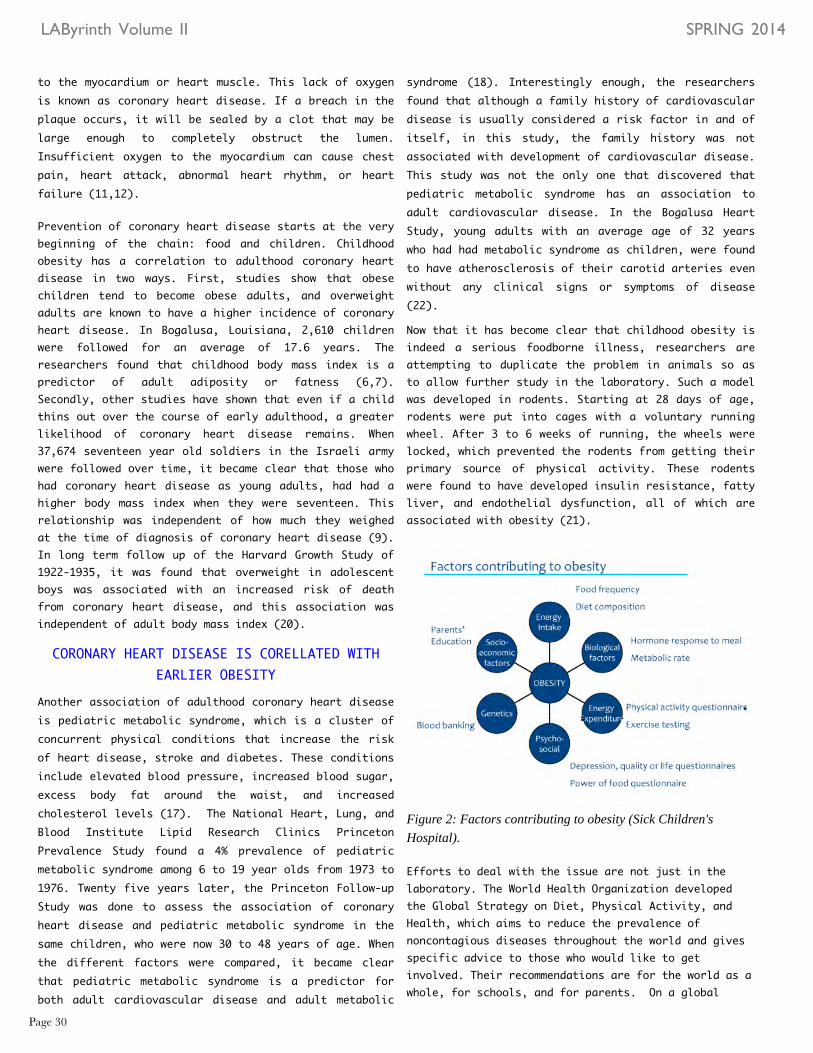
Now that it has become clear that childhood obesity is indeed a serious foodborne illness, researchers are attempting to duplicate the problem in animals so as to allow further study in the laboratory. Such a model was developed in rodents. Starting at 28 days of age, rodents were put into cages with a voluntary running wheel. After 3 to 6 weeks of running, the wheels were locked, which prevented the rodents from getting their primary source of physical activity. These rodents were found to have developed insulin resistance, fatty liver, and endothelial dysfunction, all of which are associated with obesity (21).
Figure 2: Factors contributing to obesity (Sick Children's Hospital).
Efforts to deal with the issue are not just in the laboratory. The World Health Organization developed the Global Strategy on Diet, Physical Activity, and Health, which aims to reduce the prevalence of noncontagious diseases throughout the world and gives specific advice to those who would like to get involved. Their recommendations are for the world as a whole, for schools, and for parents. On a global
to the myocardium or heart muscle. This lack of oxygen is known as coronary heart disease. If a breach in the plaque occurs, it will be sealed by a clot that may be large enough to completely obstruct the lumen. Insufficient oxygen to the myocardium can cause chest pain, heart attack, abnormal heart rhythm, or heart failure (11,12).
Prevention of coronary heart disease starts at the very beginning of the chain: food and children. Childhood obesity has a correlation to adulthood coronary heart disease in two ways. First, studies show that obese children tend to become obese adults, and overweight adults are known to have a higher incidence of coronary heart disease. In Bogalusa, Louisiana, 2,610 children were followed for an average of 17.6 years. The researchers found that childhood body mass index is a predictor of adult adiposity or fatness (6,7). Secondly, other studies have shown that even if a child thins out over the course of early adulthood, a greater likelihood of coronary heart disease remains. When 37,674 seventeen year old soldiers in the Israeli army were followed over time, it became clear that those who had coronary heart disease as young adults, had had a higher body mass index when they were seventeen. This relationship was independent of how much they weighed at the time of diagnosis of coronary heart disease (9). In long term follow up of the Harvard Growth Study of 1922-1935, it was found that overweight in adolescent boys was associated with an increased risk of death from coronary heart disease, and this association was independent of adult body mass index (20).
CORONARY HEART DISEASE IS CORELLATED WITH EARLIER OBESITY
Another association of adulthood coronary heart disease is pediatric metabolic syndrome, which is a cluster of concurrent physical conditions that increase the risk of heart disease, stroke and diabetes. These conditions include elevated blood pressure, increased blood sugar, excess body fat around the waist, and increased cholesterol levels (17). The National Heart, Lung, and Blood Institute Lipid Research Clinics Princeton Prevalence Study found a 4% prevalence of pediatric metabolic syndrome among 6 to 19 year olds from 1973 to 1976. Twenty five years later, the Princeton Follow-up Study was done to assess the association of coronary heart disease and pediatric metabolic syndrome in the same children, who were now 30 to 48 years of age. When the different factors were compared, it became clear that pediatric metabolic syndrome is a predictor for both adult cardiovascular disease and adult metabolic
LAByrinth Volume II SPRING 2014
Page 30

Works Cited1. "Center for Disease Control and Prevention."
Centers for Disease Control and Prevention. Centers for Disease Control and Prevention, 10 July 2013. Web. 13 Nov. 2013. <http://www.cdc.gov/healthyyouth/obesity/facts.htm>.
2. "Childhood Obesity Facts." Centers for Disease Control and Prevention. Centers for Disease Control and Prevention, 10 July 2013. Web. 13 Nov. 2013. <http://www.cdc.gov/healthyyouth/obesity/facts.htm>.
3. "Childhood overweight and obesity." WHO Childhood Overweight and Obesity. World Health Organization, n.d. Web. 3 Feb. 2014. http://www.who.int/dietphysicalactivity/childhood/en/
4. Corvalan, Camila, Ricardo Uauy, Juliana Kain, and Reynaldo Martorell. "Obesity Indicators and Cardiometabolic Status in 4-y-old Children." The American Journal of Clinical Nutrition 91 (2010): 166-174. Print.
5. Daniels, Stephen R., Donna K. Arnett, Robert H. Eckel, Samuel S. Gidding, Laura L. Hayman, Shiriki Kumanyika, Thomas N. Robinson, Barbara J. Scott, Sachiko St. Jeor, and Christine L. Williams. "Overweight in Children and Adolescents: Pathophysiology, Consequences, Prevention, and Treatment."
6.Freedman, David S. , Laura Kettel Khan, Mary K. Serdula, William H. Dietz, Sathanur R. Srinivasan, and Gerald S. Berenson. "The Relation of Childhood BMI to Adult Adiposity: The Bogalusa Heart Study." Pediatrics 115.1 (2005): 22-27. Print.
7.Freedman, David S., Laura Kettel Khan, William H. Dietz, Sathanur R. Srinivasan, and Gerald S. Berenson. "Relationship of Childhood Obesity to Coronary Heart Disease Risk Factors in Adulthood: The Bogalusa Heart Study." Pediatrics 108.3 (2001): 712-718. Print.
8."Global Strategy on Diet, Physical Activity and Health." World Health Organization. World Health Organization, n.d. Web. 5 Apr. 2014. .
9.Tirosh, Amir, Iris Shai, Assaf Rudich, Arnon Afek, Gal Dubnov-Raz, Nir Ayalon, Barak Gordon, Estela Derazne, Dorit Tzur, Ari Shamis, and Shlomo Vinker. "Adolescent BMI Trajectory and Risk of Diabetes versus Coronary Disease." New England Journal of Medicine 364.14 (2011): 1315-1324. Print.
10. "Understanding Childhood Obesity: 2011 Statistical Sourcebook." American Heart Association/American Stroke Association. American Heart Association/American Stroke Association, n.d. Web. 13 Nov. 2013. .
11. "What Is Atherosclerosis?." - NHLBI, NIH. National Heart, Lung, and Blood Institute, n.d. Web. 2 Feb. 2014.
12. "What Is Coronary Heart Disease?." - NHLBI, NIH. National Heart, Lung, and Blood Institute, n.d. Web. 4 Feb. 2014.
13. "A-Z Index." Healthy Weight: Assessing Your Weight: BMI: Child and Teen Calculator. Centers for Disease Control and Prevention, n.d. Web. 8 Apr. 2014.
14. "Childhood Obesity Facts." Centers for Disease Control and Prevention. Centers for Disease Control and Prevention, 28 Mar. 2014. Web. 7 Apr. 2014.
15. "Childhood overweight and obesity." WHO. World Health Organization, n.d. Web. 8 Apr. 2014.
16. "How Are Overweight and Obesity Diagnosed?." - NHLBI, NIH. National Heart, Lung, and Blood Institute, n.d. Web. 8 Apr. 2014.
17. "Metabolic syndrome." Definition. Mayo Clinic, n.d. Web. 7 Apr. 2014.
18. Morrison, John A., Lisa Aronson Friedman, and Courtney Gray-McGuire. "Metabolic Syndrome in Childhood Predicts Adult Cardiovascular Disease 25 Years Later: The Princeton Lipid Research Clinics Follow-up Study." Pediatrics 120.2 (2007): 340-345. Print.
LAByrinth Volume II SPRING 2014
Page 31
level, the World Health Organization has the job of promoting and publicizing their strategy by implementing recommendations for countries, providing support for member states that implement programs, supporting research, and establishing networks to build up research and training. WHO recommends that schools teach children how to live a healthy life primarily by way of example. To increase the availability of healthy food, programs should provide healthy food options for the students. Physical education classes should vary in activities to maximize student interest and noncompetitive physical extracurricular activities should be encouraged. (8).
Childhood obesity is recognized as more than just an aesthetic issue; it is deeply rooted in the smoldering embers of the coronary arteries, waiting to burst forth into flames of adulthood coronary heart disease. Therefore, to improve global health we must increase worldwide awareness and take action to preserve the future of our children. After all, are they not the heart of the matter?

Page 32

Is there any correlation between Yahadus and the study of forensic science, a science that is usually related to crime scenes? Interestingly, there are numerous applications of forensic science to our daily lives and history. One major event in our history was Yetzias Mizrayim: the Exodus from Egypt. Many archeologists have found artifacts in the Red Sea that could be from this occurrence. However, as Jews, we do not rely upon scientific evidence for our faith. Even without such “evidence,” we know that Hashem performed a great miracle by emancipating us from Egypt and splitting the sea. The objects found within and in the proximity of the Red sea are merely there, to help us understand the context of our history giving us tangible objects as forensic support.
The story of the Yetzias Mizrayim, is discussed in Sefer Shemos, primarily in Parashas Bishalach, perekim די ' and וט '. The exact location of the splitting of the Red Sea is unclear. According to the Torah, Krias Yam Suf occurred somewhere between Migdol and the Red sea. However, Migdols' exact location is unknown to us. Many commentaries believe that the crossing took place by the Yam Suf HaMaravi- the Western side of the Red Sea by the Gulf of Suez, while others believe that the Jews crossed over the dry land until the Yam Suf Hamizrachi, by the Gulf of Aquaba on the beach of Nuweiba. Ron Wyatt, a noted archeologist, suggests that the crossing is referring to the beach of Nuweiba.
Before delving into this specific topic, let us answer the question: what is forensic evidence? Forensic evidence, according to the definition given by Max M. Houck and Jay A. Siegal is "the science of associating people, places and things involved in criminal activities" (3-4) to be used as evidence in numerous court cases. There are many different types of evidence: hairs, fibers, glass, fractured objects, shoe and tire impressions, fire accelerants, fingerprints,
THE SECRETS OF YAM SUF
By: Esther Rothman* '15gunshot residue, toolmarks to mention a few. Although Kriyas Yam Suf, was not "a criminal event," the artifacts that archeologists have discovered can be categorized into different types of forensic evidence; archeological evidence, those pertaining to the scientific study and analysis of historic people through artifacts; anthropological evidence, those pertaining to remains of the skeletal system; and questioned documents, those pertaining to any surface with linguistic or numerical markings.
What type of archeological evidence was found by the Yam Suf? On two separate occasions, while scuba diving, researchers discovered archeological evidence from Kriyas Yam Suf. In 1998, Ron Wyatt and in 2000, Ross Patterson, Viveka Ponten, Michael Redman, Aaron Sen, and Tor Larsen, discovered chariot wheels and axels, many of which were covered in coral. The wheels contained four, six, and eight spokes (see Figure 1). Wyatt was able to date these artifacts back to Yetzias Mizrayim. Wyatt took his discovery to Nassif Mohammed Hassan, the director of antiques in Cairo, who confirmed that the wheels with four, six, and eight spokes were the prevalent style of chariot wheels used during the eighteenth dynasty in Egypt, which was the time period in which the splitting of the sea occurred.
F i g u r e 1 : Chariot wheels and axels from Yam Suf found in 1998 and 2000.
LAByrinth Volume II SPRING 2014
Page 33* Esther was one of the winners of the Jerusalem Science Competition; She traveled to Chicago to present the reserach summarized in this paper at the JSC Award Ceremony i..Chicago.

Figure 2: The bone From the Red Sea, left, is covered in coral, and the bone found in an Egyptian tomb, right, is not covered in coral. Thus, it could not be determined whether the bone is the remains of an Egyptian who drowned in the sea.
sea" (Shemos, Perek 15:4). On this pasuk, Rashi and the Mechilta explain that the pillar of cloud that Hashem provided, turned the ground underneath the Egyptians into mud, and the horses became 'unshod-' their hoofs fell off, which would account for the discovery of the horse hooves. Interestingly, with respect to the human bone that was found, the Torah says:" דחא-דע ,םהב ראשנ-אל " - "not one of them remained alive" (Shemos, Perek 14:28) and " ץרא ,ומעלבת " -" The earth swallowed them" (Shemos, Perek 15:12). On this pasuk, the Midrash explains that the earth swallowed the Egyptians and interred them into the depths of the earth. Therefore, the Midrash's explanation clarifies why there is not a multitude of bones at the bottom of the Red sea and why even those recovered are literally combined with the earth.
A final piece of evidence that I found particularly interesting, is the two identical granite pillars discovered by Wyatt (see Figure 3). Both pillars are 14 feet high, 3 feet in diameter, and made of red granite. One pillar was found in 1978 in Nuweiba, however, its inscriptions were washed away. A second pillar was found in 1984 by the Gulf of Aqaba, by the Yam Suf, on the Saudi Arabian side. Fortunately, this pillar has its inscriptions intact. The words inscribed on the pillar are written in Ancient Hebrew- words such as Mitzrayim, Edom and Sholomo. The Name of Hashem also appears on it. These pillars can serve as questioned documents, because they contain linguistic or numerical markings whose source or author is unknown. Wyatt and his team believe that since his name is etched on the pillar, Shlomo Hamelech, built it to commemorate and to thank Hashem for Yetizas Mizrayim. As supporting proof, they site two sources: first, Shlomo's port city was at Gulf of Aqaba as it says in Melachim Alef: " רבג-ןויצעב המלש למה השע ינאו
Additionally, this chariot style is seen in the chariots found in King Tutankhamen' tomb, as well as in art and hieroglyphics that were etched during that same time period.
These wheels are examples of forensic archeological evidence; determining their source of origin due to their situ. However, are these the same chariot wheels from Yetzias Mizrayim according to the Torah? In Sefer Shemos, Perek חכ:'די ', it says " בכרה-תא וסכיו
םישרפה-תאו "- "and the waters covered the chariots and the cavalry," and " ויתבכרמ ןפא תא ,רסיו "- "And He removed the wheels of their chariots" (Shemos, Perek 14:25). On this pasuk Rashi quotes a Midrash that Hashem placed hot coals on the floor of the sea and set fire to the chariots, causing the wooden fillings of the chariots to burn and the wheels to fall off; this explains why there are remains of the wheels alone and not whole chariots in the Red sea.
Another interesting discovery regarding the chariot wheels was that the scuba divers found gold wheels which did not disintegrate in the ocean or become covered with coral, since coral does not grow on gold. It is likely that these wheels originated in Egypt. In Sefer Shemos, the Torah explicitly states that the Egyptians had a lot of gold and silver " םירצממ ,ולאשיו,
בהז ילכו ףסכ-ילכ " - "and they requested of the Egyptians silver articles and gold articles" (Shemos, Perek 12:35).
FORENSIC ANTHROPOLOGICAL EVIDENCE COULD NOT UNDERGO CARBON DATING
In addition to the wheels, Ron Wyatt, Bill Fry and members of their teams also found horse hooves, horse skeletons and important forensic anthropological evidence at the bottom of the Red Sea in the form of human rib cages and a human femur bone. Like all forensic evidence, Wyatt attempted to determine the origin of this femur bone. The bone was analyzed at the Department of Osteology at Stockham University and found it to be a human femur bone from the right leg of a 165 to 170 centimeter man. Unfortunately, minerals and coral replaced the bone because it was in the Red sea for a myriad of years, so it could not undergo carbon dating (see Figure 2).
The Torah perspective is as follows. Regarding the horse hooves and bones, the Torah says " המר ובכרו סוס םיב " - "horse and its riders He threw into the
sea" (Shemos, Perek 15:1), and" ףוס-םיב ועבט ,וישלש רחבמו " - "his, Pharaoh's, elite, officers were sunk in the Red
LAByrinth Volume II SPRING 2014
Page 34

ףוס-םי תפש-לע ,תולא-תא רשא " - "and king Solomon made a navy of ships in Ezion-geber, which is beside Elot, on the shore of the Red Sea" (Melachim Aleph, 9:26).
Secondly, the pillars were made of red granite, a mineral available in Egypt on the other side of the Gulf, meaning on the Egyptian side. Shlomo would have only been able to obtain this mineral if he had connections with Egypt. Interestingly, It says in Melachim that Shlomo married the daughter of Pharaoh-" הערפ-תב-תא חקיו ;םירצמ למ הערפ-תא ,המלש ןתחתיו " - " And Solomon became allied to Pharaoh king of Egypt by marriage, and took Pharaoh's daughter " (Melachim Aleph, 3:1); his ties with Egypt would have enabled him to acquire the granite necessary to form these structures.
Figure 3: A pillar made of red granite.
As seen from these artifacts, forensic evidence is not simply applicable to crime scenes, but we can extend its methodologies and logic to help us further understand our history. The chariot wheels, horse hooves, horse bones, and human bones found at the bottom of the Red Sea, as well as the ancient granite pillars, help us understand the Great Miracle that Hashem performed and enable us to be that much more appreciative of His infinite kindness.
Works Cited1."Crossing the Red Sea." Flood. N.p., n.d. Web. 6
Mar. 2014. <http://www.cps.org.rs/Innerpeace/Creation/redsea.html>.
2.Houck, Max M., and Jay A. Siegel. Fundamentals of Forensic Science. Amsterdam: Elsevier/Academic Press, 2006. Print.
3."Red Sea Update." Anchor Stone International. N.p., 27 Mar. 2007. Web. 6 Mar. 2014. <http://anchorstone.com/29-discoveries/Red-Sea-Crossing/133-red-sea-update.html>.
4."The Red Crossing Site." Wyatt Archaeological Research - Official Site of Ron Wyatt's Discoveries. N.p., n.d. Web. 6 Mar. 2014. <http://wyattmuseum.com/red-sea-crossing.htm>.
LAByrinth Volume II SPRING 2014
Page 35

Page 36

The new mother turns over in her bed to check on her sleeping baby. She thought she heard Miriam cry but saw that she was sound asleep. One hour later, Miriam awakens hungry, and the new mother is forced to exchange her own sleep schedule for that of her newborn baby.
Figure 1: The human circadian and associated behaviors.
Our bodies keep time without using alarms. Environmental cues serve as the marker of time, indicating to the body when to perform time dependent functions. It is our inborn clocks that cause us to feel awake or tired at the appropriate times. In addition to sleeping and wakefulness, there are many other processes that are time regulated. Hormone secretion, eating and digestion, cell regeneration, and the maintenance of body temperature are all time dependent functions. In fact, research has shown that our internal clocks are the result of basic cellular phenomena and can have dramatic ramifications on both physical and emotional health (see Figure 1).
Our circadian rhythm is a complex 24 hour cycle of different mental, physical, and behavioral body changes. The master clock, which keeps all the different cycles in sync, is found in a group of nerve cells called the suprachasmatic nucleus, or SCN (see Figure 2), of the hypothalamus in the brain (2). In these nerve cells, the circadian genes and their protein products oscillate at a steady pace, a biochemical pendulum that sweeps a steady arc. To track these rhythms, scientists turn to melatonin - a hormone
OUR INTERNAL ALARM CLOCKS:
OUR CIRCADIAN RHYTHMS
By: Miriam Schuster '14
that is secreted by the pineal gland in the brain and whose levels rise at night and then fluctuate depending on the exposure of light to the sleeper (4).
Circadian rhythms start functioning when an infant is about 10 weeks, but are not recognizable until about three months, when an infant has a set rhythm and a discernible biological clock. Researchers studying melatonin levels in newborns found that “the circadian rhythm of melatonin was not observed in the neonatal period and it appears at the same time as the appearance of adult-like sleep–wake rhythm, about 2–3 months after birth” (4). Apparently, cycles of melatonin secretion correlate strongly with sleep/wake cycles, a fact proven by ten prematurely born babies who were hospitalized 2-4 hours after birth and placed in a full-light incubator. The infants were fasting and on fluid replacement during the first day and, on the second day, fed formula and nutrition intravenously every 24 hours (1). Antibiotics were given for five days to maintain the health of the babies. The results were encouraging.
Researchers focused on two of the circadian genes - Bmal1 and CRY1 - which control negative feedback loops (see Figure 3). Bmal1 levels are low at the beginning of the subjective night, while CRY1 levels are high. CRY1 promotes transcription of Bmal1, causing Bmall levels to rise through the night and reach their peak by morning. In addition, high levels of Bmal1 inhibit CRY1, so when such levels are present, Bmal1 levels
Figure 2: Superchasmatic nucleus hypothalamus in the brain
LAByrinth Volume II SPRING 2014
Page 37

Sleeping and wakefulness are not the only functions controlled by circadian rhythms. Cell proliferation and growth are controlled by the circadian gene Period 1 (PER1). When PER1 rhythms are altered, it can result in disordered and uncontrolled cell growth, or cancer (5). Upon investigating mice with tumors, researchers saw a significant decrease in PER1 expression in areas with the PER1 gene. When these mice had the cancer, their clock genes were not working properly.
Aside from mice, many experiments were done on fruit flies, Drosophila melanogaster, which they found resemble the sleep-wake cycles of humans and have much in common regarding their circadian rhythms (6). These researchers watched the 24 hour cycle of the PER protein or Period gene, which oscillated the different biological activities throughout this 24 hour cycle. This PER gene accumulates throughout the night and translates into proteins during the day (7). When these flies were injected with different mutations that were meant to shorten or lengthen the fly’s circadian period, for example making their cycle 18 or 28 hours instead of the normal 24, the flies PER genes were able to keep going even with this mutation (7). They found that the mutation they injected into these flies messed up the regulation of dbt, which regulates the accumulation of the PER protein. This mutation promptly affected the dbt but only affected the PER protein over time. The mutated dbt regulated the degradation and breakdown of the PER protein over time (7).
Figure 4: Several cell cycle genes that regulate proliferation.Also, there are circadian proteins that maintain checkpointactivation in the presence of DNA damage.
drop, release inhibition of CRY1, and the cycle repeats. In contrast, premature newborns did not show such fluctuations. However, the preemies were under constant light and not subject to environmental lighting changes, which could have impacted the lack of development of proper rhythm. In addition, the preemies were not fed by their mothers, preventing them from patterning a circadian rhythm in synchrony with the mother. They were also fed every three hours, which inhibited a proper sleeping-hunger-feeding cycle (4). These experimental flaws demonstrate that in the absence of environmental cues, preemies do not exhibit cellular cycling of the circadian genes.
CIRCADIAN RHYTHMS CHANGE THROUGHOUT LIFETIME
During adolescence the circadian rhythm fluctuates. Often, teenagers go to sleep later and their bodies will compensate for the lack of sleep by waking up later. In addition, when the teenage body is short-changed of its optimal 8-9 hours of sleep, it becomes deprived of the environmental cues it needs for its circadian clock to function properly. If the body is lacking sleep, the rhythm will change, leading to an awake feeling late at night. At about 2:00 a.m., the circadian “crashes” for an extended period of time, which is dependent on the amount of sleep already gained. The body can also be trained to stay up later by using bright lights to confuse the circadian into “thinking” that it is day. In the morning, when the lights are turned on, the SCN responds with arousal of our body - increasing body temperature, decreasing melatonin levels and producing daytime hormones, such as cortisol. This abnormality causes the body to produce melatonin for our late nights, which lasts until the morning when we are exposed to light (3).
Figure 3: The negative feedback loops of Bmal1 and Cry1proteins.
LAByrinth Volume II SPRING 2014
Page 38

Works Cited
1. Blood Mononuclear Cells Of Preterm Neonates."Journal Of Maternal-Fetal & 6 Jan. 2014.
2. Fults, Erin. "Inside Life Science: How Our Bodies Keep Time." LiveScience. <http://www.livescience.com/13123-circadian-rhythms-obesity-diabetes- nih.html>
3. National, Sleep Foundation. "Sleep Drive and Your Body Clock." Body Clock & Sleep. N.p., n.d. Web. 23 Jan. 2014. <http://www.sleepfoundation.org/article/sleep-topics/sleep-drive-and- your-body-clock>.
4. Shinohara, Hitomi, and Hideya Kodama. "Relationship between Circadian International 53.1 (2011): 29-35. Academic Search Premier. Web. 6 Jan. 2014
5. Zhao, Ningbo, et al. "Aberrant Expression Of Clock Gene Period1 And Its Squamous Cell Carcinoma." Plos ONE 8.2 (2013): 1-9. Academic Search Premier. Web. 26 Jan. 2014
6. Rosato, Ezio, Eran Tauber, and Charalambos P Kyriacou. "European Journal of Human Genetics." Nature.com. Nature Publishing Group, n.d. Web. 6 Apr. 2014. <http://www.nature.com/ejhg/journal/v14/n
7. Preuss, Fabian , Jin Yuan-Fan, Madhavi Kalive, Shu Bao, Eric Schuenemann, Biology." National Center for Biotechnology Information. Version 24. N.p., n.d. Web. 6 Apr. 2014. <http://www.ncbi.nlm.nih.gov/pmc/articles>
LAByrinth Volume II SPRING 2014
Page 39
Research on circadian rhythm opens windows into how our genes act within our environment. Innovative research on our own biological clock can help us better understand the complexity of our systems. As we study the link between different cancers and our circadian rhythm, we can discover new cures and preventions for cancer. Adolescents can listen to their bodies’ cues and create a proper setting for sleep, so that they can feel awake during the appropriate times. And new mothers everywhere can train their preemies, who awake at erratic hours, to a set circadian rhythm, which matches their own.

Page 40

or childhood, while others may not appear until middle age or later. The different types can vary in whom they affect, which muscles they affect, and what the symptoms are; but all forms of MD grow worse as the person's muscles get weaker. Unfortunately, most people with MD eventually lose the ability to walk (8). Dr. Sachs anticipated that if he could close these channels, the symptoms induced by them would be suppressed (2).
Figure 1: Open ion channels, left, allow substances to enter the cell from the extracellular environment. Closed ion channels, right, selectively prohibit certain substances from entering the cell.
The team at the University of Buffalo did not initially focus their research on Chilean rose tarantula venom. It was only after they obtained negative results from several known drug compounds that Sachs and his research team turned to venom. They hypothesized that venoms, with their complex chemical makeup, might contain molecular compounds that could block ion channels. Sachs screened the venoms of centipedes, scorpions, and spiders, and a promising hit came from the Chilean rose tarantula (Chilean Grammostola spatulata), a relatively harmless spider that can be bought in local pet stores and that contains weak venom. They found a peptide, GsMtx-4, that could successfully turn off the channels and reduce muscular stress. By injecting a synthetic version of GsMtx-4
VENOM. The mere sound of this word alone elicits fear in the bravest of hearts. Perhaps this is because society views venom, and the spiders that produce them, as dangerous and lethal weapons — weapons that trigger long and painful deaths. And spiders, whether poisonous or benign, have become a universal means of conjuring panic in rational adults. Yet, we may have misjudged spiders, as it now seems that venom has been wrongfully accused. The properties that make venom harmful or deadly are also what make it so valuable for medicine.
Many venom toxins target the same molecules that need to be controlled to treat diseases. Since venoms are biologically active and contain many toxins, they are a natural resource for scientific investigation and contain the potential to be useful in pharmaceutical therapies. Venom works fast, is highly specific, and targets particular molecules, working the same way medicines do, to fit into and target specific molecular locks to combat illnesses. In fact, scientists are currently investigating the possibility that venom has medicinal use and that it can potentially benefit several human health conditions.
Frederick Sachs, a biophysicist at the University of Buffalo, has been investigating the use of venom found in the Chilean rose tarantula as a treatment for malfunctioning mechanosensitive ion channels on the membranes of muscle cells. Mechanosensitive ion channels are small tunnels that connect the inside of a cell to the outside world. Generally, the tunnels are closed. However, when a cell is contorted or stretched, the tunnels open and permit calcium and other substances to enter the cell (1) (see Figure 1).
The excess calcium results in the digestion of muscles from the inside out, leading to the crippling symptoms observed in those who have Muscular Dystrophy (MD). MD appears in different forms and causes muscle weakness and muscle loss. Some forms of MD appear in infancy or
ARACHNOPHILLIA
By: Hendel Seif '14
LAByrinth Volume II SPRING 2014
Page 41

venom is relatively weak, its potential to heal far outweighs its potential to harm (4). outweighs its potential to harm (4).
Muscular dystrophy and atrial fibrillation are not the only conditions this toxin modulates. Mechanosensitive channels mediate somatic sensation so this venom can also be used to alleviate pain. For instance, one of the peptide toxins isolated from the venom of the Chilean rose tarantula spider, mechano toxin 4 (GsMTx4), is known to block stretch-activated cation channels in sensory neurons. Since mechanosensitive channels in sensory neurons are thought to be molecular sensors for
mechanotransduction, such as touch and pressure, scientists wondered whether the venom might block certain forms of mechanical pain. Results suggested that GsMTx4 selectively alleviates mechanical hyperalgesia (increased sensitivity to pain), possibly by blocking mechanosensitive channels, pointing to the potential clinical use of venom as a pain treatment (3).
While the venom from the Chilean rose tarantula is not particularly harmful, the venom from the funnel-web spider (one of the most lethal spiders) is deadly. Yet, like the Chilean rose tarantula, this spider may have also been misjudged. Researchers at the University of Queensland’s Institute for Molecular Bioscience are now studying this venom as a way to kill breast cancer cells. The hope is that the complex mix of molecules in this spider’s venom, which target very specific sites, would yield some that can target cancer cells. This research is currently underway in Australia and is still in its early stages (5). Scorpion venom, as well, has been shown to bind to cancer cells in mice.
Spider venom has served various functions in natural healing and medicine. Venom molecules are used in chronic pain prevention. Yale researchers, too, are screening the funnel-web spider’s venom for toxins with potential benefits in pain medication. They will be testing the activity of 93 toxins, called atracotoxins, found in the venom, against a dozen pain receptors to discover the few that have specific activity that can inhibit ion channels involved in pain transduction. Pairs of synthesized toxin RNA and pain receptor RNA are injected inside frog eggs, inducing them to produce toxins and pain receptor proteins using instructions from the RNA. After
into lab mice with dystrophy, Sachs and his team were able to keep the mice’s ion channels closed. Concurrently, they found that the venom improved muscle activity and slowed muscle deterioration. Sachs and his research team are presently awaiting FDA approval to begin clinical trials (1).
GSMTX-4 CAN PREVENT ATRIAL FIBRILLATIONThe heart is one of the most important muscles in the body and ion channels play a huge role in its function. Further research on rabbits suggests that GsMtx-4 is useful in preventing rapid and chaotic electrical activity in the atria, a condition ordinarily caused by atrial fibrillation. Atrial fibrillation (AF) is the most common type of arrhythmia, a problem with the rate or rhythm of the heartbeat. During an arrhythmia, the heart can beat too fast, too slow, or with an irregular rhythm. AF occurs if rapid, disorganized signals cause the heart’s atria to fibrillate (contract fast and irregularly). Blood pools in the atria and is not pumped completely into the ventricles; the heart’s upper and lower chambers do not work together appropriately. AF can increase the risk of a stroke and, in some people, can cause chest pain or heart failure, especially if the heart rhythm is very rapid (7) (see Figure 2).
Led by Sachs, a team of US and German scientists performed further research to determine whether insect venom could be used to block ion channels, thereby preventing cells from swelling and triggering atrial fibrillation. The team electrically stimulated the hearts of rabbits into arrhythmia and then suppressed the abnormal rhythm with extracts of the venom. It was also demonstrated that the peptide had no effect on healthy hearts, suggesting that side effects of this treatment would be minimal. The research on GsMtx-4 is especially exciting because it targets treating the symptoms of atrial fibrillation, rather than its causes. Thus, this research can eventually lead to preventative treatment for those at risk of stroke and heart failure. Moreover, since the
LAByrinth Volume II SPRING 2014
Page 42
Figure 2: Top shows normal rhythm, bottom shows faster, irregular, disorganized rhythm.

Works Cited
1. "Good Venom - University at Buffalo."Good Venom - University at Buffalo. N.p., n.d. Web. 1 Jan. 2014.
2. “Medical Moment: Using Spider Venom to Slow Down Duchenne Muscular Dystrophy.” WNDU RSS. N.p., n.d. Web. 30 Dec. 2013
3. Park, Seung Pyo, et al. "A Tarantula Spider Toxin, Gsmtx4, Reduces Mechanical And Neuropathic Pain." Pain 137.1 (2008): 208-217. MEDLINE. Web. 1 Jan. 2014.
4. "Spider Venom Might Prevent Atrial Fibrillation - Western Journal of Medicine."NCBI. N.p., n.d. Web. 30 Dec. 2013.
5.“Academia.edu." SPIDER VENOM. N.p., n.d. Web. 17 Mar. 2014.
6."Menu." Yale Daily News. N.p., n.d. Web. 17 Mar. 2014.
7."What Is Atrial Fibrillation." - NHLBI, NIH. N.p., n.d. Web. 15 Mar. 2014.
8."Muscular Dystrophy: MedlinePlus." U.S National Library of Medicine. U.S. National Library of Medicine, n.d. Web. 16 Mar. 2014.
After waiting two days for the frog eggs to produce the toxins and pain receptors, they are screened. A machine was assembled to measure the receptor's activities in the eggs. Pain receptors are channels that allow charged particles to pass into a cell, therefore, generating an electrical current. If little or no electric current is detected in the frog eggs, it means the pain receptors may have been blocked (6) (see Figure 3)
It is clear that a creature once regarded as threatening can have many beneficial uses for humankind. Indeed, there is the potential for every organism on our planet to provide us with the solution to any particular obstacle of survival, if we would only look. Thus, it is especially important that we, as a species, guard the biodiversity found in our ecosphere, so that we can keep our tool chest fully stocked. Although nature presents many dangers, it also carries the solutions to endless problems in the field of medicine, engineering and just about any other human endeavor. It is our responsibility to discover these solutions and to invite the possibility that they may present themselves in places å we least expect them. What better example than a venomous spider who metamorphasized from an instrument of pain to a mechanism of healing?
Figure 3 Three models depicting how a change in the voltage in the extracellular environment caused by an outwardly flow of positive ions causes the subsequent closing of the channels to further penetration ofions. The left shows the open configuration of the ion channel; the right shows the ion channel in the closed position, preventing the further influx of positive ions.
LAByrinth Volume II SPRING 2014
Page 43

Page 44

We have all felt that we’ve lost ourselves, to some degree. Our daily existential crisis? Our ability to lose ourselves in work? Our mid-life crisis, during which we feel a drive to change our course, reach new goals, and design a better person? We have all felt some degree of dissociation from the creature we know as ourselves, but patients with multiple personality disorder, or Dissociative Identity Disorder, go through severe dissociation, breaking the connection between the victim's thoughts and actions, often leaving no memory behind.
Dissociative Identity Disorder (DID) is often found in the past of the patients. It usually stems from childhood or adolescence, from past of neglect, physical and/or emotional abuse, or a traumatic event. As defined by the DSM-IV, DID is characterized by the presence of two or more alternate personalities, each split from the other. Each personality is unique, each its own race, gender, and/ or age. They have distinct mannerisms, postures, gestures, and speech patterns, making each one discernable from the others. The alters can, and often do, present physical differences- such as allergies, right/ left hand dominance, and vision prescriptions. When the victim is under the control of one of the alters, they often do not remember what has occurred. They develop a degree of amnesia, for the periods when they are out of control of their own minds. Many psychiatrists and psychologists describe the process as boarding- in which the alters rotate throughout the rooms in a house (psyche). All alters stay in their respective “bedrooms”, until they are summoned to the “living room”. Applied to the psyche, the alternate personalities lurk in the background, waiting until they’re needed to come forward, bringing their characteristics and habits with them (2;3;5).
While Dissociative Identity Disorder fills many of the same qualifications of other dissociative disorders, as well as many non-dissociative mental disorders, the combination of symptoms of DID is unique. The most common symptoms, found in about a third to a half of all victims include: changing levels of motor and
WHICH ME AM I?
By: Zahava Sokolow '16chamental function (from well to nearly disabled), undue pain/ migraines, derealization and depersonalization, depression/ volatile behaviors, unexplained changes in habits or patterns, anxiety, long-term amnesia, and hallucinations. Many of these symptoms are linked to the alternate personalities, as well as the constant changes in mindset (1).
Figure 1: An enlarged hypothalamus, the prominent physical
indicator of DID.
A number of people with mental illnesses complain of experiencing dissociation- which is defined by the DSM-IV as a disturbance in thinking, awareness, identity, conciousness, or memory. It is more severe than ordinary absentmindedness or forgetfulness, and is not associated with any underlying cause for memory deficits, or altered personality (i.e, neuroloical illnesses, substance or alcohol abuse, etc.). Dissociation can last anywhere from a few moments, and up to an extended period of separation. During these periods, many people lose motor function and regulation, and report “supernatural” or “out of body” experiences. Others lose control of emotions and actions during these episodes, and seem out of character. Some victims have memory of these moments,
LAByrinth Volume II SPRING 2014
Page 45

aroma therapy, which can allow the patient to express feelings and deal with memories. Clinical hypnosis is a treatment technique that uses intense relaxation, concentration, and attention to achieve an altered state of consciousness or awarenes.. It allows the victim to explore thoughts, feelings, and memories they have, which they have not yet processed themselves (1;2;4;5).
Treatment for Dissociative Identity Disorder is crucial, and without treatment, physical, emotional, and mental harm can be caused to the victim. It is a chronic, serious disease, with many complications in cerebral and daily functions. Patients with dID are at great risk for the following; suicide attempts, self-harm, violence, substance abuse, and victimization by others. So long as treatment is provided in a timely, effective fashion, the outlook for those with DID is positive. Patients generally respond well to treatment; however, treatment can be a long, painstaking process. Some patients develop a form of Stockholm Syndrome, where they are imprisoned by their own minds, yet are reluctant to leave. They form bonds with the alternate personalities, and are reluctant to destroy them, as they are helpful in the process of coping with the damage caused by the trauma. To improve the outlook, as well as the mindet of the patient, other mental illnesses must be treated- such as substance abuse, eating disorders, depression, and anxiety should be treated respectively, to create a healthier envoronment, and improve chances of complete recovery (2;5).
Figure 2: Obvious and Hidden Signs of D.I.D.
The effects of Dissociative Identity Disorder are unimaginable, with lives being torn apart, families put through an unimaginable situation, and people losing their sense of identity and stability. Possible cures have been proposed, yet funding for this disorder is limited. Testing and drug trials are ongoing, but not many. They include: attempts to blend the alternate personalities, shrink the hypothalamus, alter the different parts of the brain, and eliminate these alternate personalites. At this moment, though, no cures have been close to reaching their goal- to
and appear disoriented as they end, while others cannot back into their dominant person. All of these factors make dissociation a disturbing and unsettling experience. There is an association between traumatic events and and the process of dissociation. The hypothesis of many scientists is that dissociation is the brain’s method of processing overwhelming stimuli- or the pain caused by traumatic memories. Many people who have experienced such an event, like physical or emotional abuse, develop alternate personalities to block out the memories of their victimization, which has a sharp similarity to PTSD- post traumatic stress disorder. Oftentimes, PTSD is a form of DID, to a lesser extent, utilizing the same treatmenbt courses for both disorders (2;3;5).
DIAGNOSIS OF D.I.D MUST TAKE INTO ACCOUNT A VARIETY FACTORS
The diagnosis of Dissociative Identity Disorder is complicated (see Figure 2), often done through process of elimination, rather than there being a clear checklist. If symptoms are present, the doctor evaluates the patient based on medical and general history, as well as a physical examination.The doctor often uses blood tests, X-Rays, and other diagnostic tests, to rule out any other physical deformation or illness. Certain conditions—including brain diseases, head injuries, drug and alcohol intoxication, and sleep deprivation—can lead to symptoms similar to those of dissociative disorders, including amnesia. In fact, it is amnesia or a sense of lost time that most often prompts a person with DID to seek treatment. He or she might otherwise be totally unaware of the disorder. If no illness is found, the person is often referred to a psychologist, who can aid mental illness, and can minimize the effects of the alters. Psychiatrists utilize a specifically designed interview, personality assessment, and/ or hypnosis to find evidence of dissociation. Once the diagnosis is confirmed, treatment can begin. Because there is no cure, the goal of treatment is to unite the alters, to relieve systems, and to ensure the safety of the patient. It aims to to help the patients safely express and process pain and memories, develop life skills and coping mechanisms, restore functioning, and improve relationships. Options for treatment include: Psychotherapy, utilizing psychological techniques to design comuunication of conflicts and problem solving. Cognitive therapy, which focuses on changing dysfunctional thinking patterns. Medication, which can relieve symptoms such as depression, eating problems, and anxiety. Family therapy, which can educate the family and prevent further damage to the victim or family. Creative therapies, such as art, music, or
LAByrinth Volume II SPRING 2014
Page 46

The effects of Dissociative Identity Disorder are unimaginable, with lives being torn apart, families put through an unimaginable situation, and people losing their sense of identity and stability. Possible cures have been proposed, yet funding for this disorder is limited. Testing and drug trials are ongoing, but not many. They include: attempts to blend the alternate personalities, shrink the hypothalamus, alter the different parts of the brain, and eliminate these alternate personalites. At this moment, though, no cures have been close to reaching their goal- to reverse the damage done by trauma.
Recent studies show that symptoms improve with age. The average age for development of alters has been set at 5.9 years old, while they only seem to stretch through age thirty five. After middle age, the rate of changing between alters slows, often allowing the person to go through a week, or even more, without alternating
personalities (1;3;4;5).
While we often doubt ourselves, or our knowledge of ourselves, those with Dissociative Identity Disorder are often left with no stability, no guarantee, and no idea of who or what they are. Our existential crisis, our mid-life crisis, our doubt- they are all based on the idea of ourselves which we have. You cannot change what you do not know about. They do not know what they love to do, what their favorite foods are, what their name is, or where they live, because those things are constantly subject to change. “Bipolar disorder, Dissociative Identity Disorder, mental illnesses? They have taken away freedom. They have taken away pride. Because those who suffer from them do not know what the next minute brings, who they are, or what they want. They are no longer a person,” said Deborah Haddock [author of the DID Handbook]. As science advances, we can only hope to reach that goal, to restore those identities, to let everyone know who they are, and who they will forever be.
LAByrinth Volume II SPRING 2014
Page 47
Works Cited1. Dissociative Identity Disorder (Multiple
Personality Disorder). (2012, July 19). Retrieved January 1, 2014, from WebMD: http://www.webmd.com/mental-health/dissociative-identity-disorder-multiple-personality-disorder
2. Dryden-Edwards, R. (2012, June 22). Dissociative Identity Disorder. Retrieved January 1, 2014, from MedicineNet: http://www.medicinenet.com/dissociative_identity_disorder/page5.htm
3. Kogan, A. (2011, November 30). The Critical Optimist. Retrieved December 17, 2013, from Psychology Today: http://www.psychologytoday.com/blog/the-gene-factor/201111/is-your-personality-in-your-genes
4. Lanius, R. A. (2010). Emotion Modulation in PTSD: Evidence of a Dissociative Subtype. The American Journal of Psychiatry , 640-645.
5. Walpert, S. (2011, September 1320). News Releases: UCLA Newsroom. Retrieved January 13, 2014, from UCLA Newsroom: http://newsroom.ucla.edu/portal/ucla/ucla-life-


![[Shoshana Felman] Writing and Madness Literature (Bookos.org)](https://static.fdocuments.us/doc/165x107/557213d6497959fc0b93244c/shoshana-felman-writing-and-madness-literature-bookosorg.jpg)


