Short Title: Skin AMPs protect juvenile leopard frogs Skin peptides
27
The Journal of Experimental Biology – ACCEPTED AUTHOR MANUSCRIPT Short Title: Skin AMPs protect juvenile leopard frogs Skin peptides protect juvenile leopard frogs (Rana pipiens) against chytridiomycosis James D. Pask 1 , Tawnya L. Cary 2 , and Louise A. Rollins-Smith 1,3,4* 1 Department of Pathology, Microbiology and Immunology, Vanderbilt University School of Medicine, Nashville, TN 37232 USA 2 Department of Zoology, University of Wisconsin-Madison, Madison, WI 53706 USA 3 Department of Pediatrics, Vanderbilt University School of Medicine, Nashville, TN 37232 USA 4 Department of Biological Sciences, Vanderbilt University, Nashville, TN 37235 USA *Corresponding author. Mailing address: Department of Pathology, Microbiology and Immunology, A-5301 Medical Center North, Vanderbilt University School of Medicine, Nashville, TN 37232 USA; Phone: (615) 343-4119; E-mail: [email protected] Keywords: amphibian, antimicrobial peptides, Batrachochytrium dendrobatidis, chytridiomycosis, Rana pipiens, skin defenses http://jeb.biologists.org/lookup/doi/10.1242/jeb.084145 Access the most recent version at J Exp Biol Advance Online Articles. First posted online on 11 April 2013 as doi:10.1242/jeb.084145 Copyright (C) 2013. Published by The Company of Biologists Ltd http://jeb.biologists.org/lookup/doi/10.1242/jeb.084145 Access the most recent version at First posted online on 11 April 2013 as 10.1242/jeb.084145
Transcript of Short Title: Skin AMPs protect juvenile leopard frogs Skin peptides
The
Jou
rnal
of
Exp
erim
enta
l Bio
logy
– A
CC
EPT
ED
AU
TH
OR
MA
NU
SCR
IPT
1
Short Title: Skin AMPs protect juvenile leopard frogs 1
2
3
4
5
Skin peptides protect juvenile leopard frogs (Rana pipiens) against chytridiomycosis 6
7
James D. Pask1, Tawnya L. Cary2, and Louise A. Rollins-Smith1,3,4* 8
9
10
1Department of Pathology, Microbiology and Immunology, Vanderbilt University School of 11
Medicine, Nashville, TN 37232 USA 12
2Department of Zoology, University of Wisconsin-Madison, Madison, WI 53706 USA 13
3Department of Pediatrics, Vanderbilt University School of Medicine, Nashville, TN 37232 USA 14
4Department of Biological Sciences, Vanderbilt University, Nashville, TN 37235 USA 15
16
17
18
19
20
21
22
*Corresponding author. Mailing address: Department of Pathology, Microbiology and 23
Immunology, A-5301 Medical Center North, Vanderbilt University School of Medicine, 24
Nashville, TN 37232 USA; Phone: (615) 343-4119; E-mail: [email protected] 25
26
Keywords: amphibian, antimicrobial peptides, Batrachochytrium dendrobatidis, 27
chytridiomycosis, Rana pipiens, skin defenses 28
29
30
31
http://jeb.biologists.org/lookup/doi/10.1242/jeb.084145Access the most recent version at J Exp Biol Advance Online Articles. First posted online on 11 April 2013 as doi:10.1242/jeb.084145
Copyright (C) 2013. Published by The Company of Biologists Ltd
http://jeb.biologists.org/lookup/doi/10.1242/jeb.084145Access the most recent version at First posted online on 11 April 2013 as 10.1242/jeb.084145

The
Jou
rnal
of
Exp
erim
enta
l Bio
logy
– A
CC
EPT
ED
AU
TH
OR
MA
NU
SCR
IPT
2
SUMMARY 32
One issue of great concern for the scientific community is the continuing loss of diverse 33
amphibian species on a global scale. Amphibian populations around the world are experiencing 34
serious losses due to the chytrid fungus, Batrachochytrium dendrobatidis. This pathogen 35
colonizes the skin leading to disruption of ionic balance and eventual cardiac arrest. In many 36
species, antimicrobial peptides secreted into the mucus are thought to contribute to protection 37
against colonization by skin pathogens. Although it is generally thought that antimicrobial 38
peptides are an important component of innate immune defenses against B. dendrobatidis, much 39
of the current evidence relies on correlations between effective antimicrobial peptide defenses 40
and species survival. There have been few studies to directly demonstrate that antimicrobial 41
peptides play a role. Using the northern leopard frog, Rana pipiens, we show here that injection 42
of norepinephrine brings about a long-term depletion of skin peptides (initial concentrations do 43
not recover until after day 56). When peptide stores recovered, the renewed peptides were 44
similar in composition to the initial peptides by MALDI-TOF mass spectrometry and in activity 45
against B. dendrobatidis determined by growth inhibition assays. Newly metamorphosed 46
froglets depleted of their peptide stores and exposed to B. dendrobatidis died more rapidly than 47
B. dendrobatidis-exposed froglets with their peptides intact. Thus, antimicrobial peptides in the 48
skin mucus appear to provide some resistance to B. dendrobatidis infections, and it is important 49
for biologists to recognize that this defense is especially important for newly metamorphosed 50
frogs in which the adaptive immune system is still immature. 51
52
INTRODUCTION 53
Antimicrobial peptides (AMPs) are an important component of the innate immune system 54
of vertebrates and play important roles as the first line of defense at mucosal barriers (Zasloff, 55
2002; Hancock et al., 2012). Like many other frog species, the skin of R. pipiens has two sets of 56
distinctive glands (Noble and Noble, 1944; Bovbjerg, 1963). Mucus glands produce a material 57
rich in heavily glycosylated mucins (Schumacher et al., 1944) and mucopolysaccharides 58
(Duellman and Treub, 1986), which are continuously released to keep the skin moist. AMPs and 59
other defensive peptides are produced in granular glands (also called poison glands) within the 60
dermal layer of the skin. The contents of the granular glands empty into the thin layer of mucus 61
produced independently by the mucus glands (Gammill et al., 2012; reviewed in Rollins-Smith et 62
The
Jou
rnal
of
Exp
erim
enta
l Bio
logy
– A
CC
EPT
ED
AU
TH
OR
MA
NU
SCR
IPT
3
al., 2011). We recently showed that in R. pipiens, the AMPs are continuously released in low 63
amounts and persist for several hours after release. Thus, there appears to be a steady flow of 64
low amounts of AMPs to deter skin pathogens (Pask et al., 2012). 65
The skin of many amphibian species has been shown to produce a diverse array of AMPs 66
that have activity against gram positive and gram negative bacteria, viruses, and fungi (Nicolas 67
and Mor, 1995; Pukala et al., 2006; Rollins-Smith, 2009). Each species has its own distinctive 68
repertoire of AMPs (reviewed by Conlon et al., 2004). The peptides are synthesized as precursor 69
peptides with a signal sequence and an acidic propiece that are cleaved to release the mature 70
active peptide before or at the time of secretion (reviewed in Amiche et al, 1999). The peptides 71
are tightly packed into the enveloped granules within the syncytial structure of the granular 72
glands poised for release (Bovbjerg, 1963; Dockray and Hopkins, 1975; reviewed in Amiche et 73
al., 1999). The glands are surrounded by a layer of myoepithelial cells innervated by 74
sympathetic nerves (Sjoberg and Flock, 1976). Following alarm or injury, the sympathetic 75
nervous system is activated, neurotransmitters engage the adrenergic receptors (Benson and 76
Hadley, 1969; Holmes and Balls, 1978), and the contents of the gland are released onto the 77
surface of the skin (Dockray and Hopkins, 1975). When the glands are stimulated, the granules 78
containing peptides are released in a what has been described as a “holocrine” fashion (Dockray 79
and Hopkins, 1975). However, this terminology suggests that the cell membranes of the 80
producing cells are disrupted, and the entire contents are extruded. Our own studies suggest that 81
most physiological responses to normal stresses would result in release of some, but not all, of 82
the contents of the glands; and the structure of the gland would persist to enable restoration of 83
peptide synthesis (Ramsey et al., 2010; Gammill et al., 2012). Because the myoepithelial cells 84
surrounding the glands have alpha-adreneric receptors, they can be experimentally induced to 85
release their contents by injection of norepinephrine (Dockray and Hopkins, 1975). 86
Worldwide amphibian declines and extinctions have been linked to an emerging 87
infectious disease, chytridiomycosis, (Skerratt, et al., 2007; Collins, 2010) which is caused by the 88
fungus Batrachochytrium dendrobatidis (B. dendrobatidis) (Berger et al., 1998; Longcore et al., 89
1999; Pessier et al., 1999). Because B. dendrobatidis colonizes the keratinized epithelium of the 90
skin, we investigated the possible role of AMPs as a mechanism to provide some protection from 91
infection by this pathogen. Accumulating evidence suggests that AMPs released into the mucus 92
provide the first line of defense against this pathogen (Rollins-Smith and Conlon, 2005; Rollins-93

The
Jou
rnal
of
Exp
erim
enta
l Bio
logy
– A
CC
EPT
ED
AU
TH
OR
MA
NU
SCR
IPT
4
Smith, 2009; Ramsey et al., 2010; Pask et al., 2012; reviewed in Rollins-Smith et al., 2011). 94
Water -borne zoospores of B. dendrobatidis adhere to the skin, form a germination tube, and 95
migrate into the stratum granulosum to mature in the protected environment of the host cell 96
(Greenspan et al., 2012; Van Rooij et al., 2012). After maturing into a zoosporangium, a 97
discharge papillus opens, and new zoospores move out to infect a new individual or the same 98
individual at other sites (Berger et al., 1998, 2005; Longcore et al., 1999; Pessier et al., 1999). 99
During infection or re-infection, zoospores would encounter the chemical defenses of the host 100
mucus. Those defenses include AMPs, lysozymes, mucosal antibodies, and bacterial metabolites 101
(reviewed in Rollins-Smith et al., 2011). 102
B. dendrobatidis not only infects adult frogs but also colonizes the mouthparts of tadpoles 103
(Berger et al., 1998; Rachowicz and Vredenburg, 2004). When a tadpole undergoes 104
metamorphosis, the newly developing adult-type skin provides an appropriate environment for B. 105
dendrobatidis at the same time that the adaptive immune system is suppressed (reviewed in 106
Rollins-Smith, 1998; Rollins-Smith et al., 2011) and much of the organism’s energy is spent 107
completing metamorphosis. Metamorphosis is driven by the concerted actions of thyroid 108
hormones and corticosteroid hormones (reviewed in Denver 2009; 2013). The elevated 109
corticosteroid hormones at metamorphosis induce apoptosis of thymocyte and splenocyte 110
populations (Rollins-Smith et al., 1997; Barker et al., 1997) resulting in a temporary suppression 111
of immune responses during the climax of metamorphosis (reviewed in Rollins-Smith, 1998). At 112
the same time, the thyroid hormones drive differentiation of larval skin to the adult pattern of 113
keratinized epithelium (reviewed in Miller 1996). Only the keratinized epithelium of the tadpole 114
mouthparts and the adult skin are colonized by B. dendrobatidis (Berger et al., 1998). 115
In B. dendrobatidis-infected populations, metamorphosing frogs appear to be the most 116
susceptible stage for infection ( Berger et al., 1998; Bosch et al., 2001; Rachowicz & 117
Vredenburg, 2004; Rachowicz et al., 2006; Tobler & Schmidt, 2010; Walker et al., 2010). Thus, 118
we hypothesized that during the period around the time of metamorphosis, AMP defenses in the 119
skin would be critical to provide some degree of protection against pathogens like B. 120
dendrobatidis. Here, we describe an experimental treatment to deplete skin peptides in adults 121
and metamorphosing juveniles and show that recovery of peptides is very slow after maximal 122
depletion. Recovered AMPs were identical in composition to previously secreted AMPs and 123

The
Jou
rnal
of
Exp
erim
enta
l Bio
logy
– A
CC
EPT
ED
AU
TH
OR
MA
NU
SCR
IPT
5
retained activity against B. dendrobatidis. Depletion of AMPs in newly metamorphosed 124
juveniles negatively impacted survival following exposure to B. dendrobatidis. 125
126
MATERIALS AND METHODS 127
Frogs 128
Adult Rana pipiens (Schreber, 1872), measuring between 2 to 2.5 inches, were purchased 129
from Connecticut Valley Biological Supply (Southampton, MA, USA). Frogs were housed in 130
groups of 5-6 in polystyrene containers measuring 17.5 x 9.5 x 8 inches containing dechlorinated 131
tap water at 20-24°C. Juvenile R. pipiens were reared to the age of 3-6 months post-132
metamorphosis in chytrid-free conditions by T. L. C. from eggs provided by Nasco (Fort 133
Atkinson, WI, USA) and transported to Vanderbilt University. At Vanderbilt, these frogs were 134
housed in groups in the same polystyrene containers used for adults. For the B. dendrobatidis 135
infection studies, the juveniles were housed individually in sterile 9 oz plastic Gladware® 136
containers in dechlorinated tap water at 20-24°C. Containers were placed at an incline, allowing 137
the frogs to choose a wet or dry area. Adults and juveniles were fed live crickets (pinhead 138
crickets and mini mealworms for juveniles), and their water was changed three times weekly 139
(adults) or five times weekly (juveniles). All protocols were approved by the Vanderbilt 140
University Medical Center Institutional Animal Care and Use Committee. 141
142
Determination of the dose of norepinephrine necessary to deplete skin peptides stores 143
To determine the amount of norepinephrine necessary to maximally deplete skin 144
peptides, groups of adult frogs were injected with 0 [N = 8], 2 [N = 8], 20 [N= 6], 40 [N= 9], or 145
80 [N= 9] nmol/g of norepinephrine-HCl (Sigma, St. Louis, MO, USA) dissolved in amphibian 146
phosphate buffered saline (APBS) as previously described (Rollins-Smith et al., 2006; Ramsey et 147
al., 2010; Pask et al., 2012). Peptides released into collection buffer were collected and 148
quantified as described below. 149
150
Kinetics of skin peptide renewal 151
To determine the time necessary for recovery of skin peptides following norepinephrine-152
induced peptide-depletion, a large group of frogs was injected with 40 nmol norepinephrine/g [N 153
= 25] or 20 nmol norepinephrine/g [N = 26], and a subset of the animals was injected with the 154

The
Jou
rnal
of
Exp
erim
enta
l Bio
logy
– A
CC
EPT
ED
AU
TH
OR
MA
NU
SCR
IPT
6
same dose at days 3, 10, 20, 30, 40, and 50 [N = 4 or 5 at each time point]. Peptides were 155
collected and quantified at each time point. 156
157
Peptide collection, partial purification, and quantification 158
Frogs were injected in the dorsal lymph sac with APBS or norepinephrine dissolved in 159
APBS and placed in collection buffer for 15 minutes as previously described (Rollins-Smith et 160
al., 2006; Ramsey et al., 2010; Pask et al., 2012). After 15 minutes, the frog was removed and 161
the buffer was acidified by adding trifluoroacetic acid to a final concentration of 1%. Peptides 162
were partially enriched over C18 Sep-Paks (Waters Corporation, Milford, MA, USA) as 163
previously described and quantified using the MicroBCA™ (bicinchonic acid) assay (Pierce, 164
Rockford, IL, USA) according to manufacturer’s instructions except that the peptide bradykinin 165
(RPPGFSPFR) (Sigma Chemical, St. Louis, MO, USA) was used as a standard (Rollins-Smith et 166
al., 2006; Ramsey et al., 2010; Pask et al., 2012). The weight of each frog was determined at the 167
time of the injection, and total induced peptides were quantified. To estimate the total amount of 168
peptides recoverable in the mucus, the surface area was calculated according to the method of 169
McClanahan and Baldwin (1969) [Surface area = 9.9 (weight in grams)0.56]. The thickness of the 170
mucus was assumed to be 50 µm (Brucker et al., 2008), and therefore the volume of mucus 171
covering one cm2 of skin would be 5 µl. As a result, the total peptides (µg) per cm2 X 200 = 172
total µg/ml in mucus (Ramsey et al., 2010; Pask et al., 2012). 173
174
Skin histology 175
A section of dorsal skin, ventral skin, and the region of skin termed the dorsal plicae 176
(Bovbjerg, 1963) from 3 peptide-depleted and 3 APBS-injected frogs was removed after 177
euthanasia and fixed in 10% buffered formalin for 48 hours. The skin sections were processed, 178
embedded in paraffin, and stained with hematoxylin and eosin (H&E) by the Vanderbilt 179
Immunohistochemistry Core in order to visualize granular glands. Approximately 10 sections 180
from each skin sample (60 total) were examined. Stained slides were photographed using an 181
Olympus BX41 microscope with an Olympus DP71 camera and DP Controller software, version 182
3.1.1.267 (Olympus Corporation, Center Valley, PA, USA). 183
184
Matrix-assisted laser desorption time of flight (MALDI-TOF) mass spectrometry (MS) 185
The
Jou
rnal
of
Exp
erim
enta
l Bio
logy
– A
CC
EPT
ED
AU
TH
OR
MA
NU
SCR
IPT
7
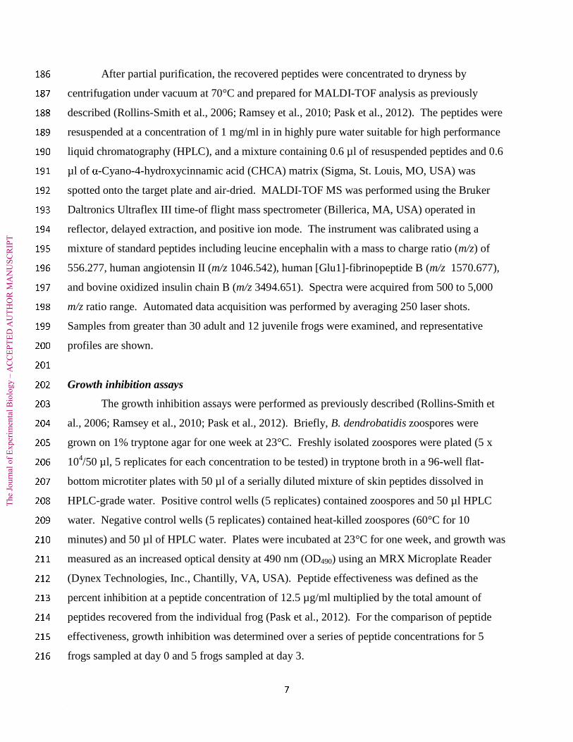
After partial purification, the recovered peptides were concentrated to dryness by 186
centrifugation under vacuum at 70°C and prepared for MALDI-TOF analysis as previously 187
described (Rollins-Smith et al., 2006; Ramsey et al., 2010; Pask et al., 2012). The peptides were 188
resuspended at a concentration of 1 mg/ml in in highly pure water suitable for high performance 189
liquid chromatography (HPLC), and a mixture containing 0.6 µl of resuspended peptides and 0.6 190
µl of α-Cyano-4-hydroxycinnamic acid (CHCA) matrix (Sigma, St. Louis, MO, USA) was 191
spotted onto the target plate and air-dried. MALDI-TOF MS was performed using the Bruker 192
Daltronics Ultraflex III time-of flight mass spectrometer (Billerica, MA, USA) operated in 193
reflector, delayed extraction, and positive ion mode. The instrument was calibrated using a 194
mixture of standard peptides including leucine encephalin with a mass to charge ratio (m/z) of 195
556.277, human angiotensin II (m/z 1046.542), human [Glu1]-fibrinopeptide B (m/z 1570.677), 196
and bovine oxidized insulin chain B (m/z 3494.651). Spectra were acquired from 500 to 5,000 197
m/z ratio range. Automated data acquisition was performed by averaging 250 laser shots. 198
Samples from greater than 30 adult and 12 juvenile frogs were examined, and representative 199
profiles are shown. 200
201
Growth inhibition assays 202
The growth inhibition assays were performed as previously described (Rollins-Smith et 203
al., 2006; Ramsey et al., 2010; Pask et al., 2012). Briefly, B. dendrobatidis zoospores were 204
grown on 1% tryptone agar for one week at 23°C. Freshly isolated zoospores were plated (5 x 205
104/50 µl, 5 replicates for each concentration to be tested) in tryptone broth in a 96-well flat-206
bottom microtiter plates with 50 µl of a serially diluted mixture of skin peptides dissolved in 207
HPLC-grade water. Positive control wells (5 replicates) contained zoospores and 50 µl HPLC 208
water. Negative control wells (5 replicates) contained heat-killed zoospores (60°C for 10 209
minutes) and 50 µl of HPLC water. Plates were incubated at 23°C for one week, and growth was 210
measured as an increased optical density at 490 nm (OD490) using an MRX Microplate Reader 211
(Dynex Technologies, Inc., Chantilly, VA, USA). Peptide effectiveness was defined as the 212
percent inhibition at a peptide concentration of 12.5 µg/ml multiplied by the total amount of 213
peptides recovered from the individual frog (Pask et al., 2012). For the comparison of peptide 214
effectiveness, growth inhibition was determined over a series of peptide concentrations for 5 215
frogs sampled at day 0 and 5 frogs sampled at day 3. 216

The
Jou
rnal
of
Exp
erim
enta
l Bio
logy
– A
CC
EPT
ED
AU
TH
OR
MA
NU
SCR
IPT
8
217
Quantification of B. dendrobatidis zoospores on the skin 218
Frogs were swabbed with a sterile cotton swab ten times on the abdomen, legs, and each foot 219
according to established protocols (Boyle et al., 2004; Hyatt et al., 2007). Swabs were placed 220
into tubes and DNA was extracted and amplified by probe-based quantitative polymerase chain 221
reactions (qPCR) as previously described (Ramsey et al., 2010). For this assay, there was a 222
“no template control” containing all reaction components except the DNA template, a positive 223
control containing DNA extracted from a swab loaded with a known number of zoospores, and a 224
negative control containing DNA extracted from a sterile swab. All skin swab samples and 225
controls were run in triplicate, and Ct (cycle threshold) values were averaged between the three 226
wells. 227
228
Exposure of post-metamorphic froglets to B. dendrobatidis 229
B. dendrobatidis-free R. pipiens at 3-6 months post-metamorphosis were reared from 230
eggs and were thus naïve to B. dendrobatidis. Frogs were swabbed before the experiment to 231
ensure they were B. dendrobatidis-free. All 27 juveniles used in this experiment were negative. 232
The experiment consisted of four experimental groups: 1) Frogs injected with 40 nmol 233
norepinephrine/g and again 2 days later with 20 nmol norepinephrine/g to deplete their granular 234
glands and exposed to 104 zoospores of isolate JEL275 (Carey et al., 2006) [N = 8]; 2) Frogs 235
injected with an equivalent volume of APBS and exposed to 104 zoospores [N = 8]; 3) Frogs 236
injected with 40 nmol norepinephrine /g and again 2 days later with 20 nmol norepinephrine/g 237
and maintained as groups 1 and 2 without further manipulation [N = 3]; 4) Frogs injected with 238
APBS and maintained as groups 1-3 without further manipulations [N = 8]. B. dendrobatidis 239
exposure lasted for two days until the water on all frogs was changed and zoospores flushed out. 240
After exposure, the frogs were examined for signs for illness or death every day and swabbed at 241
10 days post exposure. 242
243
Statistical comparisons 244
Peptide concentrations, optical density determinations of B. dendrobatidis growth 245
(OD490), relative peptide effectiveness, and zoospore equivalents determined by qPCR were 246
averaged for each group, and the mean values ± standard errors (M ± SE) were compared by 247

The
Jou
rnal
of
Exp
erim
enta
l Bio
logy
– A
CC
EPT
ED
AU
TH
OR
MA
NU
SCR
IPT
9
one-way analysis of variance (ANOVA) after log transformation (Tukey post hoc test) or by 248
Student’s t test following log transformation to meet the assumptions of normal distribution and 249
homogeneity of variances for parametric statistics. Percent survival of juveniles at 10 days post 250
exposure to B. dendrobatidis was compared among experimental groups by a Chi-Square test. A 251
p value of ≤ 0.05 was considered statistically significant. The number of animals or replicates is 252
shown in the figure legends. 253
254
RESULTS 255
Determination of the dose of norepinephrine necessary to deplete skin peptides stores 256
To determine the importance of AMPs in B. dendrobatidis infection, it was necessary to 257
deplete the stores of AMPs in granular glands. Prior to this study, the concentration necessary to 258
fully deplete glands had not been determined. With increasing concentrations of norepinephrine, 259
greater concentrations of total peptides were recovered in a dose-dependent fashion. The 260
concentration required to maximally deplete peptides was 40 nmol norepinephrine/g (Fig. 1). 261
Although injection of 80 nmol norepinephrine/g induced slightly more peptides than the 40 262
nmol/g dose, the difference was not statistically significant. 263
264
Renewal of depleted skin peptides 265
In preparation for studies to determine the effects of skin peptide-depletion on 266
susceptibility to experimental infections with B. dendrobatidis, it was important to determine for 267
how long peptide contents of granular glands remained depleted. When peptides were depleted 268
by one injection of 40 nmol norepinephrine/g, some residual peptides could be recovered at each 269
time point tested, but peptide stores had not recovered to initial levels as late as 56 days after the 270
first injection (Fig. 2A). Following one injection of 20 nmol norepinephrine/g, peptide stores 271
were still significantly depleted until day 40 but recovered to initial levels after 50 days (Fig. 272
2B). To determine the effects of the norepinephrine-injection on gland morphology, histology 273
was performed. Dorsal skin sections were the most informative because animals were injected in 274
an area adjacent to the complex of skin glands termed the dermal plicae (Bovbjerg, 1963). The 275
morphology of intact glands is shown in a skin section from an APBS-injected frog. The glands 276
are full of bright red granules by H&E staining (Fig. 2C). In contrast, gland morphology 277
following an injection with 40 nmol norepinephrine/g was quite different. The granular glands 278

The
Jou
rnal
of
Exp
erim
enta
l Bio
logy
– A
CC
EPT
ED
AU
TH
OR
MA
NU
SCR
IPT
10
were empty of the eosinophilic granules, but they did not show significant structural damage 279
(Fig. 2D). 280
281
Characterization of recovered peptides by MALDI-TOF mass spectrometry 282
To determine whether the profile of AMPs that were newly synthesized following 283
granular gland depletion was compositionally the same or different from the initial profile of 284
peptides, peptides induced at days 0, 3, and 60 were compared by MALDI-TOF mass 285
spectrometry. Shown in Figure 3a are representative MALDI-TOF spectra. The profiles are 286
very similar to each other with the three expected brevinin peptides (brevinin-1Pb, brevinin-1Pd, 287
and brevinin-1Pa) present in order of relative intensity. Also present is a strong signal for 288
ranatuerin-2P and another defensive peptide, bradykinin (Fig. 3A). When comparing the activity 289
of the recovered peptides from different time points, the ability to inhibit B. dendrobatidis 290
growth was comparable between the initial peptides (Day 0, Fig. 3B) and the peptides recovered 291
at other time points (Day 3 shown, Fig. 3C). Note that both sets of peptides showed a minimal 292
inhibitory concentration (MIC) of about 50 µg/ml. Given that the activity of peptides from day 0 293
and 3 were not different, peptides recovered at day 60 were not tested for growth inhibitory 294
activity. 295
296
Role of AMPs in protecting Rana pipiens juveniles 297
Young postmetamorphic R. pipiens were used in these experiments because they had 298
been reared in a laboratory free of the presence of B. dendrobatidis and were thus naïve to the 299
pathogen. MALDI-TOF analysis was performed on partially purified AMPs from these juvenile 300
frogs to confirm that they have a normal complement of AMPs at this life stage. Because the 301
parents of these frogs were collected in the state of Minnesota or Wisconsin, the juvenile frogs 302
expressed a slightly different profile of AMPs characteristic of populations in this region 303
(Tennessen et al., 2009). This profile includes brevinin-1Pa, brevinin-1Pb, brevinin-1Pe, and 304
ranatuerin-2P (Fig. 4A). The peptides recovered from the juvenile frogs showed excellent 305
capacity to inhibit growth of B. dendrobatidis with an approximate MIC of 50-100 µg/ml (Range 306
25-250 µg/ml, M ± SE = 96 ± 27 µg/ml), comparable to that of peptides from adult frogs (Fig. 307
4B). When these postmetamorphic frogs were depleted of their peptides by two injections of 308
norepinephrine and exposed to B. dendrobatidis in their water (NE+Bd), they did not survive as 309

The
Jou
rnal
of
Exp
erim
enta
l Bio
logy
– A
CC
EPT
ED
AU
TH
OR
MA
NU
SCR
IPT
11
well as B. dendrobatidis-exposed animals which had an intact set of peptides (APBS+Bd). 310
Among the peptide-depleted and B. dendrobatidis-exposed frogs, only 3/8 had survived to day 311
10 while 8/8 APBS-injected control frogs that were exposed to B. dendrobatidis were alive. 312
Other control groups that were norepinephrine-treated (3/3) or APBS-injected and not exposed to 313
B. dendrobatidis (8/8) also survived to day 10 (Fig. 4C). Among frogs surviving at 10 days post-314
exposure to B. dendrobatidis, remaining peptide-depleted frogs had significantly higher numbers 315
of zoospores on the skin than APBS-injected and B. dendrobatidis-exposed frogs (Fig. 4D). 316
Frogs not exposed to B. dendrobatidis had no evidence of B. dendrobatidis infection determined 317
by qPCR. 318
319
DISCUSSION 320
The current study supports previous work that showed a correlation between the survival 321
of amphibians exposed to B. dendrobatidis with beneficial AMPs (Woodhams et al., 2006a, 322
2006b, 2007). It also supports a previous study showing that depletion of skin peptides in 323
Xenopus laevis (naturally resistant to chytridiomycosis) resulted in increased pathogen burden 324
(Ramsey et al., 2010). 325
326
Development of a method to deplete skin peptides. 327
Using adult northern leopard frogs (Rana pipiens) as a model, we developed a protocol to 328
deplete skin AMPs by injection of norepinephrine and observed the relatively slow renewal of 329
the same set of peptides over a period of approximately two months. We then applied the same 330
methods to deplete skin peptides from juvenile leopard frogs raised under laboratory conditions 331
to insure that they were not exposed to B. dendrobatidis. This allowed us to examine the role of 332
skin peptides in protection from B. dendrobatidis infections free of the potential complication of 333
mucosal antibodies if the frogs had previously been exposed to B. dendrobatidis (Ramsey et al., 334
2010). Although one injection of 40 nmol norepinephrine/g significantly reduced available skin 335
peptides, some peptides were recovered at three days after the first injection. We previously 336
showed in X. laevis that two norepinephrine injections separated by two days were necessary to 337
completely deplete skin peptides (Gammill et al., 2012). Thus, for the experimental infection 338
study with juvenile R. pipiens, two norepinephrine injections were used. 339
340

The
Jou
rnal
of
Exp
erim
enta
l Bio
logy
– A
CC
EPT
ED
AU
TH
OR
MA
NU
SCR
IPT
12
Kinetics of peptide recovery and characteristics of the recovered peptides 341
Previous studies of peptide recovery following norepinephrine injection examined 342
relatively mild inductions, and recovery was examined after a very short time. In Xenopus 343
laevis, recovery of peptides following a mild norepinephrine injection (0.5 to 1 nmol/g) was 344
detected by fast atom bombardment mass spectrometry. The full complement of expected 345
peptides was detected within two to six days (Giovannini et al., 1987) suggesting that recovery is 346
very rapid or peptides were not depleted by this injection. Following injection of a slightly 347
higher concentration of norepinephrine (3 nmol/g), gland morphology was not completely 348
restored for two weeks as determined by histology and electron microscopy (Dockray and 349
Hopkins, 1975). This suggested that a higher concentration of norepinephrine is necessary to 350
more completely deplete peptides. We have shown that in X. laevis, two injections of 80 nmol/g 351
within two days were necessary to completely deplete peptides (Gammill et al., 2012), and 352
peptide recovery following depletion with 80 nmol/g required seven to nine weeks (Ramsey et 353
al., 2010). In the current study, we showed that peptide recovery after nearly complete depletion 354
of R. pipiens peptides (injected with 40 nmol/g norepinephrine) required greater than 56 days 355
(Fig. 2). By MALDI-TOF MS analysis, the profile of recovered peptides at days 0, 3, and 60 356
were identical. When concentrated, the peptides had similar growth inhibitory activity at days 0 357
and 3 (Fig. 3B and 3C). This finding demonstrates that although the amount of AMPs present in 358
the granular glands was reduced at day 3, the same relative mixture of peptides was present and 359
effective against B. dendrobatidis. The relative effectiveness of the AMP mixture at day 3 360
versus day 0 showed that the relative effectiveness at day 3 was greatly diminished reflecting the 361
reduced amounts of peptides that can be induced following their depletion three days earlier (Fig. 362
3D). 363
364
Immune defenses against B. dendrobatidis in juvenile frogs 365
Our previous study of immune defenses against B. dendrobatidis in X. laevis, showed that 366
both innate immune defenses (AMPs) and adaptive lymphocyte-mediated defenses play a role in 367
protection of this highly resistant species (Ramsey et al, 2010). The development of immune 368
defenses in R. pipiens is not well studied, but we can make some informed assumptions about the 369
maturation of the immune system based on studies of X. laevis. In Xenopus, adult-type 370
recognition of minor histocompatibility antigens and development of high affinity antibodies 371

The
Jou
rnal
of
Exp
erim
enta
l Bio
logy
– A
CC
EPT
ED
AU
TH
OR
MA
NU
SCR
IPT
13
develops within 1-2 months post-metamorphosis (DiMarzo and Cohen, 1982; Rollins-Smith et 372
al. 1988; Hsu and Du Pasquier 1984; Hsu and Du Pasquier, 1992; reviewed in Rollins-Smith, 373
1998). Presence of high affinity IgY antibodies in Rana catesbeiana juveniles at 3 months post-374
metamorphosis (Du Pasquier and Haimovich, 1976) suggests that the adult B cell population in 375
ranid frogs develops during the postmetamorphic lymphocyte expansion just as it does in 376
Xenopus. Thus, we are confident that the juvenile R. pipiens used in our studies at 3-6 months 377
post-metamorphosis had an adult-type immune system. Much less is known about the ontogeny 378
of expression of the antimicrobial peptide defenses. However, previous studies in R. pipiens 379
showed that granular glands are completely developed in the dermal plicae of R. pipiens at the 380
conclusion of metamorphosis (Bovbjerg, 1963). Thus, we assumed that the expected 381
antimicrobial peptides would be present at 3-6 months post-metamorphosis when the chytrid-382
exposure experiments were conducted. To confirm that the AMP repertoire was complete, we 383
examined the peptides present in twelve of these young frogs and found the expected AMPs in 384
the majority of these animals (9/12) as shown in Fig. 4A. Thus, the normal complement of 385
AMPs had developed in these young frogs, and the peptide depletion protocols developed in 386
larger adults were effective in depleting the peptide reserves of the younger frogs. 387
Although the most direct effect of injection of norepinephrine at the relatively high 388
concentrations used in our experiments is the depletion of skin peptide reserves, the 389
norepinephrine could also have other immunosuppressive effects such as activation of 390
corticosterone release (Shepherd and Holzwarth, 2001). Future studies will determine whether 391
there are other immunosuppressive effects of norepinephrine-treatment. This very strong 392
norepinephrine stimulus to deplete peptides was a pharmacological treatment to impair the skin 393
peptide defenses. It is unlikely that natural stresses in nature would result in such significant 394
long-term depletion of skin peptides. We showed previously (Pask et al., 2012) that the stress to 395
a frog of being chased for ten minutes resulted in the total peptide release of about 6,400 µg of 396
peptides/ml of mucus comparable to that observed following the injection of 2 nmol/g of 397
norepinephrine in the experiments presented here (Fig. 1). Thus, the treatment to deplete 398
peptides (40 nmol/g norepinephrine), which induced about 90,000 µg of peptides/ml of mucus 399
far exceeds the amount that would naturally be released. 400
401
AMP protection of naïve juvenile frogs exposed to B. dendrobatidis 402
The
Jou
rnal
of
Exp
erim
enta
l Bio
logy
– A
CC
EPT
ED
AU
TH
OR
MA
NU
SCR
IPT
14
Metamorphosis in amphibians is a time of fasting due to the extensive reorganization of 403
the digestive tract (reviewed in Fox, 1981). In Xenopus, tadpoles are fasting after stage 62 but 404
their glucose levels in body fluids do not decrease (Hanke and Leist, 1971). As suggested by 405
Jaudet and Hatey (1984), glycogen, lipids, and proteins are probably converted to glucose 406
(Hanke and Leist, 1971, Gunesch, 1974). Elevation of corticosteroids hormones is observed 407
during metamorphosis of all amphibian species studied (reviewed in White and Nicoll, 1981; 408
Kikuyama et al., 1993; Denver, 2009). Therefore, it is likely that a tradeoff may occur that 409
diverts energy from other physiological processes to the tissue and organ remodeling that occurs 410
at metamorphosis because the organism has a finite amount of energy (reviewed in Rollins-411
Smith and Woodhams, 2012). One of these systems affected is the immune system. Wood frogs 412
(Rana sylvatica) develop in temporary ponds, which may dry up in an unpredictable fashion. In 413
conditions that simulated quickly drying ponds, tadpoles metamorphosed earlier and appeared to 414
experience a tradeoff in decreased total leukocyte counts and diminished cellular immune 415
response (skin swelling in response to a plant lectin) (Gervasi and Foufopoulos, 2008). 416
Precocious metamorphosis induced by administration of thyroid hormones limited lymphocyte 417
development and impaired allograft rejection responses in X. laevis (Rollins-Smith et al., 1988). 418
During metamorphosis the immune system undergoes involution due to elevated corticosteroid 419
hormones (reviewed in Rollins-Smith, 1998), and new metamorphs are particularly susceptible 420
to diseases including chytridiomycosis (Bosch et al., 2001; Rachowicz & Vredenburg, 2004; 421
Rachowicz et al., 2006; Tobler & Schmidt, 2010; Walker et al., 2010). 422
To assess the role of AMPs in protecting naïve juvenile frogs, froglets at 3-6 months 423
post-metamorphosis, which expressed a normal complement of AMPs capable of inhibiting 424
growth of B. dendrobatidis at this life stage, were depleted of their peptides and exposed to a 425
limited number of B. dendrobatidis zoospores. Frogs that had their peptide stores intact survived 426
the experimental infection significantly better than those that were peptide depleted. Peptide- 427
depleted juveniles that survived the infection had significantly higher B. dendrobatidis zoospore 428
loads compared to the peptide intact animals. In a previous study, we showed that AMPs are 429
constitutively expressed by adult R. pipiens at low levels that are sufficient to inhibit B. 430
dendrobatidis growth in vitro (Pask et al., 2012). The current study demonstrates that those 431
constitutively released AMPs provide critical protection for the most susceptible life stage 432
against B. dendrobatidis. 433

The
Jou
rnal
of
Exp
erim
enta
l Bio
logy
– A
CC
EPT
ED
AU
TH
OR
MA
NU
SCR
IPT
15
434
List of symbols and abbreviations 435
ANOVA, analysis of variance, AMPs, antimicrobial peptides; APBS, amphibian phosphate 436
buffered saline; Bd, B. dendrobatidis; Ct values, cycle threshold is defined as the number of 437
cycles necessary for the fluorescence signal to exceed background; CHCA, α-Cyano-4-438
hydroxycinnamic acid; H&E, hematoxylin and eosin; HPLC, high performance liquid 439
chromatography; M ± SE, mean values ± standard errors; MALDI-TOF, matrix-assisted laser 440
desorption time-of-flight; MIC, minimal inhibitory concentration; MS, mass spectrometry; NE, 441
norepinephrine; qPCR, quantitative polymerase chain reactions. 442
443
Acknowledgements 444
We thank William Karasov, Department of Forest and Wildlife Ecology, University of 445
Wisconsin-Madison, Madison, Wisconsin, for assistance in raising Rana pipiens froglets in a 446
chytrid-free environment. We acknowledge the assistance of the Vanderbilt Mass Spectrometry 447
Core and the Vanderbilt Immunohistochemistry Core. 448
449
Funding 450
This research was supported by Grants IOS-0619536, IOS-0843207, and IOS-1121758 from the 451
National Science Foundation of the USA (to L. R-S). 452
453
1. Amiche, M., Seon, A. A., Pierre, T. N., and Nicolas, P. (1999). The dermaseptin 454
precursors: a protein family with a common preproregion and a variable C-terminal 455
antimicrobial domain. FEBS Lett. 456, 352-256. 456
2. Barker, K.S., Davis, A.T., Li, B., and Rollins-Smith, L.A. (1997). In vitro studies of 457
spontaneous and corticosteroid-induced apoptosis of lymphocyte populations in 458
metamorphosing frogs/RU486 inhibition. Brain Behav. Immun. 11, 119-131. 459
3. Berger, L., Speare, R., Daszak, P., Green, D. E., Cunningham, A. A., Goggin, C. L., 460
Slocombe, R., Ragan, M.A ., Hyatt, A. D., McDonald, K. R., et al. (1998). 461
Chytridiomycosis causes amphibian mortality associated with population declines in the 462
rain forests of Australia and Central America. Proc. Natl. Acad. Sci .USA 95, 9031-9036. 463

The
Jou
rnal
of
Exp
erim
enta
l Bio
logy
– A
CC
EPT
ED
AU
TH
OR
MA
NU
SCR
IPT
16
4. Berger, L., Hyatt, A. D., Speare, R., and Longcore, J. E. (2005). Life cycle stages of 464
the amphibian chytrid Batrachochytrium dendrobatidis. Dis. Aquat. Org. 68, 51-63. 465
5. Bosch, J., Martínez-Solano, I. and García-París, M. (2001). Evidence of a chytrid 466
fungus infection involved in the decline of the common midwife toad (Alytes 467
obstetricans) in protected areas of central Spain. Biol. Conserv. 97, 331-337. 468
6. Bovbjerg, A. M. (1963). Development of the glands of the dermal plicae in Rana 469
pipiens. J. Morphol. 113, 321-243. 470
7. Boyle, D. G., Boyle, D. B., Olsen, V., Morgan, J. A. T., and Hyatt, A. D. (2004). Rapid 471
quantitative detection of chytridiomycosis (Batrachochytrium dendrobatidis) in 472
amphibian samples using real-time Taqman PCR assay. Dis. Aquat. Org. 60, 141-148. 473
8. Brucker, R. M., Harris, R. N., Schwantes, C. R., Gallaher, T. N., Flaherty, D. C., 474
Lam, B. A., and Minbiole, K. P. C. (2008) Amphibian chemical defense: Antifungal 475
metabolites of the microsymbiont Janthinobacterium lividum on the salamander 476
Plethodon cinereus. J. Chem. Ecol. 34, 1422-1429. 477
9. Carey, C., Bruzgul, J. E., Livo, L. J., Walling, M. L., Kuehl, K. A., Dixon, B. F., 478
Pessier, A. P., Alford, R. A., and Rogers K. B. (2006). Experimental exposures of 479
boreal toads (Bufo boreas to a pathogenic chytrid fungus (Batrachochytrium 480
dendrobatidis). EcoHealth 3, 5-21. 481
10. Collins, J. P. (2010). Amphibian decline and extinction: What we know and what we 482
need to learn. Dis. Aquat. Organ. 92, 93-99. 483
11. Denver R. J. (2009). Stress hormones mediate environment-genotype interactions during 484
amphibian development. Gen Comp. Endocrinol. 164, 20-31. 485
12. Denver R. J. (2013). Neuroendocrinology of amphibian metamorphosis. Curr. Top. Dev. 486
Biol. 103, 195-227. 487
13. DiMarzo, S. J. and Cohen, N. (1982). Immunogenetic aspects of in vivo allotolerance 488
induction during the ontogeny of Xenopus laevis. Immunogenetics 16, 103-116. 489
14. Dockray, G. J. and Hopkins. C. R. (1975). Caerulein secretion by dermal glands in 490
Xenopus laevis. J. Cell Biol. 64, 724-733. 491
15. Du Pasquier, L. and Haimovich, J. (1976). The antibody response during amphibian 492
ontogeny. Immunogenetics 3, 381-391. 493

The
Jou
rnal
of
Exp
erim
enta
l Bio
logy
– A
CC
EPT
ED
AU
TH
OR
MA
NU
SCR
IPT
17
16. Duellman, W. E. and Treub L. (1986) Biology of amphibians. New York, NY. 494
McGraw-Hill. 495
17. Fox, H. (1981). Cytological and morphological changes during amphibian 496
metamorphosis. In Metamorphosis (ed. L. I. Gilbert and E. Frieden), pp. 327-362. New 497
York, NY: Plenum Press. 498
18. Gammill, W. M., Fites, J. S., and Rollins-Smith, L. A. (2012). Norepinephrine 499
depletion of antimicrobial peptides from the skin glands of Xenopus laevis. Dev. Comp. 500
Immunol. 37, 19-27. 501
19. Gervasi, S.S. and Foufopoulos, J. (2008). Costs of plasticity: responses to desiccation 502
decrease post-metamorphic immune function in a pond-breeding amphibian. Funct. Ecol. 503
22, 100-108. 504
20. Greenspan, S. E., Longcore, J. E., and Calhoun, A. J. K. (2012). Host invasion by 505
Batrachochytrium dendrobatidis: Fungal and epidermal ultrastructure in model anurans. 506
Dis. Aquat. Organ. 100, 201-210. 507
21. Gunesch, K.D. (1974). Changes of the lipid metabolism in the clawed toad (Xenopus 508
laevis Daudin) induced by ACTH, corticosteroids and insulin. Zool. Jahrb. Abt. Allg. 509
Zool. Physiol. Tiere. 78, 108-127 510
22. Hancock, R. E., Nijnik, A., and Pilpott, D. J. (2012). Modulating immunity as a 511
therapy for bacterial infections. Nat. Rev. Microbiol. 10, 243-254. 512
23. Hanke, W. and Leist, K. H. (1971). The effect of ACTH and corticosteroids on 513
carbohydrate metabolism during metamorphosis of Xenopus laevis. Gen. Comp. 514
Endocrinol. 17, 137-148. 515
24. Hsu, E. and Du Pasquier, L. (1984). Ontogeny of the immune system in Xenopus II. 516
Antibody repertoire differences between larvae and adults. Differentiation 28, 116-122. 517
25. Hsu, E. and Du Pasquier, L. (1992). Changes in the amphibian antibody repertoire are 518
correlated with metamorphosis and not with age or size. Develop. Immunol. 2, 1-6. 519
26. Hyatt, A. D., Boyle, D. G., Olsen, V., Boyle, D. B., Berger, L., Obendorf, D., Dalton, 520
A., Kriger, K., Hero, M., Hines, H., et al. (2007). Diagnostic assays and sampling 521
protocols for the detection of Batrachochytrium dendrobatidis. Dis. Aquat. Org. 73, 175-522
192. 523

The
Jou
rnal
of
Exp
erim
enta
l Bio
logy
– A
CC
EPT
ED
AU
TH
OR
MA
NU
SCR
IPT
18
27. Jaudet G. J. and Hatey, J. H. (1984). Variations in aldosterone and corticosterone 524
plasma levels during metamorphosis in Xenopus laevis tadpoles. Gen. Comp. Endocrinol 525
56, 59-65. 526
28. Kikuyama S., Kawamura K., Tanaka, S., and Yamamoto K. (1993). Aspects of 527
amphibian metamorphosis: Hormonal control. Int. Rev. Cytol. 145, 105-148. 528
29. Longcore, J. E., Pessier, A. P., and Nichols, D. K. (1999). Batrachochytrium 529
dendrobatidis gen. et sp. nov., a chytrid pathogenic to amphibians. Mycologia 91, 219-530
227. 531
30. McClanahan, L. J. and Baldwin, R. (1969). Rate of water uptake through the 532
integument of the desert toad, Bufo punctatus. Comp. Biochem. Physiol. 28, 381-389. 533
31. Miller L. (1996). Hormone-induced changes in keratin gene expression during 534
amphibian skin metamorphosis. In Metamorphosis: Postembryonic Reprogramming of 535
Gene Expression in Amphibian and Insect Cells (ed. L. I. Gilbert, B. G. Atkinson, and J. 536
Tata), pp. 599-624. New York, NY: Academic Press. 537
32. Nicolas, P. and Mor, A. (1995). Peptides as weapons against microorganisms in the 538
chemical defense system of vertebrates. Ann. Rev. Microbiol. 49, 277-304. 539
33. Noble, G. E. and Noble, E. R. (1944). On the histology of frog skin glands. Trans. Am. 540
Microsc. Soc. 63, 254-263. 541
34. Pask, J. D., Woodhams, D. C. and Rollins-Smith, L. A. (2012). The ebb and flow of 542
antimicrobial skin peptides defends northern leopard frogs (Rana pipiens) against 543
chytridiomycosis. Global Change Biol. 18, 1231-1238. 544
35. Pessier, A. P., Nichols, D. K., Longcore, J. E., and Fuller, M.S. (1999). Cutaneous 545
chytridiomycosis in poison dart frogs (Dendrobates spp.) and White’s tree frogs (Litoria 546
caerulea). J. Vet. Diagn. Invest. 11, 194-199. 547
36. Pukala, T., Bowie, J. H., Maselli, V. M., Musgrave, I. F., and Tyler, M. J. (2006). 548
Host-defence peptides from the glandular secretions of amphibians: structure and 549
activity. Nat. Prod. Rep. 23, 368-393. 550
37. Rachowicz, L. J. and Vredenburg, V. T. (2004). Transmission of Batrachochytrium 551
dendrobatidis within and between amphibian life stages. Dis. Aquat. Organ. 61, 75-83. 552
The
Jou
rnal
of
Exp
erim
enta
l Bio
logy
– A
CC
EPT
ED
AU
TH
OR
MA
NU
SCR
IPT
19
38. Rachowicz, L. J., Knapp, R. A., Morgan, J. A. T., Stice, M. J., Vredenburg, V. T., 553
Parker, J. M., and Briggs, C. J. (2006). Emerging infectious disease as a proximate 554
cause of amphibian mass mortality. Ecology 87, 1671-1683. 555
39. Ramsey, J. P., Reinert, L. K., Harper, L. K., Woodhams, D. C., and Rollins-Smith, 556
L. A. (2010). Innate and adaptive immune defenses against a fungus linked to global 557
amphibian declines in the South African clawed frog, Xenopus laevis. Infect. Immun. 78, 558
3981-3992. 559
40. Rollins-Smith, L. A. (1998). Metamorphosis and the amphibian immune system. 560
Immunol. Rev. 166, 221-230. 561
41. Rollins-Smith, L.A. (2009). The role of amphibian antimicrobial peptides in protection 562
of amphibians from pathogens linked to global amphibian declines. Biochim. Biophys. 563
Acta. 1788, 1593-1599. 564
42. Rollins-Smith, L. A. and Conlon, J. M. (2005). Antimicrobial peptide defenses against 565
chytridiomycosis, an emerging infectious disease of amphibian populations. Dev. Comp. 566
Immunol. 29, 589-598. 567
43. Rollins-Smith, L.A., and Woodhams D.C. (2012). Amphibian immunity: Staying in 568
tune with the environment. In Eco-Immunology (ed. G.E. Demas and R.J Nelson), pp. 92-569
143. Oxford, United Kingdom: Oxford University Press. 570
44. Rollins-Smith, L.A., Barker, K.S., and Davis, A.T. (1997). Involvement of 571
glucocorticoids in the reorganization of the amphibian immune system at metamorphosis. 572
Dev. Immunol. 5, 145-152. 573
45. Rollins-Smith, L. A., Parsons, S. C. V. and Cohen, N. (1988). Effects of thyroxine-574
driven precocious metamorphosis on maturation of adult-type allograft rejection 575
responses in early throidectomized frogs. Differentiation 37, 180-185. 576
46. Rollins-Smith, L. A., Ramsey, J. P., Pask, J. D., Reinert, L. K., and Woodhams, D. 577
C. (2011). Amphibian immune defenses against chytridiomycosis: Impacts of changing 578
environments. Integr. Comp. Biol. 51, 552-562. 579
47. Rollins-Smith, L. A., Reinert, L. K., O’Leary C. J., Houston, L. E., and Woodhams, 580
D .C. (2005). Antimicrobial Peptide Defenses in Amphibian Skin. Integr. Comp. Biol. 581
45, 137-142. 582
The
Jou
rnal
of
Exp
erim
enta
l Bio
logy
– A
CC
EPT
ED
AU
TH
OR
MA
NU
SCR
IPT
20
48. Rollins-Smith, L. A., Woodhams, D. C., Reinert, L. K., Vredenburg, V. T., Briggs, 583
C. J., Nielsen, P. F., and Conlon, J. M. (2006). Antimicrobial peptide defenses of the 584
mountain yellow-legged frog (Rana muscosa). Dev. Comp. Immunol. 30, 831-842. 585
49. Schumacher, U., Adam, W., Hauser, F., Probst, J. C., and Hoffmann, W. (1994). 586
Molecular anatomy of a skin gland: Histochemical and biochemical investigations on the 587
mucous glands of Xenopus laevis. J. Histochem. Cytochem. 42, 57-65. 588
50. Shepherd, S. P. and Holzwarth, M. A. (2001). Chromaffin-adrenocortical cell 589
interactions: effects of chromaffin cell activation in adrenal cell cocultures. Am. J. 590
Physiol. Cell Physiol. 280, C61-C71. 591
51. Skerratt L. F., Berger, L., Speare, R., Cashins, S., McDonald, K. R., Phillott, A. D., 592
Hines, H. B., and Kenyon, N. (2007). Spread of chytridiomycosis has caused the rapid 593
global decline and extinction of frogs. EcoHealth 4, 125-134. 594
52. Tennessen, J. A., Woodhams, D. C., Chaurand, P., Reinert, L. K., Billheimer, D., 595
Shyr, Y., Caprioli, R. M., Blouin, M. S., and Rollins-Smith, L. A. (2009). Variations 596
in the expressed antimicrobial peptide repertoire of northern leopard frog (Rana pipiens) 597
populations suggest intraspecies differences in resistance to pathogens. Dev. Comp. 598
Immunol. 33, 1247-1257. 599
53. Tobler, U., and Schmidt, B. R. (2010). Within-and among-population variation in 600
chytridiomycosis-induced mortality in the toad Alytes obstetricans. PLoS One 5, e1092. 601
54. Van Rooij, P., Martel, A., D’Herde, K., Brutyn, M., Croubels, S., Ducatelle, R., 602
Haesebrouck, F., and Pasmans F. (2012). Germ tube mediated invasion of 603
Batrachochytrium dendrobatidis in amphibian skin is host dependent. PLoS One 7(7), 604
e41481. 605
55. Walker, S. F., Bosch, J., Gomez, V., Garner, T. W. J., Cunningham, A. A., 606
Schmeller, D. S., Ninyerola, M., Henk, D. A., Ginestet, C., Christian-Philippe, A., et 607
al. (2010). Factors driving pathogenicity vs. prevalence of amphibian panzootic 608
chytridiomycosis in Iberia. Ecol. Lett. 13, 372-382. 609
56. Woodhams, D. C., Ardipradja, K., Alford, R. A., Marantelli, G., Reinert, L. K., and 610
Rollins-Smith, L. A. (2007). Resistance to chytridiomycosis varies among amphibian 611
species and is correlated with skin peptide defenses. Anim. Conserv. 10, 409-417. 612
57. Woodhams, D. C., Rollins-Smith, L. A., Carey, C., Reinert, L., Tyler, M. J., and 613

The
Jou
rnal
of
Exp
erim
enta
l Bio
logy
– A
CC
EPT
ED
AU
TH
OR
MA
NU
SCR
IPT
21
Alford, R. A. (2006). Population trends associated with antimicrobial peptide defenses 614
against chytridiomycosis in Australian frogs. Oecologia 146, 531-540. 615
58. Woodhams, D. C., Voyles, J., Lips, K. R., Carey, C., and Rollins-Smith, L. A. (2006). 616
Predicted disease susceptibility in a Panamanian amphibian assemblage based on skin 617
peptide defenses. J. Wildl. Dis. 42, 207-218. 618
59. Zasloff, M. (2002). Antimicrobial peptides of multicellular organisms. Nature 415, 389-619
395. 620
621
Figure legends 622
Figure 1. Dose-response to norepinephrine injections. Frogs (N = 6 to 9 per group) were 623
injected with APBS [N = 8] or 2 [N = 8], 20 [N = 6], 40 [N = 9] or 80 [N = 6] nmol 624
norepinephrine/g. *All concentrations of norepinephrine induced more peptides than those 625
recovered from APBS-injected frogs by one-way ANOVA after log transformation, Tukey post 626
hoc test, p ≤ 0.01. Concentrations of peptides recovered from frogs injected with 2, 20, and 40 627
nmol norepinephrine/g were significantly different from each other and from APBS controls by 628
one-way ANOVA after log transformation, Tukey post hoc test, p ≤ 0.05. 629
630
Figure 2. Recovery of peptides following injection of norepinephrine. (A) 40 nmol 631
norepinephrine/g injected at each time point [N = 25 at day 0; N = 4 or 5 at other time points]. 632
*Peptide concentrations significantly reduced in comparison with those collected at day 0 by 633
two-tailed Student’s t-test after log transformation, p < 0.02. (B) 20 nmol norepinephrine/g 634
injected at each time point [N = 26 at day 0; N = 4 or 5 at other time points]. *Peptide 635
concentrations significantly reduced in comparison with those collected at day 0 by two-tailed 636
Student’s t-test after log transformation, p < 0.025; **significantly reduced by one-tailed 637
Student’s t-test, p ≤ 0.05. (C) Skin from APBS-injected control frog. (D) Skin from frog injected 638
with 40 nmol/g of norepinephrine showing intact but largely empty granular glands. Granular 639
glands are indicated with the letter “G”. 640
641
Figure 3. Characterization of skin peptides recovered at days 0, 3, and 60 after norepinephrine 642
induction. (A) Representative MALDI-TOF profiles of peptides recovered after injection of 40 643
nmol norepinephrine/g at day 0 (top), day 3 (middle) and day 60 (bottom) (N = 5 frogs examined 644

The
Jou
rnal
of
Exp
erim
enta
l Bio
logy
– A
CC
EPT
ED
AU
TH
OR
MA
NU
SCR
IPT
22
at each time point). (B) Growth inhibition of B. dendrobatidis zoospores by peptides recovered at 645
day 0 after norepinephrine injection. (C) Growth inhibition of B. dendrobatidis zoospores by 646
peptides recovered at day 3 after norepinephrine injection. For (B, C), MIC = 50 µg/ml. 647
*Growth was significantly less than that of positive control wells by two-tailed Student’s t-test 648
after log transformation, p ≤ 0.05 (5 replicates for each concentration of peptide, negative 649
control, and positive control wells). (D) Peptide effectiveness of the mixture of peptides 650
recovered at day 0 and day 3. Peptide effectiveness at day 3 was significantly decreased in 651
comparison with that observed for peptides collected at day 0 by two-tailed Student’s t test after 652
log transformation, p ≤ 0.05 (N =5 frogs sampled at day 0 and day 3). 653
654
Figure 4. Peptide-depletion induced by norepinephrine increases susceptibility of 655
postmetamorphic juveniles to B. dendrobatidis. (A) Representative MALDI-TOF profile of 656
hydrophobic skin peptides from juvenile frogs including antimicrobial peptides brevinin-1Pa, 657
brevinin-1Pb, brevinin-1Pe, and ranaturin-2P (representative of 12 individual frogs). (B) Growth 658
inhibition of B. dendrobatidis zoospores by skin peptides from juvenile frogs (representative of 659
12 individual frogs tested). MIC = 50 µg/ml. *Growth was significantly less than that of positive 660
control wells by two-tailed Student’s t-test after log transformation, p ≤ 0.05 (5 replicates for 661
each concentration of peptide, negative control, and positive control wells). (C) Percent survival 662
of postmetamorphic juveniles peptide-depleted and exposed to B. dendrobatidis (NE+Bd, N = 8), 663
APBS-injected and exposed to B. dendrobatidis (APBS+Bd, N = 8), norepinephrine-injected but 664
not exposed to B. dendrobatidis (NE, no Bd, N =3), or APBS injected but not exposed to B. 665
dendrobatidis (APBS, no Bd, N =8). Percent survival of peptide-depleted and B. dendrobatidis-666
exposed frogs was significantly less than that of APBS-injected controls exposed or not exposed 667
to B. dendrobatidis by Chi-Square test, p ≤ 0.05 and also significantly less than peptide-depleted 668
frogs not exposed to B. dendrobatidis, p ≤ 0.1. (D) Infection intensity (zoospore equivalents + 1) 669
on the skin of surviving frogs at day 10 that were APBS-injected but not exposed to B. 670
dendrobatidis (Bd) (APBS, no Bd, N = 8) or injected with norepinephrine (NE) and not exposed 671
to Bd (NE, no Bd, N = 2) or APBS-injected and exposed to Bd (APBS+ Bd, N = 7) or injected 672
with NE and exposed to Bd (NE+Bd, N = 3). The data represent the mean log zoospore 673
equivalents + 1 ± standard error. *Greater number of zoospore equivalents present on the skin of 674
The
Jou
rnal
of
Exp
erim
enta
l Bio
logy
– A
CC
EPT
ED
AU
TH
OR
MA
NU
SCR
IPT
23
NE+Bd frogs compared to APBS+Bd frogs by a one-tailed Student’s t-test after log 675
transformation, p = 0.0352. 676

The
Jou
rnal
of
Exp
erim
enta
l Bio
logy
– A
CC
EPT
ED
AU
TH
OR
MA
NU
SCR
IPT
0
20
40
60
80
100
120
APBS 2 20 40 80
To
tal p
epti
des
(
g/m
l o
f m
ucu
s x
10-3
)
Concentration of norepinephrine (nmol/g)
*
*
* *

The
Jou
rnal
of
Exp
erim
enta
l Bio
logy
– A
CC
EPT
ED
AU
TH
OR
MA
NU
SCR
IPT
0
2
4
6
8
10
0 3 10 20 30 40 50 60
To
tal p
epti
des
(
g/m
l in
mu
cus
X 1
0-4)
Days of recovery
**
*
****
0
2
4
6
8
10
12
0 3 10 20 30 40 56
To
tal
pep
tid
es
(g
/ml
in m
ucu
s X
10-
4)
Days of recovery
**
** *
*
A
B
C
D
G G G
G GG
GG

The
Jou
rnal
of
Exp
erim
enta
l Bio
logy
– A
CC
EPT
ED
AU
TH
OR
MA
NU
SCR
IPT
A
0.00
0.02
0.04
0.06
0.08
0.10
0 500 250 100 50 25 12.5 6.2 3.1 1.6
OD
49
0
Peptide Concentration (g/ml)
Pos. controlNeg. controlPeptides* * * * *
*
700 1260 1820 2380 2940 3500Mass (m/z)
4265.2
0102030405060708090
100
% I
nte
nsi
ty
Bradykinin1060
Brevinin-1Pb2577
Brevinin-1Pd2569
Brevinin-1Pa2563
Ranatuerin-2P3000
700 1260 1820 2380 2940 3500Mass (m/z)
122.8
0102030405060708090
100
% I
nte
nsi
ty
Bradykinin1060
Brevinin-1Pb2577Brevinin-1Pd2569
Brevinin-1Pa2563
Ranatuerin-2P3000
700 1260 1820 2380 2940 3500Mass (m/z)
1113.4
0102030405060708090
100
% I
nte
nsi
ty
Bradykinin1060
Brevinin-1Pb2577Brevinin-1Pd2569
Brevinin-1Pa2563 Ranatuerin-2P3000
B C
0.00
0.05
0.10
0.15
0.20
0.25
0 250 50 12.5 3.1
OD
49
0
Peptide concentration (g/ml)
Pos. controlNeg. controlPeptides* * * *
*
*
0
10
20
30
40
50
60
Day 0 Day 3
Pep
tid
e ef
fect
iven
ess
(X 1
0-5
)
*
D

The
Jou
rnal
of
Exp
erim
enta
l Bio
logy
– A
CC
EPT
ED
AU
TH
OR
MA
NU
SCR
IPT
700 1260 1820 2380 2940 3500Mass (m/z)
1.0E+4
0102030405060708090
100
% I
nte
nsi
ty
Ranatuerin-2P 3000
Brevinin-1Pb 2577
Brevinin-1Pe 2595Brevinin-1Pa
2563
A
0
10
20
30
40
50
60
70
APBS,no Bd
NE, noBd
APBS+Bd
NE+Bd
Bd
infe
ctio
n in
ten
sity
(z
sp e
qu
ival
ents
+1)
*
[1] [1]
D
0
20
40
60
80
100
120
1 2 3 4 5 6 7 8 9 10 11
Per
cen
t su
rviv
al
Days
NE+Bd
APBS+Bd
NE, no Bd
APBS, no Bd
C
0.000
0.005
0.010
0.015
0.020
0.025
0 500 250 100 50 25 12.56.22 3.1 1.6
OD
49
0
Peptide concentration (g/ml)
Pos.controlNeg.control*
*
***
B
Bradykinin 1060