Short Segment -cap draft August 23rdcopy
15
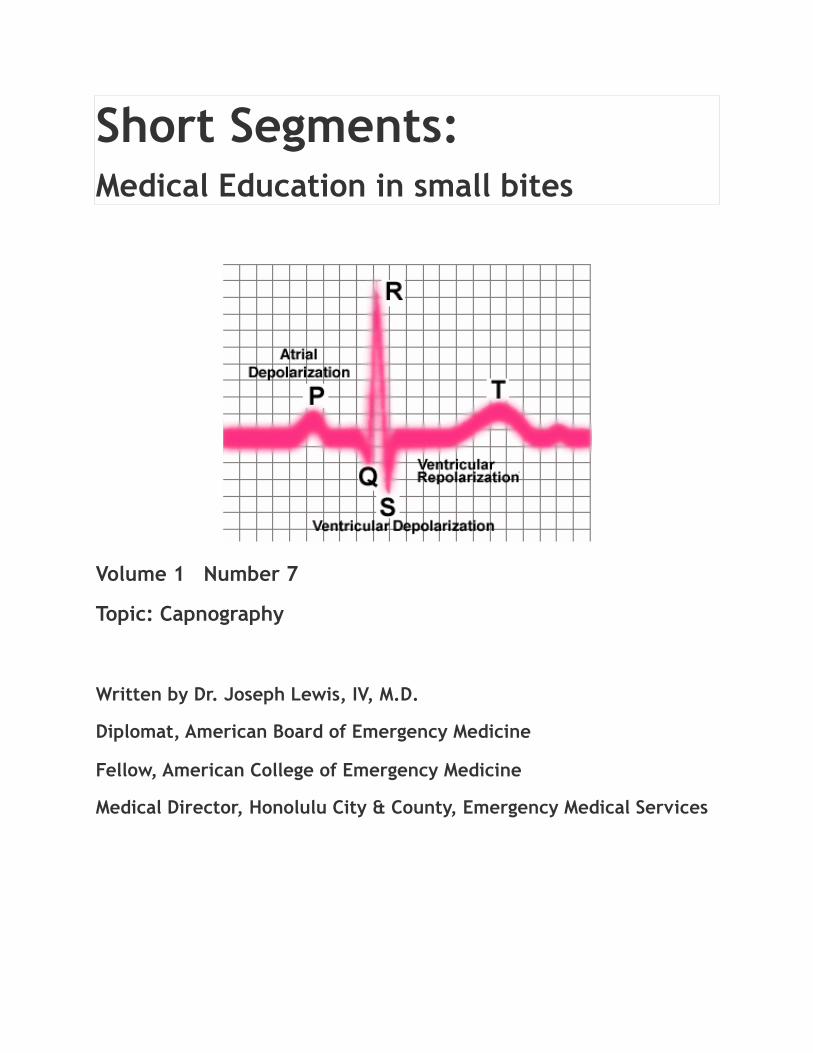
Short Segments: Medical Education in small bites Volume 1 Number 7 Topic: Capnography Written by Dr. Joseph Lewis, IV, M.D. Diplomat, American Board of Emergency Medicine Fellow, American College of Emergency Medicine Medical Director, Honolulu City & County, Emergency Medical Services
Transcript of Short Segment -cap draft August 23rdcopy
Short Segments:Medical Education in small bites
Volume 1 Number 7
Topic: Capnography
Written by Dr. Joseph Lewis, IV, M.D.
Diplomat, American Board of Emergency Medicine
Fellow, American College of Emergency Medicine
Medical Director, Honolulu City & County, Emergency Medical Services

Learning Objectives:
1. Know the six vital signs, how we measure them and why the tools we use for measurement are important.
2. Understand the difference between oxygenation and ventilation
3. Define oxygenation
4. Define ventilation
5. Define pulse oximetry
6. Define capnography
7. Recognize a normal capnography wave
8. Recognize an abnormal Capnography wave
9. Know the normal numerical values for capnography
10. Recognize a Capnography wave for a hyperventilating patient
11. Recognize a Capnography wave for a hypoventilation patient
12. Recognize the Capnography wave of an apneic patient
13. Recognize the Capnography wave of an asthma patient
14. Recognize improvement in your asthma patient by the Capnography wave
15. Recognize the Capnography Wave of a successful intubation
16. Recognize the Capnography Wave of an unsuccessful intubation
17. Recognize the Capnography wave of a code patient with return of perfusing pulse
18. Understand how fast the capnography device responds to apnea versus how slow the pulse oximetry device responds in a patient your giving pain medicine like fentanyl or sedative like Ativan or Valium.
19. Be amazed at how useful this tool is!
1.Know the six vital signs, how we measure them and why the tools we
use for measurement are important.
The Six Vital Signs.
Traditionally there are four vital signs: 1. Temperature, 2. Blood Pressure, 3. Heart Rate and 4. Respiratory Rate.
Tools.
We use our assessment skills of sight, sound, touch and thought to assess patients. We can feel a patient’s hot skin, we can see their pale skin, we can feel their fast faint pulse and we will think this patient is sick. But our tools like a thermometer, BP cuff and cardiac monitor give us numerical data like temperature 103, BP 70/palp and HR 144 and visual data like a narrow complex tachycardia. This numerical and visual data tells us a lot more then our assessment skills alone. It also allows us to provide the right care in the right amounts. Remember if we can measure the problem, we can measure the solution.
The fifth vital sign is Pulse Oximetry which measures oxygenation and number six is Capnography which measures ventilation.
In the same way you can use your assessment skills to assess a patient’s oxygenation and ventilation, but pulse oximetry and capnography give us numerical data and visual data. These tools allow us to measure the problem and apply the right amount of the solution.
2. Understand the difference between oxygenation and ventilation
Oxygenation Versus VentilationOxygenation is how we get oxygen to the tissue.
Oxygen is inhaled into the lungs where gas exchange occurs at the capillary-alveolar membrane. Oxygen is transported to the tissues through the blood stream.
Pulse oximetry measures oxygenation.
Ventilation (the movement of air) is how we get rid of carbon dioxide.
At the cellular level, oxygen and glucose combine to produce energy. Carbon dioxide, a waste product of this process diffuses into the blood. Carbon dioxide is carried back through the blood and exhaled by the lungs through the alveoli.
Capnography measures ventilation

3. What doe a normal Capnography wave look like and what does it mean?
ETCO2 35-45 mm Hg is the normal value for capnography
Look at the labelled diagram below, it looks complicated, but when you know what means
its easy to understand.
Capongraphy measures CO2 in your breath.
When you inhale it reads low because you don't blow air when you inhale.
But when you exhale it goes up because your giving off CO2.
Exhale takes time so it goes up and plateausit goes up to maximal exhalation and as
A_B is the pause after a breath, your lungs are full.
BC is the start of exhalation.
C-D is the full exhalation with D being the peak volume of carbon dioxide.
DE is inspration you taking a breath in and no carbon dioxide is going out.
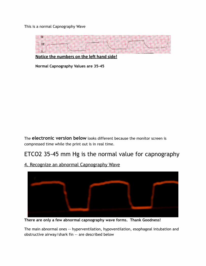
This is a normal Capnography Wave
The electronic version below looks different because the monitor screen is compressed time while the print out is in real time.
ETCO2 35-45 mm Hg is the normal value for capnography
4. Recognize an abnormal Capnography Wave
There are only a few abnormal capnography wave forms. Thank Goodness!
The main abnormal ones -- hyperventilation, hypoventilation, esophageal intubation and obstructive airway/shark fin -- are described below
Notice the numbers on the left hand side!
Normal Capnography Values are 35-45
There are two types of abnormal values in Caponography:
1. ETCO2 Less Than 35 mmHg = "Hyperventilation causes Hypocapnia"
2. ETC02 Greater Than 45 mmHg = "Hypoventilation causes Hypercapnia"
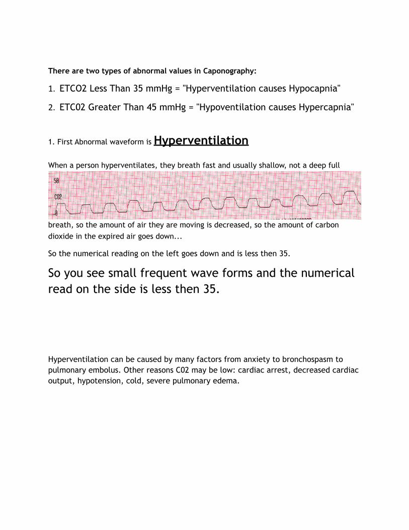
1. First Abnormal waveform is Hyperventilation
When a person hyperventilates, they breath fast and usually shallow, not a deep full
breath, so the amount of air they are moving is decreased, so the amount of carbon dioxide in the expired air goes down...
So the numerical reading on the left goes down and is less then 35.
So you see small frequent wave forms and the numerical read on the side is less then 35.
Hyperventilation can be caused by many factors from anxiety to bronchospasm to pulmonary embolus. Other reasons C02 may be low: cardiac arrest, decreased cardiac output, hypotension, cold, severe pulmonary edema.
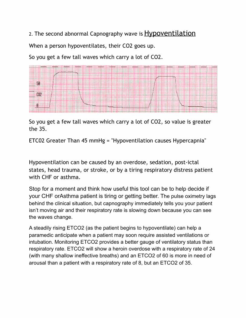
2. The second abnormal Capnography wave is Hypoventilation
When a person hypoventilates, their CO2 goes up.
So you get a few tall waves which carry a lot of CO2.
So you get a few tall waves which carry a lot of CO2, so value is greater the 35.
ETC02 Greater Than 45 mmHg = "Hypoventilation causes Hypercapnia"
Hypoventilation can be caused by an overdose, sedation, post-ictal states, head trauma, or stroke, or by a tiring respiratory distress patient with CHF or asthma.
Stop for a moment and think how useful this tool can be to help decide if your CHF orAsthma patient is tiring or getting better. The pulse oximetry lags behind the clinical situation, but capnography immediately tells you your patient isn’t moving air and their respiratory rate is slowing down because you can see the waves change.
A steadily rising ETCO2 (as the patient begins to hypoventilate) can help a paramedic anticipate when a patient may soon require assisted ventilations or intubation. Monitoring ETCO2 provides a better gauge of ventilatory status than respiratory rate. ETCO2 will show a heroin overdose with a respiratory rate of 24 (with many shallow ineffective breaths) and an ETCO2 of 60 is more in need of arousal than a patient with a respiratory rate of 8, but an ETCO2 of 35.

3. The third wave form is *Esophageal Intubation
Reasons ETCO2 is zero: The tube is in the esophagus.*The stomach doesn’t exhale so the wave form is zero and stays flat.
NOTE: Greatly prolonged down time prior to initiation of CPR or cases or Massive pulmonary embolism where blood flow to the lungs is completely blocked may have a flat wave form. Also, in patients in arrest, CPR is necessary to generate a waveform.
Caution: In patients with a prolonged down time, the ETCO2 reading may be so low (sometimes less than 6mm HG) that some monitor's apnea alarms may go off even though the monitor is still providing an ETCO2 reading and a small wave form. If the apnea alarm goes off and you continue to bag without resistance and have equal lung sounds and negative epigatric sounds, do not automatically pull your tube. A small but distinct square wave form along with even a marginal EtCO2 reading is still verification the tube is in the trachea.
Successful Intubation
A good wave form indicating the presence of CO2 ensures the ET tube is in the trachea
A good wave form indicating the presence of CO2 ensures the ET tube is in the trachea.

4. The fourth Obstructive Capnography Pattern
Bronchospasm will produce a characteristic “shark fin” wave form as the patient has to struggle to exhale, creating a sloping “B-‐C” upstroke
ETCO2 in Asthma, COPD
End-tidal CO2 monitoring on non-intubated patients is an excellent way to assess the severity of Asthma/COPD, and the effectiveness of treatment. Successful treatment will lessen or eliminate the shark fin shape and return the ETCO2 to normal range (Patient below: capnogram on arrival, after start of 1st DuoNeb, after two Duonebs.
Asthma values change with severity.
Mild asthma, the CO2 will drop (below 35) as the patient hyperventilates to compensate. As the asthma worsens, the C02 levels will rise to normal. When the asthma becomes severe, and the patient is tiring and has little air movement, the C02 numbers will rise to dangerous levels (above 60).

Bonus Material
Capnography can also be used for combitubes and LMAs.
Paramedics should document their use of continuous ETCO2 monitoring and attach wave form strips to their PCRs. Print a strip on intubation, periodically during care and transport, and then just prior to moving the patient from your stretcher to the hospital table and then immediately after transfer. This will timestamp and document your tube as good.
Cautions: Imperfect positioning of nasal cannula capnofilters may cause distorted readings. Unique nasal anatomy, obstructed nares and mouth breathers may skew results and/or require repositioning of cannula. Also, oxygen by mask may lower the reading by 10% or more.
Capnography during chest compressions shows that chest compressions ventilate the patient!
Paramedics who utilize this method during cardiac arrests with cardiac compressions continuing while they intubate may see CPR oscillations on the monitor screen immediately upon intubating, replaced by larger wave forms once the ambu-bag has been attached and ventilations begun. The oscillations provide proof that compressions alone can produce some ventilation.
Note: You must still assess for equal lung sounds. Capnography cannot detect right main-stem intubations.

Return of Spontaneous Circulation (ROSC)
ETCO2 can be the first sign of return of spontaneous circulation (ROSC). During a cardiac arrest, if you see the CO2 number shoot up, stop CPR and check for pulses.
End-tidal CO2 will often overshoot baseline values when circulation is restored due to carbon dioxide washout from the tissues.
A recent study found the ETCO2 shot up on average 13.5 mmHg with sudden ROSC before settling into a normal range.-Grmec S, Krizmaric M, Mally S, Kozelj A, Spindler M, Lesnik B.,Resuscitation. 2006 Dec 8
Note: Each bar represents 30 seconds.
“End-tidal CO2 monitoring during cardiac arrest is a safe and effective noninvasive indicator of cardiac output during CPR and may be an early indicator of ROSC in intubated patients.” - American Heart Association Guidelines 2005 CPR and ECG
Loss of Spontaneous CirculationIn a resuscitated patient, if you see the stabilized ETCO2 number significantly drop in a person with ROSC, immediately check pulses. You may have to restart CPR.
The graph below demonstrates three episodes of ROSC, followed by loss of circulation during a cardiac arrest:

Monitoring Sedated Patients (Pain Medicines/Fentanyl or Morphine)
Capnography should be used to monitor any patients receiving pain management or sedation (enough to alter their mental status) for evidence of hypoventilation and/or apnea. In the graph below, the respiratory rate decreases as the ETCO2 rises, and the patient suffers apnea, all the while the SPO2 remains stable. Note your Pulse oximetry reading doesn't drop down with apnea because pulse oximetry measures oxygenation and one breath gives you plenty of oxygen, even if you stop breathing it stays normal for almost a minute. Remember pulse oximetry doesn't measure carbon dioxide, so it isn't reliable to monitor ventilation. But when you stop breathing, just like when you inhale your capnography reading goes right to zero. capnography is a
very sensitive monitor of ventilation. Note: Each bar represents thirty seconds.
Sedated, Intubated Patients Capnography is also essential in sedated, intubated patients. A small notch in the wave form indicates the patient is beginning to arouse from sedation, starting to breathe on their own, and will need additional medication to prevent them from "bucking" the tube.
Hypoxic Drive
Capnography will show the hypoxic drive in COPD "retainers." ETCO2 readings will steadily rise, alerting you to cut back on the oxygen before the patient becomes obtunded. Since it has been estimated that only 5% of COPDers have a hypoxic drive, monitoring capnography will also allow you to maintain sufficient oxygen levels in the majority of tachypneic COPDers without worry that they will hypoventilate.
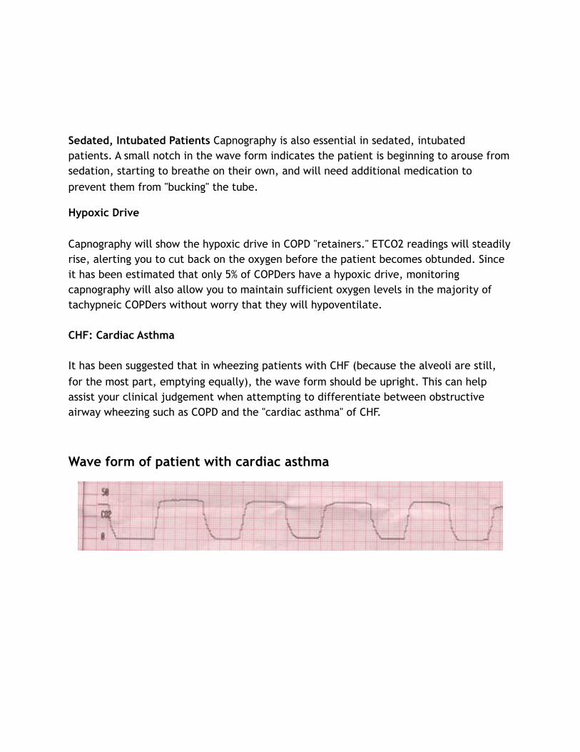
CHF: Cardiac Asthma
It has been suggested that in wheezing patients with CHF (because the alveoli are still, for the most part, emptying equally), the wave form should be upright. This can help assist your clinical judgement when attempting to differentiate between obstructive airway wheezing such as COPD and the "cardiac asthma" of CHF.
Wave form of patient with cardiac asthma

Ventilating Head Injured Patients
Capnography can help paramedics avoid hyperventilation in intubated head injured
patients. Hyperventilation decreases intracranial pressure by decreasing intracranial
blood flow. The decreased cerebral blood flow may result in cerebral ischemia.
“Recent evidence suggests hyperventilation leads to ischemia almost immediately...current models of
both ischemic and TBI suggest an immediate period during which the brain is especially vulnerable to
secondary insults. This underscores the importance of avoiding hyperventilation in the prehospital
environment.” --Capnography as a Guide to Ventilation in the Field, D.P. Davis, Gravenstein,
Capnography: Clinical Perspectives, Cambridge Press, 2004
In a study of 291 intubated head injured patients, 144 had ETCO2 monitoring. Patients with ETCO2
monitoring had lower incidence of inadvertant severe hyperventilation (5.6%) than those without
ETCO2 monitoring (13.4%). Patients in both groups with severe hyperventilation had significantly higher
mortality (56%) than those without (30%). –Davis, The Use of Quantitative End-Tidal Capnometry to
Avoid Inadvertant Severe Hyperventilation in Patients with Head Injury After Paramedic Rapid Sequence
Intubation, Journal of Trauma, April 2004
“A target value of 35 mmHg is recommended...The propensity of prehospital personnel to use
excessively high respiratory rates suggests that the number of breaths per minute should be decreased.
On the other hand, the mounting evidence against tidal volumes in excessive of 10cc/kg especially in
the absence of peep, would suggest the hypocapnia be addressed by lower volume ventilation.” – --
Capnography as a Guide to Ventilation in the Field, D.P. Davis, Gravenstein, Capnography: Clinical
Perspectives, Cambridge Press, 2004

References
***
1. Burton, Does End-Tidal Carbon Dioxide Monitoring Detect Respiratory Events Prior to Current Sedation Monitoring Practices, Academic Emergency Medicine, May 2006
2. Capnography as a Guide to Ventilation in the Field, D.P. Davis, Gravenstein, Capnography: Clinical Perspectives, Cambridge Press, 2004
3. The Use of Quantitative End-Tidal Capnometry to Avoid Inadvertant Severe Hyperventilation in Patients with Head Injury After Paramedic Rapid Sequence Intubation, Journal of Trauma, April 2004
4. Multiple studies have confirmed the sloping shape correlates to bronchospasm and obstructive lung disease. “The analysis of the capnogram’s shape is a quantitative method for evaluating the severity of bronchospasm.” –You, Expiratory capnography in asthma: evaluation of various shape indicies, European Respiratory Journal, Feb, 1994
5. In a 2006 published study of 60 patients undergoing sedation, in 14 of 17 patients who suffered acute respiratory events, ETCO2 monitoring flagged a problem before changes in SPO2 or observed changes in respiratory rate. “End-tidal carbon dioxide monitoring of patients undergoing PSA detected many clinically significant acute respiratory events before standard ED monitoring practice did so. The majority of acute respiratory events noted in this trial occurred before changes in SP02 or observed hypoventilation and apnea.” - -Burton, Does End-Tidal
6. 10 Things Every Paramedic Should Know About CapnographyPeter Canning, EMT-PDecember 29, 2007 (Version 6.3)