SHOCK in Paediatric Trauma - hkcpn.com · Definition of Shock ... Impaired cardiac contractility...
60
SHOCK in Paediatric Trauma Speaker: Tang Sze Kit, (Fellow, Hong Kong College of Paediatric Nursing, MSc, MN, BN) Date: 26/4/2016 Time: 18:30- 19:30 Venue: Paediatrics & Adolescent Ambulatory Centre, G/F, Block C, Yan Chai Hospital
Transcript of SHOCK in Paediatric Trauma - hkcpn.com · Definition of Shock ... Impaired cardiac contractility...

SHOCK in Paediatric Trauma
Speaker: Tang Sze Kit,
(Fellow, Hong Kong College of Paediatric Nursing,
MSc, MN, BN)
Date: 26/4/2016
Time: 18:30- 19:30
Venue: Paediatrics & Adolescent Ambulatory Centre, G/F,
Block C, Yan Chai Hospital

Content
Definition of shock
Classification and phase of shock
Recognition of Shock
Shock management
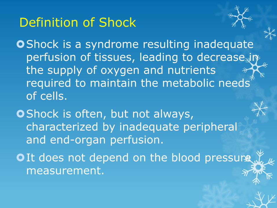
Definition of Shock
Shock is a syndrome resulting inadequate perfusion of tissues, leading to decrease in the supply of oxygen and nutrients required to maintain the metabolic needs of cells.
Shock is often, but not always, characterized by inadequate peripheral and end-organ perfusion.
It does not depend on the blood pressure measurement.

Definition of Shock
All types of shock can result in impaired function of vital organ, such as brain and kidneys.
If prolonged, it leads to multiple organ failure and death.
It can caused by any serious disease and injury.

Phase of Shock
Normal Cardiac Output Normal blood pressure
Cardiac output Blood pressure
Cardiac output Blood pressure
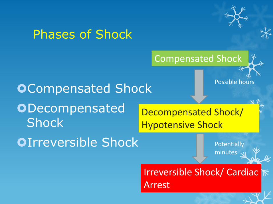
Phases of Shock
Compensated Shock
Decompensated Shock
Irreversible Shock
Compensated Shock
Decompensated Shock/ Hypotensive Shock
Irreversible Shock/ Cardiac Arrest
Possible hours
Potentially minutes

Compensated Shock
A clinical state of tissue perfusion that is inadequate to meet metabolic demand in the presence of blood pressure within the normal range.
When oxygen delivery is limited, compensatory mechanism by to maintain normal blood flow to the brain and heart.

Common Signs of Compensated Shock
Compensatory Mechanism
Area Sign
Increased heart rate Heart Tachycardia
Increased SVR
Skin Cold, pale, mottled, diaphoretic
Peripheral circulation
Delayed capillary refill
Pulse Weak peripheral pulse; narrow pulse pressure (increase diastolic pressure)
Increase renal and splanchnic vascular resistance (redistribution of blood flow away from these areas)
Kidney Oliguria ( decrease urine output)
Intestine Vomiting, ileus

Decompensated Shock
Hypotension (I.e. systolic blood pressure (SBP) > 5th percentile for age.
According to the AHA (2010), hypotension is characterized by the following limits of systolic blood pressure (SBP):
Age Lower limit of Blood pressure (systolic) mmHg
Neonate (0- 28 days) < 60
Infant (1 to 12 months) < 70
Children (1 to 10 years) 70 + (2 x age in years)
Children (> 10 years) < 90

Classification of Shock
Shock can result from:
Inadequate blood volume or oxygen-carrying capacity (hypovolemic shock including hemorrhagic shock).
Inappropriate distribution of blood volume and flow (distributive shock).
Impaired cardiac contractility (cardiogenic shock).
Obstructed blood flow (obstructive shock).

Classification of Shock
Shock can be categorized into 4 types:
Hypovolemic shock
Cardiogenic shock
Distributive shock
Obstructive shock
Shock in Trauma Patient:
Haemorrhagic shock
non-haemorrhagic shock

Classification of Shock Classification Etiology Underlying Pathology
Hypovolemic -Haemorrhage -Burns
-Whole blood loss - Plasma Loss
Cardiogenic -Myocardial infarction -Dysrhythmias -Blunt cardiac injury
-Loss of cardiac contractility -Reduced cardiac output -Loss of cardiac contractility
Obstructive -Cardiac temponade -Tension pneumothroax -Tension haemothroax
-Compression of heart with obstruction to atrial filling -Mediatstinal shift with obstruction to atrial filling -Combination of above
Distributive -Neurogenic shock -Anaphylactic shock -Septic shock
-Venous pooling- maldistribution of blood volume -Shunting in microcirculation and in later stages- decrease in venous resistance -Poor distribution of blood flow

Classification of Shock

Haemorrhagic Shock
Most common cause of shock after injury
Haemorrhage is defined as acute loss of circulating blood volume.
Causes of significant blood loss contributing to haemorrhagic shock are:
Major vessel disruption
Massive haemothoraces
Liver or spleen injuries
Pelvic fractures
Extremity fractures
Amputation

Classification of haemorrhagic shock in children
Class I Class II Class III Class IV
Blood Loss <15% 15-25% 26-39% >40%
CVS Normal to HR Normal BP/P
HR Normal BP Peripheral pulse
HR Hypotension Thready periperal pulses
HR Profound hypotension, Absent peripheral pulse, Thready central pulses
Resp Normal Rate Tachypnea Moderate tachypnea
Severe tachypnea
CNS Slightly anxious
Irritable, confused, combative
Diminished response to pain
Coma

Classification of haemorrhagic shock in children
Class I Class II Class III Class IV
Blood Loss <15% 15-25% 26-39% >40%
Skin Warm, pink Normal cap refill
Cool extremities, Mottling, Delayed cap refill
Cool extremities, Mottling, Delayed cap refill
Cool extremities Pallor Cyanosis
Renal Normal urine output
Oliguria Urine SG
Oliguria BUN
Anuria
Acid-base Normal pH Normal pH
Metabolic acidosis
Metabolic acidosis

Non-haemorrhage Shock
Cardiogenic shock- blunt cardiac injury, cardiac tamponade, air embolus, myocardial infarction associated with the patient’s injury
Tension pneumothroax
Neurogenic shock
Septic shock

Recognition of Shock

Primary Assessment & Resuscitation
A – Airway maintenance + C – spine control
B – Breathing
C – Circulation + Haemorrhage control
D – Disability (neurological exam)

Airway Assessment + C-spine immobilization
Clear/ patent
Maintainable with head positioning
Not maintainable without intubation

Breathing Assessment
Respiratory rate
Respiratory effort/ mechanics
Breath sounds/ air entry/ tidal volume
Skin color and pulse oximetry

Primary Assessment & Resuscitation
A – Airway maintenance + C – spine control
B – Breathing
C – Circulation + Haemorrhage control
D – Disability (neurological exam)

Recognition of Shock
Assessment
Level of consciousness
Pulse: quality, rate and regularity
Skin colour
Capillary refill (? > 2 sec)
Skin temperature
External haemorrhage
Measure blood pressure
Urine output

Pulses
Thready pulses are felt, as cardiac output falls and systemic vascular resistance increases.
Loss of central pulse is considered as cardiac arrest

Skin Perfusion
Evaluation of temperature, capillary refill and color
Cold extremities when cardiac output decreases
Mottling, pallor and peripheral cyanosis when skin perfusion is poor
Prolonged capillary refill when cardiac output decreases

Evaluation of skin perfusion
Temperature of extremities forearms and legs
Capillary refill time
Colour
Pink Pale Blue
Mottled
Weak Perfusion
Worsening Perfusion

Perfusion - Kidneys
Evaluation of urine output
Urine output less than 1ml/kg/hr in the absence of renal disease is a sign of poor renal perfusion or hypovolaemia
Urine output
Infant At least 1ml/kg/hr Preschool
Adolescent

Evaluation of Disability - Brain
Evaluation of responsiveness
Use of AVPU scale
A - alert
V- response to voice
P- response to pain
U- unresponsiveness
Glasgow Coma Scale
Muscle tone
Papillary response

Modified Glasgow Coma Scale for Infants and Children
Child (> 2 yr) Infant (0-2 yr) Score
Eye Opening Spontaneous Spontaneous 4
To verbal stimuli To verbal stimuli 3
To pain only To pain only 2
No response No response 1
Verbal Response Oriented, appropriate Coos and babbles 5
Confused Irritable Cries 4
Inappropriate words Cries to pain 3
Incomprehensible words or non-specific sounds
Moans to pain 2
No response No response 1

Modified Glasgow Coma Scale for Infants and Children
Child (> 2 yr) Infant (0-2 yr) Score
Motor Response
Obeys commands Moves spontaneously and purposefully
6
Localized painful stimulus
Withdraws to touch 5
Withdraws in response to pain
Withdraws in response to pain
4
Flexion in response to pain
Decorticate posturing (abnormal flexion)
3
Extension in response to pain
Decerebrate posturing (abnormal extension)
2
No response No response 1

Blood Pressure: Normal blood pressure like heart rate, there is a wide range of value within the normal range
Age Systolic BP ( mmHg) Diastolic BP (mmHg)
Female Male Female Male
Neonate (1day) 60 - 76 60 - 74 31 - 45 30 - 44
Neonate (4day) 67 - 83 68 - 84 37 - 53 35 - 53
Infant (1 month) 73 - 91 74 - 94 36 - 56 37 - 55
Infant (3 month) 78 - 100 81 – 103 44 – 64 45 - 65
Infant (6 month) 82 - 102 87 - 105 46 – 66 48 - 68
Infant (1 year) 68 - 104 67 - 103 22 - 60 20 - 58
Child (2 years) 71 - 105 70 - 106 27 - 65 25 - 63
Child (7 years) 79 - 113 79 - 115 39 - 77 38 - 78
Adolescent (15years)
93 - 127 95 - 131 47 - 85 45 - 85
Range from 33rd - 67th percentile in the first year of life and from 5th – 95th percentile for systolic and diastolic BP according to age and gender
Normal BP in Paediatrics

According to the AHA (2010), hypotension is characterized by the following limits of systolic blood pressure (SBP):
Age Lower limit of Blood pressure (systolic) mmHg
Neonate (0- 28 days) < 60
Infant (1 to 12 months) < 70
Children (1 to 10 years) 70 + (2 x age in years)
Children (> 10 years) < 90

Recognition of Shock
Assessment
Diagnostic Procedure
Chest radiograph- to determine the presence of hemothorax/ pneumothorax
Pelvic radiograph- to locate fracture
Femur radiograph if fracture is suspected
Laboratory Studies
Venous blood sample for typing
Urinalysis including specific gravity
Arterial pH, PaO2, PaCO2 and base deficit

Fundamentals of Shock Management
• The acute treatment of shock focuses on restoring oxygen delivery to the tissues and improving the balance between tissue perfusion and metabolic demand.
• The acute treatment of shock includes:
– Optimizing oxygen content of the blood
– Improving volume and distribution of cardiac output
– Reducing oxygen demand
– Correcting metabolic derangements
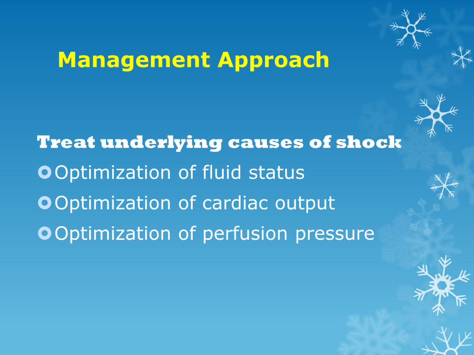
Management Approach
Treat underlying causes of shock
Optimization of fluid status
Optimization of cardiac output
Optimization of perfusion pressure
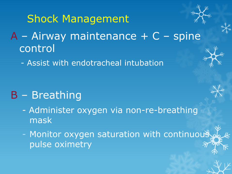
Shock Management
A – Airway maintenance + C – spine control
- Assist with endotracheal intubation
B – Breathing
- Administer oxygen via non-re-breathing mask
- Monitor oxygen saturation with continuous pulse oximetry

Circulation
Intervention
Haemorrhage control- any uncontrolled external bleeding
Fluid resuscitation: 2 large-bore IV with Ringer’s Lactate solution/ Normal Saline (20ml/kg)
Lab-test – type and screen, CBP, chemical, toxicology
Blood replacement as needed


Response to initial fluid resuscitation
Rapid response
Transient response
No response
Vital signs Return to normal
Transient improvement, recurrence of HR and BP
Remain abnormal
Estimated blood loss
Minimal (10-20%)
Moderate & ongoing (20-40%)
Severe (>40%)
Needs for more crystalloid
Low High High
Blood preparation
Type & cross-match
Type- specific Emergency blood release
Need for operative intervention
Possibly Likely Highly likely
Early presence of surgeon
Yes Yes Yes

Not responding to fluid challenge ??
Identify cardiogenic event
Using inotropes/ vasopressors/ or both
Perform echocardiogram

Inotropes/ Vasopressors
Dopamine
Dobutamine
Adrenaline
Noradrenaline

INOTROPES
Acts on HR & cardiac contractility

Vasopressors
Acts on peripheral vascular tones Either vasoconstriction/ vasodilatation

Inotropes
DRUGS Receptors Actions
Dobutamine Beta 1/2 ^ HR/ SV
Peripheral vasodilatation
Dopamine Alpha 1/ beta 1 ^ SV/ vasoconstriction
Adrenaline Alpha 1
Beta 1/2
^ HR/ SV
vasoconstriction

Vasoactive Agents
DRUGS Receptors Actions
Noradrenaline Alpha 1 Peripheral vasoconstriction
Phenylephrine Alpha 1 Peripheral vasoconstriction

Epinephrine (Adrenaline)
Both - and - adrenergic effects
- adrenergic action (vasoconstriction) increase systemic vascular resistance and elevates the systolic and diastolic blood pressure
- adrenergic action increases myocardial contractility and heart rate and relaxes smooth muscles in the skeletal muscle vascular bed and in bronchi

Epinephrine (Adrenaline)
Indications:
Cardiac arrest
Symptomatic bradycardia unresponsive to ventilation and oxygen administration
Hypotension not related to volume depletion
Dose:
IV / IO
Initial 1:10,000 at 0.1ml/kg
Subsequent same dose
ET
1:1,000 at 0.1ml/kg
For neonate, 1:10,000 at 0.1-0.3ml/kg
Q3-5min

Epinephrine (Adrenaline)
Side effect:
High dose atrial tachycardia or VT
Arrhythmia, chest pain, nervousness, restlessness
Extravasation local ischaemia

Dopamine Hydrochloride
An endogenous catechloamine with complex cardiovascular effects
Indications
Inadequate cardiac output
BP
splanchnic blood flow & urine output
2 – 5ug/kg/min vasodilatory effects .Little direct
cardiac action
renal, splanchnic, coronary and
cerebral blood flow
5-10ug/kg/min cardiac contractility
10-20ug/kg/min Vasoconstriction, BP, AR
> 20ug/kg/min Vasoconstriction, No further inotropic
effect

Dopamine Hydrochloride
Side effect:
BP, HR
Arrhythmia ( ventricular ectopic, SVT, VT)
Ectopic heart beats, conduction abnormalities
Extravasation tissue necrosis & gangrene of extremities

Dobutamine Hydrochloride
Synthetic catecholamine possessing relatively selective action a -adrenergic receptors which effects including cardiac contractility and heart rate, often with mild dilation of the peripheral vascular bed
Indications
Myocardial dysfunction( e.g. severe congestive heart failure)
Inadequate cardiac output, particularly in patients with elevated systemic or pulmonary vascular resistance
Poor perfusion despite adequate intravascular volume
Post resuscitation myocardial dysfunction

Dobutamine Hydrochloride
Dose
2-20 g/kg/min.
often started at 5-10 g/kg/min
IV infusion into large vein
Side effect: HR, BP, nausea, vomiting
Tachyarrhythmias or ectopic beats
Extravasation tissue ischemia & necrosis

Noradrenaline
Both - and - adrenergic effects
- adrenergic action (vasoconstriction) increase systemic vascular resistance and elevates the systolic and diastolic blood pressure
Most often used after volume repletion and administration of other inotropes have been ineffective in raising the BP
Noradrenaline
Dose: 0.15- 0.1microgram/kg/minutes
The rate of infusion is titrated up to 1 microgram/kg/min
Side effect:
High dose ischemic injury of the extremities

Circulation
Intervention
Insert an indwelling urinary catheter
ECG
Pericardial tamponade- pericardiocentesis
ED thoracotomy / surgery

Evaluation and ongoing assessment
Reassessing patient response and avoiding complications
Control haemorrhage surgical intervention
Monitoring end- organ perfusion
Monitor urine output
Collaborating with other trauma team members as diagnostic studies and physical assessment identify the cause and source of haemorrhage
Monitor temperature to determine hypthermia

Therapeutic End Points
Clinical Improvement:
Normal heart rate and blood pressure for age
Normal pulse
Capillary refill time < 2 seconds
Warm extremities
Normal mental status
Urine output >1ml/kg/hr
Decrease serum lactate
Decrease base deficit

Key Message
Early Recognition of shock and intervention is critical in halting progression from compensated to hypotensive shock to cardiopulmonary failure
Treatment goal for shock is to prevent end-organ injury and halt the progression of cardiopulmonary failure and cardiac arrest

The End Question & suggestion are welcome

Reference and Bibliography American Academy of Pediatrics (2000). Pediatric Education
for Prehospital Professionals.
American College of Surgeon (2007) Advanced Trauma Life Support
American Heart Association (2011). Paediatric Advanced Life Support- provider manual
Emergency Nurses Association (2000). Trauma Nursing Core Course
Fuhrman B P et al (2011)Paediatric Critical Care (4th edition) Philadelphia: Elsevier Saunders
Hazinski M. F. (1999) Manual of Pediatric Critical Care. St Louis: Mosby
Ranjit S (2010) Manual of Paediatric Emergency & Critical Care . 2nd Edition. New Deihi: Paras Medical Publisher