Shiv Kumar Singh - Journal of Anaesthesia & Critical...
15
43 | Journal of Anaesthesia and Critical Care Case Reports | Volume 3 | Issue 2 | May-Aug | 2017 | Page 43-57 The Anaesthetist Society Buddha said “There is no wealth like knowledge, and no poverty like ignorance.” Facebook group “The Anaesthetist” was born on 21 st February 2012 by the initiative of Dr. Vivek Gupta and moderated by Dr. Shiv Kumar Singh. Over the months the group grew steadily in numbers, and people were joining from all over the world, but most were from the Indian Subcontinent and the Middle East. The members from the West were again mostly of eastern origin. There were some members from the European and South American countries as well, who do regularly participate in the activities. In less than a year we were more than 1000 members strong, and in the past 3 years, our strength has grown to more than 5000 dedicated members. On 25 th October 2013, we launched the logo competition. Aashish Jain was the 1 st person to submit his entry, and this initiated many more to participate with great enthusiasm. The creativity exhibited by the members who took part really surprised me. The competition was won by Sunny Bhasin. The logo of the society signifies the activities of an anaesthetist. In November 2013, Hetal Vadera organized a Regional Anaesthesia workshop at Rajkot; this was the first time that many of the members from the group met face to face. The workshop was a great success and was the catalyst that started the reaction of stronger friendship and camaraderie among the members. The workshop created enthusiasm among members to use RA as part of multimodal analgesia and improve the quality of care for their patients. On 5 th November 2013, the idea of TACON (The Anaesthetist Conference) was floated and by 5 th December, within a month, the Venue (India International Centre [IIC]) was booked for the conference by Anil Sharma who took up the challenge. On 24 th March 2014, the group got registered as a society, The Anaesthetist Society or TAS as most members call it. The TACON 2014 was held at the prestigious IIC on the 14 th and 15 th November. It was a grand success with members from all over the world interacting academically and socially, the bond was further strengthened. Since the next TACON would be in 2016, it was decided that we would hold few workshops in 2015 and “Nerve Blocks for The Masses” Rajkot 2015 is 1 st of these workshops where we will discuss and demonstrate loss of resistance (LOR) and peripheral nerve stimulator guided blocks. This workshop will remind the members the first unofficial meeting of the TASians. This booklet has been specifically produced for the members to practice blocks that can be done by anyone anywhere even in the remotest of the places. Good Luck The TAS CORE COMMITTEE Introduction to Loss of Resistance Technique The LOR techniques rely on using blunt or short-beveled needles, which provide a good feedback (pops or clicks) when they pass through the various fascia. There are some simple blocks that can be performed with just a needle and some local anaesthetics (LA). These blocks, if done properly, provide good analgesia Journal of Anaesthesia and Critical Care Case Reports 2017 May-Aug;3(2):43-57 © 2017 by Journal of Anaesthesia and Critical Care Case Reports| Available on www.jaccr.com | This is an Open Access article distributed under the terms of the Creative Commons Attribution Non-Commercial License (http://creativecommons.org/licenses/by-nc/3.0) which permits unrestricted non-commercial use, distribution, and reproduction in any medium, provided the original work is properly cited. Nerve Blocks for the Masses: Loss of Resistance Blocks Shiv Kumar Singh Royal Liverpool University Hospitals Prescot Street Liverpool L7 8XP Shiv Kumar Singh Address of Correspondence: Shiv Kumar Singh Royal Liverpool University Hospitals Prescot Street Liverpool L7 8XP. E-mail: [email protected]
Transcript of Shiv Kumar Singh - Journal of Anaesthesia & Critical...

43 | Journal of Anaesthesia and Critical Care Case Reports | Volume 3 | Issue 2 | May-Aug | 2017 | Page 43-57
The Anaesthetist Society
Buddha said “There is no wealth like knowledge, and no poverty like ignorance.”
Facebook group “The Anaesthetist” was born on 21st February 2012 by the initiative of Dr. Vivek Gupta and moderated by Dr. Shiv Kumar Singh. Over the months the group grew steadily in numbers, and people were joining from all over the world, but most were from the Indian Subcontinent and the Middle East. The members from the West were again mostly of eastern origin. There were some members from the European and South American countries as well, who do regularly participate in the activities. In less than a year we were more than 1000 members strong, and in the past 3 years, our strength has grown to more than 5000 dedicated members.
On 25th October 2013, we launched the logo competition. Aashish Jain was the 1st person to submit his entry, and this initiated many more to participate with great enthusiasm. The creativity exhibited by the members who took part really surprised me. The competition was won by Sunny Bhasin. The logo of the society signifies the activities of an anaesthetist.
In November 2013, Hetal Vadera organized a Regional Anaesthesia workshop at Rajkot; this was the first time that many of the members from the group met face to face. The workshop was a great success and was the catalyst that started the reaction of stronger friendship and camaraderie among the members.
The workshop created enthusiasm among members to use RA as part of multimodal analgesia and improve the quality of care for their patients.
On 5th November 2013, the idea of TACON (The Anaesthetist Conference) was floated and by 5th December, within a month, the Venue (India International Centre [IIC]) was booked for the conference by Anil Sharma who took up the challenge.
On 24th March 2014, the group got registered as a society, The Anaesthetist Society or TAS as most members call it. The TACON 2014 was held at the prestigious IIC on the 14th and 15th November. It was a grand success with members from all over the world interacting academically and socially, the bond was further strengthened.
Since the next TACON would be in 2016, it was decided that we would hold few workshops in 2015 and “Nerve Blocks for The Masses” Rajkot 2015 is 1st of these workshops where we will discuss and demonstrate loss of resistance (LOR) and peripheral nerve stimulator guided blocks. This workshop will remind the members the first unofficial meeting of the TASians.
This booklet has been specifically produced for the members to practice blocks that can be done by anyone anywhere even in the remotest of the places.
Good Luck
The TAS CORE COMMITTEE
Introduction to Loss of Resistance Technique
The LOR techniques rely on using blunt or short-beveled needles, which provide a good feedback (pops or clicks) when they pass through the various fascia. There are some simple blocks that can be performed with just a needle and some local anaesthetics (LA). These blocks, if done properly, provide good analgesia
Journal of Anaesthesia and Critical Care Case Reports 2017 May-Aug;3(2):43-57
© 2017 by Journal of Anaesthesia and Critical Care Case Reports| Available on www.jaccr.com | This is an Open Access article distributed under the terms of the Creative Commons Attribution Non-Commercial License
(http://creativecommons.org/licenses/by-nc/3.0) which permits unrestricted non-commercial use, distribution, and reproduction in any medium, provided the original work is properly cited.
Nerve Blocks for the Masses: Loss of Resistance Blocks
Shiv Kumar Singh
Royal Liverpool University Hospitals Prescot Street Liverpool L7 8XP
Shiv Kumar Singh
Address of Correspondence: Shiv Kumar Singh Royal Liverpool University Hospitals Prescot Street Liverpool L7 8XP. E-mail: [email protected]

Singh www.jaccr.com
44 | Journal of Anaesthesia and Critical Care Case Reports | Volume 3 | Issue 2 | May-Aug | 2017 | Page 43-57
for most common procedures on the abdomen and for fracture neck of femurs. These blocks include; rectus sheath block (RSB), transversus abdominis plane (TAP), ilioinguinal (IIN), and fascia iliaca compartment block (FICB).
The success of these blocks depends on two main things, one understanding the surgical anatomy and second on understanding the “cushion effect” that occurs at the skin level while using LOR needles.
It also important to understand that these blocks provide relief only from somatic pain and multimodal analgesia must be provided to all patients.
These are “volume blocks” and one should always use LA within its safe limits. Volume can be increased using lower concentrations.
In this booklet, we describe the common blocks and techniques that can used to provide analgesia in some of the commonly performed surgical procedures such as lower segment Cesarean section, appendectomy, inguinal hernia, bladder surgeries, open prostatectomy, and even for laparotomies. FICB can be used for hip fractures, total hip replacements, fracture patella, total knee replacement, and skin grafts.
Conflict of Interest: Nil. Source of Support: None
How to Cite this Article
Singh SK. Nerve blocks for the masses: Loss of resistance blocks. Journal of Anaesthesia and Critical Care Case Reports May-Aug 2017;3(2):43-57.

Singh www.jaccr.com
45 | Journal of Anaesthesia and Critical Care Case Reports | Volume 3 | Issue 2 | May-Aug | 2017 | Page 43-57
LOR Needles
There are a number of needles that can be used for LOR blocks. Some are commercially available; others are available in the theaters can be modified for the LOR blocks.
Short Bevel Needles
Short beveled needles are commercially available and provide very good tactile feel of the LOR (Pop) when they pass through the fascia. These needles also come with an extension tubing and hence are easy to handle and prevent displacement while injecting the LA.
Needles Available in OTs that can be used for LOR Blocks
Epidural (Tuohy) needles blunt drawing up needles and pencil or bullet point spinal needles can also be used successfully for these blocks. Of these the Tuohy and drawing up needles are not only of large gauge but also lot more blunt and hence may cause more trauma to the tissue. Spinal needles with blunt tip (bullet or pencil point) of 22-25 G provide a good tactile feel and can be used comfortably for LOR blocks.
Using a Hypodermic Needle for LOR Blocks
Hypodermic needles are sharp and are designed to pass through the skin with minimal damage or pain and hence cannot be used without modification for LOR blocks.
How Do We Do That?
There are two (maybe more) common methods that we use. After attaching a green needle to a syringe and with the bevel facing inward, scrape it against the inner wall of a sterile glass ampoule till the tip bends toward the bevel causing it to form a “parrot beak.”
The second method is using two sterile needles. The LOR needle is of a smaller gauge than the second one. The tip of the LOR needle is inserted into the larger gauge needle and is bent like a beak. The bent tip is pointing in the direction of the bevel rather than away from it.
People have used other method of blunting the needle, but these two techniques give better control over how much the tip can be blunted or modified so that the tissue damage is minimal when used for LOR blocks.
Loss of resistance Needles

Singh www.jaccr.com
46 | Journal of Anaesthesia and Critical Care Case Reports | Volume 3 | Issue 2 | May-Aug | 2017 | Page 43-57
LOR Needles and Cushion Effect
The needles used for the LOR are blunt or short beveled and therefore a considerable amount of force is needed for the tip of the needle to pierce the skin. This large amount of force tends to obliterate the cushion of subcutaneous fat lying under the skin, the “cushion effect” and as the skin barrier is breached, the needle passes not only though the skin but also through the subcutaneous tissues and the fascia. This may lead to feeling “pops” in a wrong/deeper planes and deposition of LA in the wrong place with failure and sometime complications (like femoral nerve block while performing hernia blocks), something which is not expected.
Preventing the “Cushion Effect”
There are mainly three ways in which the “cushion effect” can be prevented.
In the 1st method, a small nick in the skin can also be made with a sharper bigger needle or a surgical blade, before introducing the LOR needle through the skin.
In the 2nd method, described by us in an article published in Anaesthesia, we use “needle through needle” technique. In this technique, the LOR needle is passed through a larger sharp needle (the introducer).
In the last method, we assume that the needle has overshot due to the “cushion effect” and all we do is once the needle has pierced the skin, we withdraw the needle back till the tip of the needle is just under it and then start feeling the LOR.
In all cases, before feeling the LOR, it is always nice to feel the bounce on the fascia. Some people like to “scratch” the fascia before feeling the “pop.”
Using Blunted Hypodermic Needle and Obliterating Cushion Effect
With the homemade LOR hypodermic needle, it is important to know how to use it effectively. The needle is initially kept almost parallel to the skin till the sharp end of the tip is through the skin. The angle is then changed to a complete vertical position (90° to the skin) and advanced through the tissues to feel for the “bounce and the pop.”

Singh www.jaccr.com
47 | Journal of Anaesthesia and Critical Care Case Reports | Volume 3 | Issue 2 | May-Aug | 2017 | Page 43-57
Anatomy of the Transversus Abdominis PlaneIn the human body, on each side of the midline, there are four principle muscles. Three of these are flat muscles, arranged in layers in the lateral part of the abdominal wall. External oblique is the most superficial, internal oblique lies deep to it, and the deepest layer is transversus abdominis. The TAP lies under the internal oblique fascia and over the transversus abdominis muscle. This fascia is not adherent to the internal oblique muscle layer and binds down the nerves on its deep surface to the transversus abdominis layer, and for this reason, it is easy to deposit LA over the fascia leading to failure of the block.
Anatomy of the Transversus Abdominis Plane
The intercostal nerves (T7-11
), subcostal nerves (T12
), IIN, and iliohypogastric nerves (L
1) that contribute to the innervation of
the anterior abdominal wall run with their accompanying blood vessels in the TAP. These nerves also supply the costal parts of diaphragm, related parietal pleura and the parietal peritoneum. Most of these nerves that course through the TAP give rise to the muscular and lateral cutaneous branches (ilioinguinal nerve has no lateral cutaneous branch) and after providing motor innervation to the rectus muscle and to pyramidalis these nerves pierce the RSB and end as anterior cutaneous nerves.
TAP Block
TAP block can be used for any surgery involving the lower abdominal wall. This includes bowel surgery, cesarean section, appendicectomy, hernia repair, umbilical surgery, and gynecological surgery.
A single injection can achieve sensory block over a wide area of the abdominal wall. The block has been shown to be useful in the upper abdominal surgery, but the upper extent of the block and its use in the upper abdominal surgery are controversial. TAP block is particularly useful for cases when an epidural is contraindicated or refused.
TAP block can be administered at various points between the costal margin and the iliac crest; classically in the “triangle of petit,” midpoint on the midaxillary line, just under the costal margin or just above the iliac crest.
The block can be performed unilaterally (e.g., appendicectomy, renal transplant, and hernia repairs), or bilaterally when the incision crosses the midline (e.g., Pfannenstiel incision and laparoscopic surgeries). A single injection can be used, or a catheter inserted for several days of analgesic benefit. TAP block also has a role as rescue analgesia on awake post-operative patients who did not receive blocks before abdominal surgery or the analgesia technique used intraoperatively failed.
TAP block can be combined with RSB above the umbilicus to complete analgesia for midline laparotomies.
Nerves in the transversus abdominis plane
LOR Blocks: Transversus Abdominis Block

Singh www.jaccr.com
48 | Journal of Anaesthesia and Critical Care Case Reports | Volume 3 | Issue 2 | May-Aug | 2017 | Page 43-57
TAP Block
The aim of TAP block is to deposit a large amount of LA in the TAP that lies below the internal oblique muscle and above the transversus abdominis muscle.
Even though the literature describes the block to be performed in the so-called lumbar triangle (“Petit’s triangle”), we prefer to use the midaxillary line and a point midway between the costal margin and the iliac crest. Once the skin is cleaned and draped, a short bevel or blunt needle is connected to a syringe with 20 ml of LA. Once the skin barrier is breached (requires a large force), the needle is withdrawn back so that the tip lies just under the skin. The needle is advanced through the external oblique, and a first “pop” sensation is felt when the needle enters the plane between the external oblique and internal oblique. We tend to actively look for a ‘bounce’ of the needle on the fascia before feeling the “pop.” Further advancement of the needle results in a second “pop” after it passes through the internal oblique fascia into the TAP. At this point, after careful aspiration, 20-30 ml of long-acting LA is injected in 5 ml aliquots. It is important that the “pops” are distinctly felt, to the extent that people watching you should be able to see the sudden movement of the needle through the plane.
Sometimes it is important to change the angle of the needle in the caudad direction to feel these clicks/pops. While using bilateral blocks, it is important to keep in mind the toxic limits of the LA as recommended by the manufactures.
The landmarks: TAP block
The schematic diagram of TAP block

Singh www.jaccr.com
49 | Journal of Anaesthesia and Critical Care Case Reports | Volume 3 | Issue 2 | May-Aug | 2017 | Page 43-57
Rectus Sheath and Rectus Abdominis Muscle
The RSB is the strong, incomplete fibrous compartment of the rectus abdominis and pyramidalis muscles. Also found in the RSB are the superior and inferior epigastric arteries and veins, lymphatic vessels, and distal portions of the thoracoabdominal nerves (abdominal portions of the anterior rami of spinal nerves T
7-12).
The sheath is formed by the decussation and interweaving of the aponeuroses of the flat abdominal muscles. The external oblique aponeurosis contributes to the anterior wall of the sheath throughout its length.
The superior two-thirds of the internal oblique aponeurosis splits into two layers, or laminae, at the lateral border of the rectus abdominis; one lamina passing anterior to the muscle and the other passing posterior to it. The anterior lamina joins the aponeurosis of the external oblique to form the anterior layer of the RSB. The posterior lamina joins the aponeurosis of the transverse abdominal muscle to form the posterior layer of the RSB.
The Schematic Diagram of TAP Block
Beginning at approximately one-third of the distance from the umbilicus to the pubic crest, the aponeuroses of the three flat muscles pass anterior to the rectus abdominis to form the anterior layer of the RSB, leaving only the relatively thin transversalis fascia to cover the rectus abdominis posteriorly.
Rectus Sheath Block
The RSB is mostly used for analgesia after umbilical or incisional hernia repairs and supraumbilical surgical incisions. It is also used to supplement TAP block for complete analgesia following large laparotomy incision that extends from the xiphisternum to the symphysis pubis.
The aim of this technique is to block the terminal branches of the upper intercostal nerves (often missed by TAP block) which run in-between the internal oblique and transversus abdominis muscles to penetrate the posterior wall of the rectus abdominis muscle and end in an anterior cutaneous branch supplying the skin of the umbilical area.
For this block, large amount of LA is often required as it is mostly performed bilaterally. The technique is similar to the TAP block. The point of insertion is 2-3 cm lateral to linea alba, midway between the xiphisternum and the umbilicus. Using a short bevel or a blunt needle, once the skin barrier is breached, the needle is withdrawn and re-advanced. The needle first passes through anterior RSB and then through the rectus abdominis muscle till resistance is felt over the post wall of the RSB, bouncing the needle on the fascia will confirm the correct location. Once the position is confirmed, injection of 15-20 ml LA is made in 5 ml aliquots. The procedure is repeated on the opposite side of the midline.
Nerves under rectus muscle and above post rectus sheath
LA deposited under the rectus abdominis muscle and above the post rectus sheath
LOR Blocks: Rectus Sheath Block

Singh www.jaccr.com
50 | Journal of Anaesthesia and Critical Care Case Reports | Volume 3 | Issue 2 | May-Aug | 2017 | Page 43-57
The Ilioinguinal and Iliohypogastric Nerves
The first lumbar nerve divides into upper and lower branches, the iliohypogastric and ilioinguinal nerves. Both the Ilioinguinal (IIN) and iliohypogastric (IHN) nerves travels in the transversus abdominis plane, the later divides into two terminal branches just above the iliac crest, the lateral cutaneous branch supplies the upper lateral part of the gluteal region and the anterior cutaneous branch supplies the suprapubic region.
The IIN leaves the neurovascular plane by piercing internal oblique above the iliac crest. It continues between the two obliques muscles and accompanies the spermatic cord (or round ligament of the uterus) in the inguinal canal.
Ilioinguinal and Iliohypogastric Nerve Blocks
Inguinal herniorrhaphy and orchidopexy remain the most common indications for this block. Bilateral blocks have also being used for obstetrics and gynaecological procedures that utilise lower abdominal incisions (e.g., Pfannenstiel incision). In our practice, we also use bilateral blocks for endovascular abdominal aneurysm repairs (EVAR).
Classical IIN Block
There are many approaches to this block; classical approach uses a landmark technique which blocks the nerves once they have separated into the different fascial layers. Using a short beveled or a blunt needle, an injection is made at a point 2 cm medial and 2 cm cephalad (some go 2 cm caudad) to the anterior superior iliac spine (ASIS).
Once the needle is through the skin, as the needle is advance an initial “pop” sensation is felt as it penetrates the external oblique aponeurosis, around 5-10 ml of LA is injected. The needle is then inserted deeper until a second ‘pop’ is felt penetrating the internal oblique, and a further 5-10 ml of LA is injected to block the iliohypogastric nerve. A subcutaneous injection of 3-5 ml can
also be made at the point of entry, to block any remaining sensory supply from the intercostal and subcostal nerve.
Classical inguinal nerves block
(1) Rectus abdominis, (2) EO, (3) IIN, (4) IO, (5) IHN, (6) LCFN, (7) femoral nerve, (8) ilium
Ilioinguinal and Iliohypogastric Nerve Blocks in TA Plane
The approach we follow is based on a study by Eichenberger. Our injection point is about 5 cm cranial and 5 cm posterior to the ASIS. At this point, both the iliohypogastric and the ilioinguinal nerves lie between the internal oblique and transverse abdominal muscle. This has been confirmed by cadaveric studies conducted as far back as 1952 and more recently in 2008.
Once the needle is through the skin, LOR is felt 1st through the EO fascia and then through the IO fascia. At this point after careful aspiration, 20 ml of LA in 5 ml aliquots is injected.
There are many anatomical variations with these nerves, and hence it is not a bad idea to inject some LA under the EO Fascia as well.
LOR Blocks: IIN Block

Singh www.jaccr.com
51 | Journal of Anaesthesia and Critical Care Case Reports | Volume 3 | Issue 2 | May-Aug | 2017 | Page 43-57
Few ml should always be injected under the subcutaneous tissue to catch the lateral cutaneous branches from the subcostal nerve.
The schematic diagram of IIN block in TA plane

Singh www.jaccr.com
52 | Journal of Anaesthesia and Critical Care Case Reports | Volume 3 | Issue 2 | May-Aug | 2017 | Page 43-57
LOR Blocks: Fascia lliaca Compartment Block
Fascia Iliaca Compartment
The fascia iliaca compartment is a potential space with the following limits; anteriorly it is covered by the posterior surface of the fascia iliaca and posteriorly, it is limited by the anterior surface of the iliacus muscle and the psoas major muscle. Medially the compartment is continuous with the space between the quadratus lumborum muscle and its fascia. The compartment spans from the lower thoracic vertebrae to the anterior thigh.
The fascia iliaca lines the posterior abdomen and pelvis, covering psoas major and iliacus muscle and it forms the posterior wall of femoral sheath, containing the femoral vessels. In the femoral triangle, it is covered by fascia lata, blending with it further distally. The fascial covering of the iliopsoas is thin superiorly, becoming significantly thicker as it reaches the level of the inguinal ligament. This thickness provides a great deal of resistance and a large “pop” as a needle tip is passed through the fascia. The femoral nerve enters the thigh beneath the inguinal ligament lying, within the fascia iliaca compartment, on iliopsoas lateral to the femoral sheath.
Fascia Iliaca Compartment Block
The FICB is a simple and inexpensive method to provide perioperative analgesia in patients with painful conditions affecting the thigh, the hip joint and/or the femur. The most common indication for this block is analgesia for fracture neck of femurs.
The landmarks for this block are; ASIS, the pubic tubercle and the inguinal ligament. Once the inguinal ligament is marked, it is trisected, and then a point one fingerbreadth (1 cm) below the junction of the medial 2/3rd and the lateral 1/3rd is marked. This is the point of the needle insertion.
A short bevel/blunt needle is introduced at this point, and once it has pierced the skin the tip would be lying on the fascia lata, passing through this layer provides the first “pop.” Once the needle passes through the fascia lata, it is advanced further till resistance can be felt over the fascia iliaca, once the second “pop” is felt 30-40 ml of LA is deposited to fill the potential space. The fluid travels cephalad (applying distal pressure helps) beneath the fascia and contacts the nerves of the lumbar plexus, which are located there. The nerves that are mainly blocked are the femoral nerve and the lateral femoral cutaneous nerve but occasionally the obturator nerves may also be blocked.
It is important to eliminate the “cushion effect” in the elderly, and this can easily be done by “needle through needle” technique or by pinching the lax skin and then introducing the needle.
The landmarks
Pinching the skin technique
The schematic diagram of FICB
Needle through needle technique using an introducer
Singh www.jaccr.com
53 | Journal of Anaesthesia and Critical Care Case Reports | Volume 3 | Issue 2 | May-Aug | 2017 | Page 43-57
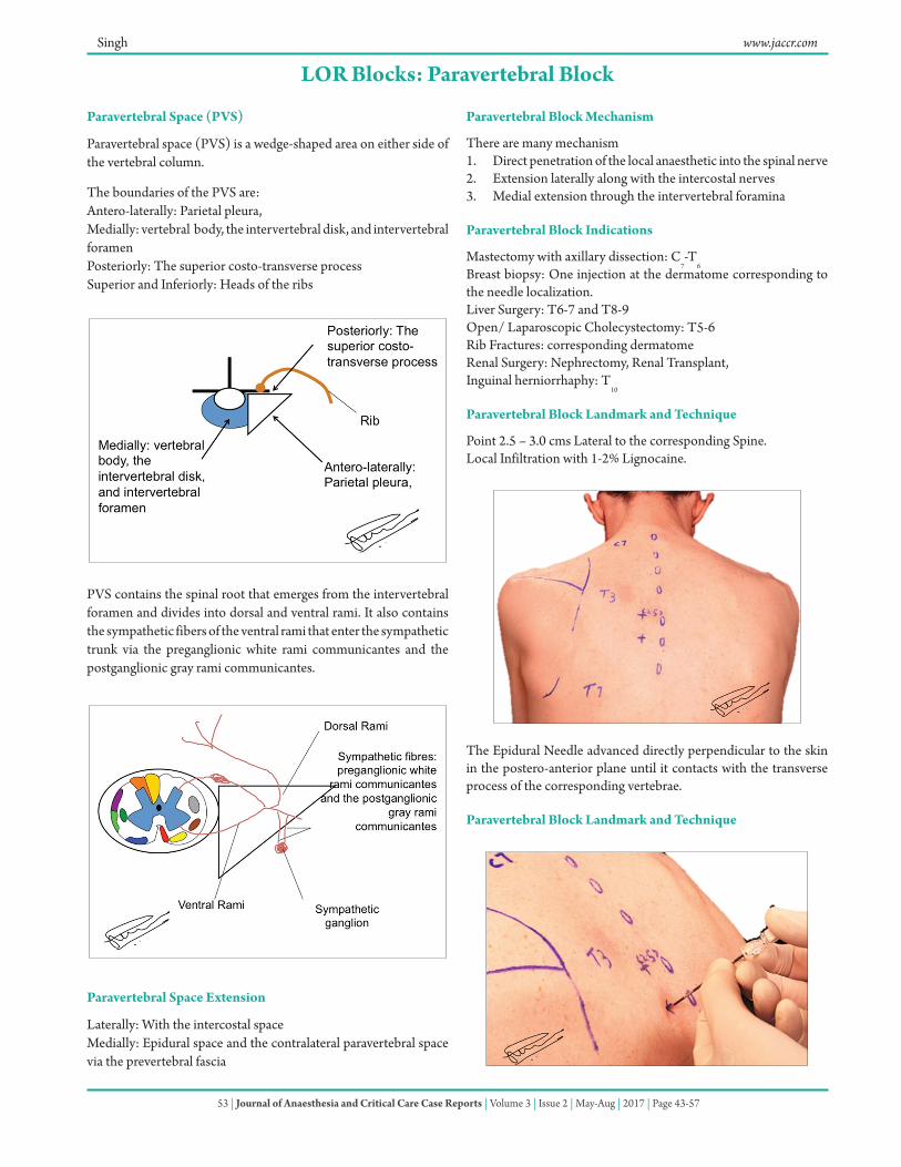
Paravertebral Space (PVS)
Paravertebral space (PVS) is a wedge-shaped area on either side of the vertebral column.
The boundaries of the PVS are:Antero-laterally: Parietal pleura, Medially: vertebral body, the intervertebral disk, and intervertebral foramenPosteriorly: The superior costo-transverse processSuperior and Inferiorly: Heads of the ribs
PVS contains the spinal root that emerges from the intervertebral foramen and divides into dorsal and ventral rami. It also contains the sympathetic fibers of the ventral rami that enter the sympathetic trunk via the preganglionic white rami communicantes and the postganglionic gray rami communicantes.
Paravertebral Space Extension
Laterally: With the intercostal space Medially: Epidural space and the contralateral paravertebral space via the prevertebral fascia
Paravertebral Block Mechanism
There are many mechanism 1. Direct penetration of the local anaesthetic into the spinal nerve2. Extension laterally along with the intercostal nerves 3. Medial extension through the intervertebral foramina
Paravertebral Block Indications
Mastectomy with axillary dissection: C7-T
6Breast biopsy: One injection at the dermatome corresponding to the needle localization.Liver Surgery: T6-7 and T8-9 Open/ Laparoscopic Cholecystectomy: T5-6Rib Fractures: corresponding dermatomeRenal Surgery: Nephrectomy, Renal Transplant, Inguinal herniorrhaphy: T
10
Paravertebral Block Landmark and Technique
Point 2.5 – 3.0 cms Lateral to the corresponding Spine.Local Infiltration with 1-2% Lignocaine.
The Epidural Needle advanced directly perpendicular to the skin in the postero-anterior plane until it contacts with the transverse process of the corresponding vertebrae.
Paravertebral Block Landmark and Technique
LOR Blocks: Paravertebral Block
Singh www.jaccr.com
54 | Journal of Anaesthesia and Critical Care Case Reports | Volume 3 | Issue 2 | May-Aug | 2017 | Page 43-57
Loss of Resistance Syringe is then attached to the needle and it is “walked off ” infero-laterally while advancing it by around 10 – 15 mm. As the costotransverse ligament is pierced and a “Pop” is felt with Loss of Resistance to Air/ Saline.
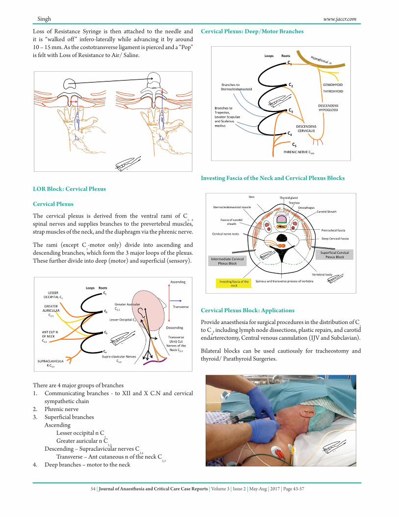
LOR Block: Cervical Plexus
Cervical Plexus
The cervical plexus is derived from the ventral rami of C1- 4
spinal nerves and supplies branches to the prevertebral muscles, strap muscles of the neck, and the diaphragm via the phrenic nerve.
The rami (except C1-motor only) divide into ascending and
descending branches, which form the 3 major loops of the plexus. These further divide into deep (motor) and superficial (sensory).
There are 4 major groups of branches1. Communicating branches - to XII and X C.N and cervical
sympathetic chain2. Phrenic nerve3. Superficial branches Ascending
Lesser occipital n C2
Greater auricular n C2,3
Descending – Supraclavicular nerves C3,4
Transverse – Ant cutaneous n of the neck C2,3
4. Deep branches – motor to the neck
Cervical Plexus: Deep/Motor Branches
Investing Fascia of the Neck and Cervical Plexus Blocks
Cervical Plexus Block: Applications
Provide anaesthesia for surgical procedures in the distribution of C2
to C4, including lymph node dissections, plastic repairs, and carotid
endarterectomy, Central venous cannulation (IJV and Subclavian).
Bilateral blocks can be used cautiously for tracheostomy and thyroid/ Parathyroid Surgeries.

Singh www.jaccr.com
55 | Journal of Anaesthesia and Critical Care Case Reports | Volume 3 | Issue 2 | May-Aug | 2017 | Page 43-57
Cervical Plexus Block: Superficial Branches
The superficial cervical plexus is blocked at the midpoint of the posterior border of the sternocleidomastoid muscle.
A skin wheal is made at this point, and a 22-gauge, 4-cm needle is advanced, injecting 5 mL of solution along the posterior border and medial surface of the sternocleidomastoid muscle.
It is possible to block the accessory nerve with this injection, resulting in temporary ipsilateral trapezius muscle paralysis.
Intermediate Cervical Plexus Block Using LOR Technique
The cervical plexus both the superficial and deep (muscular) branches can be easily blocked using this simple LOR technique.The landmark for this block is same as that for superficial plexus block i.e. the midpoint of the posterior border of the sternocleidomastoid muscle.
A skin wheal is made at this point, and a 22-gauge, 4-cm needle is advanced at the midpoint of SCM keeping the needle almost
perpendicular to the skin. Once the needle tip is through the skin a resistance is felt over the investing fascia of the neck, feel for a bounce and then a “pop” as the needle tip pierces the fascia. 10 -12 mls of 0.25 -0.5% of (Levo) bupivIcaine is injected incrementally.

Singh www.jaccr.com
56 | Journal of Anaesthesia and Critical Care Case Reports | Volume 3 | Issue 2 | May-Aug | 2017 | Page 43-57
LOR Blocks for Surgeries
Incision: Pfannenstiel
Dermatomes and myotomes involved: L1 and L
2
Preferred Block: Ilioinguinal and Iliohypogastric block in the TA Plane.
LA: These are volume blocks and each side will require minimum of 0.3 – 0.4 ml/kg of LA to soak the nerves. Ropivicaine or (Levo) Bupivicaine within its safe toxic limits can be used for these blocks.
Inguinal Hernia Repair, Orchidopexy
Incision: Parallel and above the Inguinal Ligament.
Dermatomes and myotomes involved: L1 and L
2
Preferred Block: Ilioinguinal and Iliohypogastric block in the TA
Plane. The Genital Branch of the Genitofemoral Nerve also needs to be blocked for complete analgesia.
Laparotomy
Incision: Midline
Dermatomes and myotomes involved: T6 – L
1/L
2
Preferred Block: Bilateral Rectus Sheath and TAP blocks
Upper Abdominal Laparotomy (Du Perforation)
Incision: Vertical Upper Abdominal (Para) median Incision
Dermatomes and myotomes involved: Terminal Branches of T6
– T10
.
Preferred Block: Rectus Sheath Block
Laparotomy
Incision: Paramedian Incision

Singh www.jaccr.com
57 | Journal of Anaesthesia and Critical Care Case Reports | Volume 3 | Issue 2 | May-Aug | 2017 | Page 43-57
Dermatomes and myotomes involved: T6 – L
1/L
2
Preferred Block: Unilateral Rectus Sheath and TAP block
Biliary (Open Cholecystectomy) or Hepatic Procedures
Incision: Kocher, Left Subcostal Incision
Dermatomes and myotomes involved: T6 – T
8/T
10
Preferred Block: Combined Right sided TAP and Rectus Sheath Block. It is preferable to give Bilateral Rectus Sheath Block as the incision may extend on to the left side. Paravertebral Block at multiple level can also be done and provide better quality analgesia.
Appendicectomy
Incision: MCBurney and its Modifications
Dermatomes and myotomes involved: T10
– L1/L
2
Preferred Block: Right sided TAP block
Laparoscopy Cholecystectomy
Incision: Paraumblical camera port and Ports at the epigastric and right flanks
Dermatomes and myotomes involved: T6-10
Preferred Block: Bilateral Rectus Sheath and Righted Sided TAP Block
Laparoscopic Hernia Repairs
Incision: Paraumblical Camera Ports and Ports on either flanks
Dermatomes and myotomes involved: T10
– L1/L
2
Preferred Block: Bilateral Paraumblical Rectus Sheath and TAP Blocks
Pancreatic and Major Gastric Surgery
Incision: Roof Top/ Mercedes Benz Incision
Dermatomes and myotomes involved: T6 – T
10
Preferred Block: Bilateral Rectus Sheath and Subcostal TAP Blocks