
Shigella ProU in osmotic tolerance and virulence · Shigella ProU in osmotic tolerance and...
35
Shigella ProU in osmotic tolerance and virulence 1 1 The Shigella ProU System is Required for Osmotic Tolerance and Virulence 2 3 Running title: Shigella ProU in osmotic tolerance and virulence 4 5 Rasha Y. Mahmoud 1, 2 , Wenqin Li 1 , Ramadan A. Eldomany 3 , Mohamed Emara 2 , Jun Yu 1 # 6 7 1 Strathclyde Institute of Pharmacy and Biomedical Sciences (SIPBS), University of Strathclyde, 8 Glasgow, UK 9 2 Department of Microbiology and Immunology, Faculty of Pharmacy, Helwan University, Cairo, 10 Egypt 11 3 Department of Microbiology and Immunology, Faculty of Pharmacy, Kafr Elsheikh University, 12 Kafr Elsheikh, Egypt 13 14 #Address correspondence to 15 Jun Yu ([email protected]) 16 Tel: 0044 141 548 2137 17 18 19 20 21 22 23
Transcript of Shigella ProU in osmotic tolerance and virulence · Shigella ProU in osmotic tolerance and...

Shigella ProU in osmotic tolerance and virulence
1
1
The Shigella ProU System is Required for Osmotic Tolerance and Virulence 2
3
Running title: Shigella ProU in osmotic tolerance and virulence 4
5
Rasha Y. Mahmoud1, 2, Wenqin Li1, Ramadan A. Eldomany3, Mohamed Emara2, Jun Yu1 # 6
7
1Strathclyde Institute of Pharmacy and Biomedical Sciences (SIPBS), University of Strathclyde, 8
Glasgow, UK 9
2Department of Microbiology and Immunology, Faculty of Pharmacy, Helwan University, Cairo, 10
Egypt 11
3Department of Microbiology and Immunology, Faculty of Pharmacy, Kafr Elsheikh University, 12
Kafr Elsheikh, Egypt 13
14
#Address correspondence to 15
Jun Yu ([email protected]) 16
Tel: 0044 141 548 2137 17
18
19
20
21
22
23

Shigella ProU in osmotic tolerance and virulence
2
Abstract 24
To cope with hyperosmotic stress encountered in the environments and in the host, the 25
pathogenic microbes use diverse transport systems to obtain osmoprotectants. To study the 26
role of Shigella sonnei ProU system in response to hyperosmotic stress and virulence, we 27
constructed deletion and complementation strains of proV and used an RNAi approach to 28
silence the whole ProU operon. We compared the response between wild type and the 29
mutants to the hyperosmotic pressure in vitro, and assessed virulence properties of the 30
mutants using gentamicin protection assay as well as a Galleria mellonella moth larvae model. 31
In response to osmotic stress by either NaCl or KCl, S. sonnei highly up-regulates transcription 32
of pro genes. Supplementation of betaine greatly elevates the growth of the wild type S. sonnei 33
but not the proV mutants in M9 medium containing 0.2 M NaCl or 0.2 M KCl. The proV mutants 34
are also defective in intracellular growth compared with the wild type. The moth larvae model 35
of G. mellonella shows that either deletion of proV gene or knockdown of pro genes transcripts 36
by RNAi significantly attenuates virulence. ProU system in S. sonnei is required to cope with 37
osmotic stress for survival and multiplication in vitro and ex vivo, and for infection. 38
Keywords: Shigella sonnei, ProU, RNAi, osmotic tolerance, osmoprotectants 39
40

Shigella ProU in osmotic tolerance and virulence
3
Introduction 41
Shigella is a facultative intracellular Gram-negative pathogen, known as the etiologic 42
agent of bacillary dysentery since the 1890s. Although Shigella was defined a genus with four 43
species S. dysenteriae, S. flexneri, S. boydii and S. sonnei in the 1950s 1, it has become clear that 44
they are pathogenic lineages of Escherichia coli of multiple origins 2. The primary transmission 45
is the fecal-oral route, so it is life threatening in developing countries because of poor 46
sanitation. Shigella strains are among the most prevalent causative agents of moderate-to-47
severe diarrhea, and especially affect children under 5 year old in developing countries 3. The 48
widespread of multiple antibiotic resistant strains, has made Shigella treatment increasingly 49
difficult and there is urgent need for vaccine development 4. Shigella is highly invasive to the 50
colon and the rectum and it has the ability to proliferate in the cell cytoplasm and trigger the 51
host pro-inflammatory response. It causes variable clinical manifestations ranging from short 52
term illness, typically watery diarrhea, to a long lasting one manifesting with fever, bloody 53
diarrhea with intestinal cramps and mucopurulent feces 5. 54
Keeping a stable osmotic balance between the cell cytoplasm and the outer environment 55
is an important challenge to all cell types, especially the unicellular organisms. For bacteria, the 56
high surface area to total volume ratios makes them vulnerable when they encounter osmotic 57
stress; bacteria could tolerate the osmolarity changes in the environment through either 58
solutes efflux or water movement across the cytoplasmic membrane 6. The external osmolarity 59
changes are translated by the microorganisms to an adaptation process to protect themselves 60
against turgor; this happens by a rapid K+ ion influx through specific transporters, and at the 61
same time, microorganisms produce counter ions like glutamate 7. However, high intracellular 62
concentration of K+ and glutamate only support microbial adaptation to moderately high 63
osmolarity. At very high osmolarity, further accumulation of K+ and glutamate becomes 64

Shigella ProU in osmotic tolerance and virulence
4
impossible for growth, and therefore, bacteria exploit less deleterious compounds called 65
osmoprotectants, like polyoles (trehalose), amino acids (proline), and methyl-amines (glycine 66
betaine) 7. Osmoprotectants can accumulate intracellularly via cellular uptake or synthesis 67
from their precursors. Glycine betaine is one of the most important osmoprotectants for 68
bacteria 7. 69
For pathogenic bacteria, osmoregulation is a very important factor for establishing 70
infection. For example, Staphylococcus aureus has a PutP proline transport system that helps in 71
host tissue colonization 8-10. Other studies have found a link between osmotic stress and the 72
expression of virulence genes in Pseudomonas aeruginosa 11, 12 and between the transport of 73
compatible solutes and colonization in the pathogen in Listeria monocytogenes 13, 14. In 74
Salmonella enteric serovar typhimurium and E. coli K-12, osmoprotectants are mainly 75
accumulated through the ProP and ProU transport systems 15-19. 76
ProP is a member of the major facilitator superfamily of permeases, and it is known as a 77
symporter 20. ProP is of low affinity for proline and glycine betaine, it has Km of ≈ 0.1 mM for 78
both 20. ProU system efficiently scavenges glycine betaine 21, Km of ≈ 1 µM 16, as well as proline 79
betaine for bacteria to cope extreme osmotic stress 22, 23. It is composed of three proteins, i.e., 80
ProV, ProW, and ProX, which are encoded by an operon proVWX 18, of which ProV belongs to 81
the ATP-binding cassette (ABC) superfamily 24, 25. ProX is a periplasmic soluble substrate-82
binding protein 19, 26, 27 which could bind and deliver glycine betaine to the inner membrane 83
protein ProW, whereas ProV hydrolyses ATP providing energy for transporting substrates 84
against the concentration gradient 24, 25. 85
In 2005, Lucchini and his colleagues 28, analyzed genomic expression of S. flexneri during 86
infection by DNA microarray; pro genes were found highly up-regulated in both epithelial HeLa 87
and macrophage-like U937 cells; in particular, proV was up-regulated by 57-fold in U937 cells 88

Shigella ProU in osmotic tolerance and virulence
5
28. Besides, it was found that the level of ProU transcription in E. coli is induced upon exposure 89
to hyperosmotic stress 29. These findings suggested that Shigella faced extreme osmotic stress 90
in the host cell cytosol and up-regulation of ProU was necessary for Shigella to cope with this 91
hostile cellular niche, allowing Shigella intracellular survival and growth to establish infection. 92
Additionally, the orthologous ProXVWZ system in Mycobacterium tuberculosis has been shown 93
to actively transport glycine betaine into macrophages, which contributed to early steps in 94
colonization of the cellular niche 30. Hence, we have investigated the impact of the ProU 95
system on osmotic stress response and pathogenesis of S. sonnei. Our data have shown that 96
the ProU transport system is important for S. sonnei to cope with hyperosmotic stress in the 97
host cell cytoplasm, and for rapid intracellular proliferation to establish infection. 98
Results 99
proV deletion and complementation 100
In order to study the function of ProU we decided to construct a proV deletion mutant 101
since proV is the most highly up-regulated gene inside host cells and the first gene in the 102
proVWX operon 28. ProV deletion was constructed in wild type S. sonnei strain (20071599) 31 103
using the phage λ Red recombination system 32, which involved two steps. In the first step, a 104
Kanamycin cassette replaced the wild type proV gene via homologous recombination. In the 105
second step, the Kanamycin gene was looped out using pCP20 plasmid, leaving a scar of 102 bp 106
(Fig. S1A). For complementation of the resultant ΔproV mutant strain, the entire proV coding 107
sequence was amplified with primers (c & d Table S1), and cloned into pGEM-T-Easy (ampR), 108
with the 5’-end of the proV coding sequence facing the lacZ promoter. The resultant clone was 109
transformed into the ΔproV mutant (Fig.S1B). All proV constructs for mutagenesis as well as 110
complementation were confirmed by PCR and agarose gel electrophoresis. All PCR products 111
were further confirmed by DNA sequencing using primers c & d (Table S1). 112

Shigella ProU in osmotic tolerance and virulence
6
ProU is required to cope with hyperosmotic stress in vitro 113
Because the ProU system is known to be required for E. coli to cope with extreme 114
osmotic stress 22, 23, we tested whether this was also the case for S. sonnei in vitro. We first 115
measured growth of the wild type, ΔproV and the complemented ΔproV/pProV strains in M9 116
medium supplemented with 0.3 M NaCl or 0.3 M KCl by measuring the optimal density 117
(OD600nm) each one hour till the late stationary phase (10 hours). While the wild type S. sonnei 118
grew well, the ΔproV strain struggled to grow in the presence of 0.3 M of NaCl or KCl (Fig. 1A 119
and 1B). Obviously, all the tested strains were able to grow properly in M9 medium without 120
any supplements (Fig. S2). Overexpression of ProV in trans not only reversed the growth defect 121
of the ΔproV strain, but also made the strain grow faster than the wild type. This suggested 122
that deletion of proV is solely responsible for the slow growth phenotype of the mutant and 123
also suggested that excess ProV made the system more effective and that ProU may also 124
transport osmoprotectants other than betaine, such as glutamine or K+, available in the 125
medium to facilitate bacterial growth (Fig. 1A and 1B). We further tested the growth of wild 126
type and the ΔproV strains in M9 medium supplemented with 0.2 M NaCl or 0.2 M KCl. Under 127
these milder conditions, the ΔproV strain grew equally well as the wild type strain up to 6 128
hours. However, the growth of the ΔproV strain ceased at 6 hours and remained flat to 10 129
hours. In contrast, the wild type grew steadily, reaching OD600nm = 0.7 and 0.8 in M9 130
supplemented with 0.2 M NaCl and 0.2 M KCl, respectively (Fig. 1C and 1D). Thus, in the 131
presence of 0.2 M NaCl, growth of ΔproV was more than 1.5-fold reduced (OD600nm 0.4 vs. 0.7) 132
compared to the wild type at the stationary phase 10 hours (Fig. 1C). In the presence of 0.2 M 133
KCl, there was a 2-fold reduction of ΔproV growth (OD600nm 0.4 vs. 0.8) compared to the wild 134
type at the stationary phase (Fig. 1D). 135
Shigella ProU in osmotic tolerance and virulence
7
Betaine is a well-known compatible solute that is a preferable osmoprotectant for many 136
organisms 33. We therefore tested the ability of both wild type and the ΔproV mutant in 137
utilizing betaine under hyperosmotic conditions. In M9 medium containing 0.2 M NaCl or 0.2 M 138
KCl, supplementation of betaine (500 µM) significantly elevated the growth of the wild type 139
(Fig. 1C & 1D, S3). In contrast, the growth suppression of the ΔproV mutant persisted in the 140
presence of 500 µM betaine (Fig. 1C, 1D, S3). Taken together, these data demonstrated that a 141
functional ProU system is required to transport betaine for S. sonnei, and that betaine can 142
correct the growth defect of the wild type induced by high concentrations of NaCl and KCl. In 143
contrast, the growth of the ΔproV mutant remained severely impaired under high osmotic 144
stress, and could not be corrected by addition of betaine in the medium (Fig. 1C, 1D). 145
proV paralogues are able to compensate the loss of proV 146
As shown in Fig. 1C and 1D, the ΔproV strain grew equally well as the wild type strain in the 147
first 6 hours albeit its growth was ceased thereafter. This result suggested that the ProU 148
system was functional at least in the first 6 hours when proV is removed. Given the fact that 149
proV encodes an ATPase and all ATPase of the ABC superfamily share highly conserved 150
sequence and structure (39), we reasoned that ATPase from other transporting systems may 151
compensate for the loss of proV. Using ProV as query sequence we identified putative 25 152
ATPases (Table S3), belonging to other transport systems in the S. sonnei SSO46 genome 153
(http://www.mgc.ac.cn/ShiBASE/Search.htm). We cloned three of them in random, oppF, glnQ 154
and malK, which are responsible for transporting oligopeptides, glutamate, and maltose, 155
respectively. Over-expressing each of them was able to complement the ΔproV strain for 156
better growth in LB broth supplemented with 0.2 M NaCl although OppF appeared less 157
effective than GlnQ and MalK (Fig. S5). These data support our hypothesis that removal of proV 158
only rendered ProU system partially inactive, and prompted us to construct mutants with a 159
Shigella ProU in osmotic tolerance and virulence
8
deletion of the whole proVWX operon or in proX or proW. However, despite repeated efforts 160
we were unable to obtain a ΔproVWX, or ΔproX or ΔproW strain with intact virulence plasmid, 161
which would allow formation of small and smooth red colonies on Congo red agar. All strains 162
harboring these deletions formed large pale and rough colonies (data not shown). This 163
suggested that ProU is rather important to maintain the genome stability due to its role in 164
transporting important osmoprotectants or other yet unraveled functions. S. sonnei must 165
rearrange its genome when completely loosing ProU. Hence, we searched for an alternative 166
approach for knockdown of the ProU system. 167
Silencing the proVWX operon using RNAi 168
Tchurikov and his colleagues 34 described an RNAi methodology to knockdown gene 169
expression in E. coli. According to their work 34 Mirlon (which has the inverted sequence of the 170
gene of interest), is the most potent in silencing the target genes compared to two other 171
constructs (Paralon and Antilon). We adopted described Mirlon approach to knockdown ProU 172
system in S. sonnei. We created a piece of dsDNA, by annealing two oligonucleotides of 87 173
bases each, which spand 45 and 42 base pairs of the promoter and 5’-end of proV coding 174
sequence, respectively (Table S1). This piece of dsDNA was cloned into pGEM-T-Easy (ampR), 175
with the 3’-end of the proV coding sequence facing the lacZ promoter, which would drive the 176
transcription of RNA molecule with inverted sequences to the proV mRNA, hence termed 177
MirproV RNA 34. We tested the impact of MirproV on the expression of proV and proX, which 178
are the first and last genes, respectively, in the ProU operon, by qRT-PCR using primers (k & l; 179
m & n respectively, Table S1). The house-keeping gene, cysG, was used as an internal control 180
using primers (o & p, Table S1) 35. Figure 2A shows the results of qRT-PCR after normalisation 181
with transcripts of cysG and using transcripts from wild type strain as calibrator. The transcripts 182

Shigella ProU in osmotic tolerance and virulence
9
of both proV and proX were significantly reduced as a result of MirproV RNA expression, which 183
indicated that the RNAi construct in MirproV was successful in ProU attenuation. 184
Similar to the proV mutant, the MirproV strain grew well as the wild type in M9 without 185
supplements (Fig. S2) but became intolerant to hyperosmolarity; its growth was severely 186
compromised in M9 medium containing either 0.3 M NaCl or 0.3 M KCl (Fig. 1A, 1B). In M9 187
supplemented with 0.2 M NaCl, the MirproV strain had a long lag phase of 4 hours (Fig. 1E). 188
This was in contrast with the ΔproV strain, which grew equally well as the wild type strain 189
during this phase of growth under this condition (Fig. 1C). Thus, MirproV was more effective in 190
inactivating the ProU system. Moreover, addition of betaine to M9 medium containing 0.2 M 191
NaCl or 0.2 M KCl failed to rescue growth of the MirproV strain (Fig. 1E, 1F, S3). Thus, 192
expression of MirproV is effective in blocking ProU function and betaine transport. 193
By linear regression analysis, the significance of inhibition of wild type, proV and MirproV 194
mutants growth by 0.2 M NaCl and 0.2 M KCl supplements was identified (Fig. S3). 195
Furthermore, paired t-test was performed to compare optical density (OD600nm) at the 196
stationary for wild type, ΔproV and MirproV in presence and absence of betaine (Fig. S4). The 197
results showed that betaine supplementation significantly increased the optical density for 198
wild type but not for either mutant strains (Fig. S4). 199
To gain insight into the transcriptional regulation of ProU and its inactivation by MirproV, 200
we isolated total RNA from wild type and the MirproV strain from M9 cultures containing 201
either 0.3M NaCl or 0.3M KCl and performed qRT-PCR for transcripts of proV and proX. Again 202
the house-keeping gene cysG was used as an internal control for normalisation. As expected, 203
the wild type responded to the osmotic stress of NaCl or KCl by massive upregulation of proV 204
and proX transcripts. The MirproV strain also responded, but at a significantly lower level (Fig. 205
2B). These data demonstrated that the ProU system was inducible under high osmotic stress 206

Shigella ProU in osmotic tolerance and virulence
10
and that MirproV RNA effectively knocked down ProU transcripts which in turn rendered S. 207
sonnei intolerant to hyperosmolarity. 208
Inactivation of ProU does not disrupt the type three secretion system (TTSS) 209
Shigella strains possess a type III secretion system (TTSS), which is essential for cell-210
invasion, phagosome escape and intracellular replication 36. Therefore, we ought to separate 211
the roles of TTSS and ProU with regard to cell invasion and intracellular growth. We first 212
wanted to confirm that the mutant strains that we were going to test for virulence maintained 213
an intact TTSS. The presence of TTSS genes in the ΔproV strain was analyzed by PCR using 214
primers e & f to amplify ipaB and primers i & j to amplify mxiD (Table S1); both ipaB and mxiD 215
were found intact in wild type and the mutant strain (Fig. S6). Further, the production and 216
secretion of IpaB and IpaC proteins were investigated by Western blotting using Congo red as 217
an environmental cue in vitro as described previously 37. Because it is known that MxiD is 218
required for type III secretion, we constructed a ΔmxiD mutant as a negative control for Ipa 219
secretion, using the same approach as for ΔproV construction with primers (g & h; Table S1). 220
Similar levels of IpaB and IpaC were detected in the cell lysates and supernatants of the wild 221
type, the ΔproV, the MirproV and the complemented ΔproV/pProV strains whereas Ipa 222
proteins were detected only in cell lysate but not supernatant of the ΔmxiD mutant (Fig. 3). We 223
therefore conclude that removal of proV or knockdown of transcripts did not impair IpaB and 224
IpaC production or secretion – TTSS is functional in the ΔproV and the MirproV strains. 225
ProU is required for S. sonnei intracellular growth (ex vivo) 226
Evidence via DNA microarray analysis indicated that the host cell cytoplasm is a hostile 227
environment which exposes bacteria to hyperosmotic stress 28. We therefore hypothesized that 228
removal of proV or reduction of proVWX transcription by RNAi would adversely influence S. 229
sonnei intracellular growth. To test our hypothesis, the HEK293 cell line, a good model for 230
Shigella ProU in osmotic tolerance and virulence
11
testing Shigella invasion 38, was used as a host to perform gentamicin protection assay to test 231
intracellular growth of the wild type, the ΔproV and the MirproV strains. As anticipated, we 232
observed a significant drop in intracellular bacterial burden with the ∆proV mutant compared 233
to the wild type 2 hours post infection (Fig. 4A). Noticeably, the MirproV strain had a 234
significantly reduced intracellular bacterial burden, compared to the ΔproV mutant. This 235
suggested that suppression of proVWX operon was more effective than mutating proV gene 236
alone because ProV paralogues may compensate the loss of ProV (Fig. S5). As anticipated, 237
expressing ProV in trans restored intracellular growth of the ΔproV mutant, demonstrating that 238
the deletion of proV had no polar effect on the expression of proW and proX downstream; 239
removal of proV was solely responsible for the defective intracellular growth observed with the 240
ΔproV mutant. 241
To further test if the ProU system is required for intracellular growth not invasion per se, 242
we performed a time course analysis of intracellular growth after 1, 2, 3, and 4 hours using 243
gentamicin protection assay. At each time point, cells were lysed with Triton X-100, and cell-244
lysates were plated out on LB agar, and incubated at 37 oC overnight and enumerated by 245
colony count. The wild type grew rapidly with a doubling time of approximately 38 min from 1 246
hour onwards whereas the ∆proV and the MirproV strains had a doubling time of 60 and 90 247
min, respectively (Fig. 4B, Fig S7). 248
To gather further evidence for ProU requirement for intracellular growth we exploited 249
flow cytometry. We transformed all strains with a plasmid that expressed EGFP, and infected 250
HEK293 cells with these green bacteria. As shown in Figure 4C, 38% of the host cells infected 251
with wild type strain emitted green fluorescence 4 hours post infection, indicating that green 252
bacteria were metabolically active inside host cells at this time point. Noticeably, some cells 253
had very strong EGFP signals (fluorescence intensity above 102 units), demonstrating that these 254

Shigella ProU in osmotic tolerance and virulence
12
cells harbored large numbers of green bacteria as a result of rapid bacterial intracellular 255
growth. In contrast to cells infected with wild type, there were less than 4% of cells infected 256
with the ∆proV mutant emitted green fluorescence (Fig. 4C), indicating impaired intracellular 257
growth. We left cell infection overnight and repeated flow cytometry and this showed that 258
43% of cells infected with wild type emitted EGFP signals, (Fig. 4D vs 4C). It was noticeable that 259
a smaller percentage of cells could emit high fluorescent above 102 scale compared to that of 4 260
hours post infection (Fig. 4D vs. 4C). Presumably, host cells harboring large numbers of green 261
bacteria were dead following the overnight infection. Consistent with this was the notion that 262
more cells were emitting low fluorescence (intensity of 101 to 102 units), suggesting that cells 263
harboring lower bacteria burden survived overnight infection. Noticeably also, there was an 264
increase in green fluorescence in host cells that have been infected with the ∆proV mutant 265
overnight, suggesting limited remaining intracellular growth of this strain (Fig. 4D). Taken 266
together, ProU is required for intracellular growth of S. sonnei in host cells. 267
ProU is requried for virulence in the Galleria mellonella larvae model (in vivo) 268
The great moth G. mellonella larvae have become a popular in vivo model for assessing 269
bacterial virulence 39, 40. We recently adopted this model to assess Shigella virulence and found 270
it comparable with the widely accepted Sereny test 41. 271
Here we exploited this model to compare the virulence of ΔproV and MirproV strains 272
with the wild type S. sonnei. Larvae were challenged with 105 CFU of each strain; 10 larvae per 273
group. Mock-infection was done using the same volume of saline. All groups of larvae were 274
observed daily for five days and dead larvae suffered from melanization and loss of motility. As 275
shown in Figure 5A, wild type and the complemented strain (ΔproV/pProV) were able to kill 276
90% and 80% of larvae, respectively, in 1 day and the remaining larvae died at the third day. 277
Using the same dose of the ΔproV strain, only 40% of larvae died at the first day, and 30% died 278

Shigella ProU in osmotic tolerance and virulence
13
at the second day while the rest died at the third day. With the MirproV strain, a dose of 105 279
CFU only caused 20% of death at the first day, 10% at the second day and other 10% at the 280
fourth day, while 60% of the whole population survived to the end of the study (Fig. 5A). These 281
data were consistent with those of Figure 4 that MirproV was more effective than proV 282
deletion in inactivating the ProU system. 283
Our recent study has demonstrated that infection of larvae hemocytes is an important 284
mechanism for larvae-killing by S. sonnei 41. Therefore, we infected moth larvae with EGFP-285
expressing strains (Table S2) and isolated hemocytes from infected and mock-infected larvae 286
for flow cytometry. Larvae were infected with 106 CFU of each strain, and mock-infection was 287
done with saline. Hemocytes were isolated from larvae 4 hours post infection. We used 288
hemocytes from mock-infected larvae to set gates defining EGFP negative and positive 289
populations (Fig. 5B). It was apparent that wild type strain colonized more hemocytes giving 290
rise to two populations of hemocytes with low and high EGFP intensity, whereas the ΔproV 291
strain colonized less numbers of hemocytes with one population of high-EGFP expressing 292
hemocytes at this time point. 293
Discussion 294
In pathogenic bacteria, osmoregulation is a very important mechanism not only for 295
survival during environmental osmotic stress, but also in establishment of infection. Examples 296
of osmoregulatory systems important for virulence include the Staphylococcus aureus PutP 8-10, 297
the Pseudomonas aeruginosa PlcH 11, 12, and Listeria monocytogenes OpuC 13, 14. 298
The ProU transport system is widely distributed in bacteria. Previous studies have firmly 299
established its role in environmental survival under hyperosmolarity in E. coli, but whether it is 300
required in the context of infection remained unknown 7. By studying the orthologous 301
ProXVWZ system in M. tuberculosis, Price and his colleagues have demonstrated the 302

Shigella ProU in osmotic tolerance and virulence
14
importance of this system in osmotolerance in vitro and during early host cell colonization 30. 303
Here, we have presented compelling evidence for the first time that the Shigella ProU system 304
is required for coping with hyperosmolarity both in vitro (Fig. 1) and ex vivo (Fig. 4), and for 305
virulence in a moth larvae infection model (Fig. 5). We have also demonstrated that ProU is 306
highly up-regulated under hyperosmotic conditions (Fig. 2). This is consistent with the findings 307
for S. flexneri by Lucchini and co-workers 28; proV is up-regulated up to 57-fold inside the host 308
cell cytoplasm compared to growth in LB broth. Lucchini suggested that the overall different 309
ionic composition in the host cell cytosol is the cue that triggers this up-regulation, because the 310
host cell cytosol should have similar osmolarity as LB broth 28. Supporting evidence for this 311
hypothesis is the notion that transcription of the phoRB regulon and mgtA is also up-regulated 312
inside host cells compared to their transcription in LB broth. phoRB is required for transporting 313
phosphates and mgtA is required for transporting Mg2+; their up-regulation indicates shortages 314
of phosphates and Mg2+ in the host cell cytosol. M. tuberculosis colonizes and modifies 315
phagosome by inhibiting acidification 42. The exact cue for up-regulation of the M. tuberculosis 316
ProXVWZ system has not been identified so far 30. Nevertheless, it is clear that both the 317
phagosome and the host cell cytosol impose osmotic stress on invading microbes (30 and this 318
study). In both these cellular niches, the microbes use the ProU system to transport glycine 319
betaine to survive and multiply. Glycine betaine is an important free cytoplasmic constituent of 320
eukaryotic cells 43, and it is present at 20 to 60 μM in human serum 44. Hence, it is not 321
surprising that pathogens like M. tuberculosis 45 and S. sonnei exploit glycine betaine as an 322
osmoprotectant during growth within the host cells. 323
ProV encodes an ATPase of the ProU system. Deletion of proV caused significantly slowed 324
growth in hyperosmotic media, and complementation by expressing ProV in trans restored 325
wild type rate of growth (Fig. 1A, 1B). These data reinforce that ProU is an energy dependent 326
Shigella ProU in osmotic tolerance and virulence
15
transport system, and ∆proV is solely responsible for the slow growth phenotype of the 327
deletion mutant. However, the ∆proV mutant grew well in early phase in the presence of 0.2 M 328
NaCl and 0.2 M KCl (Fig. 1C, 1D). This result suggested that the ProU system is, at least in part, 329
still functional when proV is removed. By overexpression of three paralogues of proV in trans, 330
we have demonstrated that ATPases from other transport systems are able to compensate for 331
the loss of ProV for better growth (Fig. S5). 332
The MirproV strain does not significantly respond to betaine supplementation in M9 333
medium with 0.2 M NaCl or 0.2 M KCl for better growth (Fig. 1E, 1F). These data suggest that 334
MirproV could effectively attenuate ProU system. Even though the MirproV strain produced 335
more transcripts of proV and proX in response to 0.3 M NaCl and 0.3 M KCl (Fig. 2B) compared 336
to its transcripts in M9 medium without any supplements (Fig. 2A), these levels of responses 337
were not sufficient to reverse the growth defect (Fig. 1E, 1F; S3, S4). The effectiveness of 338
MirproV was also demonstrated in gentamicin protection assay; the MirproV strain was more 339
severely defective than the ∆proV mutant in the intracellular growth (Fig. 4A, 4B). Moreover, 340
the possibility of ‘off-target’ effects by MirproV is low; there are only 4 low score hits when the 341
42 bp proV sequence is used as query to blast the S. sonnei SSO46 genome (Table S4). 342
Noticeably, mxiJ is one of the hits; mxiJ encodes an essential component of TTSS 46However, 343
like the ∆proV strain, the MirproV strain produces and secretes Ipa proteins (Fig. 3), suggesting 344
that any ‘off-target’ hits, if they occur, do not significantly change the strain’s osmotolerance 345
or virulence. 346
Our inability to obtain strains with intact virulence plasmid with deletion of the whole 347
proVWX operon, or proX or proW strongly suggests the ProU system plays an important role in 348
Shigella genome stability. Previous studies have shown that S. sonnei could frequently become 349
avirulent by losing its 120-megadalton virulence plasmid 47. Besides, it is well known that both 350
Shigella ProU in osmotic tolerance and virulence
16
ProU and the TTSS genes are controlled by the same negative regulator: the H-NS protein and 351
this regulatory link provided an additional rational for exploring the possibility of altered TTSS 352
expression upon ProU attenuation 48, 49. So, it was very important to test the stability of this 353
plasmid in wild type and the mutant strains under investigation (ΔproV, MirproV and the 354
complemented ΔproV/pProV). Our data show ΔproV strain possesses key plasmid borne 355
virulence genes, ipaB and mxiD. Furthermore, all strains produce and secrete IpaB and IpaC 356
proteins, except ∆mxiD strain that produces but does not secrete Ipa proteins (Fig. 3). Hence, 357
we can conclude that inactivation of the ProU system by either deletion of proV or RNAi 358
approach does not impair genetic stability and function of the virulence plasmid. 359
Finally, we have once again demonstrated the usefulness of the moth larvae model in 360
assessing bacterial virulence, which enabled us to establish a link between osmotolerance and 361
virulence. 362
Conclusions 363
Altogether, we conclude that the ProU system is important for S. sonnei to tolerate 364
hyperosmotic stress in vitro, as well as for survival and proliferation of the bacteria in the 365
stressful intracellular niche. Silencing of the whole proVWX operon is more effective than 366
deleting proV alone because proV paralogues may compensate for the loss of proV. Last but 367
not least, the G. mellonella larvae model is a cost-effective and good model for studying 368
Shigella virulence, and reflects results of more established models such as the Sereny test. 369
Materials and Methods 370
Bacterial strains and growth conditions 371
The wild type Shigella strain used in this study was S. sonnei strain 20071599 31 and 372
mutants thereof. Bacteria were routinely grown on Congo red TSA plates or in liquid LB at 37 373
oC. To obtain EGFP expressing bacteria, strains were transformed with pGEMT-Easy (AmpR) 374

Shigella ProU in osmotic tolerance and virulence
17
containing EGFP. Afterwards, 100 μg/ml ampicillin was added for selection of plasmid 375
containing strains. All primers and strains used in this study are listed in Tables S1 and S2 376
respectively. 377
Genetic engineering 378
Construction of the mutant bearing in-frame deletion in proV gene was done using phage 379
λ Red recombination system 32. Construction of the mutant bearing in-frame deletion in proV 380
gene was done using phage λ Red recombination system 50. Both IpaB and IpaC were detected 381
using the monoclonal antibodies H16 (anti-IpaB) and J22 (anti- IpaC), respectively 50, followed 382
by incubation with Alexa Fluor 680 goat anti-mouse IgG (H+L), and the images were visualized 383
using a UV scanner at 700 nm. 384
In vitro osmotolerance test 385
M9 medium was prepared, autoclaved and supplemented with filtered 200 µl/100ml of 1 386
M MgSO4, 4% glucose, 12.5 µg/ml of Nicotinic acid, 45 µg/ml of L-methionine, and 20 µg/ml of 387
L-Tryptophan. Routinely, all strains were grown on M9 agar overnight, smooth colonies were 388
picked and grown in M9 broth 2 hours before the start of the experiment. Then, 96-well plates 389
were set for growth curve; each well contained 200 μl M9 medium with or without 390
supplements: 0.2 and 0.3 M NaCl or 0.2 and 0.3 M KCl in presence and absence of 500 µM 391
betaine. Each culture condition was tested in triplicates for each bacterial strain, and three 392
independent experiments were carried out. Afterwards, 107 CFU from each of the strains were 393
added to the 96-well plates. Wells without bacterial inocula were used as a blank, while wells 394
without supplements were used to study the growth curve of Shigella strains in M9 medium. 395
The plates were incubated in a shaker incubator at 37 oC, and the OD600nm was recorded each 1 396
hour. 397
Quantitative real-time polymerase chain reaction (QRT-PCR) 398

Shigella ProU in osmotic tolerance and virulence
18
Wild type and MirproV strains were grown in M9 minimal medium to mid-log phase. 399
Total RNA was isolated using RNA isolation kit (Bioline). The house-keeping gene, cycG, was 400
used as an internal control 35. To establish standard curves for each gene primers k & l; m & n 401
and o & p (Table S1) were used to amplify proV, proX and cycG genes, respectively, and serial 402
dilutions (from 100 to 108 molecules/µl) of the genome DNA was used as templates using 403
SYBR-Green QRT-PCR kit on Rotor Gene 6000 (Qiagen). Triplicate RNA samples from triplicate 404
cultures (n = 3) were used to prepare cDNAs, which were quantified by the same PCR 405
procedure. The amplification curves of proV and proX were normalized with that of cycG, and 406
quantification was calculated using the standard curves. Changes in gene expression between 407
wild type (set as calibrator) and the MirproV strains were calculated using the 2-ΔΔct method 408
and proprietary software in the Rotor Gene instrument (version 1.7.34) 51. To analyze the 409
impact of high osmotic stress on ProU expression both wild type and MirproV strains were 410
grown in M9 supplemented with 0.3 M NaCl or 0.3 M KCl. The levels of transcripts from wild 411
type and MirproV grown in M9 without salt supplements were used as calibrators. The 412
transcripts of house-keeping gene, cycG, were again used for normalization to calculate the 2-413
ΔΔct 51. 414
Gentamicin protection assay 415
HEK293 (human embryonic kidney stem) cells were seeded and cultured until 416
approximately 80% confluence in 24-well plates and S. sonnei bacteria were added to the cell 417
monolayers at a multiplicity of infection (MOI) of 10. The plates were centrifuged at 700 xg for 418
10 min at 22 oC. The plates were incubated for 40 min at 37 oC under 5% CO2 atmosphere to 419
allow bacterial invasion into host cells. Thereafter, cell monolayers were washed twice with 420
PBS, DMEM containing gentamicin (50 µg/ml) was added, and the plates were incubated for 421
required time intervals before terminating infection. Cells were washed 3 times using PBS and 422

Shigella ProU in osmotic tolerance and virulence
19
lysed with Triton X-100 (0.1 % in H2O) for 10 min. Cell lysates were serially diluted, plated on LB 423
agar, incubated at 37 oC overnight and enumerated by colony count. 424
Infection of Galleria mellonella larvae 425
Smooth red colonies of S. sonnei strains were selected from Congo red TSA plates, grown 426
for 3 to 5 hours in LB broth to prepare bacterial suspensions. Each group of 10 healthy larvae 427
of approximately similar size were injected with 10 µl (105 CFU) of each bacterial suspension. 428
The mock-infection group received sterile PBS instead of bacteria. All larvae were incubated at 429
37 oC 52 and observed for 5 days post infection 41. The experiments have been repeated three 430
times for results confirmation and the averages have been used in Kaplan-Meier survival 431
curves. 432
Flow cytometry 433
For HEK293 cells, infection was terminated at appropriate time intervals (either 4 hours 434
or overnight); cells were washed with saline twice and then trypsinized for 1 min after which 435
the DMEM medium was added. Cells were spun down and cell pellets were resuspended in 436
saline containing 4% (v/v) paraformaldehyde for fixation. 437
For the wax moth larvae, 4 hours post-infection with 106 CFU bacteria, hemocytes from 438
10 larvae were collected by incision between two segments near larvae tail to avoid gut 439
interruption. Hemocytes were collected in 1 ml of sterile PBS and the processing was within 10 440
min to prevent clotting 52. Cells were centrifuged at 500 xg for 10 min at room temperature, 441
and were resuspended in 1 ml PBS containing 4% (v/v) paraformaldehyde for fixation. 442
Cells were vigorously mixed and then were used for the flow-cytometry. Lasers emitting 443
at 488 nm was set for the detecting signals in FITC channel, which overlap with EGFP 444
excitation. Data acquisition was performed using Kaluza™ software (Beckman Coulter, Inc). The 445
experiments have been repeated three times for results confirmation. 446

Shigella ProU in osmotic tolerance and virulence
20
Statistics 447
The growth curves in Fig. 1 are x/y blotting using triplicates for each reading and the error bars 448
are for SD. In Fig 2, unpaired t test has been used to compare the expression of different genes 449
using 2-ΔΔct method. In Fig 4, unpaired t test has been used to compare the % intracellular CFU 450
of different Shigella strains; the growth curve of intracellular growth of Shigella strains is x/y 451
blotting using triplicates for each reading and the error bars are for SD. 452
Competing interests 453
The Authors declare no conflict of interest. 454
AUTHOR CONTRIBUTIONS 455
Conceived and designed experiments: JY 456
Performed the experiments: RYM WL 457
Analyzed the data: RYM JY 458
Contributed reagents/materials/analysis tools: JY ME ERA 459
Wrote the paper: RYM JY 460
461
Acknowledgement 462
We are grateful for technical assistance from Clare Harding, Namrta Bhopal, Aliyah Shafiq 463
and Jade Slaven. We also thank Gareth Westrop and Anne-Marie Krachler for critical reading 464
and comments on the manuscript. Rasha Y. Mahmoud is supported by the Egyptian Culture 465
Council under the Channel Scheme. 466
467

Shigella ProU in osmotic tolerance and virulence
21
References 468
1. Hale TL. Genetic basis of virulence in Shigella species. Microbiol Rev 1991; 55:206-24. 469
2. Peng J, Zhang X, Yang J, Wang J, Yang E, Bin W, Wei C, Sun M, Jin Q. The use of comparative genomic 470
hybridization to characterize genome dynamics and diversity among the serotypes of Shigella. BMC 471
Genomics 2006; 7:218. 472
3. von Seidlein L, Kim DR, Ali M, Lee H, Wang X, Thiem VD, Canh do G, Chaicumpa W, Agtini MD, 473
Hossain A, et al. A multicentre study of Shigella diarrhoea in six Asian countries: disease burden, clinical 474
manifestations, and microbiology. PLoS Med 2006; 3:e353. 475
4. Sansonetti PJ. Slaying the Hydra all at once or head by head? Nat Med 1998; 4:499-500. 476
5. Sansonetti PJ. Microbes and microbial toxins: paradigms for microbial-mucosal interactions III. 477
Shigellosis: from symptoms to molecular pathogenesis. Am J Physiol Gastrointest Liver Physiol 2001; 478
280:G319-23. 479
6. Arakawa T, Timasheff SN. The stabilization of proteins by osmolytes. Biophys J 1985; 47:411-4. 480
7. Lucht JM, Bremer E. Adaptation of Escherichia coli to high osmolarity environments: osmoregulation 481
of the high-affinity glycine betaine transport system proU. FEMS Microbiol Rev 1994; 14:3-20. 482
8. Schwan WR, Coulter SN, Ng EY, Langhorne MH, Ritchie HD, Brody LL, Westbrock-Wadman S, Bayer 483
AS, Folger KR, Stover CK. Identification and characterization of the PutP proline permease that 484
contributes to in vivo survival of Staphylococcus aureus in animal models. Infect Immun 1998; 66:567-485
72. 486
9. Bayer AS, Coulter SN, Stover CK, Schwan WR. Impact of the high-affinity proline permease gene 487
(putP) on the virulence of Staphylococcus aureus in experimental endocarditis. Infect Immun 1999; 488
67:740-4. 489
10. Schwan WR, Wetzel KJ, Gomez TS, Stiles MA, Beitlich BD, Grunwald S. Low-proline environments 490
impair growth, proline transport and in vivo survival of Staphylococcus aureus strain-specific putP 491
mutants. Microbiology 2004; 150:1055-61. 492

Shigella ProU in osmotic tolerance and virulence
22
11. D'Souza-Ault MR, Smith LT, Smith GM. Roles of N-acetylglutaminylglutamine amide and glycine 493
betaine in adaptation of Pseudomonas aeruginosa to osmotic stress. Appl Environ Microbiol 1993; 494
59:473-8. 495
12. Sage AE, Vasil ML. Osmoprotectant-dependent expression of plcH, encoding the hemolytic 496
phospholipase C, is subject to novel catabolite repression control in Pseudomonas aeruginosa PAO1. J 497
Bacteriol 1997; 179:4874-81. 498
13. Sleator RD, Wouters J, Gahan CG, Abee T, Hill C. Analysis of the role of OpuC, an osmolyte transport 499
system, in salt tolerance and virulence potential of Listeria monocytogenes. Appl Environ Microbiol 500
2001; 67:2692-8. 501
14. Wemekamp-Kamphuis HH, Wouters JA, Sleator RD, Gahan CG, Hill C, Abee T. Multiple deletions of 502
the osmolyte transporters BetL, Gbu, and OpuC of Listeria monocytogenes affect virulence and growth 503
at high osmolarity. Appl Environ Microbiol 2002; 68:4710-6. 504
15. Frossard SM, Khan AA, Warrick EC, Gately JM, Hanson AD, Oldham ML, Sanders DA, Csonka LN. 505
Identification of a third osmoprotectant transport system, the osmU system, in Salmonella enterica. J 506
Bacteriol 2012; 194:3861-71. 507
16. Cairney J, Booth IR, Higgins CF. Osmoregulation of gene expression in Salmonella typhimurium: proU 508
encodes an osmotically induced betaine transport system. J Bacteriol 1985; 164:1224-32. 509
17. Dunlap VJ, Csonka LN. Osmotic regulation of L-proline transport in Salmonella typhimurium. J 510
Bacteriol 1985; 163:296-304. 511
18. Gowrishankar J. Nucleotide sequence of the osmoregulatory proU operon of Escherichia coli. J 512
Bacteriol 1989; 171:1923-31. 513
19. May G, Faatz E, Villarejo M, Bremer E. Binding protein dependent transport of glycine betaine and 514
its osmotic regulation in Escherichia coli K12. Mol Gen Genet 1986; 205:225-33. 515
20. MacMillan SV, Alexander DA, Culham DE, Kunte HJ, Marshall EV, Rochon D, Wood JM. The ion 516
coupling and organic substrate specificities of osmoregulatory transporter ProP in Escherichia coli. 517
Biochim Biophys Acta 1999; 1420:30-44. 518

Shigella ProU in osmotic tolerance and virulence
23
21. Ko R, Smith LT, Smith GM. Glycine betaine confers enhanced osmotolerance and cryotolerance on 519
Listeria monocytogenes. J Bacteriol 1994; 176:426-31. 520
22. Gouesbet G, Jebbar M, Talibart R, Bernard T, Blanco C. Pipecolic acid is an osmoprotectant for 521
Escherichia coli taken up by the general osmoporters ProU and ProP. Microbiology 1994; 140 ( Pt 522
9):2415-22. 523
23. Haardt M, Kempf B, Faatz E, Bremer E. The osmoprotectant proline betaine is a major substrate for 524
the binding-protein-dependent transport system ProU of Escherichia coli K-12. Mol Gen Genet 1995; 525
246:783-6. 526
24. Doige CA, Ames GF. ATP-dependent transport systems in bacteria and humans: relevance to cystic 527
fibrosis and multidrug resistance. Annu Rev Microbiol 1993; 47:291-319. 528
25. Higgins CF. ABC transporters: from microorganisms to man. Annu Rev Cell Biol 1992; 8:67-113. 529
26. Barron A, Jung JU, Villarejo M. Purification and characterization of a glycine betaine binding protein 530
from Escherichia coli. J Biol Chem 1987; 262:11841-6. 531
27. Higgins CF, Sutherland L, Cairney J, Booth IR. The osmotically regulated proU locus of Salmonella 532
typhimurium encodes a periplasmic betaine-binding protein. J Gen Microbiol 1987; 133:305-10. 533
28. Lucchini S, Liu H, Jin Q, Hinton JC, Yu J. Transcriptional adaptation of Shigella flexneri during 534
infection of macrophages and epithelial cells: insights into the strategies of a cytosolic bacterial 535
pathogen. Infect Immun 2005; 73:88-102. 536
29. Gowrishankar J, Manna D. How is osmotic regulation of transcription of the Escherichia coli proU 537
operon achieved? A review and a model. Genetica 1996; 97:363-78. 538
30. Price CT, Bukka A, Cynamon M, Graham JE. Glycine betaine uptake by the ProXVWZ ABC transporter 539
contributes to the ability of Mycobacterium tuberculosis to initiate growth in human macrophages. J 540
Bacteriol 2008; 190:3955-61. 541
31. Xu D, Yang X, Wang D, Yu J, Wang Y. Surface display of the HPV L1 capsid protein by the 542
autotransporter Shigella IcsA. J Microbiol 2014; 52:77-82. 543
Shigella ProU in osmotic tolerance and virulence
24
32. Datsenko KA, Wanner BL. One-step inactivation of chromosomal genes in Escherichia coli K-12 using 544
PCR products. Proc Natl Acad Sci U S A 2000; 97:6640-5. 545
33. Haardt M, Bremer E. Use of phoA and lacZ fusions to study the membrane topology of ProW, a 546
component of the osmoregulated ProU transport system of Escherichia coli. J Bacteriol 1996; 178:5370-547
81. 548
34. Tchurikov NA, Chistyakova LG, Zavilgelsky GB, Manukhov IV, Chernov BK, Golova YB. Gene-specific 549
silencing by expression of parallel complementary RNA in Escherichia coli. J Biol Chem 2000; 275:26523-550
9. 551
35. Zhou K, Zhou L, Lim Q, Zou R, Stephanopoulos G, Too HP. Novel reference genes for quantifying 552
transcriptional responses of Escherichia coli to protein overexpression by quantitative PCR. BMC Mol 553
Biol 2011; 12:18. 554
36. Carayol N, Tran Van Nhieu G. Tips and tricks about Shigella invasion of epithelial cells. Curr Opin 555
Microbiol 2013; 16:32-7. 556
37. Bahrani FK, Sansonetti PJ, Parsot C. Secretion of Ipa proteins by Shigella flexneri: inducer molecules 557
and kinetics of activation. Infection and immunity 1997; 65:4005-10. 558
38. Paciello I, Silipo A, Lembo-Fazio L, Curcuru L, Zumsteg A, Noel G, Ciancarella V, Sturiale L, Molinaro 559
A, Bernardini ML. Intracellular Shigella remodels its LPS to dampen the innate immune recognition and 560
evade inflammasome activation. Proc Natl Acad Sci U S A 2013; 110:E4345-54. 561
39. Leuko S, Raivio TL. Mutations that impact the enteropathogenic Escherichia coli Cpx envelope stress 562
response attenuate virulence in Galleria mellonella. Infect Immun 2012; 80:3077-85. 563
40. Viegas SC, Mil-Homens D, Fialho AM, Arraiano CM. The virulence of Salmonella enterica Serovar 564
Typhimurium in the insect model Galleria mellonella is impaired by mutations in RNase E and RNase III. 565
Appl Environ Microbiol 2013; 79:6124-33. 566
41. Mahmoud RY, Stones DH, Li W, Emara M, El-Domany RA, Wang D, Wang Y, Krachler AM, Yu J. The 567
Multivalent Adhesion Molecule SSO1327 plays a key role in Shigella sonnei pathogenesis. Mol Microbiol 568
2015; 99:658-73. 569

Shigella ProU in osmotic tolerance and virulence
25
42. Wong D, Bach H, Sun J, Hmama Z, Av-Gay Y. Mycobacterium tuberculosis protein tyrosine 570
phosphatase (PtpA) excludes host vacuolar-H+-ATPase to inhibit phagosome acidification. Proc Natl 571
Acad Sci U S A 2011; 108:19371-6. 572
43. Denkert C, Warskulat U, Hensel F, Haussinger D. Osmolyte strategy in human monocytes and 573
macrophages: involvement of p38MAPK in hyperosmotic induction of betaine and myoinositol 574
transporters. Arch Biochem Biophys 1998; 354:172-80. 575
44. Melse-Boonstra A, Holm PI, Ueland PM, Olthof M, Clarke R, Verhoef P. Betaine concentration as a 576
determinant of fasting total homocysteine concentrations and the effect of folic acid supplementation 577
on betaine concentrations. Am J Clin Nutr 2005; 81:1378-82. 578
45. Metris A, George SM, Mulholland F, Carter AT, Baranyi J. Metabolic shift of Escherichia coli under 579
salt stress in the presence of glycine betaine. Appl Environ Microbiol 2014; 80:4745-56. 580
46. Allaoui A, Sansonetti PJ, Parsot C. MxiJ, a lipoprotein involved in secretion of Shigella Ipa invasins, is 581
homologous to YscJ, a secretion factor of the Yersinia Yop proteins. J Bacteriol 1992; 174:7661-9. 582
47. Sansonetti PJ, Kopecko DJ, Formal SB. Shigella sonnei plasmids: evidence that a large plasmid is 583
necessary for virulence. Infect Immun 1981; 34:75-83. 584
48. Dagberg B, Uhlin BE. Regulation of virulence-associated plasmid genes in enteroinvasive Escherichia 585
coli. J Bacteriol 1992; 174:7606-12. 586
49. Kane KA, Dorman CJ. Rational design of an artificial genetic switch: Co-option of the H-NS-repressed 587
proU operon by the VirB virulence master regulator. J Bacteriol 2011; 193:5950-60. 588
50. Yu J, Edwards-Jones B, Neyrolles O, Kroll JS. Key role for DsbA in cell-to-cell spread of Shigella 589
flexneri, permitting secretion of Ipa proteins into interepithelial protrusions. Infect Immun 2000; 590
68:6449-56. 591
51. Bustin SA, Benes V, Garson JA, Hellemans J, Huggett J, Kubista M, Mueller R, Nolan T, Pfaffl MW, 592
Shipley GL, et al. The MIQE guidelines: minimum information for publication of quantitative real-time 593
PCR experiments. Clin Chem 2009; 55:611-22. 594

Shigella ProU in osmotic tolerance and virulence
26
52. Harding CR, Schroeder GN, Collins JW, Frankel G. Use of Galleria mellonella as a model organism to 595
study Legionella pneumophila infection. J Vis Exp 2013:e50964. 596
597
598

Shigella ProU in osmotic tolerance and virulence
27
Figures Legends 599
Figure 1. S. sonnei growth under various osmotic conditions 600
Growth curves of wild type (triangle), proV mutant (square) , MirproV (trigonal) and ∆proV 601
/pProV strains (circle) in M9 medium supplemented with 0.3 M NaCl (A), 0.3 M KCl (B), 0.2 M 602
NaCl (C, E) and 0.2 M KCl (D, F) in the presence (open symbols) or absence (closed symbols) of 603
500 µM betaine (C, D, E, F). The results are means of three successive wells ± standard 604
deviation (n=3). 605
Figure 2. qRT-PCR analysis for both proV and proX transcripts in both wild type and MirproV 606
(A) qRT-PCR analysis of wild type and MirproV strains growth in M9 medium without 607
supplement. Levels of proV and proX transcripts in wild type was set as calibrator (zero) and 608
levels of transcripts in MirproV strain were calculated using the 2-ΔΔct method; a significant 609
drop of proV and proX transcripts was observed in MirproV strains. Levels of transcription 610
between proV and proX also significantly differ in the MirproV strain (* p < 0.05). 611
(B) qRT-PCR analysis for the expression of proV (grey columns) and proX (open columns) genes 612
in both wild type and MirproV grown in hyperosmotic media. The difference in proV transcripts 613
between wild type and MirproV strains in presence of both 0.3 M NaCl and KCl is highly 614
significant (*** p < 0.0001; grey columns); and the difference in proX transcripts between wild 615
type and MirproV in the presence of 0.3 M NaCl or 0.3 M KCl are highly significant (*** p = 616
0.0007, and ** p = 0.0025, respectively). 617
All the results are means of three successive groups ± standard deviation (n=3). 618
Figure 3. Deletion of proV doesn’t disturb the function of TTSS 619
S. sonnei wild type, the complemented strain ΔproV/PproV, ΔmxiD, ΔproV and MirproV strains 620
were grown to mid-log phase, and TTSS secretion was induced with Congo red. Total proteins 621

Shigella ProU in osmotic tolerance and virulence
28
from cell lysates (A) and culture supernatants (B) were separated on SDS-PAGE and IpaB and 622
IpaC were detected with anti-IpB and anti-IpaC antibodies. 623
Figure 4. Testing the intracellular growth of the two mutation approaches in vitro using 624
HEK293 cells 625
(A) Intracellular growth of S. sonnei 2 hours post infection (MOI of 10). Intracellular CFU of the 626
wild type were taken as 100%, and intracellular CFU from strains ΔproV, MirproV and 627
ΔproV/pProV were expressed as percentages to that of wild type. Each value is the mean of 628
three independent determinations ± standard deviation. The level of significance was 629
determined using unpaired t-test, Asterisks (****) indicate p-values < 0.0001, (***) means p-630
values = 0.0003. (B) Time course of intracellular growth of S. sonnei (MOI of 10). At indicated 631
time interval post infection, cells were lysed and intracellular CFU were determined by plating 632
on agar. Each value is the mean of triplicates ± standard deviation (n=3). Doubling time for 633
each strain is calculated by linear regression analysis (Fig. S7). (C, D) Overlay of the flow 634
cytometry analysis of cells were infected with ΔproV (dark grey), wild type (black) or mock-635
infected (light grey). Both S. sonnei strains were expressing EGFP. Controls are cells mock-636
infected with saline. Analysis was done 4 hours post infection (C) or overnight (D). Gate A 637
depicts populations of cells emit EGFP signals. 638
Figure 5. Testing the two mutation approaches in vivo using G. mellonella larvae 639
(A) Fraction survival of G. mellonella larvae model challenged by 105 CFU of wild type S. sonnei 640
strain 20071599 (grey square), the complemented strain (ΔproV/pProV) (black circle), ΔproV 641
(black triangle) and the MirproV (black trigonal), using saline as a control (crosses). The 642
observation lasted for 5 days. The results are means of three successive groups (n=10 larvae). 643
(B) Overlaid histogram of the flow cytometry analysis of hemocytes isolated from G. mellonella 644
larvae mock-infected as a control (light grey), challenged by S. sonnei wild strain 20071599 645

Shigella ProU in osmotic tolerance and virulence
29
(dark grey) or by ΔproV (black). Both S. sonnei strains were expressing EGFP. Hemocytes were 646
isolated 4 hours post infection for analysis; gate A depicts hemocytes emit GFP signals. 647
648
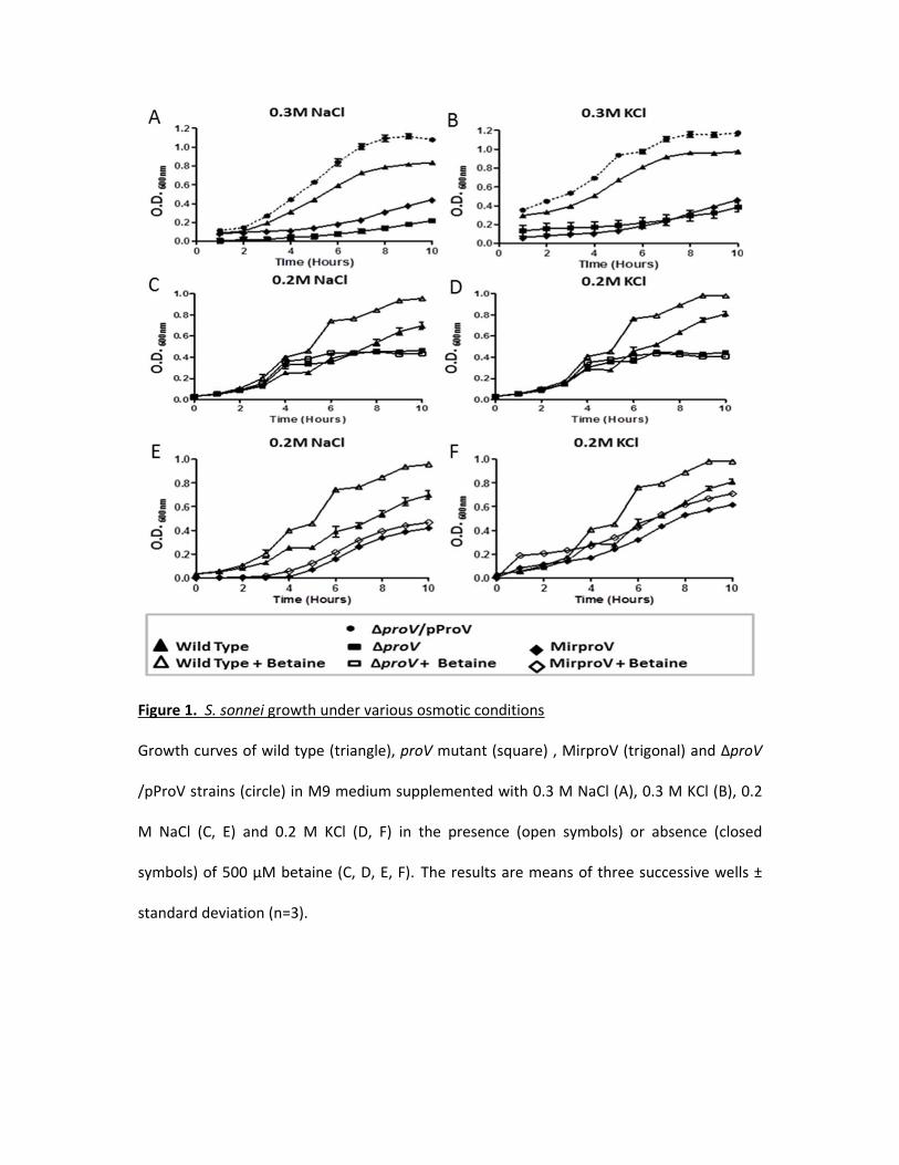
Figure 1. S. sonnei growth under various osmotic conditions
Growth curves of wild type (triangle), proV mutant (square) , MirproV (trigonal) and ∆proV
/pProV strains (circle) in M9 medium supplemented with 0.3 M NaCl (A), 0.3 M KCl (B), 0.2
M NaCl (C, E) and 0.2 M KCl (D, F) in the presence (open symbols) or absence (closed
symbols) of 500 µM betaine (C, D, E, F). The results are means of three successive wells ±
standard deviation (n=3).

Figure 2. qRT-PCR analysis for both proV and proX transcripts in both wild type and MirproV
(A) qRT-PCR analysis of wild type and MirproV strains growth in M9 medium without
supplement. Levels of proV and proX transcripts in wild type was set as calibrator (zero) and
levels of transcripts in MirproV strain were calculated using the 2-ΔΔct method; a significant
drop of proV and proX transcripts was observed in MirproV strains. Levels of transcription
between proV and proX also significantly differ in the MirproV strain (* p < 0.05).
(B) qRT-PCR analysis for the expression of proV (grey columns) and proX (open columns)
genes in both wild type and MirproV grown in hyperosmotic media. The difference in proV
transcripts between wild type and MirproV strains in presence of both 0.3 M NaCl and KCl is
highly significant (*** p < 0.0001; grey columns); and the difference in proX transcripts
between wild type and MirproV in the presence of 0.3 M NaCl or 0.3 M KCl are highly
significant (*** p = 0.0007, and ** p = 0.0025, respectively).
All the results are means of three successive groups ± standard deviation (n=3).

Figure 3. Deletion of proV doesn’t disturb the function of TTSS
S. sonnei wild type, the complemented strain ΔproV/PproV, ΔmxiD, ΔproV and MirproV
strains were grown to mid-log phase, and TTSS secretion was induced with Congo red. Total
proteins from cell lysates (A) and culture supernatants (B) were separated on SDS-PAGE and
IpaB and IpaC were detected with anti-IpB and anti-IpaC antibodies.

Figure 4. Testing the intracellular growth of the two mutation approaches in vitro using
HEK293 cells
(A) Intracellular growth of S. sonnei 2 hours post infection (MOI of 10). Intracellular CFU of
the wild type were taken as 100%, and intracellular CFU from strains ΔproV, MirproV and
ΔproV/pProV were expressed as percentages to that of wild type. Each value is the mean of
three independent determinations ± standard deviation. The level of significance was
determined using unpaired t-test, Asterisks (****) indicate p-values < 0.0001, (***) means
p-values = 0.0003. (B) Time course of intracellular growth of S. sonnei (MOI of 10). At
indicated time interval post infection, cells were lysed and intracellular CFU were
determined by plating on agar. Each value is the mean of triplicates ± standard deviation
(n=3). Doubling time for each strain is calculated by linear regression analysis (Fig. S7). (C, D)
Overlay of the flow cytometry analysis of cells were infected with ΔproV (dark grey), wild

type (black) or mock-infected (light grey). Both S. sonnei strains were expressing EGFP.
Controls are cells mock-infected with saline. Analysis was done 4 hours post infection (C) or
overnight (D). Gate A depicts populations of cells emit EGFP signals.

Figure 5. Testing the two mutation approaches in vivo using G. mellonella larvae
(A) Fraction survival of G. mellonella larvae model challenged by 105 CFU of wild type S.
sonnei strain 20071599 (grey square), the complemented strain (ΔproV/pProV) (black circle),
ΔproV (black triangle) and the MirproV (black trigonal), using saline as a control (crosses).
The observation lasted for 5 days. The results are means of three successive groups (n=10
larvae). (B) Overlaid histogram of the flow cytometry analysis of hemocytes isolated from G.
mellonella larvae mock-infected as a control (light grey), challenged by S. sonnei wild strain
20071599 (dark grey) or by ΔproV (black). Both S. sonnei strains were expressing EGFP.
Hemocytes were isolated 4 hours post infection for analysis; gate A depicts hemocytes emit
GFP signals.


