SHEET REINFORCEMENT 1. THE ORGANIZATION OF … · SHEET REINFORCEMENT 1. THE ORGANIZATION OF THE...
10
Name: Class: Date: REINFORCEMENT 1. THE ORGANIZATION OF THE HUMAN BODY 1 Label the levels of organization. Then complete the sentence: write highest, lowest, higher, lower. The level of organization is the organism; the level of organization is the cell. Tissues are a level than organs, but organ systems are a level than organs. SHEET 1 24 BIOLOGY AND GEOLOGY 3. Photocopiable material © 2015 Santillana Educación, S. L.
Transcript of SHEET REINFORCEMENT 1. THE ORGANIZATION OF … · SHEET REINFORCEMENT 1. THE ORGANIZATION OF THE...

Name: Class: Date:
REINFORCEMENT 1. THE ORGANIZATION OF THE HUMAN BODY
1 Label the levels of organization. Then complete the sentence: write highest, lowest, higher, lower.
The level of organization is the organism; the
level of organization is the cell. Tissues are a level than
organs, but organ systems are a level than organs.
SHEET
1
24 BIOLOGY AND GEOLOGY 3. Photocopiable material © 2015 Santillana Educación, S. L.

Name: Class: Date:
REINFORCEMENT 1. THE ORGANIZATION OF THE HUMAN BODY
1 Write the name of each cell: eukaryotic or prokaryotic. Then label its parts.
cell
cell
SHEET
2
25BIOLOGY AND GEOLOGY 3. Photocopiable material © 2015 Santillana Educación, S. L.
Name: Class: Date:
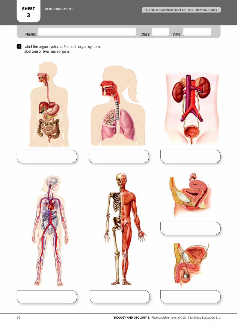
REINFORCEMENT
1 Label the organ systems. For each organ system, label one or two main organs.
SHEET
31. THE ORGANIZATION OF THE HUMAN BODY
26 BIOLOGY AND GEOLOGY 3. Photocopiable material © 2015 Santillana Educación, S. L.

Name: Class: Date:
REINFORCEMENT AND EXTENSION
1. THE ORGANIZATION OF THE HUMAN BODYSHEET
4
1 Complete the summary.
a. All living things are organized into
of organization.
b. The cell is the unit, the unit
of all living things. organisms
are made up of one cell.
organisms are made up of many cells.
c. Cells can be such as bacteria,
or such as those in human beings.
Eukaryotic cells have three structures:
.
d. A tissue is a group of
that work together
to perform a specific function. There are four types:
e. A group of different tissues that work together to perform
a specific function is called an .
f. An is a group
of organs that work together to carry out one or more
functions.
g. The organ systems involved in nutrition are the:
.
h. The organ systems involved in interaction are the:
.
i. The organ systems involved in reproduction are the
systems.
SCIENTIFIC ANALYSIS The origin of eukaryotic cellsThe Serial Endosymbiotic Theory (SET) was developed by biologist Lynn Margulis. According to this theory, eukaryotic cells evolved from ancient prokaryotic cells between 2 and 1.5 billion years ago. The original prokaryotic cell lost its cell wall and became larger. As a result, the surface of the cell membrane increased, improving phagocytic capacity. In later stages, a pronucleus formed. These cells could ingest other smaller, free-living aerobic or photosynthetic prokaryotes and form symbiotic relationships with them.
According to the SET theory, successive symbiotic associations explain the presence of cell organelles such as mitochondria and chloroplasts in eukaryotic cells.
2 What do these terms mean: pronucleus, phagocytic, aerobic and symbiotic?
3 What does the SET theory try to explain?
4 Explain why this theory is called the Serial Endosymbiosis Theory.
5 Search for information on SET. Explain why two prokaryotic organisms were part of the first symbiotic association.
a. The prokaryotic cell loses
its cell wall.
b. The surface of the membrane
increases and inner membranes
are formed.
c. A pronucleus is formed.
Endosymbiosis with spirochete
bacteria could be the origin of
flagella.
d. Association with an aerobic
prokaryote could be the origin
of mitochondria.
e. Association with a
photosynthetic prokaryote could
be the origin of chloroplasts.
27BIOLOGY AND GEOLOGY 3. Photocopiable material © 2015 Santillana Educación, S. L.
Name: Class: Date:
REINFORCEMENTSHEET
51. THE ORGANIZATION OF THE HUMAN BODY
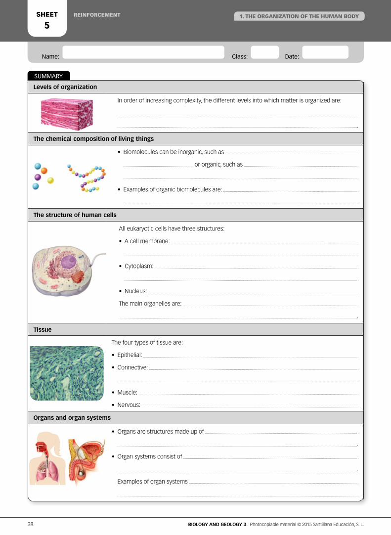
SUMMARY
Levels of organization
In order of increasing complexity, the different levels into which matter is organized are:
.
The chemical composition of living things
• Biomolecules can be inorganic, such as
or organic, such as
• Examples of organic biomolecules are:
The structure of human cells
All eukaryotic cells have three structures:
• A cell membrane:
• Cytoplasm:
• Nucleus:
The main organelles are:
.
Tissue
The four types of tissue are:
• Epithelial:
• Connective:
• Muscle:
• Nervous:
Organs and organ systems
• Organs are structures made up of
.
• Organ systems consist of
.
Examples of organ systems
28 BIOLOGY AND GEOLOGY 3. Photocopiable material © 2015 Santillana Educación, S. L.

REINFORCEMENT 1. THE ORGANIZATION OF THE HUMAN BODYSHEET
6
In multicellular organisms, the zygote and the cells produced during early cell divisions are embryonic stem cells: they can form complete new organisms. These embryonic stem cells can develop into different types of cells.
There are approximately 200 types of cells in the human body. During cell differentiation, cells acquire the appropriate morphological and chemical characteristics to perform specific functions.
Muscle cell Neuron Epithelial cell Blood cell Connective cell
CELL DIFFERENTIATION AND TRANSPLANTS
ACTIVITIES
1 Answer the questions. a. Explain what embryonic stem cells are. b. When a cell specializes to carry out different functions, it
loses its ability to become a new organism. Explain why this happens.
2 Describe the characteristics of each of the cells above. Find out the function of each cell.
3 Make correlations between the morphological and physiological characteristics of each cell and the cell's function.
4 Why was Japanese professor Shinya Yamanaka awarded the Nobel Prize for Medicine in 2012? a. What are iPS stem cells? b. Why are these cells important?
5 Explain the meaning of the following: Mature cells can be reprogrammed to become pluripotent cells.
Organ donation campaign
Task: Prepare posters on organ donation.
Work in groups of five. Divide up the work in your group.
Group A: Who can be a donor? Which organs can be donated? Why is umbilical cord blood donated? What is living organ donation?
Group B: What are the criteria for organ and tissue transplants? What side effects does the patient experience? Why does rejection occur?
Group C: How are organ donations and transplants handled in Spain? What is a donor card and what is it for? Do organ donors receive any benefits?
Group D: Report on innovative transplant solutions, for example: stem cells and regenerative medicine, kidney paired transplants, a registry of intended kidney donor–recipient pairs.
COOPERATIVE PROJECT
29BIOLOGY AND GEOLOGY 3. Photocopiable material © 2015 Santillana Educación, S. L.
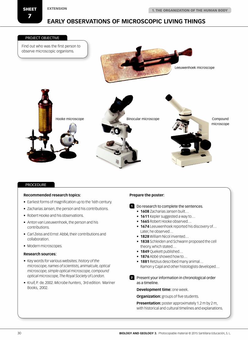
EARLY OBSERVATIONS OF MICROSCOPIC LIVING THINGS
EXTENSION
Find out who was the first person to observe microscopic organisms.
Recommended research topics:
• Earliest forms of magnification up to the 16th century.
• Zacharias Jansen, the person and his contributions.
• Robert Hooke and his observations.
• Anton van Leeuwenhoek, the person and his contributions.
• Carl Zeiss and Ernst Abbé, their contributions and collaboration.
• Modern microscopes.
Research sources:
• Key words for various websites: history of the microscope, names of scientists, animalcule, optical microscope, simple optical microscope, compound optical microscope, The Royal Society of London.
• Kruif, P. de. 2002. Microbe hunters, 3rd edition. Mariner Books, 2002.
Prepare the poster:
1 Do research to complete the sentences. • 1608 Zacharias Jansen built… • 1611 Kepler suggested a way to… • 1665 Robert Hooke observed… • 1674 Leeuwenhoek reported his discovery of… Later, he observed… • 1828 William Nicol invented… • 1838 Schleiden and Schwann proposed the cell theory, which stated… • 1849 Quekett published… • 1876 Abbé showed how to… • 1881 Retzius described many animal… Ramon y Cajal and other histologists developed…
2 Present your information in chronological order as a timeline.
Development time: one week.
Organization: groups of five students.
Presentation: poster approximately 1.2 m by 2 m, with historical and cultural timelines and explanations.
PROCEDURE
PROJECT OBJECTIVE
Leeuwenhoek microscope
Hooke microscope Binocular microscope Compound
microscope
1. THE ORGANIZATION OF THE HUMAN BODYSHEET
7
30 BIOLOGY AND GEOLOGY 3. Photocopiable material © 2015 Santillana Educación, S. L.

ACTIVITIES
1 Make a fact file for four types of tissues:
a. Name of tissue.
b. Subtypes of these tissues.
c. Name of the principal cells in the tissue.
d. Function of the tissue in the human body.
e. Ability to regenerate or not after a destructive process.
f. The type of tumor that is produced when there is uncontrolled cell division.
g. Non-cellular elements, if any, that form part of the tissue.
INTERPRETATION OF TISSUE SECTIONS
EXTENSION 1. THE ORGANIZATION OF THE HUMAN BODYSHEET
8
• Adipose tissue. Adipose cells (adipocytes) are spherical when isolated, but polygonal when close together. They vary in size from about 50 to 150 microns. The nucleus appears flat and is not always visible because it is pushed to one side of the cell by the large vacuole of lipids (fats). A thin outer layer surrounds each cell. The extracellular matrix is made up of reticular fibres (type III collagen).
• Skeletal muscle tissue. A longitudinal section shows long parallel fibres (cells) and alternating light and dark bands (striations). During embryonic development, each skeletal muscle cell is formed by the fusion of many stem cells. In adults, skeletal muscle fibres (cells) are actually syncytia containing many nuclei. Each cell is surrounded by an outer layer: a cell membrane (sarcolemma). The cytoplasm of a muscle fibre is called sarcoplasm.
• Cardiac muscle tissue. Cells appear elliptical in a transverse section. In a longitudinal section, the branching fibres (cells) and nuclei are visible. Cells have a central nucleus. Between cells there are thick fibrocollagenous membranes. Desmosomes are structures that hold the cells together.
• Osseous or bone tissue. The basic unit called the osteon is visible in a transverse section. In the centre of each osteon is the Haversian or central canal. Concentric layers of bone matrix surround the canal. The osteocytes within the bone matrix are organized around the central canal. Osteocytes have cytoplasmic extensions that connect them to other osteocytes to obtain nutrients. Osteocytes are enclosed in small cavities within the mineralized bone matrix.
• Nervous tissue (cerebral cortex). Neuron cell bodies are visible including some axons and dentrites. Neurons vary in size and shape according to their function. In most of the cerebral cortex, there are six layers with different types of cells. Capillaries are abundant. The darkly stained circles are the nuclei of glial cells; oligodendrocytes are the most visible. The background is a network of neuronal and glial cell processes (axons and dendrites).
• Smooth muscle tissue. Long fibres (cells) are visible. The cytoplasm is abundant and pink. Many cells have no visible nucleus. When visible, nuclei are elongated and centrally located.
31BIOLOGY AND GEOLOGY 3. Photocopiable material © 2015 Santillana Educación, S. L.

Name: Class: Date:
1 Read and write prokaryotic cells or eukaryotic cells:
a. They are the largest and most complex cells.
b. Their genetic material is dispersed in the cytoplasm.
c. The only organelles they contain are ribosomes.
d. They have a nucleus and a nucleolus.
e. They contain mitochondria.
f. Bacterial cells.
2 Make pairs of related concepts: use these words. Explain why they are related.
Glucose – Protein – DNA – Polysaccharide – RNA – Amino acids – Glycerol – Fatty acid
3 Define metabolism, anabolism and catabolism. Are the following reactions anabolic or catabolic?
a. amino acids + energy → proteinsb. glucose → inorganic molecules + energy
4 Label the diagram and answer the questions.
a. What type of cell is it?
b. Which two organelles are visible?
c. What are the membranes like?
d. What is the difference between chromatin and chromosomes?
ASSESSMENT1
32 BIOLOGY AND GEOLOGY 3. Photocopiable material © 2015 Santillana Educación, S. L.
Name: Class: Date:
5 What is the relationship between the endoplasmic reticulum, the Golgi apparatus and secretory vesicles?
6 Complete the table.
Cells Tissue and type Tissue function
Red blood cells
Osteocytes
Neurons
Chondrocytes
7 What are the differences between tissues and organs? And between organs and organ systems? Give examples.
8 Write the parts of the body where each tissue is found.
Endothelium Epidermis Connective Smooth muscle Nervous Cartilaginous
9 Name the organ systems involved in nutrition. Name two parts or organs in each system and their function.
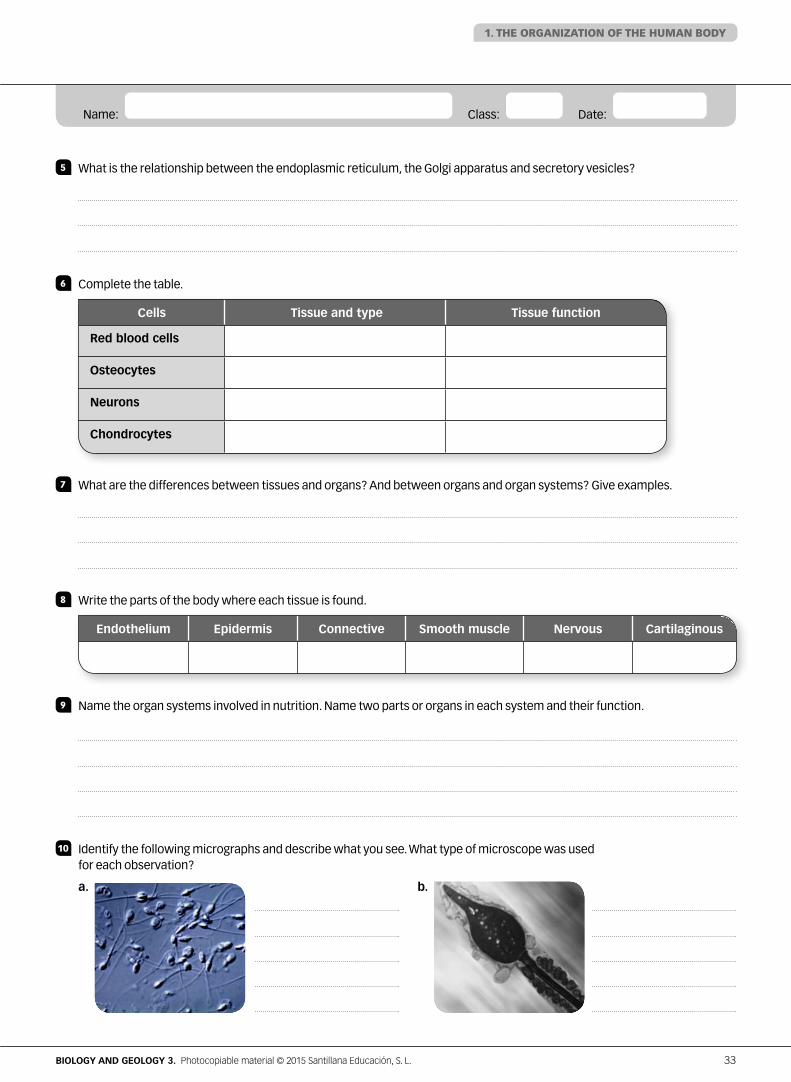
10 Identify the following micrographs and describe what you see. What type of microscope was used for each observation?
a. b.
1. THE ORGANIZATION OF THE HUMAN BODY
33BIOLOGY AND GEOLOGY 3. Photocopiable material © 2015 Santillana Educación, S. L.