Sheep Pulmonary Adenomatosis
212
SHEEP PULMONARY ADENOMATOSIS Joshua Geoffrey^Wandera, Dip.Vet.Sc., M.Sc. A Thesis submitted for the Degree of Doctor of Philosophy in the University of East Africa. HfuLZ, if70 Department of Pathology and Microbiolbgy Faculty of Veterinary Science University College Nairobi April, 1970:
Transcript of Sheep Pulmonary Adenomatosis
SHEEP PULMONARY ADENOMATOSIS
Joshua Geoffrey^Wandera, Dip.Vet.Sc., M.Sc.
A Thesis submitted for the Degree of Doctor of
Philosophy in the University of East Africa.
H fu L Z , i f 7 0
Department of Pathology and Microbiolbgy
Faculty of Veterinary Science
University College Nairobi
April, 1970:
3rd April, 1970.
EXTERNAL EXAMINERS
Prof. F.K. Ramsey, D.V.M., Ph.D. Professor and Head,Department of Veterinary Pathology Iowa State University AMES, IOWA.
Prof. E. Weiss,Dr.med.Vet Professor and Head, Department of Veterinary PathologyJustus-Liebig Universit&t 63 Giessen, WEST GERMANY.
INTERNAL EXAMINERS
Dr. H. Krauss, Dr.med.Vet. Universit&tsdozent Justus-Liebig Universit&t Reader in Dept, of Pathology and Microbiology, Faculty of Veterinary Science UNIVERSITY COLLEGE NAIROBI.
Dr.G.M. Mugera, Dip.Vet.Sc., M.Sc., Ph.D.,
Ag. Head Department of Veteri nary Pathology & Microbiology UNIVERSITY COLLEGE NAIROBI.
(ii)
VA C K N O W L E D G E M E N T S
The author wishes to express appreciation to his principal
supervisor, Professor Dr. E. Weiss of Veterinar-Pathologisches
Institut - Giessen, for his invaluable, guidance, support and
encouragement throughout this study, and for his careful reading
and constructive criticisms of the manuscript.
Thanks are also due to Dr. H. Krauss for immensable assi
stance in the conduct of aetiological and electron microscopic
studies; and the very valuable discussions we had in the course
of the investigations; and to Miss R. Schirmer for technical
assistance.
The author wishes to express his sincere appreciation to
all members of the Department of Pathology and Microbiology,
of the Faculty of Veterinary Science for their generosity and
consideration and for the technical and professional aid and
guidance. The assistance of Drs. H. Frank and 0. Bwangamoi
this Department, with photography and that of Mr. Robert
Mbugua in typing the whole thesis is gratefully acknowledged -
This study was supported by grants from University College
Nairobi, and Rockefeller Foundation Grant 99/281 for research
on pneumonias of food producing animals. This financial assi
stance is gratefully acknowledged by the author.
(iii)
TABLE.. OF CONTENTS
ACKNOWLEDGEMENTS O O O 0 O 0 9 9 O O 0 9 O O O O 9 O OOCO 0 9 9 0 0 9 0
LIST OF TABLES
LIST OF FIGURES
9 O C O 0 O 9 O O O
SUMMARY
CHAPTERS:
1. INTRODUCTION
2. LITERATURE REVIEW
I Incidence
II Aetiology and Epidemiology
III Pathology
IV Economic Importance
V Geographical Origins of the Disease
3<, AETIOLOGICAL STUDIES.... ....... o o o q o o o o
I Study with chicken Embryos 0 9 0 0 0
II Isolation and Characterization of Mycoplasma species• 0 0 0 9 0 9 0 0 0 0 0 0 0 0 0 0 9
III Isolation and Serology of Chlamydiae
IV Cell Cultures from SPA Lungs
V Discussion . .0 0 0 9 0 O
o o o o o o n o
0 0 0 9 0 0 0 000
ii
Pages
vi
vii - xii
xiv - xv i
1 - 4
5
5 - 1 3
14 - 21
21 - 25
25 - 27
27 - 29
30
30 - 36
36 - 47
47 - 53
53 - 56
57 - 60
(iv)Pages
4. TRANSMISSION EXPERIMENTS ............ ....... 61
• A. MATERIALS AND METHODS ................... 61
I Experimental Animals ................ 61
II Donor (Infective) Materials ........ 62
III Experiments Performed .............. 62
Experiment 1: Transmission withNaturally infected Material .... 6 2 - 6 3
Experiment 2: Repeat Transmissionwith Naturally Infected Material 63 - 64
Experiment 3: Transmission with Egg-Passaged Material ............... 65
Experiment 4: Transmission with Myco-El asma-containing Material .... 65
IV Necropsy Procedure: Collection andSpecimen Preparation............ 66 - 67
B. R E S U L T S ............................. 67
I Experiment 1 ......................... 67 - 70
II Experiment 2 ......................... 70 - 79
III Experiments 3 & 4 79 - 80
IV Discussion ........................... 80 - 85
5. ELECTRON MICROSCOPIC STUDIES ................. 86
I Materials and Methods................. 86 - 88
II Results ............................... 88 - 93
III Discussion ........................... 93 - 96
( V )
Pages
6. GENERAL DISCUSSION............... ...... 97
I Aetiological Studies........... 97 - 100
II Clinical Response of TransmissionExperiments..................... 100 - 103
III Pathology and Pathogenesis ....... 103 - 109
IV Conclusion....................... 110 - 116
APPENDIX "A": Transmission Experiment 2:
Clinical Signs Exhibited by up
to 260 days' Observation period .... 116 - 117.
REFERENCES ................................ 117 - 135
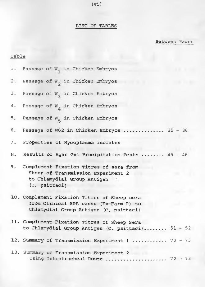
(Vi)
LIST OF TABLES
Between Pages
Table
1. Passage of in Chicken Embryos .
2. Passage of in Chicken Embryos
3. Passage of in Chicken Embryos
4. Passage of in Chicken Embryos
5. Passage of W in Chicken Embryos56. Passage of W62 in Chicken Embryos.............. 35 - 36
7. Properties of Mycoplasma isolates
8. Results of Agar Gel Precipitation Tests ....... 4 5 - 4 6
9. Complement Fixation Titres of sera fromSheep of Transmission Experiment 2 to Chlamydial Group Antigen (C. psittaci)
10. Complement Fixation Titres of Sheep serafrom Clinical SPA cases (Ex-Farm D) to Chlamydial Group Antigen (C. psittaci)
11. Complement Fixation Titres of Sheep Serato Chlamydial Group Antigen (C. psittaci)...... . 51 - 52
12. Summary of Transmission Experiment 1 ............ 72 - 73
13. Summary of Transmission Experiment 2 .Using Intratracheal Route ....... ............. 72 - 73
(vii)
14, Experiment 2: Lung Lesion Distributionand Percentage in 8 Sheep . ................ 7 2 - 7 3
15, Summary of Transmission Experiment 3 & 4 .... 7 9 - 8 0
LIST OF FIGURES
Figures
1. World Distribution of Sheep PulmonaryAdenomatosis, 1800-1969 ........ . 5 - 6
2. Distribution of SPA in Kenya, 1920-1969 ..... 1 2 - 1 3
3. From mixed culture showing macrophages andgiant cells? no evidence of intracellularinclusion bodies. H & E stain. X 125 .... 5 6 - 5 7
4. Higher magnification of Figure 3.H. & E. stain. X 315 ....... .......... . 56 - 57
5. Sketch of SPA lesion distribution, sheep 3631 68 - 69
6. Sheep 363L: Three Adenomatous foci (AF)from atelectatic alveoli. H & E stain.X 315........................................ 68 - 69
7. Compact tumour cells from another area ofsheep 3631. Cytoplasm of many cells is vacuolated. H & E stain,X 315.
Between Pages
Table (Contd.)
68 - 69
(ix)
19. Sheep 3648: Dorsal view - showingspread of adenomatous lesion ...
20. Sheep 3648: Closer-view of dorsalaspect ...... ...................
21. Sheep 3648: Ventral aspect. Very littleof the surface is normal ............ 75 - 76
22. Sheep 3613: Dorsal aspect showing noevident SPA foci in the right lung.Several glandular foci are scattered in the anterior lobes of the left l u n g ................................. 75 - 76
23. Sheep 3613: Closer-view of the leftlung showing scattered adenomatous foci ......................... 75 - 76
24. Sheep 3648: Adenomatous foci locatedin "centres" of once thickened interalveolar walls. H & E stain. X 125 ... 7 6 - 7 7
25. Sheep 3645: Two adenomatous foci (near A)located in "centres" of thickened interalveolar walls. Lendrum's stain.X 500 .. 76 - 77
26. Sheep 3645: A "sheet" of adenomatous cells
Figures (Contd. ) Between Pages
with no marked alveolar spaces.H & E stain. X 500 .................... 76 - 77
27. Sheep 3615: Compact group of adenomatouscells with vacuolated cytoplasmH & E. stain. X 500...................... 76 - 77
28. Sheep 3645: Several adenomatous nodules "suspended" by interalveolar walls whose transient thickening had subsided. There is no bronchiolar (B) involvement yet. H & E. stain. X 45 ..............
74 - 75
75 - 76
77 78
( X )
Figures (Contd.) Between Pages
29. Sheep 3645: An adenomatous nodulewhose cells are compact "suspended" by normal size interalveolar walls.H & E stain. X 315.................... 77 - 78
30. Sheep 3616: An adenomatous focus (AF)surrounded by macrophages (Ma) which had infiltrated alveolar spaces and some fused to form epithelioid cells.H & E stain. X 315..................... 78 - 79
31. Sheep 3617: An advanced lesion of SPA.Interlobular septum (S) divided thetwo groups of tumour cells. The lattersupported by thin connective tissuegrow in a papillary manner into alveolarspaces. H. & E stain. X 125 .......... 78 - 79
32. A natural case (WV) showing fibroplasiaon the right side (F) in an advanced case with apparent secondary reaction.H & E stain. X 45....................... 78 - 79
33. Sheep 3645: Intrabronchiolar proliferation, partially obliterating thelumen (L) . H & E Stain. X 315 ........ 7 8 - 7 9
34. From a natural case (1057/66): Intrabron*-chiolar proliferation with partial 78 - 79occlusion of the lumen. H & E stain. X 45.\
35. From a natural case (1156/66) showingmetastasis to tracheobronchial lymphnode. H & E stain. X 125............... 78 - 79.
36. Sheep WII: Adenomatous cells in solidclusters, a dark cell centrally located with an indented nucleus and paranuclear space. Cell organelles are few X 12,000. 90 - 91
(Xii)
Figures (Contd.) Between Pages
44. Sheep WXI: Adenomatous cell with largenucleus (N) irregular and markedly dilated rough-surfaced endoplasmic reticulum (ER). Tonofilaments arepresent around the nucleus. X 21,000.... 91 - 92
45. Sheep WXI: Three adenomatous cells liningan alveolus with many vacuoles full ofsecretion (S). There are also glycogengranules. X 15,000..... ......... . 91 - 92
46. Sheep WV: One transformed adenomatouscell protruding towards the alveolar space (AL) with numerous vacuoles (V) some of which contain granules. Microvilli are evident. X 13,800........................ 91 - 92
47. Sheep WXI: Adenomatous cell with multipleGolgi apparatus (G) dilated rough-surfaced endoplasmic reticulum and tonofilaments.X 21,000................................... 91 - 92
48. Sheep WXI: Adenomatous cell with numerousglycogen granules, irregular cell borders (cb) and multiple Golgi apparatus (G).X 21,000................................... 91 - 92
49. Sheep WII: Tumour cell with Chlamydiae (CD),.numerous free ribosomes (rb) and dilated endoplasmic reticulum (ER) . X 38,400.... 92 - 93
50. Sheep WXI: Columnar tumour cells aroundan alveolus (AL) in which are virus-like particles (vp). There are also glycogen granules and dilated endoplasmic reticulum.X 13,500.................................... 92 - 93
(xiii)
Figures (Contd.) Between Pages
51. Sheep WXI: Virus-like particles (vp)in \an alveolus (AL) close'to the microvilli of tumour cells. There are few lysosomes. X 21,000. ..................... 92 - 93
52. Sheep WXI: Adenomatous cells whose microvilli are close to aggregations of viruslike particles. X 26,700................ 92 - 93
53. Sheep WXI: Adenomatous cell possibly disintegrating with indented nucleus (N) vacuoles and aggregations of virus-like particles (vp) in the alveolus (AL) and a myelin figure (my). X 21,000............
54. Sheep WXI: Virus-like particles close to :plasma membrane which appear to be spherical to oval. Numerous irregularly shaped vacuoles (VC) on the right side.X 57,000..................................
92 - 93
92 - 93
( X i v)
S U M M A R Y
The impetus for research on sheep pulmonary adenomatosis
has been the recognition of its contagious and neoplastic nature
and, of a possible relationship with certain cancers.
Literature Review;
A detailed review of the literature revealed that the disease
has a world-wide distribution. It has been clearly identified as
a serious cause of loss on a number of sheep farms in Kenya,
South Africa, South West Africa, Bulgaria, Scotland and, in Iceland
before it was eradicated. Most countries in which the Merino breed
(or those they came in contact with) had been imported have re
corded presence of SPA. The important exceptions are Australia
and New Zealand. Despite this wide distribution there have been
gaps in our knowledge of the aetiology, pathology and pathogene
sis of the disease.
Aetiological Studies:
Chicken embryos, bacterial media and cell cultures from SPA
lungs were used in attempts to identify the aetiological agent.
Serological studies against Chlamydial antigens were also carried
out. Infective lung materials from 3 natural and 3 experimental
( X V )
clinical cases of SPA were inoculated via the yolk-sacs into
embryonating eggs. Two specimens from the natural cases and one
specimen (lung fluid) from experimental cases were passaged 18, 17
and 27 times, respectively. Constant lesions produced by these
3 specimens were haemorrhages under the skin of embryos and in
visceral organs particularly the liver and heart in those dying
3-5 days after inoculation. The specimen passaged 27 times
revealed presence of Chlamydiae.
Ten out of 14 lung tumours and fluid from clinical cases of
SPA yielded Mycoplasma when cultured on Tryptose Serum Agar (TSA)
and in Newing's Tryptose Broth (NTB). More than one isolate were
encountered in some cases. Their biochemical and serological be
haviour showed that they were different from bovine pleuropneumonia
vaccine strain and a strain of caprine pleuropneumonia used for
comparison. Four samples did not yield Mycoplasma on culturing.
Macrophage cultures from 4 adenomatous lungs maintained for 5-7
days formed many giant cells. Both macrophages and giant cells
had no intracellular inclusion bodies.
(xvi)
Although the aetiological studies failed to demonstrate the
presence of clear-cut infectious agent, a virus is still considered
to be the organism involved. Mycoplasma spp. encountered may be
opportunists that take advantage of fluid produced by the adenoma
tous lesions to proliferate and, not involved in the causation of
SPA. Serological investigation showed that Chlamydial infections•
are widespread in many sheep flocks in Kenya. High antibody titres
were found in sera from healthy sheep and, no titres in sera from
some sheep with clinical SPA.
Transmission Experiments:
Four transmission experiments were carried out. In the first
one, a 25% inoculum was prepared from adenomatous lungs of freshly
killed sheep using a pestle and mortar. Eight sheep were each
inoculated with 10 ml., 4 intratracheally and 4 into the lungs
through the thorax. Three of the sheep at days 65, 118, and 251
after inoculation had lung lesions typical of SPA but no clinical
signs. They had been inoculated intratracheally. Sheep inocu
lated intrathoracically and one intratracheally and, killed 255
days after inoculation had no lung lesions.

(xvii)
Ultra-sonic vibration was used in preparing a 57% inoculum
for the second experiment. The infective material was adenoma
tous lung tissue from freshly killed sheep. Ten sheep were all
inoculated intratracheally, 7 of them showed clinical signs
(dyspnoea, increased respiration, lung discharge, moist rales,
cough) of the disease between 107 and 260 days after inoculation.
They all had lung lesions of the disease when examined post-mortem.
An 8th sheep without clinical signs also had lung lesions of SPA.
In the 3rd and 4th experiments, infective materials passaged
in embryonating eggs and, mixed Mycoplasma isolates were used re
spectively. Inoculations carried out intratracheally in 8 and 5
sheep respectively produced no lung lesions of SPA. In Experiment
3 the sheep were observed for a year and in Experiment 4 for
between 151 and 215 days.
It was demonstrated in Experiment 2 that reproduction of
typical lesions and clinical disease of SPA was not as difficult
as previously thought. This also showed that the aetiological
agent was contained in the tumour cells. The reproduction of
clinical SPA would appear to be dose-and susceptibility-dependent
(xviii)
and not time-dependent. The incubation period would appear to
be between 6 and 8 months.
Electron Microscopic Studies:
Eight freshly killed sheep with SPA provided adenomatous
lung tissue for investigation. Several abnormalities in the* * V
tumour cells were observed. These were an enlarged polymorphic
nucleus, enlarged Golgi complex, an increase of free ribosomes.
Dilatation into vesicules of endoplasmic reticulum was evident.
There was also irregularity of cytoplasmic membranes in some
adenomatous cells. No intracellular inclusion bodies were seen.
In all, the ultrastructural changes of the adenomatous cells
had similarity to carcinomas in human beings.
Pathology and Pathogenesis:
The gross lesions encountered in 7 of the sheep in Experiment
2 with clinical SPA revealed that infection is normally via inspired
air. Anterior lobes and antero-ventral aspects of the diaphrag
matic lobes are the ones which showed the grayish adenomatous
tissue typical of SPA. This distribution shows that SPA behavesrdifferently from any other lung cancer.
(xix)
The histopathological lesions clearly revealed the multice
ntric origin and the progressive nature of this adenomatous cancer.
Within the same lung and even the same section, could be seen
adenomatous foci of less than 10 tumour cells and sheets of tumour
cells. The earliest adenomatous foci were often located in
"centres" of thickened interalveolar walls. This suggests that
epithelium of microatelectatic alveoli is the first to be involved
in transformation and proliferation in sheep that are affected.
From these early beginnings adenomatous foci increase in size
until they fuse with adjacent ones to form continuous tumour
tissue. Although no metastases were found in the pulmonary
lymph nodes of 8 sheep of Experiment 2 with typical lesions,
metastases have been found in natural cases by several workers.
To date the author has encountered 4 cases with metastases.
This study indicates the need for more research on the
aetiology of SPA and, the involvement of infectious agents
in the causation of tumours.
-1-
CHAPTER 1
INTRODUCTION
Ovine pneumonia is the most important disease of sheep in
Kenya, which has so far not been controlled, It has always been
a cause of serious losses in large flocks of sheep since the
early days of modern sheep farming. Pneumonias in sheep are
complicated in their aetiology and pathology. Their terminology
is confusing because in many instances the conditions have been
described without adequate information available to give a simple
description to classify them. The best example of confusing
terminology is the "Laikipia Lung Disease" which included many
pneumonic conditions of sheep in Kenya (WHITWORTH, 1926, METTAM,
1927, 1929f ANON, 1956), acquired because of the high incidence
of sheep pneumonia in Laikipia district of Kenya which then was
the chief sheep farming area in the country.
Despite the general similarity in the clinical picture of
ovine pneumonia, it is evident that the aetiology and pathology
of the condition is complex. WANDERA (1967a,b) classified ovine
pneumonia in Kenya on aetiological and histopatholog^' bases as:
"Cuffing pneumonia" - of still unknown aetiological
-2-
agent but possibly caused by
Chlamydiae?
"Bacterial pneumonia" being mainly represented by
enzootic sheep pneumonia and
associated with Pasteurella
haemolytica?
"Parasitic pneumonia" caused by Dictyocaulus filaria
and Muellerius capillaris?
and "Chronic progressive pneumonia" - under which are two disti
nct conditions:
Maedi - progressive interstitial
pneumonia
and
Jaagsiekte - sheep pulmonary
adenomatosis (SPA),
Both "Maedi" and "Jaagsiekte" have been encountered in Kenya,
(WANDERA, 1970) sometimes on the same farm and in one sheep.
It is because of this mixed infection in some sheep that
SHIRLAW (1959) regarded the two types as just various manifesta
tions of one disease entity.
Jaagsiekte (Dutch: Jagziekte from jagt - to drive, ziekte -
sickness, hence driving sickness) was so named as the sick sheep
-3-
in advanced stages of the disease showed accelerated respiration
as if they had been rapidly driven (Hutcheon, quoted by MITCHELL,
1915). The sheep suddenly stopped when driven hungry for breath
and died soon after. The term, Jaagsiekte, is now used syno
nymously with sheep pulmonary adenomatosis (SPA). When originally
applied by South Africa sheep farmers, it could have referred
to Maedi (Graaff-Reinet disease) also or both.
Jaagsiekte is a distinct contagious disease manifesting
itself as a malignant neoplasm with an incubation period varying
from months to 2 years dependent on unknown factors. According
to WANDERA (1968) the incubation period in some flocks in Kenya
is between 6-12 months. The chief clinical features are progre
ssive emaciation, overt respiratory distress, greatly increased
respiratory rate, moist rales, and discharge of mucinous,
frothy, and watery exudate from the lungs through the nostrils.
It is afebrile."\
Most reports of SPA are generally in accord with respect
to epidemiology, symptoms and gross pathology. There are varia
tions in histopathology and little on pathogenesis. Little is
known about the nature of the aetiologic agent apart from sugge
stive evidence incriminating a virus because of cytoplasmic
-4-
inclusion bodies (ENCHEV, 1966) and/or Mycoplasma. The pur
pose of this investigation on sheep pulmonary adenomatosis
as a distinct disease was to:-
(a) undertake aetiologicai studies,
(b) carry out transmission experiments,9
(c) study in more detail its histopathology and pathogenesis
and,
(d) study the fine structure of adenomatous cells.

-4-
inclusion bodies (ENCHEV, 1966) and/or Mycoplasma. The pur
pose of this investigation on sheep pulmonary adenomatosis
as a distinct disease was to:-
(a) undertake aetiological studies,
(b) carry out transmission experiments,
(c) study in more detail its histopathology and pathogenesis
and,
(d) study the fine structure of adenomatous cells.
-5-
CHAPTER 2
LITERATURE REVIEW
II. INCIDENCE
In the first half of this century, sheep pulmonary adeno
matosis (Jaagsiekte) and progressive interstitial pneumonia
(Maedi) were never clearly-fciistingui-sheek This confusion was
made worse by the use of various terminologies, based on native
language of the country concerned though indicative of respira
tory embarrassment, such as Lungers, Jaagsiekte and Bouhite0
While Bouhite meaning "panting" is non-specific, when first used
(AYNAUD, 1926) in France, it applied to a sheep lung condition
whose dominent characteristic was adenomatous lesions with
metastases to regional lymph nodes,, The fact that in some
countries (France, Iceland, Kenya, South Africa and U„S.A.) both
pulmonary adenomatosis and Maedi were present (WANDERA, 1970)
only created more problems. Despite variations in the descrip
tions of the adenomatous lesions of sheep pulmonary adenomatosis
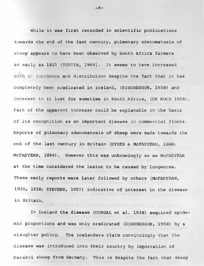
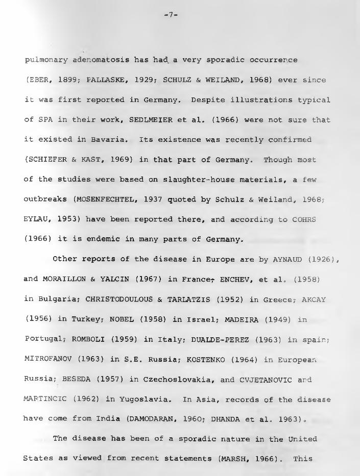
(SPA) it is one disease entity. SPA has a world-wide distri
bution (TUSTIN, 1969) as it has been recorded in more than 20
countries (Fig.l).
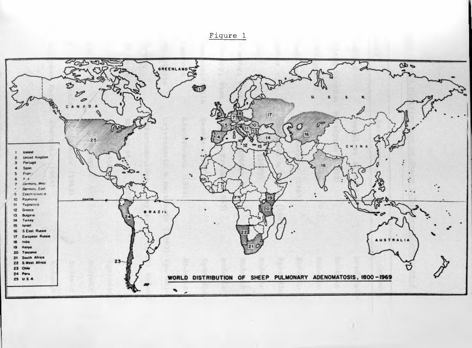
Figure 1

Fig,1: World Distribution of Sheep Pulmonary Adenomatosis
—
Country Reference Comment
1. Iceland Dungal et al. (1938) Eradicated
2. United Kingdom McFadyean (1938) Endemic
3. Portugal Madeira (1949) Endemic
4. Spain Dualde-Perez (1963) Endemic
5. France Moraillon & Yalcin (1967) Sporadic
6. Italy Romboli (1959) Endemic
7. Germany, West Cohrs (1966) Endemic
8. Germany, East Jakob & Krause (1965) Endemic
9. Czechoslovakia Beseda (1957) Sporadic
10* Roumania Adamesteanu (1969) Endemic
11, Yugoslavia Cvjetanovic & Martincic (1962) Sporadic
12* Greece Christodoulous & Tarlatzis (1952) Endemic
13„ Bulgaria Enchev et al, (1958) Endemic
14, Turkey Akcay (1956) Sporadic
15, Israel Nobel (1958) Endemic
16. S. East Russia Mitrofanov (1963) Endemic
17. European Russia Kostenko (1964) Endemic

Fig.1: (Continued).
Country Reference Comment
18. India Damodaran (1960) Sporadic
19. KENYA Shirlaw (1959) Endemic
20. Tanzania (Jakob, H., 1969 - Personal
communication) Sporadic
21. South Africa Tustin (1969) Endemic
22. South West Africa Tustin (1969) Endemic
23. Chile Schulz et al. (1965) Sporadic
24. Peru Cuba-Caparo et al. (1961) Endemic
25. U.S.A. Marsh (1966) Sporadic
-6-
While it was first recorded in scientific publications
towards the end of the last century, pulmonary adenomatosis of
sheep appears to have been observed by South Africa farmers
as early as 1825 (TUSTIN, 1969). It seems to have increased
both ir incidence and distribution despite the fact that it has
completely been eradicated in Iceland, (SIGURDSSON, 1958) and
interest in it lost for sometime in South Africa, (DE KOCK 1958),
Part of the apparent increase could be explanable on the basis
of its recognition as an important disease in commercial flocks*
Reports of pulmonary adenomatosis of sheep were made towards the
end of the last century in Britain (DYKES & MeFADYEAN, 1888?
McFADYEAN, 1894). However this was unknowingly so as McFADYEAN
at the time considered the lesion to be caused by lungworms.
These early reports were later followed by others (McFADYEAN,
1920, 1938; STEVENS, 1957) indicative of interest in the disease
in Britain.
In Iceland the disease (DUNGAL et al. 1938) acquired epide
mic proportions and was only eradicated (SIGURDSSON, 1958) by a
slaughter policy. The Icelanders claim convincingly that the
disease was introduced into their country by importation of
Karakul sheep from Germany. This is despite the fact that sheep
-7-
pulmonary adenomatosis has had, a very sporadic occurrence
(EBER, 1899? PALLASKE, 1929? SCHULZ & WEILAND, 1968) ever since
it was first reported in Germany. Despite illustrations typical
of SPA in their work, SEDLMEIER et al. (1966) were not sure that
it existed in Bavaria. Its existence was recently confirmed
(SCHIEFER & KAST, 1969) in that part of Germany. Though most
of the studies were based on slaughter-house materials, a few
outbreaks (MOSENFECHTEL, 1937 quoted by Schulz & Weiland, 1968?
EYLAU, 1953) have been reported there, and according to COHRS
(1966) it is endemic in many parts of Germany.
Other reports of the disease in Europe are by AYNAUD (1926),
and MORAILLON & YALCIN (1967) in France?- ENCHEV, et al. (1958)
in Bulgaria? CHRISTODOULOUS & TARLATZIS (1952) in Greece? AKCAY
(1956) in Turkey? NOBEL (1958) in Israel? MADEIRA (1949) in
Portugal? ROMBOLI (1959) in Italy? DUALDE-PEREZ (1963) in Spain?
MITROFANOV (1963) in S.E. Russia? KOSTENKO (1964) in European
Russia? BESEDA (1957) in Czechoslovakia, and CVJETANOVIC and
MAFTINCIC (1962) in Yugoslavia. In Asia, records of the disease
have come from India (DAMODARAN, 1960? DHANDA et al. 1963).
The disease has been of a sporadic nature in the United
States as viewed from recent statements (MARSH, 1966). This

-8-
would appear to conflict with earlier work (COWDRY & MARSH,
1928) in which emphasis was placed on the adenomatous lesions
and similarity between "Montana progressive pneumonia" and
Jaagsiekte. Materials used for the joint study were apparently
not from the same sheep as those handled earlier (MARSH, 1923a).
The description and illustrations by DAVIS (1939) on a primary
carcinoma of the lung of a sheep are undoubtedly those referra-
ble to SPA, although he was of the opinion that the two were
different. It was material supplied by this DAVIS that made
MARSH (1966) be convinced that sheep pulmonary adenomatosis exi
sted in U.S.A., especially in parts of Montana and Oregon, "Mon
tana progressive pneumonia" would appear to have been more or
less a group name covering both SPA and progressive interstitial
pneumonia (WANDERA, 1970). A report by SAVAGE (1926) on pulmo
nary cancer in sheep emanating from Winnipeg, Canada is essen
tially that of SPA. It is not clear whether the sheep in ques
tion was bred in Canada or imported from elsewhere.
SPA was first reported in Peru in 1945 although it may
have existed for a long time before this recognition. This meta
stasizing adenocarcinoma has been confirmed by CUBA-CAPARO, et
al. (1961) and also found in Chile (SCHULZ et al. 1965). One
-9-
other country which has suffered from pulmonary adenomatosis is
South Africa (COWDRY 1925, DE KOCK, 1929a). According to
D. HUTCHEON (Cit. ROBERTSON, 1904), farmers in South Africa,
especially in the then Cape Colony, were encountering a serious
disease of sheep by 1893 or even earlier. Recent investigations
(TUSTIN, 1969) suggest that the disease existed there in early
1800. This disease was called Jaagsiekte. But as reported by
ROBERTSON (1904) and MITCHELL (1915) Jaagsiekte was applied to
another chronic pneumonia. However the latter was renamed
"Graaff-Reinet disease" by DE KOCK (1929a), and differentiated
from the present synonym of SPA. The disease was so named be
cause of the number of cases emanating from the now defunct
Experimental Station at Graaff-Reinet. It has also been observed
on several farms in South West Africa. SPA has recently been re
ported in Tanzania (Jakob, H., 1969 - Personal communication) and
confirmed on the basis of slide examination by the author,
Pulmonary adenomatosis was found in certain flocks of sheep
in Kenya, (SHIRLAW 1959). Sheep of all ages and breeds were
affected, although some breeds and families within breeds are
more resistant than others. One case, (SHIRLAW, 1963 Personal
-10-
communication) was encountered in a goat, as were a few others
in India (RAJYA & SINGH, 1964), in S.E. Russia (ALIEV, 1967),
and in Peru, (CUBA-CAPARO et al. 1961). However none were en
countered in goats or cattle in Iceland (DUNGAL, 1946) on farms
where they were kept in close contact with sick sheep, These
reports of the disease in goats do indicate that this species of
animals is also affected. On the other hand they could be erro
neous and arise from the apparent inability of histopathologists
and others to differentiate between adenomatoid lesions of alve
olar epithelium and adenomatosis.
It was in the early 1920's that farmers in the Laikipia
district of Kenya became concerned about a lung disease with
heavy losses in both lambs and adult sheep. They called it
amongst other names, Laikipia lung disease. Although originally
this name may have been used to refer to what we now know as
pasteurella pneumonia in sheep, the name came to be applied
to any type of lung condition in sheep. WHITWORTH (1926) was
much more impressed by the lung abscesses and/or fibrinous
pneumonia he encountered in the affected sheep and neglected
the, ".... numerous greyish spots about a pin's head in size
on the cut surface" of the affected lungs. Though both him and
- 1 1 -
METTAM (1927) did little pathological examination of the cases
they were dealing with, their descriptions show that in addition
to fibrinous (Pasteurella) pneumonia and pulmonary abscesses
due to Corynebactenum pyogenes, some of the lungs had lesions
of Jaagsiekte.
A pneumonia of sheep said to resemble that form known as
Jaagsiekte in South Africa had previously been reported from
two centres (KENYA 1923) by sheep farmers. Unfortunately the
complaint was not investigated at the time. Later (KENYA, 1924)
more cases of pneumonia in sheep were encountered tnat were at
first thought to resemble those associated with sheep-pox.
However, some of the cases were found on farms on which sheep-
pox did not exist. Indications are that these early reports
could have been dealing with the as yet unrecognised SPA. This
is despite the claim of METTAM (1929), that he had not encountered
J aagsiekte after careful examination of many pneumonic lungs.
Mettam cited movement from one farm of 1300 sheep over a dis
tance of 40 miles in 4 days and a subsequent outbreak of a lung
disease. Ninety seven of the sheep (7.6%) died in two days but
no deaths seen in 4000 sheep found at the destination farm.
When the owner examined 15 carcases, he found extensive bilateral
- 1 2 -
consolidation of anterior lobes of both lungs, and suspected
"Laikipia lung disease," Twenty more carcases were later
examined by METTAM (1929), and in every case he found identical
lung lesions, which he thought were not those of "Laikipia
lung disease," No bacteria were isolated in any of the bila
terally consolidated lungs. He incriminated dust as being
responsible. Although dust was found in the respiratory tree
of some sheep, it is not known to cause fatal pneumonia. This
incident does suggest presence of chronic lung disease among
some of the sheep prior to their movement. The stress of stre
nuous exertion due to long driving revealed its presence.
FOTHERINGHAM (1935) showed that Laikipia lung disease
was not a single disease entity and that Jaagsiekte in fact did
exist in Kenya. It spread rapidly to other areas due to indis
criminate movement of sheep. Because of lack of organised and
continuous investigation the confusion regarding lung diseases
of sheep continued until much later when SHIRLAW (1959) showed
that Jaagsiekte as known in South Africa and Iceland did in
fact exist in Kenya. The situation in Kenya was further clari
fied by WANDERA (1968, 1970) who showed that both Jaagsiekte
and Maedi exist in Kenya - sometimes in one flock and in the
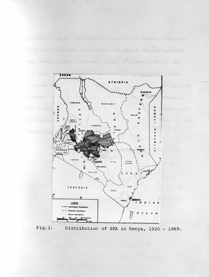
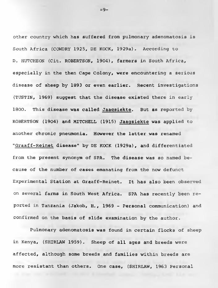
Distribution of SPA in Kenya, 1920 - 1969Fig. 2
-13-
same sheep's lungs. Laikipia, Nyandarua and Nakuru districts
of Kenya were the main areas where SPA was a serious problem
in some sheep flocks. Sporadic cases in other areas of the
country were due to movement of infected non-clinical cases.
Fig.2 shows districts of Kenya from which SPA has been reported.
The disease is still endemic in some sheep flocks in Nakuru,
Nyandarua and Laikipia Districts. The pathological description
of the disease by SHIRLAW (1959) was of mixed infections of SPA
and progressive interstitial pneumonia.
-14-
II. AETIOLOGY AND EPIDEMIOLOGY;
Jaagsiekte is a contagious disease. Its exact aetiology
has not been worked out but is probably caused by a virus. Most
of the published reports have laid emphasis on clinical signs,
epidemiology and pathology. Some suspected aetiological agents
have not been subjected to sufficient investigation. Lungworms
encountered in several cases (McFADYEAN, 1894, 1920; PALLASKE,
1929; and SEDLMEIER et al. 1966), are no longer regarded as the
cause. None of the worms or their larvae were found within the
adenomatous lesions. Secondly, there were lungs infected with
lungworms but which had no adenomatous transformations and pro
liferations. ENCHEV (1962) and DHANDA et al. (1963) failed to
observe any lung parasites or their ova after careful search,
Adenomatoid lesions produced by Muellerius capillaris (WANDERA,
1967a) can be distinguished by experienced investigators from
those of Jaagsiekte.
The non-existence of sheep pulmonary adenomatosis in
Australia where Muellerius capillaris occur and failure to
experimentally reproduce the disease (DUNGAL et al. 1938)
with lungworms discount the helminth theory. The significance
of Mycoplasma isolated by some investigators (MACKAY, 1966;
-17-
ptibility to sheep pulmonary adenomatosis, as with most infec
tious disease, is influenced by environment. Secondly resis
tance in affected flocks increases in proportion to the length
of time that they had been in contact with the infection.
DUNGAL et al. (1938) repeated contact transmission of
DE KOCK (1929b) by housing 8 sheep with diseased ones. One of
them died from the disease seven months later. Six had typical
lesions and the seventh had no lesions when killed ter. months
after contact. Clinical disease and death within 8 months were
produced by intrapulmonary inoculation of ground unfiltered
lung material of SPA in 1 of 3 sheep. However negative results
were obtained with a filtered sample inoculated in 5 sheep
after 10 months observation. In a later publication DUNGAL
(1946) demonstrated in two ways that expired air of an infected
sheep contained the infectious agent.
In the first instance sheep were maintained in an elevated
compartment 1.5 yards (146 cm.) above the heads of diseased sheep
kept in a lower compartment for 4-6 months. Hence other body excretions were excluded. Three of the eight lambs used had adenomatous lesions in their lungs. in the second instance a
diseased sheep was made to breathe through a 20% solution of
-18-
glycerine in normal saline for 30 minutes. Five millilitres of the
mixture were inoculated intratracheally and 2 ml. intrapul-
monary in three lambs. Two of the lambs developed lesions
of sheep pulmonary adenomatosis of which one showed clinical
signs 4 months after infection. In a similar experiment using
gradocol filtrate (0.9 p pore diameter) b f SPA lung suspension
he inoculated 5 ml. intratracheally into 4 lambs. Clinical
SPA disease developed in one of them, 4 months later. The other
3 killed at the same time had no lung lesions.
DUNGAL (1946) further obtained successful production of
SPA lesions following intranasal spraying of sheep with a fil
trate of bronchial secretion from an affected sheep obtained
by filtration through a Chamberland L3 filter. This method of
spraying, in addition to contact with clinical cases, was used
by MARKSON and TERLECKI (1964) by exposing 6 our-month-old
lambs to an aerosol spray consisting of a 10% suspension of
affected lung tissue. It was sprayed once a week into the air
of the loose-box in which they were kept for 2 years and 15
weeks. Spraying was again applied to another group of 6 lambs
without being kept in contact with clinical cases. Scattered
adenomatous lesions were present in the second instance in two
-19-
animals killed 380 and 864 days, and slightly larger lesions
(5-15mm. diameter) in the first instance in one sheep killed at
day 1034 after infection.
Typical lesions have also been reproduced by various
other routes (DUNGAL et al. 1938; DUNGAL, 1946; ENCHEV, 1966;
MARKSON & TERLECKI 1964; SIGURDSSON, 1958; WANDERA, 1968). These
were intranasal, intratracheal, intrapulmonary, intrapleural and
their various combinations. In South Africa, TUSTIN (1969) used
intravenous inoculation of the neoplastic cells, after their
growth in tissue culture, and their medium into 2 day-old lambs,
to produce clinical disease in one lamb which died 249 days
after infection. Advanced lung lesions were also found in the
other sheep killed after 253 days.
SHIRLAW (1959) produced typical adenomatous lesions in
rabbits. He inoculated 5 rabbits intravenously
with tiny infective lung fragments and 5 others with lung
suspensions incorporated in an agar plug. In two of the latter
and all the former rabbits, lesions suggestive of SPA were
observed in the lungs. Two of the rabbits inoculated with
lung fragments and kept for a longer time (200 days) had undoub
tedly pulmonary adenomatosis lesions. For in addition to
-20-
lymphoid hyperplasia around bronchioles and blood vessels
observed histologically, there was marked proliferation of bron-
chiolar epithelium with the formation of papillomatous tufts,
These tufts traversed and partially occluded the bronchiolar
lumina. The adenomatous process also involved alveoli and alveo
lar ducts. ZILBER et al. (1962) working in Russia produced,
after 4-6 months, multiple cysts filled with serous fluid in the
lymph nodes of mice following subcutaneous inoculation of a
Seitz-filtered filtrate of a suspension of adenomatous lung.
Taking the above experiments into consideration it is
definite that the natural mode of transmission of the disease
is aerogenous, and that spread is facilitated by close contact,
According to TUSTIN (1969) prenatal infection is of no signi
ficance if it ever takes place. Any sheep population is vulne
rable, and classic examples of an epidemic are the experiences
suffered by farmers in Iceland (SIGURDSSON, 1958) during the
latter 1930's - 1940's, in South Africa (TUSTIN, 1969) during
the 19th and early 20th centuries, and in Kenya (SHIRLAW, 1959).
However after it has appeared in a flock, the rate of infection
remains the same for 2-3 years, after which it decreases,

- 2 1 -
Incidence is increased by overcrowding as during cold periods,
or when it is hot by sheep crowding under a shade or where there
is none by placing their heads beneath the bellies of others in
order to gain some protection from the sun.
III. PATHOLOGY.
The pathology of sheep pulmonary adenomatosis has been
described by COWDRY (1925), DE KOCK (1929b), CUBA-CAPARO et al.
(1961), STAMP and NISBET (1963), MARKSON and TERLECKI (1964),
ENCHEV (1961), and WANDERA (1968). Grossly the disease is
characterised by multiple irregularly circumscribed solid no
dules .scattered throughout a large area of the lungs. These
nodules increase in number and size, eventually fusing and
giving a continuous grayish-white glandular tissue. Hence the
distinction into two anatomical forms: nodular and diffuse used
by some workers is arbitrary and erroneous and should be des
criptive only.
Affected lungs do not collapse when the chest is opened
and are increased in size three or more times their normal
weight and volume. There is usually a watery frothy fluid in
the trachea, bronchi and bronchioles, and the cut surface is
- 2 2 -
fleshy. The majority of cases tend to affect the apical and
cardiac lobes first. The pulmonary lymph nodes are sometimes
enlarged.
Histologically areas of adenomatous proliferations with
their protrusion into the alveoli and sometimes the bronchiolar
and bronchial lumina are encountered. Metastases to the tra
cheobronchial and mediastinal lymph nodes first observed by
AYNAUD (1926), have also been seen by PAREDES (1953), STAMP
& NISBET (1963), MARKSON & TELECKI (1964), CUBA-CAPARO et al.
(1961), ENCHEV (1963), SANTIAGO-LUQUE (1963), MITROFANOV (1964),
WANDERA (1967b) and TUSTIN (1969). MARTINCIC and CVJETANOVIC
(1967) encountered metastases in mediastinal and mesenteric
lymph nodes and ENCHEV (1963) observed them in the liver, spleen,
kidney and in the heart in one case. NOBEL et al. (1968) recorded
3 extrathoracic metastases - one involving the mesenteric peri
toneum, one a psoas muscle and the third, subcutaneous tissue
and musculature of the right retrofemoral region. TUSTIN (1969)
observed two SPA cases in which extensive spread of the neoplasm
to the pleura had taken place. These metastases place the dise
ase in the group of lung tumours. DE KOCK (1929a) mistakenly
-23-
classified this malignant tumour as "multiple papilliform
cyst-adenoma."
EBER (1899) was the first to recognise SPA as a neoplastic
condition: which he regarded as multiple adenomas in the lungs,
He observed the clear glandular epithelial forms. The cells
were columnar, originating from alveolar ducts and bronchioles.
It is now generally agreed (COWDRY, 1925; DE KOCK, 1929a;
CUBA-CAPARO et al. 1961), that both cuboidal and columnar
epithelial cells are involved, and do arise from alveoli, alveo
lar ducts, bronchioles, and bronchi. The initial lesion of
interalveolar wall thickening observed by COWDRY (1925),
COWDRY and MARSH (1927), SHIRLAW (1959), ENCHEV (1966) and
WANDERA (1967b, 1968), was not encountered by DE KOCK (1929a),
STAMP and NISBET (1963) and MARKSON and TERLECKI (1964). The
latter group regarded the minute adenomatous foci as the ini
tial lesions of pulmonary adenomatosis. There is no guarantee
that they had the opportunity of observing very early lesions.
As WANDERA (1968) stated, minute foci do not necessarily repre
sent beginning lesions.
-24-
Generally, in the fhst-growing SPA cases, an animal dies
before there has been time for secondary fibroplasia. In the
slow-growing SPA cases, possibly in the less susceptible sheep,
fibroplasia sets in and in addition to crowding out some of the
adenomatous cells, there is a tendency for the remaining tumour
cells to be much more cuboidal than columnar. Foci of myxomatous
tissue have been seen in a small proportion of SPA cases by
Me FADYE AN (1920), COWDRY (1925), PATTISON (1946), CUBA-CAPARO
et ai. (1961), and STAMP and NISBET (1963).
Descriptions of Jaagsiekte (ROBERTSON, 1904? MITCHELL,
1915)as a disease characterized by marked thickening of inter
alveolar walls and hyperplasia of lymphoid tissue refer to
what is now known as "Graaff-Reinet disease" (DE KOCK, 1929a).
This terminology is in reference to the now defunct Graaff-Reinet
Experimental Station from where both Robertson and Mitchell
obtained their sheep. "Graaff-Reinet disease" is akin in its
pathology to Maedi of Iceland, one aspect of "Montana progressive
pneumonia" of U.S.A. one form of "Bouhite" of France and Zwoeger-
ziekte of Netherlands (WANDERA, 1970).
Lymphoid hyperplasia round the bronchi, bronchioles and
blood vessels observed sometimes in SPA cases (WANDERA, 1967b)
-26-
some flocks in Central Sierra region. In Britain (MACKAY
and NISBET, 1966) the incidence is less than 1%, though
becoming a possible hazard of intensified sheep husbandry in
parts of that country. In Russia (ALIEV, 1967), the incidence
of SPA, especially in Azerbaijan ranged from 1.6 to 7.5^- In
Germany, clinical cases and losses in affected flocks show in
crease, such as 5% stated by PALLASKE (1954) and 22.41% encoun
tered by JAKOB and KRAUSE (1965).
Though no longer present in Iceland SPA was an epidemic
in that country. Losses in some flocks (DUNGAL et al. 1938)
ranged from 50 to 80% within 1-2 years. The disease is of
economic importance in Bulgaria. When first recorded in 1955,
ENCHEV et al. (1958) stated that affected flocks were losing
between 1-3%. Following spread to other regions of that country
(ENCHEV, 1961), an epidemic soon followed with losses on
certain farms reaching 35.5%, and is now endemic. Other countries
with serious outbreaks also had more losses from the disease,
such as 10 to 30% of affected flocks in Greece (CHRISTODOULOUS
and TARLATZIS, 1952).
-27-
At one time Kenya had up to 30% annual mortality
(SHIRLAW, 1959), from the disease alone on certain farms. It
has now dropped down to between 1 to 5% (WANDERA, unpublished
data) in endemic flocks. This could possibly be due to better
husbandry and elimination of overcrowding at night and partly
due to the fact that, as with most infectious diseases, susce
ptibility is gradually reduced in endemic flocks. The figure
for South Africa (DE KOCK, 1929b) was less than 5%. But accor
ding to TUSTIN (1969) when the disease was first introduced in
South Africa in 1800's losses of 30% and more in certain flocks
were not unusual. None of the affected sheep ever recover.
V. GEOGRAPHICAL ORIGINS OF THE DISEASE.
It is not known just how the disease started in Kenya.
However spread to some countries and within a country (KOSTENKO,
1964) can be traced to importation of healthy latently infected
sheep. While the disease may have occured in Scotland for many
years, its spread to England is associated with infected but
apparently healthy sheep from the former (STEVENS, 1957). Though
SPA was and is still largely sporadic in Germany, the Icelandic
-28-
epidemic is traced back to Karakul sheep bought from Germany
(DUNGAL, 1946) in the district of Halle in December of 1933.
Indeed cases recorded in Yugoslavia (CVJETANOVIC & MARTINCIC,
1962) are believed to have been introduced by Merino rams from
France and German Federal Republic.
For many years it was believed that in Kenya Jaagsiekte
occurred naturally in Masai sheep (SHIRLAW, 1959). However,
the available evidence (WANDERA, 1967a) shows that Masai sheep
may have been suffering from bacterial and/or parasitic pneu
monia and not Jaagsiekte. METTAM (1927) encountered the disease
in flocks of Merino sheep in Kenya on farms where no Masai sheep
had ever been. Both Jaagsiekte and Maedi could have been intro
duced by Merino sheep imported into Kenya ever since 1904 from
South Africa, TUSTIN (1969) showed that South African sheep
farmers had been losing sheep from pulmonary adenomatosis since
the 1800:s, and further produced evidence to show that the dise
ase was brought in that country with the importation of Spanish
Merino sheep.
It is hard to explain how and why the disease has not been
recorded in Australia and New Zealand. Both these two countries

-29-
imported Merino sheep direct, and, via Britain and South
Africa from Spain. It would appear, on the basis of available
circumstantial evidence, as if the original home of SPA could
be Spain and possible France and Germany. If this is probable,
is it likely that the disease could have spread with the move
ment of the Merino sheep and those that had been in-contact with
them to other countries. Of all the sheep breeds, it is only
the Merino (RYDER and STEPHENSON, 1968) which has had the widest
distribution, ever since it left its Spanish home. The chance
°f infected symptomless Merino sheep leaving Europe to South
Africa, spreading the disease there and later some asymptomatic
infected Merino sheep and their crosses being imported into
Kenya is quite feasible.
-30-
CHAPTER 3
AETI0L0GICAL STUDIES
Despite the fact that SPA is a contagious disease, its aetio-
logical agent has not been isolated and characterised. Accor
ding to SHIRLAW (1959) adenomatous lung tissue 'preparation passa
ged in chicken embryos and put back into sheep produced lung
lesions of the disease. He used yolk sac membrane and the whole
embryo of 34th passage and yolk sac membrane alone at the 56th
passage. On this basis it would appear that, irrespective of its
nature the causal agent can propagate in chicken embryos and be
transmitted to susceptible sheep.
The aims of the present studies are first to repeat SHIRLAW’S
work, and secondly to try and isolate infectious agents propagating
in embryonating eggs.
I: STUDIES WITH CHICKEN EMBRYOS
(1) Sourde and Preparation of Eggs: At the beginning of these
studies fertile eggs were obtained from a commercial flock; later,
following the establishment of a Specific Pathogen Free (SPF)
flock, in the Department of Veterinary Pathology, free of known
poultry pathogens including leukosis, such eggs were obtained
from an internal source. Eggs were incubated at 37°C., candled
-31-
at the 5th day and r.on-fertile, weak or dead ones removed. On
the 7th day, selected embryonating eggs were prepared for ino
culation.
(2) Preparation of Infective Materials: Specimens used for
passaging in chicken embryos were from sheep with clinical signs
and lesions of pulmonary adenomatosis. Lung specimens W^, W^,
were from naturally affected sheep killed and immediately prepared
for inoculation. Sample was the same as that used for infecting
sheep in the Transmission Experiment 2 of Chapter 4. and
were from sheep of the same flock as the first one. Specimers
W4 , w . and W62, used for passaging came from sheep 3616, 3615
and 3645 respectively, with experimentally produced diseaseo(Cnapter 4) . Lung specimens W and W had been frozen at -70 C.4 5
for 9 and 6 days respectively before use. Specimen W62 was
lung fluid which was collected via the nostrils by lifting up the
hind limbs and used immediately.
Inoculum of was prepared as follows:
One hundred and twenty of representative lung lesions obtained
in a sterile way was minced finely with scissors and homogenized in a
Silverstone blendor for one minute at full speed. After the first
-32-
blending about 60ml. of Eagle's medium containing 100 I„U.
penicillin, 100 ug, streptomycin, 10 0 ug. kanamycin per ml, was
added and the suspension again blended for one minute, Follo
wing centrifugation in the refrigerated MSE centrifuge for 10
minutes at 2,500 g., the supernatant was removed and stored in
an ice-bath. To the sediment 3ojnl. of the same medium was added
and the suspension subjected to ultrasonic vibration with a
Branson Sonifier Model S-125 (Branson Instr., Danburry, Conn,,
U.S.A.) for about 30 seconds at position 8 . The suspension was
next centrifuged as before, but for one hour. This second super
natant was removed and mixed with the first one, giving it a con
centration of over 55%. A sample of this suspension was used for
sheep inoculation in Experiment 2 Chapter 4. Another sample of
this preparation was diluted to 10% with the same medium and
filtered through 0.45 u Millipore, before egg inoculation,
Essentially the same method was used for preparing 10% con
centrated inocula of W^, and W _. Penicillin, streptomycin
and kanamycin were included in the medium for preparing but
kanamycin was not used in V7 , W , and W_. These four samples3 4 5were used unfiltered. Lung fluid making the inoculum of W62
was the third of the 5 fluid samples collected at intervals from
-33-
sheep 3645. This one was obtained at day 195 after infection,
The fluid was diluted 1:2 with Trisbuffer and filtered through
1 .2 microns filter to remove cells and large subcellular ele
ments. No antibiotics were used.
(3) Methods of Inoculation and Sample Collection:
Preliminary trials were made with chorioallantoic membrane,
allantoic cavity, amniotic cavity and yolk sac routes, The yolk
sac method was found to be most effective and convenient, because
of easy administration and the fact that eggs generally died more
regularly. The 7-day old embryonating eggs were inoculated with
0.2ml. through the air-cells into the yolk sac using an opening
made away from the embryos and large blood vessels after disin
fection with iodine. The lobes were sealed with "UHU" glue.
The infected embryonating eggs were again incubated at 37°C,
and candled daily. Embryo deaths within 24 hours were regarded
as accidental and disregarded while those alive by the 18th day
of incubation were killed. The allantoic-amniotic fluid (AAF)
was collected from those embryos dying between the 2nd - 1 1 th day
after infection. In some cases, liver and heart were also colle
cted. AAF and collected organs were stored at -20°C., after some
of it being filtered through sterile Millipore filters of 0,45 u
-34-
mean pore diameter for further embryo inoculations. Several
passages of and W62 were titrated in embryonated eggs. Both
dead and killed embryos were examined post-mortem for possible
gross lesions. Each original and passage samples were routinely
checked for bacteria on plates of McConkey's and blood agar, and
for Mycoplasma on Tryptose Serum Agar (TSA) and in Newing's
Tryptose Broth (NTB).
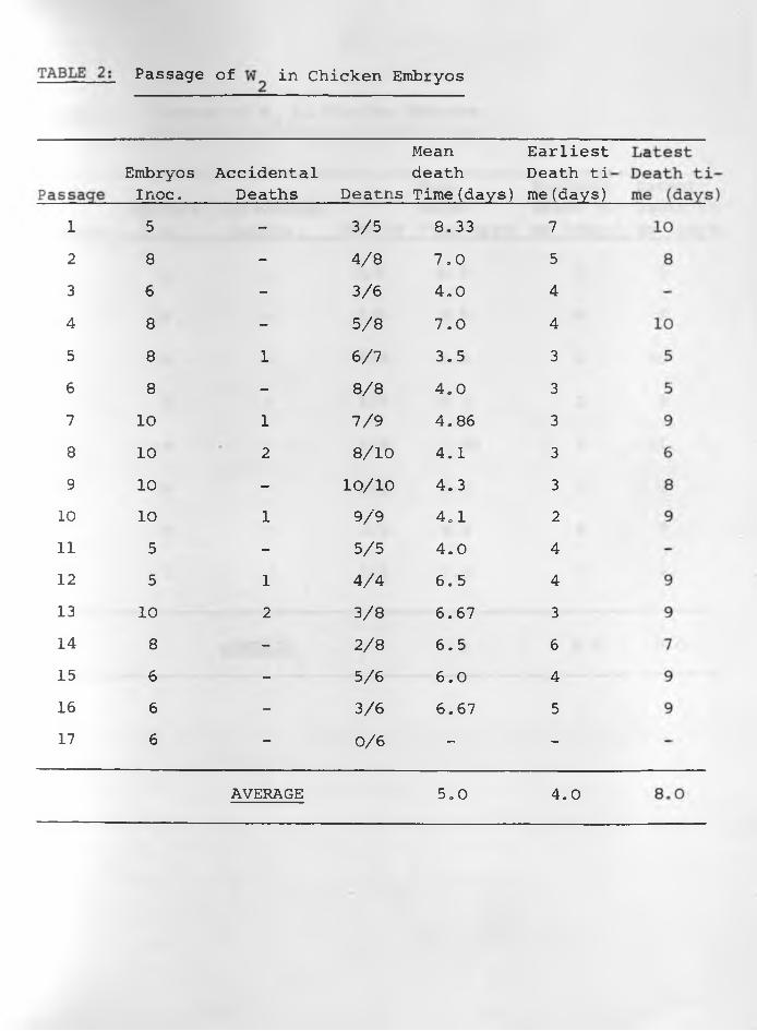
(4) Results: The data of the six sets of passage of infective
materials in embryos are indicated in TABLES: 1, 2, 3, 4, 5, 6 .
Embryos died more regularly with specimens W^, and W62. The
average mean death time was about 5.0 days for and The
earliest average death time was 3.0 days while the latest was
7.0 days in W^. For it was 4.0 days and 8.0 days respectively,
Passages of W , W and W had irregular results in all respects.3 4 5With the exception of the 1st, 11th, 14th, 15th and 16th passages
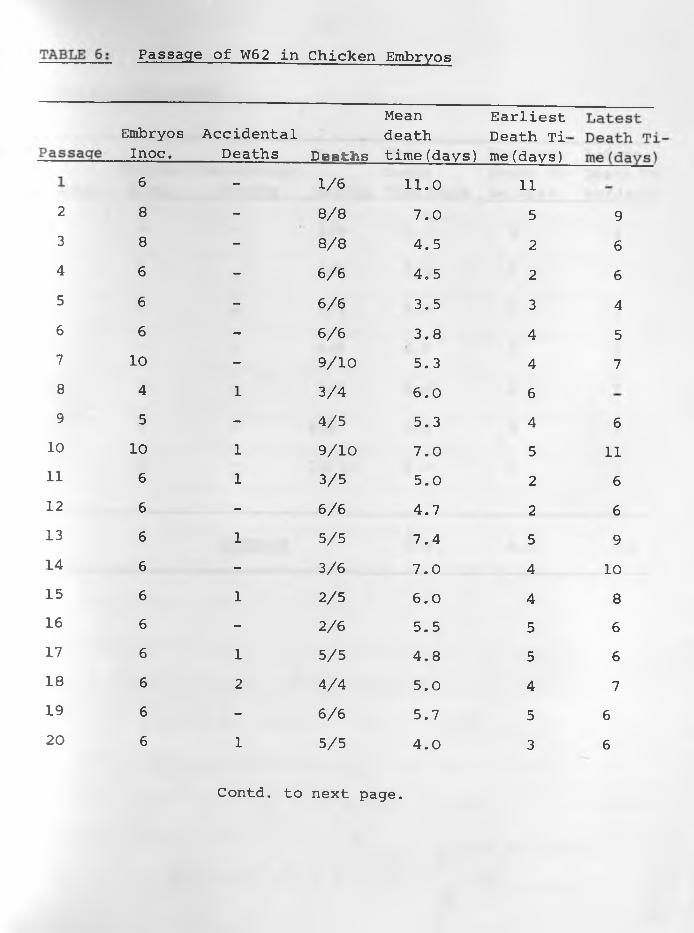
embryo deaths were consistent in all the 27 passages of W62.
The average mean death time was 5.0 days whilst the earliest and
latest average death times were 4.0 and 6 .0 days respectively.
Constant lesions caused by W^, and W62 were haemorrhages •under the skin of embryos and visceral organs especially liver
and heart in those dying 3-5 days following inoculation. Liver
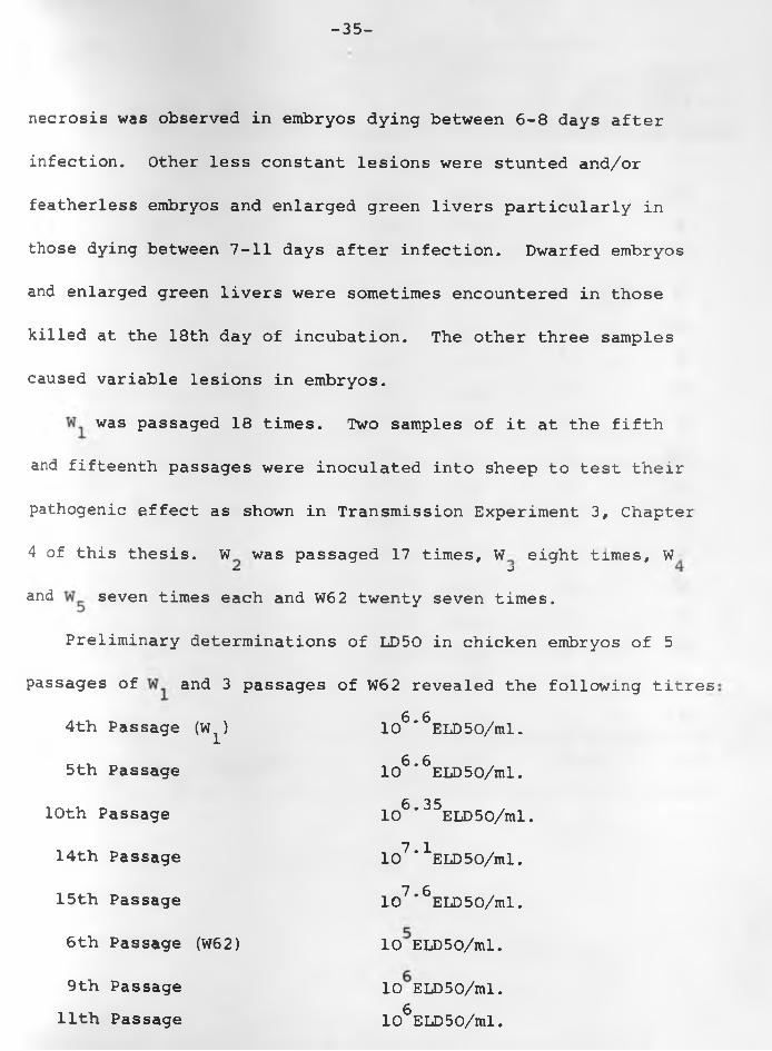
-35-
necrosis was observed in embryos dying between 6 - 8 days after
infection. Other less constant lesions were stunted and/or
featherless embryos and enlarged green livers particularly in
those dying between 7-11 days after infection. Dwarfed embryos
and enlarged green livers were sometimes encountered in those
killed at the 18th day of incubation. The other three samples
caused variable lesions in embryos.
was passaged 18 times. Two samples of it at the fifth
and fifteenth passages were inoculated into sheep to test their
pathogenic effect as shown in Transmission Experiment 3, Chapter
4 of this thesis. W was passaged 17 times, W eight times, W“ J
and seven times each and W62 twenty seven times.
Preliminary determinations of LD50 in chicken embryos of 5
passages of and 3 passages of W62 revealed the following titres
4th Passage (W )
5th Passage
10th Passage
14th Passage
15th Passage
6 th Passage (W62)
9th Passage 11th Passage
6 610 * ELD50/ml.6 610 * ELD50/ml.6 3510 * ELD50/ml.7 110 ' ELD50/ml.7 610 ' ELD50/ml.
10 ELD50/ml.
10 ELD50/ml.g
10 ELD50/ml.
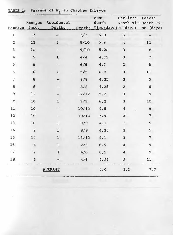
TABLE 1: Passage of in Chicken Embryos
PassageEmbryosInoc.
AccidentalDeaths Deaths
Mean death Time(days)
Earliest Death Time (days)
Latest Death Time (days)
1 7 - 2/7 6 .0 6 -2 12 2 8 / 1 0 5.9 4 10
3 10 - 9/10 5. 20 3 8
4 5 1 4/4 4.75 3 75 6 - 6 / 6 4.7 3 6
6 6 1 5/5 6 .0 3 1 1
7 8 - 8 /8 4.25 3 58 8 - 8 /8 4.25 2 6
9 12 - 1 2 / 1 2 5.2 3 910 10 1 9/9 6 .2 3 10
1 1 10 - 1 0 / 1 0 4.6 4 6
12 10 - 1 0 / 1 0 3.9 3 713 10 1 9/9 4.1 3 514 9 1 8 /8 4.25 3 515 14 1 13/13 4.1 3 716 4 1 2/3 6.5 4 917 7 1 4/6 6.5 4 918 6 — 4/6 5.25 2 1 1
AVERAGE 5.0 3.0 7.0
12
345678
9
10
11
12
1314151617
Passage of in Chicken Embryos
Mean EarliestEmbryosInoc.
AccidentalDeaths Deatns
death Time(days)
Death ti me (days)
5 - 3/5 8,33 78 - 4/8 7.0 56 - 3/6 4,0 48 - 5/8 7.0 48 1 6/7 3.5 38 - 8 /8 4.0 3
10 1 7/9 4.86 310 2 8 / 1 0 4.1 310 - 1 0 / 1 0 4.3 310 1 9/9 4.1 2
5 - 5/5 4.0 45 1 4/4 6,5 4
10 2 3/8 6.67 38 - 2 /8 6.5 6
6 - 5/6 6 .0 46 - 3/6 6.67 56 - 0 /6 — -
AVERAGE 5,0 4.0

TABLE 3 Passage of 3 in Chicken Embryos
PassageEmbryosInoc.
AccidentalDeaths Deaths
Mean Death Time(days)
Earliest Death Time (days)
Latest Death Ti me (days
1 8 - 2 /8 6.5 5 7
2 6 - 2 /6 8.5 8 9
3 8 - 5/8 6 .6 2 10
4 8 1 4/7 6 .0 2 8
5 8 - 8 /8 7.25 5 1 1
6 8 - 2 /8 5.0 3 7
7 6 - 2 /6 00 % 8 9
8 5 1 1/4 3.0 3 -
AVERAGE 6 .0 4.5 9.0
i
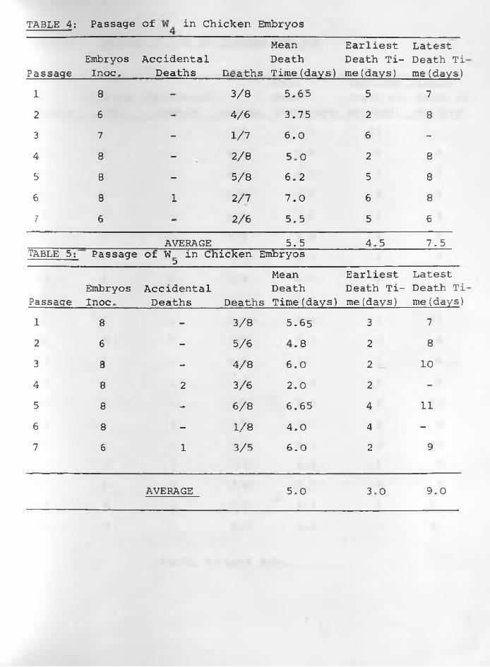
TABLE 4: Passage of in Chicken Embryos
PassageEmbryosInoc.
AccidentalDeaths Deaths
Mean Death Time(days)
Earliest Death Time (days)
Latest Death Ti' me(days)
1 8 - 3/8 5.65 5 72 6 - 4/6 3.75 2 8
3 7 - 1/7 6 .0 6 -4 8 - 2 /8
o0in 2 8
5 8 - 5/8 6 .2 5 8
6 8 1 2/7 7.0 6 8
7 6 - 2 /6 5.5 5 6
AVERAGE 5.5 4.5 7.5TABLE 5: Passage of W in Chicken Embryos 5
Mean Earliest LatestEmbryos Accidental Death Death Ti- Death Ti
Passaqe Inoc„ Deaths Deaths Time(days) me (days) me(days)1 8 - 3/8 5.65 3 72 6 - 5/6 COe 2 8
3 8 - 4/8 6 .0 2 10
4 8 2 3/6 2 .0 2 -5 8 - 6 / 8 6.65 4 1 1
6 8 - 1 / 8 4.0 4 -7 6 1 3/5 6 .0 2 9
AVERAGE 5,0 3,0 9,0
2
345678
9
10
1112
1314151617181920
96
6457
611
669
10
8
6
6
766
Passage of W62 in Chicken Embryos
EmbryosInoc.
AccidentalDeaths Deaths
Mean death time(days)
Earliest Death Tb me (days)
6 - 1 / 6 1 1 . 0 1 1
8 - 8 /8 7.0 58 - 8 /8 4.5 2
6 - 6 / 6 4.5 2
6 - 6 / 6 3.5 36 - 6 / 6 3.8 4
10 - 9/10 5.3 44 1 3/4 6 .0 6
5 - 4/5 5.3 410 1 9/10 7.0 5
6 1 3/5 5.0 2
6 - 6 / 6 4.7 2
6 1 5/5 7.4 56 - 3/6 7.0 46 1 2/5 6 .0 46 - 2 /6 5.5 56 1 5/5 4,8 56 2 4/4 5.0 46 - 6 /6 5.7 56 1 5/5 4.0 3
Contd, to next page.
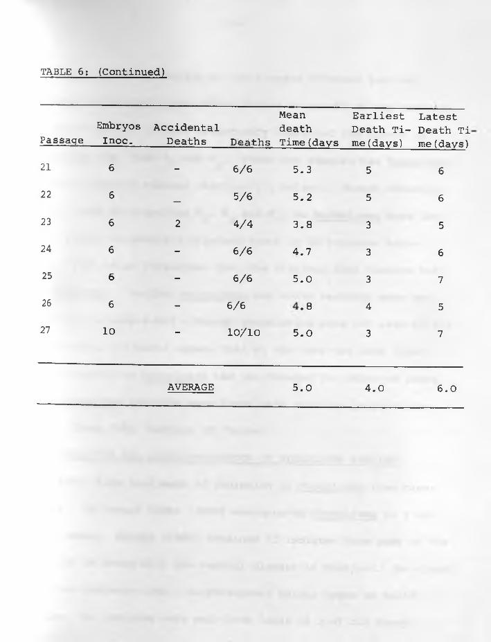
TABLE 6; (Continued)
PassaqeEmbryosInoc.
AccidentalDeaths Deaths
Mean death Time(days
Earliest Death Time (days)
Latest Death Ti me(days)
21 6 - 6 / 6 5.3 5 6
22 6—
5/6 5.2 5 6
23 6 2 4/4 3.8 3 524 6 - 6 / 6 4.7 3 6
25 6 - 6 / 6 5.0 3 726 6 - 6 /6
00• 4 527 10 — 1 0 / 1 0 5.0 3 7
AVERAGE 5.0 4.0 6 . 0
-36-
Samples of every passage of all 6 cases cultured both on
McConkey's and blood agar plates showed no growth of any bacteria.
Kanamycin appears to have effectively inhibited the growth of
Mycoplasma spp. from and These two samples had Mycoplasma
in their original tumours (Section II, below). Though kanamycin
was not used in preparing W , and W^, no Mycoplasma were iso
lated either in Newing's Tryptose Broth or on Tryptose Serum <
Agar. This is an indication that the original lung tumours had
no Mycoplasma. Neither Mycoplasma nor other bacteria were en
countered in sample W62 although antibiotics were not used in its
preparation. It would appear that at the time the lung fluid
was collected, no Mycoplasma had yet invaded the affected lungs.
Four Mycoplasma isolates were later made from the lungs of the
donor, sheep 3645 (Section II, below) .
II. ISOLATION AND CHARACTERIZATION OF MYCOPLASMA SPECIES
Reports have been made of isolation of Mycoplasma from cases
of SPA. In Israel NOBEL (1958) encountered Mycoplasma in 2 out
of 19 cases. MACKAY (1966) obtained 22 isolates from some of the
lungs of 30 sheep with the natural disease in Scotland. He classi
fied his isolates into 2 morphological colony types on solid
medium. No isolates were made from lungs of 9 of his sheep.
-37-
DEIANA and CERETTO (1967) working in Sardinia, Italy, found
antibodies against Mycoplasma isolated from SPA cases.
So far with the exception of DEIANA and CERETTO (1967) only
a secondary role has been attributed to Mycoplasma in adenomatous
lungs of sheep. It has not yet been conclusively shown that
they do not play a role in the causation of SPA, Mycoplasma
have been encountered in Maeai lungs (WANDERA, 1970), and nasal
fluid (KRAUSS, H. - 1969, unpublished data) from 40 sheep sent
for slaughter but free of any lung lesions. Mycoplasma have
further been isolated from pneumonic lungs in Australia (ANON,
1964-67) where SPA has not yet been observed. While organisms
isolated from Australia may be non-pathogenic, there is no
record in literature of respiratory-pathogenic Mycoplasma
of sheep. Hence circumstantial evidence suggests that Mycoplasma
isolated so far are not involved in causing the adenomatous
process of Jaagsiekte.
Mycoplasma were encountered during investigations of the
aetiological agent of pulmonary adenomatosis of sheep in Kenya.
Apart from COTTEW et al. (1968) in Turkey, little has been done
to elucidate the properties of Mycoplasma isolated from sheep.
-38-
The present investigation was undertaken to isolate Mycoplasma
spp. from natural and experimental cases of Jaagsiekte and study
some of their properties.
(2) Materials and Methods:
(a) For isolation and propagation of Mycoplasma: The
liquid medium used was modified Newing's Tryptose Broth (NTB)
as described by BROWN et al. (1965), made up of:
Bacto-Tryptose (Dif c6 ).... ................. . ..... 2.00%
Dextrose .................................... . 0. 50%
Sodium chloride ........................... 0,50%
Anhydrous di-sodium phosphate (Na^HPO^) .......... 0.25%
Glycerol....................................... . 0.50%
Distilled w a t e r ........................... to 100.00%
The solid medium used was Tryptose Serum Agar (TSA) as described
by DAVIES and READ (1968), and prepared in two parts as follows:
(i) Tryptose agar
Bacto-tryptose (Difco) .............. . 2.00%
Sodium chloride .................... ........ . 0.50%
Glycerol.... ...... . ... ............................ . 0. 50%
Anhydrous di-sodium phosphate (Na^PO^) ............ 0.25%
Distilled water to 100.00%
-39-
(ii) Pig Serum and Additives
Inactivated (fresh) pig s e r u m ............... 30 ml.
Dextrose ...................................... 5 ml. (10%)
Bacto-yeast extract (Difco) ................. I ml. (10%)
Crystalline penicillin G (Glaxo) ............ 100,000 Units
Thallium acetate ............................. 1 ml. (1%Soln.)
Part (i) was prepared and kept in stock until needed.
Part (ii) was prepared immediately before use and filtered through
Seitz E.K. pads before being added to the melted part (i) in the
ratio of 100 volumes part (i) to 36 volumes part (ii). In some
cases of solid medium the penicillin and thallium acetate were
omitted.
(b) Embryonated chicken eggs were from a SPF-flock. Infec
tive material was inoculated into the yolk sac after 7 days of. oincubation at 37 C. The eggs were candled daily to detected
mortality. Alantoic/amniotic fluid was collected from dead
embryos for further passage.
(c) Tissue cultures of chick embryo kidneys from 17 days
old embryos, and chick embryo fibroblasts from 1 0 days old
embryos after removing heads, legs and visceral organs were
-40-
prepared by trypsinization. Trypsin was at a concentration of
0.25% in buffer. The culture medium used was 50% Eagle's medium
in 50% Hanks' medium. For growth, 10% calf serum was added.
Lung macrophage cultures were obtained from normal lungs of
freshly killed sheep. Such lungs were first filled with Hanks'
BSS via the trachea. The opening was stoppered and the distended
lungs massaged gently for about 5 minutes. The fluid was next
poured out into a sterile container and centrifuged at 400 r.p.m.
for 10 minutes. The cells were washed once with Hanks' BSS
following sedimentation. The sedimented cells were suspended
to a concentration of 1.25% in Eagle's medium in Hanks' BSS
containing 10% tryptose phosphate broth, and 1 0% inactivated
horse serum. Antibiotics used were 20 units penicillin, 50 p g .
streptomycin and 200 ) ig . kanamycin per ml. of the suspension.
Cultures were inoculated with infective material and checked
daily for signs of a cytopathic effect. Medium was collected
at the 7th day after infection for further propagation.
(d) Primary isolation of Mycoplasma was from killed sheep
with clinical or pathological evidence of SPA. Materials
used were either directly from the lung tumours and/or,
samples of discharge from the lungs through the nostrils on
-42-
(f) Preparation of antisera for serological investigation:
Rabbits were bled for pre-inoculation sera and then inoculated
with 100-fold washed concentrate of a 24-48 hour broth culture,11 12lml. containing about 10 to 10 living organisms. A series
of 4 inoculations were made, The first 3 were done subcutaneously
using 2ml. of concentrate mixed with 2L1I, of Freund's incomplete
adjuvant, at weekly intervals, and the last one intraperitoneally■ v / °with 4ml. of concentrate only. Seven to-ten days after the last
inoculation the rabbits were bled for serum.
Hyperimmune sera against T2/33 Vaccine-strain of bovine
pleuro-pneumonia previously used in Uganda (SANDERS, 1961)
and against M, laidlawii were available for comparative study.
(g) Agar gel double precipitation reaction: The reactants of
the tests were incubated in a moist atmosphere at 37°C. for 3
days and the precipitation results recorded. Mycoplasma concen
trates, subjected to ultra-sonic vibration at 4°C. with a
Branson Sonifier Model S-125 (Branson Instr,, Danburry, Conn.,
U.S.A.) for 3 min. at position 6 were used as antigens, All anti
sera were absorbed with lyophilized NTB containing 20% (V/V) horse
serum by thoroughly mixing 200 gm. of the dry powder into lml.oimmune serum. The absorption was continued for 2 days at 4 C,
-43-
The antisera were cleared by centrifugation at lOOOg. for 10
minutes.
(h) Inoculated TSA plates were incubated at 42°, 37°, 32°, oand 22 C., for 7 days and growth recorded. Isolates growing on
serum-free tryptose agar were cloned, propagated in NTB without
serum and again grown on serum-free tryptose agar.
(i) Other tests; These included carbohydrate fermentation,
production of catalase, H^S, and reduction of methylene bine,
tetrazoliura blue, and production of haemolysis, serum liquefa
ction and haemadsorption. The medium used was NTB or TSA, glu
cose and yeast being omitted where necessary. Production of
indol was tested by use of Kovac's reagent, with an indcl pro
ducing strain of Escherichia coli as control. Haemagglutination
tests were performed with 0.5ml. of serial dilutions of washed
Mycoplasma concentrates in phosphate buffered saline and 0.5ml,
of a 0.5% horse or sheep erythrocyte suspension. Tests were in
cubated at room temperature and read when the erythrocytes had
sedimented.
(j) A mixed Mycoplasma sample from W^, W^, W6/1 and W6/'2
were used for sheep inoculation in Transmission Experiment 4,
Chapter 4 of this thesis to test their pathogenicity.
-44-
Out of 14 killed sheep with typical SPA disease, 10 had
Mycoplasma in their adenomatous lungs and/or lung secretions., Six
teen isolates were made from them; 6 from lung secretions via the
nostrils and 10 from tumours directly. Lung tumours and secretions
of 4 sheep (cases W .W W W62) yielded no Mycoplasma.J T | OThe solid medium (TSA) was found superior to liquid medium (NTB)
or embryonating eggs for the primary isolation of Mycoplasma. The
number of primary colonies could be checked and morphologically di
fferent colonies on the same plate could be cloned immediately after
outgrowth. Visible colonies appeared within 3 to 10 days after ino
culation in numbers that were identifiable by microscopic observation
and sometimes with the naked eye. Bacterial contamination was effe
ctively inhibited by the use of TSA medium for primary isolation,,
Development and passage of possible L-forms of bacteria was avoided
by passages in NTB medium without antibiotics or thallium acetate.
Mycoplasma isolates passaged in chicken embryos showed no
evidence of their pathogenicity. Embryo deaths were very sporadic
even after several passages with the isolates. The VOM strain on
the other hand killed embryos within 2 days after an initial passage
and the T^ vaccine strain within 2 to 7 days. Mycoplasma growth in
tissue cultures of embryo fibroblasts and lung macrophages produced
(3) Results;
-44-
Out of 14 killed sheep with typical SPA disease, 10 had
Mycoplasma in their adenomatous lungs and/or lung secretions. Six
teen isolates were made from them? 6 from lung secretions via the
nostrils and 10 from tumours directly. Lung tumours and secretions
of 4 sheep (cases W ,W W W62) yielded no Mycoplasma.J | 3The solid medium (TSA) was found superior to liquid medium (NTB)
or embryonating eggs for the primary isolation of Mycoplasma. The
number of primary colonies could be checked and morphologically di
fferent colonies on the same plate could be cloned immediately after
outgrowth. Visible colonies appeared within 3 to 10 days after ino
culation in numbers that were identifiable by microscopic observation
and sometimes with the naked eye. Bacterial contamination was effe
ctively inhibited by the use of TSA medium for primary isolation,,
Development and passage of possible L-forms of bacteria was avoided
by passages in NTB medium without antibiotics or thallium acetate.
Mycoplasma isolates passaged in chicken embryos showed no
evidence of their pathogenicity. Embryo deaths were very sporadic
even after several passages with the isolates. The VOM strain on
the other hand killed embryos within 2 days after an initial passage
end the vaccine strain within 2 to 7 days. Mycoplasma growth in
tissue cultures of embryo fibroblasts and lung macrophages produced
(3) Results:
-44-
Out of 14 killed sheep with typical SPA disease, 10 had
Mycoplasma in their adenomatous lungs and/or lung secretions, Six
teen isolates were made from them; 6 from lung secretions via the
nostrils and 10 from tumours directly. Lung tumours and secretions
of 4 sheep (cases W ,W W W62) yielded no Mycoplasma.J T f | J
The solid medium (TSA) was found superior to liquid medium (NTB)
or embryonating eggs for the primary isolation of Mycoplasma. The
number of primary colonies could be checked and morphologically di
fferent colonies on the same plate could be cloned immediately after
outgrowth. Visible colonies appeared within 3 to 10 days after ino
culation in numbers that were identifiable by microscopic observation,
and sometimes with the naked eye. Bacterial contamination was effe
ctively inhibited by the use of TSA medium for primary isolation.
Development and passage of possible L-forms of bacteria was avoided
hy passages in NTB medium without antibiotics or thallium acetate.
Mycoplasma isolates passaged in chicken embryos showed no
evidence of their pathogenicity. Embryo deaths were very sporadic
even after several passages with the isolates, The VOM strain on
the other hand killed embryos within 2 days after an initial passage
and the T^ vaccine strain within 2 to 7 days. Mycoplasma growth in
tissue cultures of embryo fibroblasts and lung macrophages produced
(3) Results:
-_45-
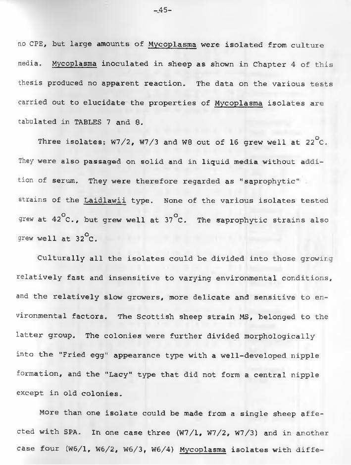
no CPE, but large amounts of Mycoplasma were isolated from culture
media. Mycoplasma inoculated in sheep as shown in Chapter 4 of this
thesis produced no apparent reaction. The data on the various tests
carried out to elucidate the properties of Mycoplasma isolates are
tabulated in TABLES 7 and 8 .
Three isolates; W7/2, W7/3 and W 8 out of 16 grew well at 22°C.
They were also passaged on solid and in liquid media without addi
tion of serum. They were therefore regarded as ’'saprophytic"
strains of the Laidlawii type. None of the various isolates tested o ogrew at 42 C., but grew well at 37 C. The saprophytic strains also
grew well at 32°C.
Culturally all the isolates could be divided into those growing
relatively fast and insensitive to varying environmental conditions,
and the relatively slow growers, more delicate and sensitive to en
vironmental factors. The Scottish sheep strain MS, belonged to the
latter group. The colonies were further divided morphologically
into the "Fried egg" appearance type with a well-developed nipple
formation, and the "Lacy" type that did not form a central nipple
except in old colonies.
More than one isolate could be made from a single sheep affe
cted with SPA. In one case three (W7/1, W7/2, W7/3) and in another case four (W6/1, W6/2, W6/3, W6/4) Mycoplasma isolates with diffe-
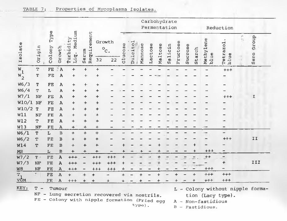
TABLE 7; Properties of Mycoplasma Isolates
C arbohydr ateFermentation Reduction
a) g +J — — ■-----------------— --------------------------------pu _ P d
Isol
ate
Orig
inEh>ido0U Gr
owth
•P Ti •H 0 TJ S •HrQ «p tr P -H>Eh PT Seru
m
s<UM•HPtQ)
GrowthoC.
Q)COou>-<
r fp .od Q)-P CO - -H 0u d •h d P <0 Q l S Lact
ose
Malt
ose
t Sal
icin
Fruc
tose
i i Suc
rose
Star
chMe
thyl
ene
blue
orH0NrdP d) -P P (D i—1& A
0PO-0p<uCA32 22 r-Ho
w i T FE ; A + + + + + +
W2T FE A + + +
W6/3 T FE A + + + —
W6/4 T L A + + +W7/1 NF FE A + + + +++ IW 1 0 / 1. NF FE A + + +W 1 0 / 2! T FE A + + + -Wll NF FE A + + + -W 1 2 T FE A + +■ -tW13 NF FE A + + +W 6 / 1 T L B + + +W 6 / 2 T FE B + + + +++ IIW14 T FE B + + -f- - + - - + - - - + - -MS L B + + + - + + - + - - - + +++ -W7/2 T r FE A +++ . - +++ +++ + - - + - - - - _ ++ -W7/3 NF FE A +++ - +++ +-f-+ + + IIIW 8 NF FE A +++ — +++ +++ + — — — + — - - ■ - +++ -Ti
7“ FE A + + — + + - + - + - + +++ +++VOM FE A +++ + + + + + — + — + — + +++ +++KEY: T - Tumour
NF - Lung secretion recovered via nostrils.FE - Colony with nipple formation (Fried
t y p e ) .
L - Colony without nipple formation (Lacy type).
A - Non-fastidious B - Fastidious.
egg
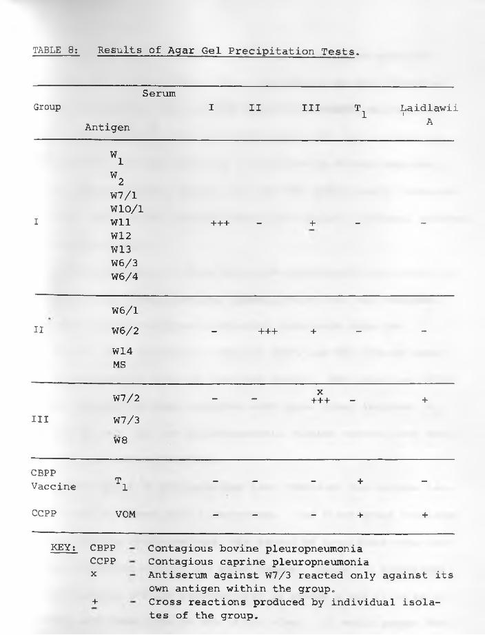
TABLE 8: Results of Agar Gel Precipitation Tests,
SerumGroup I II Ill Ti Laidlawiii
Antigen A
w iW2W7/1W10/1
I Wll +++ - + - -W12W13W6/3W6/4
W6/1II W6/2 - +++ + - -
W14MS
W7/2 - -X+++ - +
III W7/3W 8
CBPPVaccine Ti - - - + -
CCPP VOM - - - + +
KEY: CBPPCCPP x
+
Contagious bovine pleuropneumonia Contagious caprine pleuropneumonia Antiserum against W7/3 reacted only against its own antigen within the group.Cross reactions produced by individual isolates of the group.
-46-
rent properties were recovered. Most of the isolates were non
fermenters of carbohydrates. The 3 saprophytic strains fermented
glucose and maltose. Isolate W14 fermented glucose, maltose and
starch, comparable with the Scottish strain, MS, but it could be
distinguished from the latter by its failure to ferment mannose.
The two pleuropneumonia strains (T and VOM) additionally fermented
fructose. None of the isolates fermented dulcitol, lactose, mannose,
sucrose and salicin.
Alpha-haemolysis of sheep and horse erythrocytes was shown by
all types while haemadsorption, haemagglutination, and catalase,
indol and hydrogen sulphide production tests were negative,
Two of the 3 saprophytic strains (W7/2 and W 8) reduced methy
lene blue, together with the Scottish strain. The third one (W7/3)
reduced tetrazolium blue, together with three other isolates (W^,
W7/1 and W6/2). The two pleuropneumonia strains reduced both methy
lene blue and tetrazolium blue.
On the basis of precipitation test (TABLE 8 ) the various iso
lates could be placed into 3 serogroups. The first group consisted
of fast growing non-fermenters, the second of fastidious organisms
partly fermenting glucose and other sugars. Group III Mycoplasma
are saprophytic and the pathogenicity of groups I and II is as yet
unknown, and their role in SPA is not clear. It would appear that
-48-
consolidation in cases of pasteurella pneumonia in lambs about 3
weeks old.
In Section II of this Chapter no Mycoplasma were recovered from
lungs and lung secretion of four (W , W - W , W62) out of 14 sheep3 4 5
with clinical SPA. These four cases were amongst the 6 (Section I)
whose infective preparations were passaged in embryonating eggs.
While W , W , and W killed eggs sporadically, W62 caused more consi- 3 4 5stent embryo deaths over most of its passages, Samples W^, Vv_
and W62 originated from experimental animals, sheep 3616, 3615 and
3645, previously inoculated with W^. Mycoplasma had been isolated
from the latter sample but not from the former. Hence it is unli
kely that Mycoplasma play a significant role in the causation of
pulmonary adenomatosis of sheep.
The findings to date reveal that there are other microorganisms
other than Mycoplasma which cause SPA. Of the four cases which
failed to yield Mycoplasma, W62 was of particular interest. It
killed eggs regularly .and was passaged more times than the others,
It was decided to investigate this sample further for chlamydial
agents especially, taking into account SHIRLAW1S (1962) work. It
was also planned to test sera from natural and experimental SPA
cases, and from several sheep flocks in Kenya both endemic and free of SPA for antibody titres against Chlamydial group antigens.
-49-
(2) Materials and Methods;
(a) Embryonating Eggs were obtained from our own SPF-group.
(b) Infective Material; Several passage samples of W62 were
used,
(c) Egg Inoculation and Sample Preparation for Electron
Microscopy;
Embryonating eggs were inoculated after 10 days of incubation
on the chorioallantoic membrane. Following normal incubation the
embryos were killed between 3 and 7 days and the chorioallantoic
membrane harvested. The membranes were immediately cut into small
pieces and fixed for electron microscopy. For negative staining the
fixative was 1% osmium tetroxide dissolved in acetate-veronal buffer,
Followrng fixation the specimens were put into 0 .5% uranyl-acetate
dissolved in acetate-veronal buffer for 2 hours. The blocks were
dehydrated in graded acetone (25, 50 and 75% for 15 minutes each,
and 90 and 100 each 30 minutes), Embedding and the remainirg elec
tron microscopic procedures were as described in Chapter 5,
(d) Serum Samples: Sera from clinical cases of natural SPA and
from experimental cases (Transmission Experiment 2 - Chapter 4) pro
vided two sets of samples, Sera were also obtained from several
sheep flocks in Kenya: those known to be endemic for SPA, and those
from where the disease has not been reported. These were obtained
-50-
in the Mau Narok, Molo and Mau Summit areas of Nakuru District?
Eldoret area, Uasin Gishu District? Naivasha Government Farm (Nakuru
District) ? sheep at the Faculty of Veterinary Science Kabete? and
from Ngobit, Naro Moru, Nanyuki, Timau areas of Laikipia District,oThe sera were stored at -20 C. until used for CFT, All the sera
. . owere inactivated for 30 minutes at 56 C. and diluted in ten steps
from 1 : 1 0 to Is 160.
(e) Antigens: Antigens for complement fixation tests came from
three sources. The first was the Chlamydial group antigen, C 0 psi-
ttaci (PAGE, 1968) obtained from Dr. L.A. Page (of U.S.A.D.A., ARS.,
NADL, Ames, Iowa) through the services of Prof. L.H. Lauerman, at
the time a member of the Veterinary Faculty, Kabete. It was diluted
1:512 before use. The second antigen was prepared from yolk sacs
of embryos infected with W62 and dying between the 3rd and 5th day
after infection. The third antigen, (control) was prepared from
yolk sacs of uninfected chicken embryos killed at the 1 2th day of
incubation.
The second and third antigens were prepared as follows:
The yolk sacs of each set were ground in a pestle and mortar
with sand and buffered Hanks' BSS (pH 7.0) to make 25% suspensions,
The suspension of each was boiled for 30 minutes, cooled and aqueous 5% phenol added to make a final concentration of 0.5% phenol. The
-51-
suspensions were freeze-dried and stored at 4°C. until required for
tests. For the complement fixation reaction the two antigens were
further diluted 1:30.
(f) Complement: Guinea pigs from our own laboratory animals'
colony were used to provide normal serum for complement. The comp
lement used had a titre of more than 1:100. Units of 2~ - 3 of co
mplement were used throughout the tests.
(g) Complement Fixation Tests: These were carried out using
a modified method described by FULTON and DUMBELL (1949). Perspex
agglutination trays with 8 cavities were used. The reagents: anti
gen, test sera and complement were mixed in small amounts of drops.
The mixtures were kept in a cold room at 4°C. overnight. Next day,
drops of the indicator system were added and the mixtures again oincubated at 37 C. for 2 hours. All the tests were kept in wet
chambers throughout the procedure. The tests were read by placing
the agglutination trays flat on a white background with a strong
light overhead.
(3) Results: The outcome of complement fixation reactions to
serum samples from several sheep flocks in Kenya are tabulated in
TABLES 9, 10 and 11.
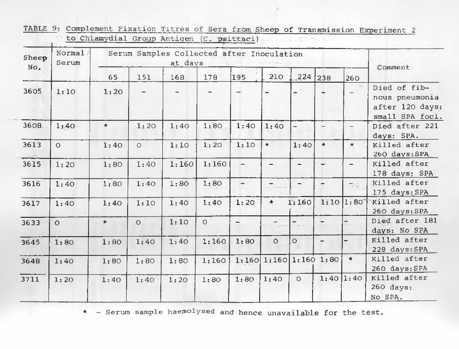
TABLE 9: Complement Fixation Titres of Sera from Sheep of Transmission Experiment 2to Chlamydial Group Antigen (C. psittaci)
SheepNT/-'*
NormalSerum
Serum Samples Collected after Inoculation at days
1NU«65 151 168 17 8 195 ; 2 1 0 ..-224 238 260
OIXUUCIl L.
3605 Is 10 1 : 2 0 — — — - #- -
Died of fib- nous pneumonia after 1 2 0 days: small SPA foci.
3608 1:40 ★ 1 : 20 1:40 1:80 1:40 1:40 - - - Died after 221 days; SPA.
3613 0 1:40 0 1 : 1 0 1 : 2 0 1 : 10 * 1:40 * * Killed after 260 days:SPA
3615 1 : 2 0 1:80 1:40 1 : 160 1 : 160 — - - — — Killed after 178 days; SPA
3616 1:40 1 : 80 1:40 1:80 1:80■
— — — — Killed after 175 days:SPA
3617 1:40 1:40 1 : 10 1:40 1:40 1 : 20 * ]Ls 160 Is 10 1:80' 'Killed after 260 days:SPA
3633 0 * 0 1 : 10 0 — — — — — Died after 181 days: No SPA
3645 1 : 80 1:80 1:40 1:40 1 : 160 1:80 0 0 — — Killed after 228 days:SPA
3648 1:40 1:80 1:80 1:80 1 : 160 1 : 160 1:160 1: 16C 1:80 * Killed after 260 days:SPA
3711 1 : 20 1:40 1:40 1 : 2 0 1:80 1:80 1:40 0 1:40 1:40 Killed after 260 days:.No SPA.
* - Serum sample haemolysed and hence unavailable for the test.
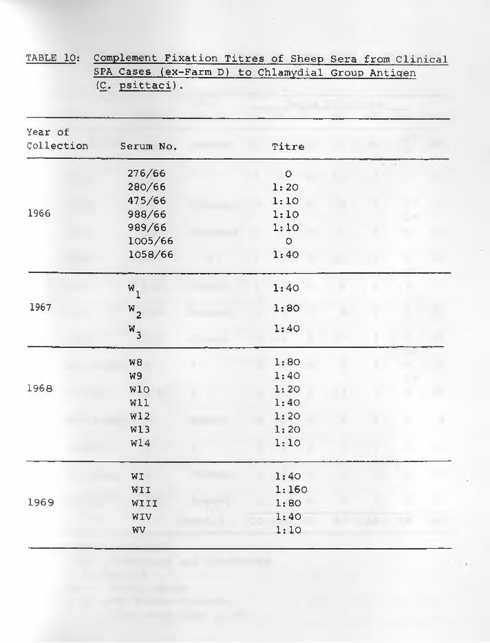
TABLE 10: Complement Fixation Titres of Sheep Sera from ClinicalSPA Cases (ex-Farm D) to Chlamydial Group Antiqen(C. psittaci),
Year ofCollection Serum No. Titre
276/66 0280/66 1 : 2 0475/66 1 : 1 0
1966 988/66 1 : 1 0989/66 1 : 1 01005/66 01058/66 1:40
w i 1:401967
W21:80
W 3 1:40
W 8 1:80W9 1:40
1968 W10 1 : 2 0Wll 1:40W 12 1 : 2 0W13 1 : 20W14 1 : 10
WI 1:40WII 1 : 160
1969 win 1:80WIV 1:40wv 1 : 10
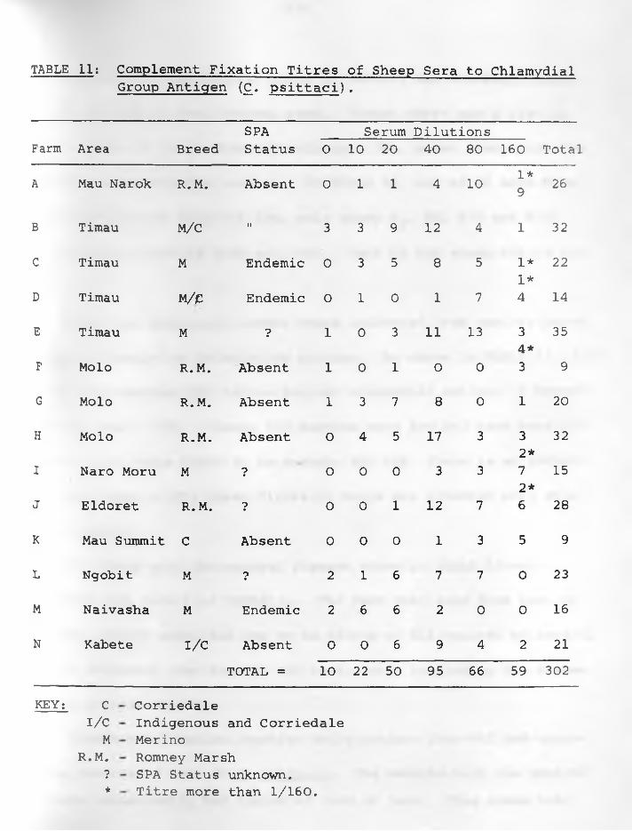
TABLE 11; Complement Fixation Titres of Sheep Sera to ChlamydialGroup Antigen (£. psittaci).
SPA Serum DilutionsFarm Area Breed Status 0 10 20 40 80 160 Total
1*A Mau Narok R.M. Absent 0 1 1 4 10 9 26
B Timau M/C II 3 3 9 12 4 1 32
C Timau M Endemic 0 3 5 8 5 1* 221*
D Timau M/e Endemic 0 1 0 1 7 4 14
E Timau M 7 1 0 3 11 13 3 354 *
F Molo R.M. Absent 1 0 1 0 0 3 9
G Molo R.M. Absent 1 3 7 8 0 1 20
H Molo R.M. Absent 0 4 5 17 3 3 322*
I Naro Moru M 7 0 0 0 3 3 7 152*
J Eldoret R.M. 7 0 0 1 12 7 6 28
K Mau Summit C Absent 0 0 0 1 3 5 9
L Ngobit M 7 2 1 6 7 7 0 23
M Naivasha M Endemic 2 6 6 2 0 0 16
N Kabete i/c Absent 0 0 6 9 4 2 21TOTAL = 10 22 50 95 66 59 302
KEY; C I/C M
R.M.7★
CorriedaleIndigenous and CorriedaleMerinoRomney MarshSPA Status unknown.Titre more than 1/160.
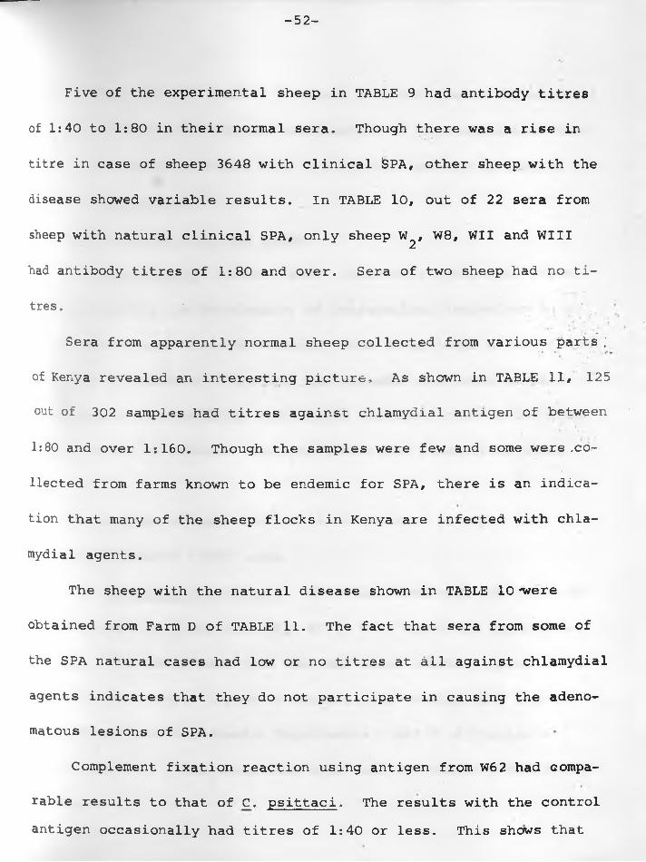
-52-
Five of the experimental sheep in TABLE 9 had antibody titres
of 1:40 to 1:80 in their normal sera. Though there was a rise in
titre in case of sheep 3648 with clinical iSPA, other sheep with the
disease showed variable results. In TABLE 10, out of 22 sera from
sheep with natural clinical SPA, only sheep W^, W8, WII and Will
had antibody titres of 1:80 and over. Sera of two sheep had no ti
tres. *
Sera from apparently normal sheep collected from various parts ;
of Kenya revealed an interesting picture, As shown in TABLE 11, 125
out of 302 samples had titres against chlamydial antigen of between
1:80 and over 1:160. Though the samples were few and some were .co
llected from farms known to be endemic for SPA, there is an indica
tion that many of the sheep flocks in Kenya are infected with chla
mydial agents.
The sheep with the natural disease shown in TABLE 10 'were
obtained from Farm D of TABLE 11. The fact that sera from some of
the SPA natural cases had low or no titres at all against chlamydial
agents indicates that they do not participate in causing the adeno
matous lesions of SPA.
Complement fixation reaction using antigen from W62 had compa
rable results to that of C. psittaci. The results with the control antigen occasionally had titres of 1:40 or less. This shows that
-53-
titres shown with the first 2 antigens were not due to egg material
as such. Electron microscopic examination of chorioallantoic memb
ranes from embryos infected with W62 revealed particles similar to
those of Chlamydiae.
IV. CELL CULTURES FROM SPA LUNGS.
(1) Introduction: MACKAY (1969) observed a cytopathic effect
characterized by the development of intranuclear inclusions in al
veolar macrophages cultured directly from sheep lungs affected with
SPA. The presence of inclusions was sporadic and found in 14 of
the 24 cases. He serially transmitted the agent causing the cyto
pathic effect in alveolar macrophage cultures from normal sheep
lungs.
The purpose of this section is to describe attempts made at
repeating MACKAY'S (1969) work.
(2) Materials and Methods: Materials used were SPA lungs ob
tained from four sheep (WI, WII, Will, WIV) which had the clinical
disease. The sheep were from the same flock (Farm D, TABLE 11) as
those used to supply infective material for studies with chicken
embryos and the Transmission Experiments 1 and 2 of Chapter 4.
Lung tissues of these sheep were also used for electron microscopy
in Chapter 5.
-54-
The tissue culture medium consisted of Eagle's medium in
Hanks5 BSS with 10% tryptose phosphate broth (Difco) and 10% un
inactivated horse serum, Antibiotics were added to a final conce
ntration cf 20 units penicillin G, 50 ug, of streptomycin and 200 ug
kanamycin per c,c.
The method for preparing cell cultures both for alveolar macro
phages and mixed cell cultures was a modification of that descri
bed by MACKAY (1969), For macrophage cultures, one SPA lung each
in the first 2 sheep was tied off and the other one filled with
Hanks- BSS via the trachea, The opening was stoppered and the
filled lung massaged gently for about 5 minutes, The fluid was
then poured into a sterile container and centrifuged at 400 r.p.m.
for 10 minutes. The cells were washed once with Hanks' BSS fo
llowing sedimentation and then diluted so as to form a 1,25% sus
pension with the medium. Each lung yielded 80-100ml. of cells.
For mixed tissue cultures, affected parts of the tied-off
lungs from the first two sheep and from Will and WIV were collected
soon after killing the animals. The specimens were chopped into
small pieces about 5 cubic mm. and placed in conical flasks. They
were immersed in 10 times their volume in a mixture of equal parts
3,7% trypsin and 1,6% pancreatin at 37°C» and gently agitated for
7 to 10 minutes. The supernatant containing dispersed cells was
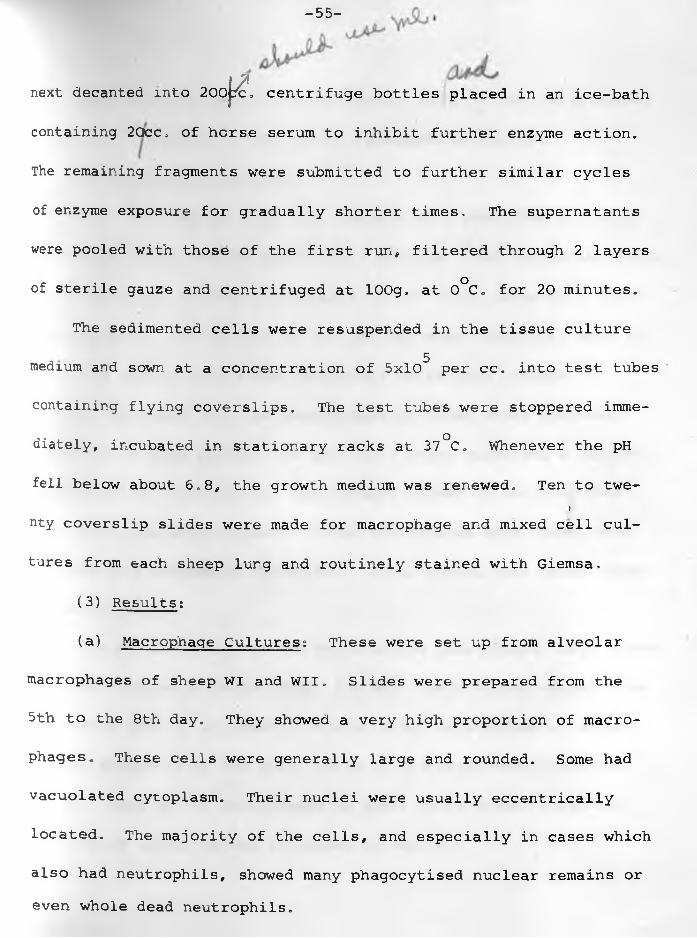
-55-
next decanted into 200K- centrifuge bottles placed in an ice-bath
containing 2Qcc. of horse serum to inhibit further enzyme action.
The remaining fragments were submitted to further similar cycles
of enzyme exposure for gradually shorter times, The supernatants
were pooled with those of the first run, filtered through 2 layers
of sterile gauze and centrifuged at lOOg, at 0°C. for 20 minutes.
The sedimented cells were resuspended in the tissue culture5medium and sown at a concentration of 5x10 per cc, into test tubes
containing flying coverslips. The test tubes were stoppered imme
diately, incubated in stationary racks at 37°C. Whenever the pH
fell below about 6.8, the growth medium was renewed. Ten to twe-i
nty coverslip slides were made for macrophage and mixed cell cul
tures from each sheep lung and routinely stained with Giemsa,
(3) Results:
(a) Macrophage Cultures: These were set up from alveolar
macrophages of sheep WI and WII. Slides were prepared from the
5th to the 8th day. They showed a very high proportion of macro
phages. These cells were generally large and rounded. Some had
vacuolated cytoplasm. Their nuclei were usually eccentrically
located. The majority of the cells, and especially in cases which
also had neutrophils, showed many phagocytised nuclear remains or even whole dead neutrophils.
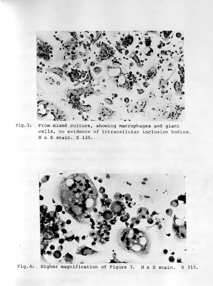
Fig.3: From mixed culture, showing macrophages and giantcells, no evidence of intracellular inclusion bodies. H & E stain. X 125.
-57-
V. DISCUSSION.
Results obtained so far demonstrate that adenomatous lung ma
terial passaged in embryonating eggs hills embryos within 4 to 5
days after infection. This was especially evident in sheep W^,
and W62, indicating that lung material from cases of SPA con
tain infective agent(s) capable of causing lesions in chicken
embryos.The fact that lung tumours and secretions of 4 sheep with ty
pical SPA yielded no Mycoplasma does point to possible non-parti
cipation of these microorganisms in the causation of this sheep
disease. While all isolated Mycoplasma could be propagated in embryonating eggs those that killed the embryos did so very spora
dically. Their growth in cell cultures of chicken embryo fibro
blasts, kidneys, and sheep lung macrophages produced no cytopathic
effect. This contrasts the claim of MACKAY (1966) which related
the cytopathic effect to the action of Mycoplasma. He had observed
plaque formation which resembled a net curtain. GREIG (1955) who
isolated Mycoplasma from nasal secretions of sheep failed to
isolate the same organisms from the lungs of some of the animals
when killed. Pneumonic lungs particularly those associated with Pasteurella spp. infection yielded Mycoplasma much more readily. However not all his pneumonic lung material of the sheep had Mycoplasma on culturing.
-58-
It is apparent in our case that in addition to the Mycoplasma,
there were other agents in SPA material responsible for the patho
logical changes seen in dead and some of the killed embryos. The
agent(s) could be viral in nature.
The Mycoplasma isolated in our study could be classified into
three serogroups, whose pathogenicity is not yet known. But as
shown under Transmission Experiment 4, Chapter 4 the encountered
isolates are of doubtful pathogenicity for sheep. They may be
opportunists that merely take advantage of the oedematous conditions
created by the adenomatous lesions to proliferate, rather than be
ing directly involved in the causation of the adenomatous process.
The significance of Chlamydiae in the causation of SPA has
not been determined. The fact that SHIRLAW (1962) found intracy-
toplasmic inclusions resembling those of Chlamydiae in smears made
from lamb pneumonia due to Pasteurella spp., and that antibody
titres were found in flocks of sheep free of SPA suggest that
Chlamydiae are widely spread in some sheep flocks in Kenya. They
are unlikely to play a pathogenic role with respect to SPA. Hence
while Chlamydiae are capable of killing embryonating eggs and
even causing grossly visible lesions, it would appear that sample
W62 contained another infectious agent in addition to a Chlamydia.
-59-
This other agent may be the one that causes the neoplastic
process characterizing SPA. SHIRLAW (1956) used this agent at the
56th passage to reproduce SPA lesions in sheep. It passes through
a Millipore filter of 450 mp. diameter. Though Mycoplasma can pass
through such a filter, they do not appear to play a pathogenic
role in SPA. SIGURDSSON (1958) discovered that the agent causing
SPA could remain viable for over 4 years in the deep freeze.
Failure of the adenomatous cells in the mixed tissue cultures
to grow may indicate that the tumour tissue in the three sheep were
not actively proliferating. On the other hand the macrophages of
the mixed cell cultures were much more active than those of the
macrophage cultures alone. These active macrophages as shown in
Figure 3 formed numerous syncytia or multinucleate cells. Some
of them had phagocytised material. The giant cell formation
could be a cytopathic effect indicating the presence of some
virus in the cultures.
Many of the phagocytised nuclear material simulate cytoplasmic
inclusions at a casual observation. Indeed as JENNINGS (^G?)
stated, many structures might be found in cells which resemble
viral inclusions and which may cause confusion. No intranuclear
- 6 0 -
inclusions nor other significant changes were observed in the
cultured cells or in Giemsa-stained smears made from the cut sur
face of the adenomatous tissue.
The intranuclear inclusions encountered by MACKAY (1969)
indicating a herpes virus and further investigated (SMITH and
MACKAY 1969) by electron microscopy, may not be the agent cau
sing pulmonary adenomatosis of sheep. The inclusions were found
in macrophages in 14 of the 24 investigated cases. No intrace
llular inclusions were found in the adenomatous cells. As shown
in Chapter 5 of this thesis no inclusions were seen in the
adenomatous cells of investigated cases.
-61-
CHAPTER 4
TRANSMISSION EXPERIMENTS
A. MATERIALS AND METHODS.
I. EXPERIMENTAL ANIMALS:
Specific Pathogen Free (SPF) sheep were not available in this
part of the world and hence could not be used. Day-old colostrum
fed or deprived lambs of the right type could not be obtained in
sufficient numbers. It was decided to obtain experimental animals
from areas that fulfilled certain minimum requirements. The
animals were 32 Corriedale and 4 other sheep. The Corriedales
were 2 ewes and 30 lambs. The latter’s age was between 4 and
15 months. They came from Molo area of Nakuru District in the
Rift Valley Province of Kenya from a farm where no cases of
pulmonary adenomatosis have been diagnosed since the farm was
established, 46 years ago. The other sheep were black-faced
Somali type from the Northern part of Kenya where there has been
no definite evidence of SPA. Their ages ranged from 6-24 months.
The sheep were de-wormed with Thiabendazole immediately they were
brought to the Faculty of Veterinary Science at Kabete.
- 62 -
II. DONOR (INFECTIVE) MATERIALS.
One farm, in Timau area of Laikipia District of Kenya (Farm D,
TABLE 11) on which SPA was endemic but on which no cases of Maedi
have been encountered by the author after examining well over 300
cases of SPA in 2 years, supplied the donor animals. For Experiment
1, the donor, 67/W/l, was one of a group of 30 lambs, 5-6 months
old which were being used for fluorosis tests by the Animal
Production Department of the Faculty of Veterinary Science. They
had previously been acquired from Farm D a month earlier. The
donor lamb had clinical signs and lesions of SPA. Infective
material 67/W/5 (designated W^) for Experiment 2 also came from
the above original source. The animal was in a group of 32 sheep
of which 27 had clinical SPA. Egg-propagated infective material
of the 5th and 15th passages of (W^/5/38 and W^/15/38) provided
the inoculum for Experiment 3. Fluid containing mixed Mycoplasma
isolates was used for Experiment 4.
III. EXPERIMENTS PERFORMED.
Experiment 1: Transmission with Naturally Infected Material.
The object of this experiment was to attempt to produce lung
lesions of SPA. The inocula was administered intratracheally or

-63-
intrathoracically. For the preparation of the infective inocula,
53.5 gm. of representative SPA lung material, 67/W/l, obtained by
sterile methods were weighed out, transferred, and minced finely
in a sterile mortar. With sterile sand, the mixture was finely
ground into an emulsion using a pestle and mortar. This emulsion
was made up to 225 ml. (1:4) with PBS containing antibiotics - 100
I.U. penicillin, 100 ug. streptomycin and 100 ig. kanamycin per
1 ml.
The content was centrifuged at about 2,500 r.p.m. for 30 minu
tes at +4°C. The supernatant was collected and kept in the refri
gerator at +4°C. for 2 hours before use. Ten millilitres of the
infective inocula were administered to each of the eight sheep
while they were held in a normal standing position. Ten millilitres
of PBS containing antibiotics was given intratracheally to each
of the 4 control sheep. Temperatures were recorded twice daily
for 5 months and then discontinued.
Experiment 2: Repeat Transmission with Naturally Infected Material.
This was a repeat of the above experiment with the purpose of
reproducing the clinical disease using differently prepared filtered
or unfiltered infective inocula administered intratracheally. The inoculum, 67/W/5, was the same as passaged in eggs in Chapter 3.
Twenty five millilitres of the supernatant were used for animal trials. Five Corriedale lambs whose normal respirations
-64-
had previously been checked for 28 days, were each inoculated with
5 ml. intratracheally. The rest of the supernatant was filtered
through a 1.2 Gelman filter. This was to get rid of cells,
large viruses and any large subcellular structures. It was diluted,
1:2 and 10 ml. of it was inoculated into 5 more Corriedale lambs
intratracheally. The 4 control animals were each inoculated intra
tracheally with 5 ml. of Eagle's medium.
Temperatures were recorded daily at 8 a.m. and 4 p.m. for 3
months, and thereafter in animals that appeared sick. The 10 lambs
were bled, for sera before inoculation, then at monthly interval
for 4 months and thereafter at 2-week intervals until the experi
ment was terminated. Those sacrificed were also bled at the time
of killing. These sera were kept for serological investigations
(Chapter 3). The sheep were examined clinically at each time of
bleeding and a record of their respirations, cough (if any) and
watery discharge from the nostrils on lifting up the hind legs was
made. The infective inoculum was checked for bacteria by cultu
ring on plates of McConkey and blood agar and later in Newing's
Tryptose Broth and Tryptose Serum Agar for presence of Mycoplasma
spp. The lungs of each sheep were checked for bacteria at slau
ghter, if this seemed warranted.
-65-
Experiment 3: Transmission with Egg-passaged Material.
The purpose of this experiment was to infect animals with SPA
material passaged in embryonating chicken eggs. The material
(allantoic and amniotic fluid) was filtered through 0.22 yx
Millipore filters. Five millilitres were administered intratra-6cheally to each of 8 sheep containing approximately 2x10 ELD 50%.
The animals were bled for sera at intervals, monthly at first and
later at about two weeks. Clinical examination was carried out at
the time of bleeding. Temperatures were recorded twice daily for
one month.
Experiment 4; Transmission with Mycoplasma-containing Material.
A mixture of Mycoplasma spp. (W^, W6/1, W6/2) isolated
from adenomatous lungs during the aetiological studies (Chapter 3)
were put back into 5 sheep. Five millilitres of the mixture were
administered intratracheally to each of 2 ewes and 3, 15-month old
wethers.
The mixed culture was 24 hours old. and were from natural
cases, the former being from the original material of Transmission
Experiment 2. W6/1 and W6/2 were from Experiment 2, sheep 3645 with
pulmonary adenomatosis. Blood was taken from them before inoculation
and at 2-weekly intervals after inoculation. Temperatures were read
at 8 a.m. and 4 p.m. each day for a month.
- 6 6 -
IV. NECROPSY PROCEDURE: COLLECTION AND SPECIMEN PREPARATION,
Sheep were killed by stunning with a humane killer and cutting
through one of the brachial arteries, In this way they bled to
death without blood aspiration. Post-mortem examination was carried
out with the sheep on their backs. Following skinning, the ventral
thoracic and abdominal walls were removed to expose visceral organs.
Portions of lung with typical SPA lesions were removed and some ostored at -70 C. for aetiological studies.
Lungs were then carefully removed and examined for gross
lesions. Selected ones were photographed and their lesion dis
tribution recorded on a diagram of the lung outline, Representa
tive specimens from all 7 lobes, cut roughly at lcc. each were
fixed in 10% buffered formal saline. In lobes that had gross
lesions, at least three specimens* covering portions of complete
consolidation, intermediate, and apparently normal lung were taken,
The tracheobronchial and mediastinal lymph nodes were routinely
preserved. All the fixed specimens were trimmed to about 3-5mm.
thick, processed by the paraffin method and sectioned at 5-6 mic
rons. All were stained with haematoxylin and eosin. Selected
lung sections were also stained with Lendrum's and May-Grunwald-
Giemsa stains for possible inclusions, and with Periodic-acid
-67-
Schiff (P.A.S.) to identify the mucin-producing capabilities of
the adenomatous cells. Several serial sections were made from each
paraffin block. This procedure was used both for experimental sheep
and sheep from natural SPA.
B. RESULTS
The data on 4 transmission experiments are summarized in
TABLES 12, 13, 14 and 15. Three out of eight sheep in experiment
1 and 8 / 1 0 of the sheep in experiment 2 developed pathological le
sions of the disease. Only those sheep with significant lung
lesions are considered in some detail.
I. Experiment 1. This is summarized in TABLE 12.
(a) Post-inoculation observations:
Record of rectal temperature for 159 days revealed no abnormalo ochange, and ranged between 38-39.4 C. (100-103 F.). However, sheep
o o3621 showed distinct pyrexia (40.6-41.7 C. or 105-107 F.) about
48 hours before death. Poor quality of hay despite supplementary
feeding with bran and dairy cubes led to nutritional oedema in
some sheep.
(b) Pathology:
Gross and microscopic lung tissue examinations of sheep 3603,
3612, 3640 and 3644 and, control sheep 3607, 3619, 3646, 3654
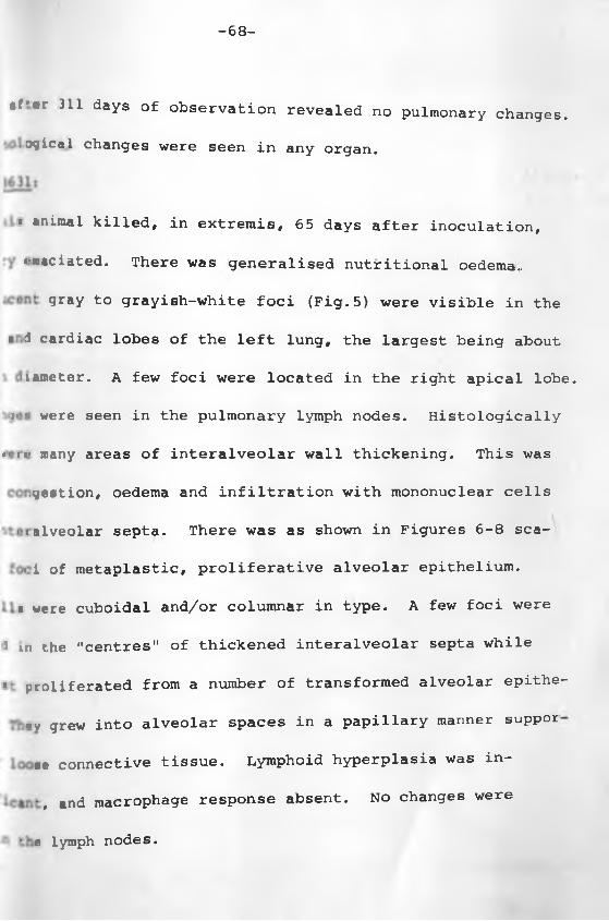
-68-
*f'*r 311 days of Observation revealed no pulmonary changes.. jgical changes were seen in any organ.
.■ animal killed, in extremis, 65 days after inoculation,
«*r.aciated. There was generalised nutritional oedema-
1 ‘ gray to grayish-white foci (Fig. 5) were visible in the
* 1 cardiac lobes of the left lung, the largest being about
:iameter. A few foci were located in the right apical lobe.
;• *. were seen in the pulmonary lymph nodes. Histologically
*< :<* many areas of interalveolar wall thickening. This was
- .geation, oedema and infiltration with mononuclear cells
< : alveolar septa. There was as shown in Figures 6-8 sca-
' >< i of metaplastic, proliferative alveolar epithelium.
• were cuboidal and/or columnar in type. A few foci were
i m the "centres" of thickened interalveolar septa while
s‘ proliferated from a number of transformed alveolar epithe-
rv grew into alveolar spaces in a papillery manner suppor
se connective tissue. Lymphoid hyperplasia was in-
*, and macrophage response absent. No changes were
• e lymph nodes.
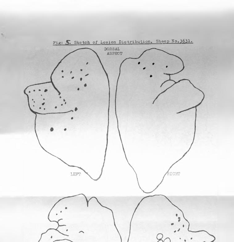
Fig; S"» Sketch of Lesion Distribution, Sheep No«3631.
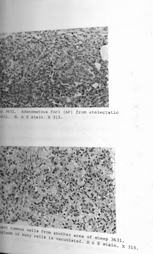
P Adenomatous foci (AF) from atelectaticoil. H. 4 E stain. X 315.
* * c u . froB -----°f -»y c.U M , ,n0thet «ea O f s h e
U Vacu°lated. ep315.
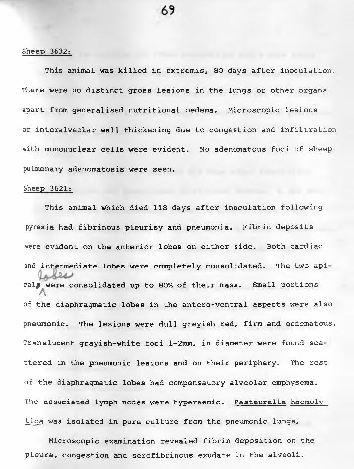
Sheep 3632:
This animal was killed in extremis, 80 days after inoculation,
There were no distinct gross lesions in the lungs or other organs
apart from generalised nutritional oedema. Microscopic lesions
of interalveolar wall thickening due to congestion and infiltration
with mononuclear cells were evident. No adenomatous foci of sheep
pulmonary adenomatosis were seen.
Sheep 3621:
This animal which died 118 days after inoculation following
pyrexia had fibrinous pleurisy and pneumonia. Fibrin deposits
were evident on the anterior lobes on either side. Both cardiac
and intermediate lobes were completely consolidated. The two api-
cal£ were consolidated up to 80% of their mass. Small portionsA
of the diaphragmatic lobes in the antero-ventral aspects were also
pneumonic. The lesions were dull greyish red, firm and oedematous.
Translucent grayish-white foci l-2mm. in diameter were found sca
ttered in the pneumonic lesions and on their periphery. The rest
of the diaphragmatic lobes had compensatory alveolar emphysema.
The associated lymph nodes were hyperaemic. Pasteurella haemoly-
tica was isolated in pure culture from the pneumonic lungs.
Microscopic examination revealed fibrin deposition on the pleura, congestion and serofibrinous exudate in the alveoli.

7o
Neutrophils, macrophages and other mononuclear cells were found
in the alveoli. Bluish staining bacterial colonies were evident.
Adenomatous foci of SPA were found scattered in areas of fibrinous
pneumonia. But there were other sections with fibrinous pneumonia
and without SPA lesions.
Sheep 3642:
This animal killed in extremis 251 days after inoculation,
showed emaciation and generalized nutritional oedema. A few small
whitish gray foci approximately l-2mm. in diameter were found in
the apical and cardiac lobes on either side. No other lesions were
grossly evident. Microscopically, lesions of interalveolar wall
thickening and adenomatous proliferations were present. Some adeno
matous foci were located in "centres" of thickened interalveolar
septa. There was also intrabronchiolar hyperplasia and moderate
lympho-reticular hyperplasia in some sections. Macrophage respo
nse was very slight.
II. Experiment 2:
(a) Clinical Findings:
Record of rectal temperature generally revealed no abnormal
change, the temperature rarely reaching 103°F. However, sheepo o3605 showed distinct pyrexia (40.6-41 C. or 105.4-106 F) about
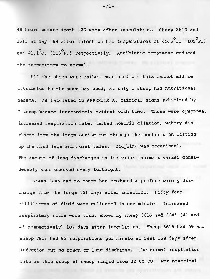
-71-
48 hours before death 120 days after inoculation. Sheep 3613 ando o3615 at day 168 after infection had temperatures of 40.6 C. (105 F.)
and 41.1°C. (106°F.) respectively. Antibiotic treatment reduced
the temperature to normal.
All the sheep were rather emaciated but this cannot all be
attributed to the poor hay used, as only 1 sheep had nutritional
oedema. As tabulated in APPENDIX A, clinical signs exhibited by
7 sheep became increasingly evident with time. These were dyspnoea,
increased respiration rate, marked nostril dilation, watery dis
charge from the lungs oozing out through the nostrils on lifting
up the hind legs and moist rales. Coughing was occasional.
The amount of lung discharges in individual animals varied consi
derably when checked every fortnight.
Sheep 3645 had no cough but produced a profuse watery dis
charge from the lungs 151 days after infection. Fifty four
millilitres of fluid were collected in one minute. Increased
respiratory rates were first shown by sheep 3616 and 3645 (40 and
43 respectively) 107 days after inoculation. Sheep 3616 had 59 and
sheep 3613 had 63 respirations per minute at rest 168 days after
infection but no cough or lung discharge. The normal respiration
rate in this group of sheep ranged from 22 to 28. For practical
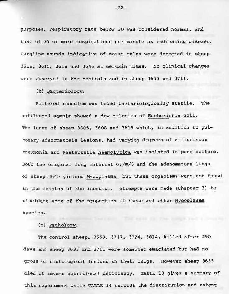
-72-
purposes, respiratory rate below 30 was considered normal, and
that of 35 or more respirations per minute as indicating disease.
Gurgling sounds indicative of moist rales were detected in sheep
3608, 3615, 3616 and 3645 at certain times. No clinical changes
were observed in the controls and in sheep 3633 and 3711.
(b) Bacteriology:
Filtered inoculum was found bacteriologically sterile. The
unfiltered sample showed a few colonies of Escherichia coli.
The lungs of sheep 3605, 3608 and 3615 which, in addition to pul
monary adenomatosis lesions, had varying degrees of a fibrinous
pneumonia and Pasteurella haemolytica was isolated in pure culture.
Both the original lung material 67/W/5 and the adenomatous lungs
of sheep 3645 yielded Mycoplasma but these organisms were not found
in the remains of the inoculum. attempts were made (Chapter 3) to
elucidate some of the properties of these and other Mycoplasma
species.
(c) Pathology:
The control sheep, 3653, 3717, 3724, 3814, killed after 290
days and sheep 3633 and 3711 were somewhat emaciated but had no
gross or histological lesions in their lungs. However sheep 3633
died of severe nutritional deficiency. TABLE 13 gives a summary of
this experiment while TABLE 14 records the distribution and extent
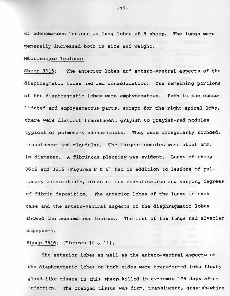
Macroscopic Lesions:
Sheep 3605: The anterior lobes and antero-ventral aspects of the
diaphragmatic lobes had red consolidation. The remaining portions
of the diaphragmatic lobes were emphysematous. Both in the conso
lidated and emphysematous parts, except for the right apidal<lobe,
there were distinct translucent grayish to grayish-red nodules
typical of pulmonary adenomatosis. They were irregularly rounded,
translucent and glandular. The largest nodules were about 5mm.
in diameter. A fibrinous pleurisy was evident. Lungs of sheep
3608 and 3615 (Figures 8 & 9) had in addition to lesions of pul
monary adenomatosis, areas of red consolidation and varying degrees
of fibrin deposition. The anterior lobes of the lungs in each
case and the antero-ventral aspects of the diaphragmatic lobes
showed the adenomatous lesions. The rest of the lungs had alveolar
emphysema.
Sheep 3616: (Figures 10 & 11) .
The anterior lobes as well as the antero-ventral aspects of
the diaphragmatic lobes on both sides were transformed into fleshy
gland-like tissue in this sheep killed in extremis 175 days after infection. The changed tissue was firm, translucent, grayish-white
of adenomatous lesions in lung lobes of 8 sheep. The lungs were
generally increased both in size and weight.
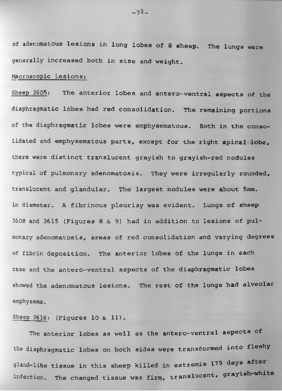
Macroscopic Lesions;
Sheep 3605: The anterior lobes and antero-ventral aspects of the
diaphragmatic lobes had red consolidation. The remaining portions
of the diaphragmatic lobes were emphysematous. Both in the conso
lidated and emphysematous parts, except for the right apidailobe,
there were distinct translucent grayish to grayish-red nodules
typical of pulmonary adenomatosis. They were irregularly rounded,
translucent and glandular. The largest nodules were about 5mm.
in diameter. A fibrinous pleurisy was evident. Lungs of sheep
3608 and 3615 (Figures 8 & 9) had in addition to lesions of pul
monary adenomatosis, areas of red consolidation and varying degrees
of fibrin deposition. The anterior lobes of the lungs in each
case and the antero-ventral aspects of the diaphragmatic lobes
showed the adenomatous lesions. The rest of the lungs had alveolar
emphysema.
Sheep 3616; (Figures 10 & 11).The anterior lobes as well as the antero-ventral aspects of
the diaphragmatic lobes on both sides were transformed into fleshy
gland-like tissue in this sheep killed in extremis 17 5 days af infection. The changed tissue was firm, translucent, grayish whi
of adenomatous lesions in lung lobes of 8 sheep. The lungs were
generally increased both in size and weight.
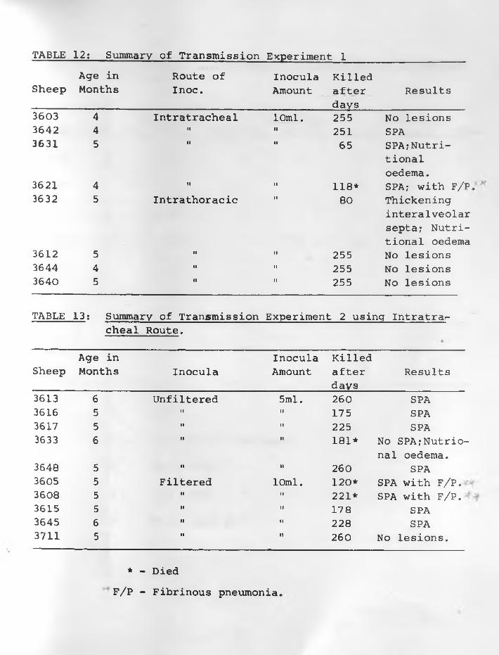
TABLE 12: Summary of Transmission Experiment 1Age in Route of Inocula Killed
Sheep Months Inoc. Amount after Resultsdays
3603 4 Intratracheal 10ml. 255 No lesions3642 4 II II 251 SPA3631 5 II II 65 SPA?Nutri-
tionaloedema.
3621 4 II II 118* SPA? with F/P.3632 5 Intrathoracic II 80 Thickening
interalveolar septa? Nutritional oedema
3612 5 II II 255 No lesions3644 4 II II 255 No lesions3640 5 II II 255 No lesions
TABLE 13: Summary of Transmission Experiment 2 using Intratra*-cheal Route.
Age in Inocula KilledSheep Months Inocula Amount after
daysResults
3613 6 Unfiltered 5ml. 260 SPA3616 5 II II 175 SPA3617 5 II II 225 SPA3633 6 II II 181* No SPA?Nutrio-
nal oedema.3648 5 II II 260 SPA3605 5 Filtered 10ml. 1 2 0 * SPA with F/P.3608 5 II II 2 2 1* SPA with F/P.3615 5 II II 178 SPA3645 6 II II 228 SPA3711 5 II II 260 No lesions.
* - DiedF/P - Fibrinous pneumonia
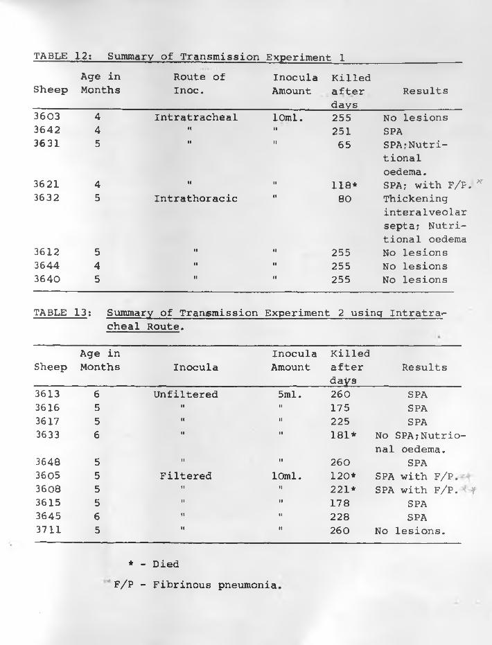
TABLE 12: Summary of Transmission Experiment 1Age in Route of Inocula Killed
Sheep Months Inoc. Amount after Resultsdays
3603 4 Intratracheal 10ml. 255 No lesions3642 4 II II 251 SPA3631 5 II II 65 SPA;Nutri-
tionaloedema.
3621 4 II II 118* SPA; with F/P. *3632 5 Intrathoracic II 80 Thickening
interalveolar septa; Nutritional oedema
3612 5 II II 255 No lesions3644 4 II II 255 No lesions3640 5 II II 255 No lesions
TABLE 13: Summary of Transmission Experiment 2 using Intratra*-cheal Route.
Age in Inocula KilledSheep Months Inocula Amount after
daysResults
3613 6 Unfiltered 5ml. 260 SPA3616 5 II II 175 SPA3617 5 II II 225 SPA3633 6 II II 181* No SPA;Nutrio-
nal oedema.3648 5 II II 260 SPA3605 5 Filtered 10ml. 1 2 0 * SPA with F/P3608 5 II II 2 2 1* SPA with F/P. ‘ f3615 5 II II 178 SPA3645 6 II II 228 SPA3711 5 m M 260 No lesions.
* - DiedF/P - Fibrinous pneumonia
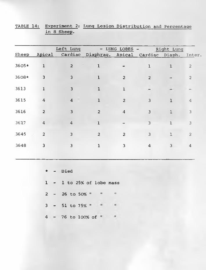
TABLE 14: Experiment 2: Lung Lesion Distribution and Percentagein 8 Sheep.
Left Lung - LUNG LOBES - Right LungSheep Apical Cardiac Diaphrag. Apical Cardiac Diaph, Inter.
3605* 1 2 1 - 1 1 2
3608* 3 3 1 2 2 - 2
3613 1 3 1 1
3615 4 4 1 2 3 1 4
3616 2 3 2 4 3 1 3
3617 4 4 1 - 3 1 3
3645 2 3 2 2 3 1 2
3648 3 3 1 3 4 3 4
* - Died
1 - 1 to 25% of lobe mass
2 - 26 to 5 0 % ...........
3 - 51 to 7 5% "
4 76 to 100% of
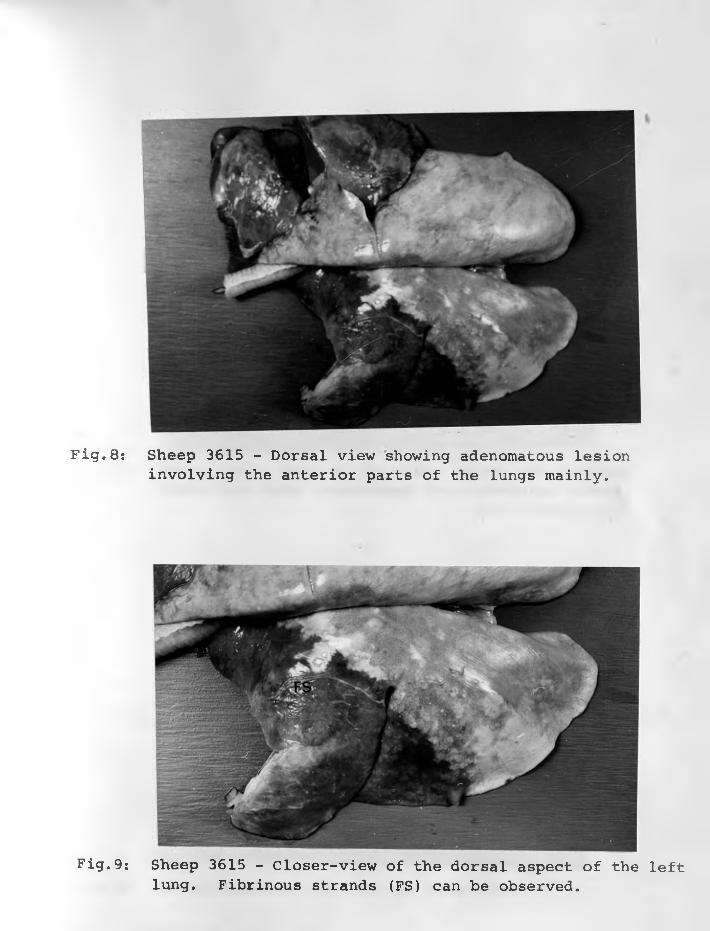
Fig.9: Sheep 3615 - Closer-view of the dorsal aspect of the leftlung. Fibrinous strands (FS) can be observed.
Fig.8 : Sheep 3615 - Dorsal view showing adenomatous lesioninvolving the anterior parts of the lungs mainly.
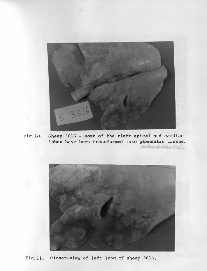
Fig.10: Sheep 3616 - Most of the right apical and cardiaclobes have been transformed into glandular tissue.
p r
Fig.11: Closer-view of left lung of sheep 3616.
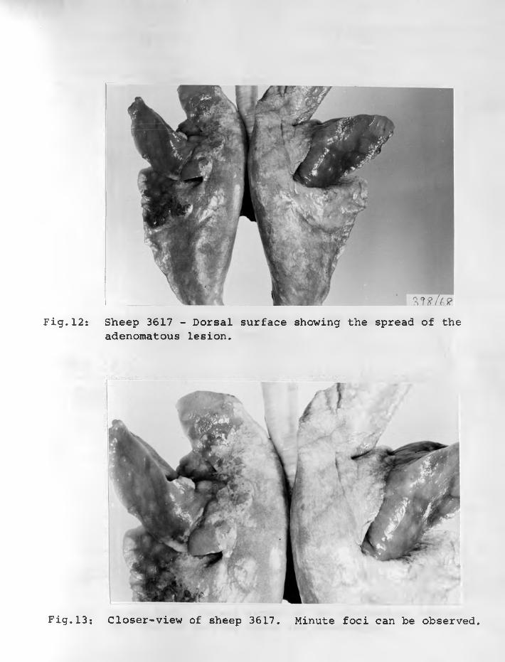
Fig.12: Sheep 3617 - Dorsal surface showing the spread of theadenomatous lesion.
Fig.13: Closer-view of sheep 3617. Minute foci can be observed.
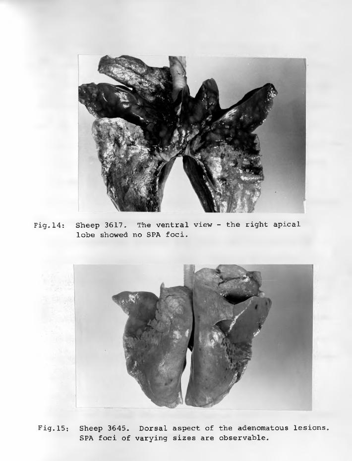
Fig.14: Sheep 3617. The ventral view - the right apicallobe showed no SPA foci.
Fig. 15*. Sheep 3645. Dorsal aspect of the adenomatous lesions. SPA foci of varying sizes are observable.
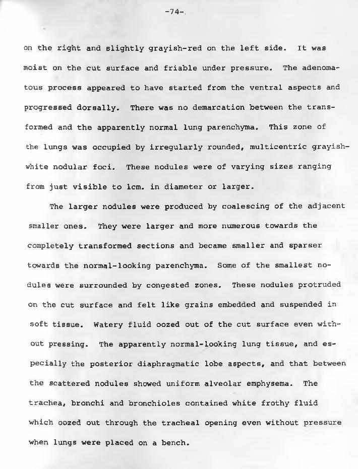
on the right and slightly grayish-red on the left side. It was
moist on the cut surface and friable under pressure. The adenoma
tous process appeared to have started from the ventral aspects and
progressed dorsally. There was no demarcation between the trans
formed and the apparently normal lung parenchyma. This zone of
the lungs was occupied by irregularly rounded, multicentric grayish-
white nodular foci. These nodules were of varying sizes ranging
from just visible to 1 cm. in diameter or larger.
The larger nodules were produced by coalescing of the adjacent
smaller ones. They were larger and more numerous towards the
completely transformed sections and became smaller and sparser
towards the normal-looking parenchyma. Some of the smallest no
dules were surrounded by congested zones. These nodules protruded
on the cut surface and felt like grains embedded and suspended in
soft tissue. Watery fluid oozed out of the cut surface even with
out pressing. The apparently normal-looking lung tissue, and es
pecially the posterior diaphragmatic lobe aspects, and that between
the scattered nodules showed uniform alveolar emphysema. The
trachea, bronchi and bronchioles contained white frothy fluid
which oozed out through the tracheal opening even without pressure
when lungs were placed on a bench.
-74-.
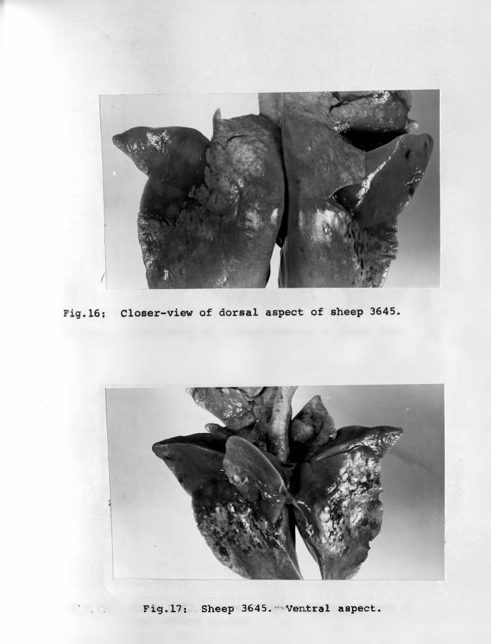
Fig.17: Sheep 3645.-Ventral aspect.
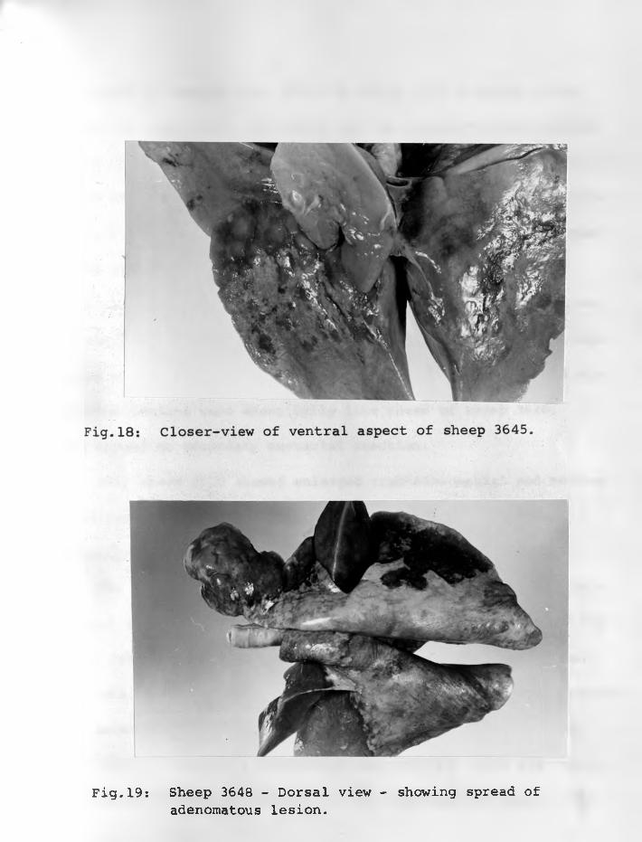
Fig.19: Sheep 3648 - Dorsal view - showing spread ofadenomatous lesion.
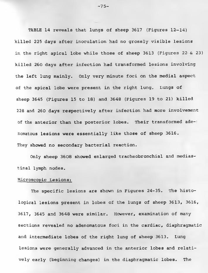
-75-
TABLE 14 reveals that lungs of sheep 3617 (Figures 12— 14)
killed 225 days after inoculation had no grossly visible lesions
in the right apical lobe while those of sheep 3613 (Figures 22- & 23)
killed 260 days after infection had transformed lesions involving
the left lung mainly. Only very minute foci on the medial aspect
of the apical lobe were present in the right lung. Lungs of
sheep 3645 (Figures 15 to 18) and 3648 (Figures 19 to 21) killed
228 and 260 days respectively after infection had more involvement
of the anterior than the posterior lobes. Their transformed ade-|
nomatous lesions were essentially like those of sheep 3616.
They showed no secondary bacterial reaction.
Only sheep 3608 showed enlarged tracheobronchial and medias
tinal lymph nodes.
Microscopic Lesions:
The specific lesions are shown in Figures 24-35. The histo
logical lesions present in lobes of the lungs of sheep 3613, 3616,
3617, 3645 and 3648 were similar. However, examination of many
sections revealed no adenomatous foci in the cardiac, diaphragmatic
and intermediate lobes of the right lung of sheep 3613. Lung
lesions were generally advanced in the anterior lobes and relati
vely early (beginning changes) in the diaphragmatic lobes. The
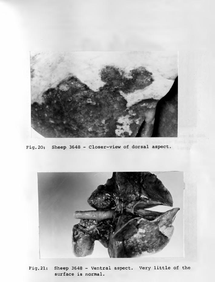
Fig.21: Sheep 3648 - Ventral aspect. Very little of thesurface is normal.
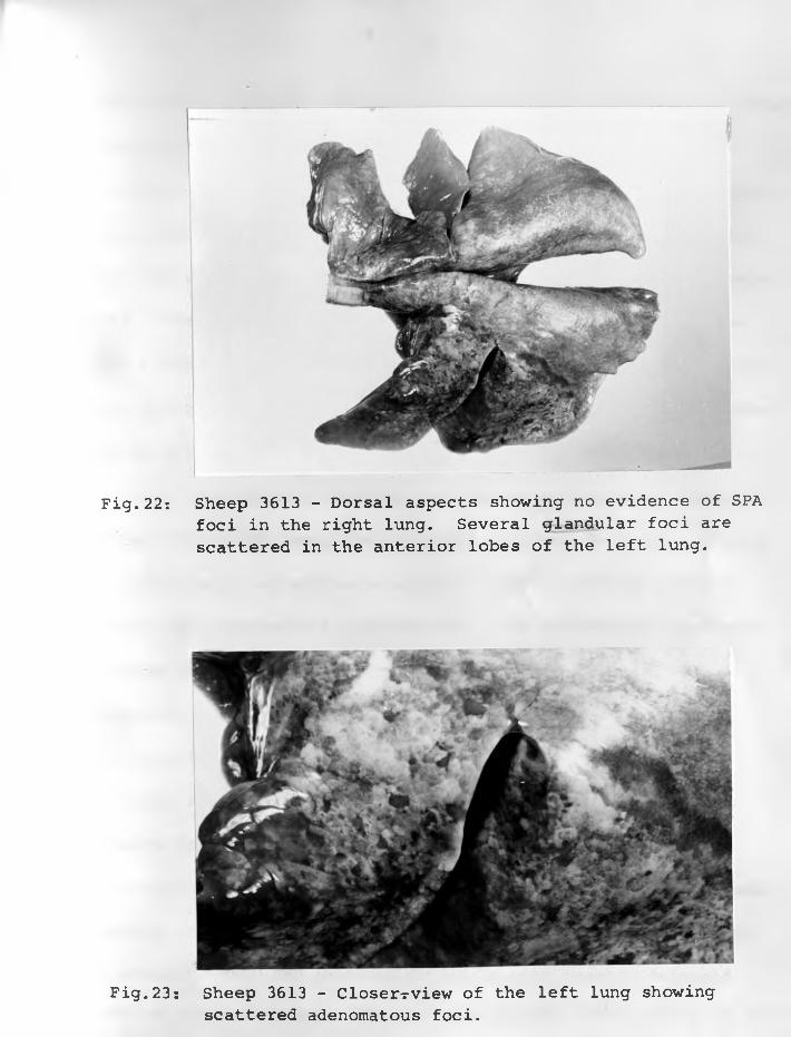
Fig.22: Sheep 3613 - Dorsal aspects showing no evidence of SPAfoci in the right lung. Several glandular foci are scattered in the anterior lobes of the left lung.
Fig. 23s Sheep 3613 - Closer-rview of the left lung showing scattered adenomatous foci.
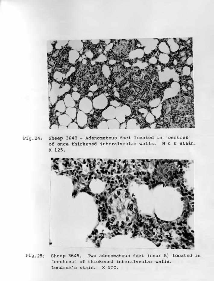
Fig.24: Sheep 3648 - Adenomatous foci located in "centres"of once thickened interalveolar walls. H & E stain. X 125.
Fig.25: Sheep 3645. Two adenomatous foci (near A) located in"centres" of thickened interalveolar walls.Lendrum's stain. X 500.

Fig.26: Sheep 3645 - A "Sheet" of adenomatous cells with no marked alveolar spaces. H & E stain. X 500.
Fig.27: Sheep 3615 - Compact group of adenomatous cells withvacuolated cytoplasm. H & E stain. X 500.
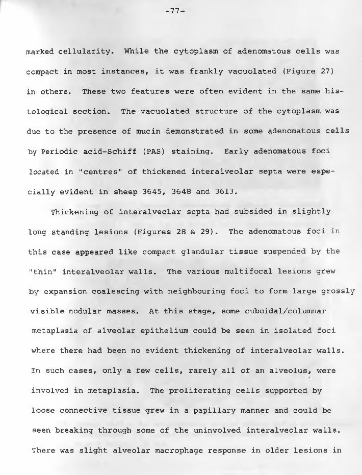
-77-
marked cellularity. While the cytoplasm of adenomatous cells was
compact in most instances, it was frankly vacuolated (Figure 27)
in others. These two features were often evident in the same his
tological section. The vacuolated structure of the cytoplasm was
due to the presence of mucin demonstrated in some adenomatous cells
by Periodic acid-Schiff (PAS) staining. Early adenomatous foci
located in "centres" of thickened interalveolar septa were espe
cially evident in sheep 3645, 3648 and 3613.
Thickening of interalveolar septa had subsided in slightly
long standing lesions (Figures 28 & 29). The adenomatous foci in
this case appeared like compact glandular tissue suspended by the
"thin" interalveolar walls. The various multifocal lesions grew
by expansion coalescing with neighbouring foci to form large grossly
visible nodular masses. At this stage, some cuboidal/columnar
metaplasia of alveolar epithelium could be seen in isolated foci
where there had been no evident thickening of interalveolar walls.
In such cases, only a few cells, rarely all of an alveolus, were
involved in metaplasia. The proliferating cells supported by
loose connective tissue grew in a papillary manner and could be
seen breaking through some of the uninvolved interalveolar walls.
There was slight alveolar macrophage response in older lesions in
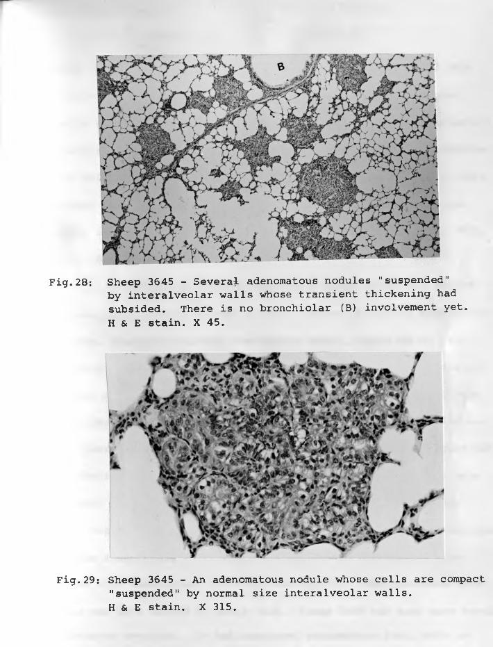
Fig.29: Sheep 3645 - An adenomatous nodule whose cells are compact "suspended" by normal size interalveolar walls.H & E stain. X 315.
Fig.28: Sheep 3645 - Several adenomatous nodules "suspended" by interalveolar walls whose transient thickening had subsided. There is no bronchiolar (B) involvement yet. H & E stain. X 45.
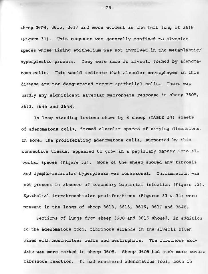
I- 78 -
sheep 3608, 3615, 3617 and more evident in the left lung of 3616
(Figure 30) . This response was generally confined to alveolar
spaces whose lining epithelium was not involved in the metaplastic/
hyperplastic process. They were rare in alveoli formed by adenoma
tous cells. This would indicate that alveolar macrophages in this
disease are not desquamated tumour epithelial cells. There was
hardly any significant alveolar macrophage response in sheep 3605,
3613, 3645 and 3648.
In long-standing lesions shown by 8 sheep (TABLE 14) sheets
of adenomatous cells, formed alveolar spaces of varying dimensions,
In some, the proliferating adenomatous cells, supported by thin
connective tissue, appeared to grow in a papillary manner into al
veolar spaces (Figure 31). None of the sheep showed any fibrosis
and lympho-reticular hyperplasia was occasional. Inflammation was
not present in absence of secondary bacterial infection (Figure 32).
Epithelial intrabronchiolar proliferations (Figures 33 & 34) were
present in the lungs of sheep 3613, 3615, 3616, 3617 and 3648.
Sections of lungs from sheep 3608 and 3615 showed, in addition
to the adenomatous foci, fibrinous strands in the alveoli often
mixed with mononuclear cells and neutrophils. The fibrinous exu
date was more marked in sheep 3608. Sheep 3605 had much more severe fibrinous reaction. It had scattered adenomatous foci, both in
s ’4 .s:_• . . .
* * v* • ■,*/ i*W\a ^I * V► ,'■• vi
Hr % *\ 'i >• <«•!
/ J l * * , -;;, " *r%**' S»* . - - •
•„>'• ••> ■'•*■ ,-r* . S'. * •<• 9 .*v#|. /’ r / M a ;'...
'St v
jp V ^
« »0
%V • '
« » 9 ,2 <t \ * j - ’• ■ <S - ; V . *r ■ X. 1 %> -
r • • * •, ’ >^ %i# 4 V/ . «- -* '• • J t? , . J-‘
Fig. 30: Sheep 3616. An adenomatous -focus (AF) surrounded bymacrophages— (Ma) which had infiltrated alveo-lair -spaces and some fused -to form epithelioid cells. H & E stain. X 315.
Fig.31: Sheep 3617. Ar^advanced lesion of SPA.Interlobular septum(S) divided by two groups of tumour cells. The latter supported oy thin connective tissue grow in a papillary manner into alveolar spaces. H & E stain. X 125.
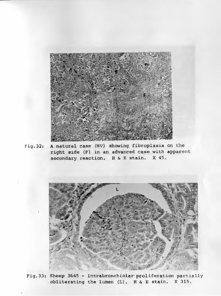
Fig.32: A natural case (WV) showing fibroplasia on theright side (F) in an advanced case with apparent secondary reaction. H & E stain. X 45.
Fig.33: Sheep 3645 - Intrabronchiolar- proliferation partially obliterating the lumen (L). H & E stain. X 315.

Fig.34: From a natural case (1057/66). Intrabronchiolarproliferation with partial occlusion of lumen.H. & E. stain. X 45.
Fig.35: From a natural case (1156/66) showing metastasis totracheobronchial lymph node. H & E stain. X 125.
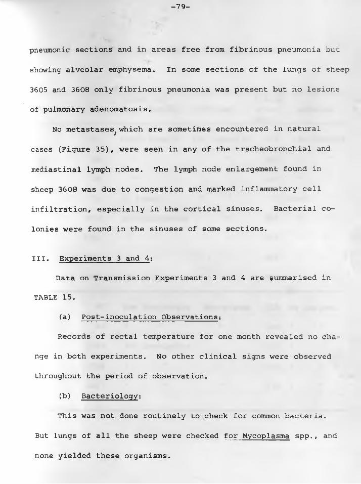
-79-
pneuraonic sections and in areas free from fibrinous pneumonia but.
showing alveolar emphysema. In some sections of the lungs of sheep
3605 and 3608 only fibrinous pneumonia was present but no lesions
of pulmonary adenomatosis.
No metastases^ which are sometimes encountered in natural
cases (Figure 35) , were seen in any of the tracheobronchial and
mediastinal lymph nodes. The lymph node enlargement found in
sheep 3608 was due to congestion and marked inflammatory cell
infiltration, especially in the cortical sinuses. Bacterial co
lonies were found in the sinuses of some sections.
III. Experiments 3 and 4;
Data on Transmission Experiments 3 and 4 are summarised in
TABLE 15.
(a) Post-inoculation Observations:
Records of rectal temperature for one month revealed no cha
nge in both experiments. No other clinical signs were observed
throughout the period of observation.
(b) Bacteriology;
This was not done routinely to check for common bacteria.
But lungs of all the sheep were checked for Mycoplasma spp,, and
none yielded these organisms.
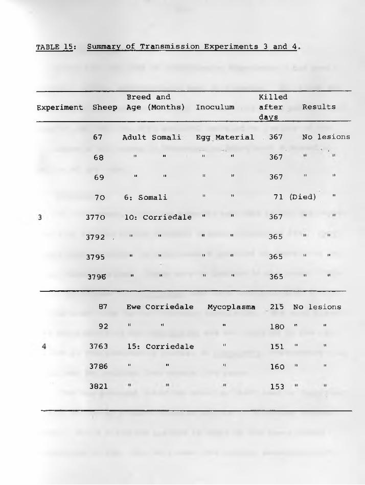
TABLE 15: Summary of Transmission Experiments 3 and 4
Breed andExperiment Sheep Age (Months) Inoculum
Killedafter Results days __________
67 Adult Somali Egg
68 II II II
69 II II II
70 6 : Somali II
3770 10: Corriedale II
3792 . II II II
3795 II II II
3796 II II II
Material 367 No lesions
" 367
" 367 " "
" 71 (Died) "
" 367 "
" 365
" 365 “ "
" 365 " "
87 Ewe Corriedale Mycoplasma 215 No lesions
92 II II II 180 II II
3763 15: Corriedale II 151 II II
3786 II II II 160 II II
3821 153
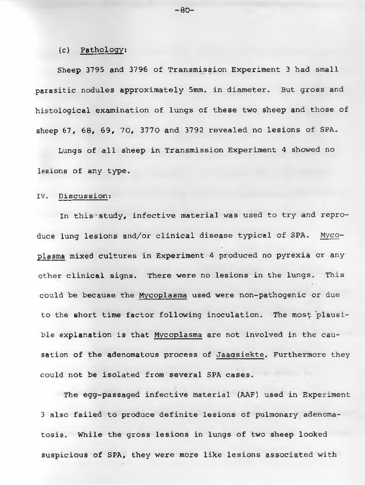
- 80 -
(c) Pathology:
Sheep 3795 and 3796 of Transmission Experiment 3 had small
parasitic nodules approximately 5mm. in diameter. But gross and
histological examination of lungs of these two sheep and those of
sheep 67, 6 8, 69, 70, 3770 and 3792 revealed no lesions of SPA.
Lungs of all sheep in Transmission Experiment 4 showed no
lesions of any type.
IV. Discussion:
In this'study, infective material was used to try and repro
duce lung lesions and/or clinical disease typical of SPA. Myco
plasma mixed cultures in Experiment 4 produced no pyrexia or any
other clinical signs. There were no lesions in the lungs. This
could be because the Mycoplasma used were non-pathogenic or due
to the short time factor following inoculation. The most plausi
ble explanation is that Mycoplasma are not involved in the cau
sation of the adenomatous process of Jaagsiekte. Furthermore they
could not be isolated from several SPA cases.
The egg-passaged infective material (AAF) used in Experiment
3 also failed to produce definite lesions of pulmonary adenoma
tosis. While the gross lesions in lungs of two sheep looked
suspicious of SPA, they were more like lesions associated with
- 81 -
Muellerius capillaris infection. However, this infective material
would appear to have contained some other agent in addition to the
Mycoplasma. This agent may have been responsible for the regular
embryo deaths in passages of W^, and W62 (Chapter 3) compared
to the irregular response to Mycoplasma alone. Whether this
agent is the one which causes metaplasia of alveolar epithelium
of sheep's lungs is not certain. If it were then the apparent
failure to produce typical lung lesions of pulmonary adenomatosis
could be because the titre of the agent was below the minimum
necessary for the stimulation of the adenomatous process.
In Transmission Experiment 2 lung lesions of the disease
were produced in 8/10 (TABLE 13) of the animals. Such lesions
were similar to those of natural disease in sheep. Clinical signs
typical of pulmonary adenomatosis of sheep were exhibited by 7/10
of them as shown in APPENDIX "A". The lung lesions were bilateral,
contrary to the findings of CUBA-CAPARO et al. (1961) who en
countered only 24/60 of the cases. This finding which is
in agreement with the observations of WANDERA (1967b) in natural
cases confirms absence of any lobar preference. The symptoms were
dyspnoea, increased respiration rate, moist rales, watery dischar
ges from the lungs and occasional dough. Clinical examinations
- 82 -
had been carried out while sheep were at rest. It was not possi
ble to observe cough in many of them for they were not under 24ihours continuous observation for the duration of the experiment.
There was no difference in outcome between sheep inoculated with
filtered and those with unfiltered material.
None of the 3 sheep in Transmission Experiment 1 with SPA
lesions showed any clinical signs as the transformed pulmonary
parenchyma was insufficient to interfere with normal lung func-
tioning. The three were out of a group of four sheep inoculated
intratracheally. Sheep 3632 with pulmonary interalveolar wall
thickening may have developed adenomatous lung lesions had it
lived long, and sheep 3631 would perhaps have developed clinical
disease had it lived long. The difference in the results of
Transmission Experiments 1 and 2 does reflect on the way the
inocula were prepared.• In the second experiment, the material
was of a high concentration. The adenomatous cells had been
subjected to much more disruption by ultra-sonic vibration.
Hence a higher proportion of infectious agent was released in
the medium. This was not the case in the preparation of Experi
ment 1 inoculum.
The results of Experiment 2 show that intratracheal route is the most suitable, and that it is the most likely portal of
- 83-
entry in natural infection. Despite the fact that the number of
animals involved is small to be statistically significant, this
experiment demonstrates that it is possible to reproduce the cli
nical disease within 5-8 months after inoculation. The productiont.
of SPA lesions in affected sheep would appear to be dose-dependent
and susceptibility-dependent and not time-dependent.
Though the sheep were not killed in series, the gross and
microscopic lung lesions observed showed changes at different sta
ges of development and hence of significance in the pathogenesis
of the condition. From this it was apparent that the transmissible
agent having infected alveolar epithelium may have acted as an
irritant leading to congestion and infiltration with mononuclear
cells into the interalveolar septa. The result of this reaction
was thickening of the latter. The micro-atelectatic alveoli
were involved in this thickening. This was a transient change
and while it may not be specific for pulmonary adenomatosis, the
later histogenetic changes appear to be connected with it.
Following a latent period of varying length, the agent
induced changes in the normally modified squamous epithelial cells.
These cells became endowed irreversibly with the potentiality to
initiate the primary stages of cancer formation. The agent next
- 84 -
stimuated or triggered off transformed cells to proliferate. This
process first started in epithelium of collapsed alveoli, possibly
because they were more susceptible to injury than that of the
patent ones. These adenomatous cells formed minute foci which in
turn formed large nodules grossly visible by both expansion and
confluence. The uninvolved alveoli were later infiltrated with
macrophages in some cases. The macrophage response appeared to
reduce the rate of hyperplasia of the adenomatous cells and even
tually stop it completely, for some of the small adenomatous foci
remained "static" and surrounded by the alveolar macrophages,
In some areas these "static" adenomatous foci may be surrounded by
alveolar emphysema. These minute adenomatous foci have mistakenly
been equated with the earliest changes.
To some degree epithelium of bronchioles and bronchi was
also involved in this hyperplastic process in some sheep. Macro
phage response cannot be regarded as part of this adenomatous
process as only a few sheep showed it. However it might be
associated with secondary infection by Mycoplasma. Fibroplasia
was absent and lympho-reticular hyperplasia was occasional/. In
flammation was only present in some cases (sheep 3605, 3608,
3617) with secondary bacterial infection. None of the sheep had any metastases to the regional lymph nodes. But this does not
-85-
disqualify the disease as being essentially neoplastic and trans
missible, as metastases have been encountered in some cases of the
natural disease by the author of this thesis0 In all the cases,
the tendency was for the adenomatous process to begin in the ante
rior lobes and gradually proceed posteriorly.
-86-
CHAPTER 5
ELECTRON MICROSCOPIC STUDIES
As discussed in Chapter 3, Mycoplasma of rather doubtful
significance were present in some SPA lungs. Because of this and
of the fact that agents involved in the causation of pulmonary
adenomatosis have not been isolated and characterized, it was
decided to carry out electron microscopic studies. Materials to
be investigated were adenomatous lungs from sheep with the natural
disease of SPA. This study was intended to reveal the presence
if any of agents that could be associated with sheep pulmonary
adenomatosis and consequently the ultra-structure of the adenoma
tous cells. Gross and histological examination of the diseased
lungs showed them to have typical lesions of SPA,
I. MATERIALS AND METHODS:
Lung; specimens from 8 sheep with SPA (WI - WVI, WXI and
WXII) were used for the study. Diseased sheep came from the same
flock (Farm D, Chapter 3) as those that supplied infective mate
rial for Chapters 3 and 4. Lungs from healthy sheep were inc
luded as controls. Materials were fixed according to the method
of ITO-KARNOVSKY (1968). In this method the fixative was pre
pared as follows:
-87-
4pn. of paraformaldehyde were added to 10 0ml, of distilled
water which was then heated to near boiling, The solution
was cleared by adding IN NaOH dropwise and shaking well.
The clear formaldehyde solution was mixed with an equal amount
of 0.2 M phosphate buffer* pH 7,2. 0.0 * trinitrophenol was
added and the solution stored in the refrigerator. For ma
king the final fixative* 2 > glutaraldehyde was added to the
stock solution just before use,
Immediately after killing the animals and removing the lungs,
small pieces of the specimens were fixed in this "Yellow fix" for
2 hours at room temperature. This was followed by 3 changes of
5 minutes each in 0.2 M cold phosphate buffer. They were then
post-fixed in cold ik osmium tetroxide in 0,2 M phosphate buffer
for about 4 hours. After rinsing for 15 minutes with 3 changes
in physiological saline in the refrigerator* they were dehydrated
For embedding* DURCUPAN ACM (FLUKA AG., BUCHS SG* Switzerland)
was used. The processing was through the following dilutions:
Durcupan i/Acetone in dilution of 1:3 for 1 hour*
oin acetone at 4 C. through
and 50* 70* 90* 100, and 100 percent for 30 minutes each.
Durcupan i/Acetone in dilution of 2:2 for 1 hour*
-88-
Durcupan i/Acetone in dilution of 3:1 for 1 hour.
The tissues were then passed through Durcupan I for 4 hours
at 56°C. with one change at 2 hours and through Durcupan II
for 2 hours at 56°C. The tissues were then placed in poly-
sterene capsules,, Durcupan II added and subjected to harde- oning at 60 C for 48 hours.
Sections were cut with glass knives on the OM U2 (REICHERT)
ultramicrotome. Thick sections of approximately 1 u were stained
with toluidine blue for light microscopy for orientation of theorespective tissue blocks, Thin sections (600-900 A ) for electron
microscopy were picked up on formvar-coated copper grids* stained
with uranyl acetate for 6 minutes and lead citrate for 2 minutes*
and examined in a CARL ZEISS EM 9A electron microscope,
II. RESULTS:
1. Light Microscopy: All the lung sections examined from the
8 sheep with the natural disease revealed the adenomatous cells
characteristic of the disease reproduced in Chapter 4. Some of
the foci* especially in two animals* appeared to be long-starding*
as groups of adenomatous cells were interspersed and even surrou
nded by increasing number of proliferating fibroblasts, Adenoma
tous foci of this nature could perhaps be regarded as chronic “static" adenomatous tissue,
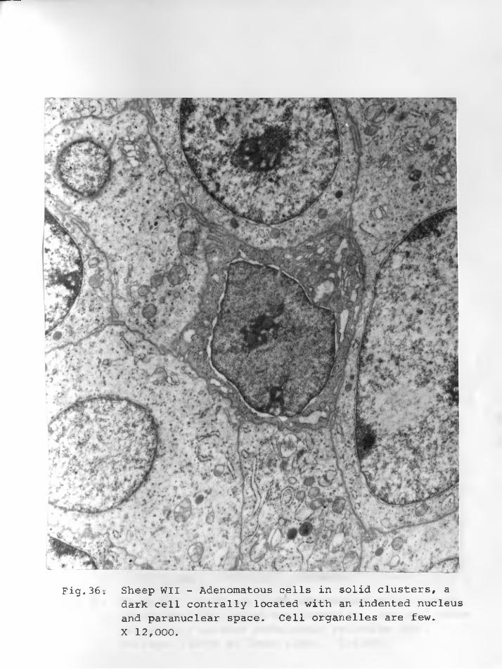
Fig.36: Sheep WII - Adenomatous cells in solid clusters, adark cell contrally located with an indented nucleus and paranuclear space. Cell organelles are few.X 12,000.
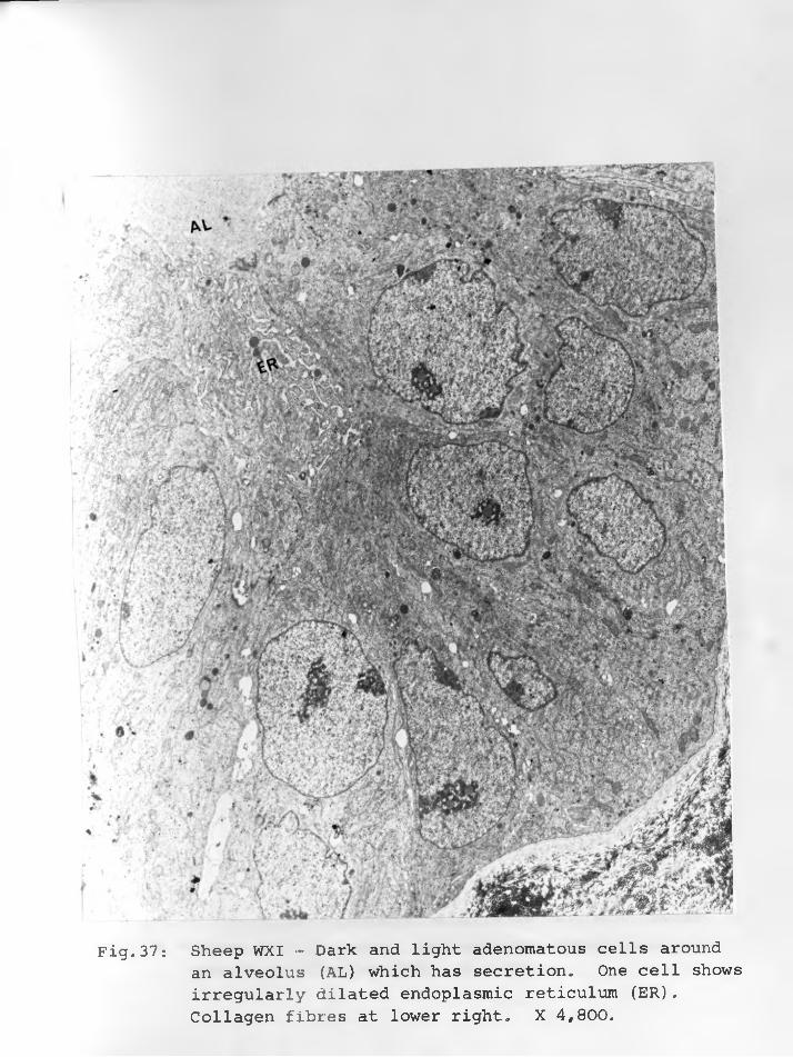
Fig.37: Sheep WXI - Dark and light adenomatous cells aroundan alveolus (AL) which has secretion. One cell shows irregularly dilated endoplasmic reticulum (ER). Collagen fibres at lower right. X 4,800,
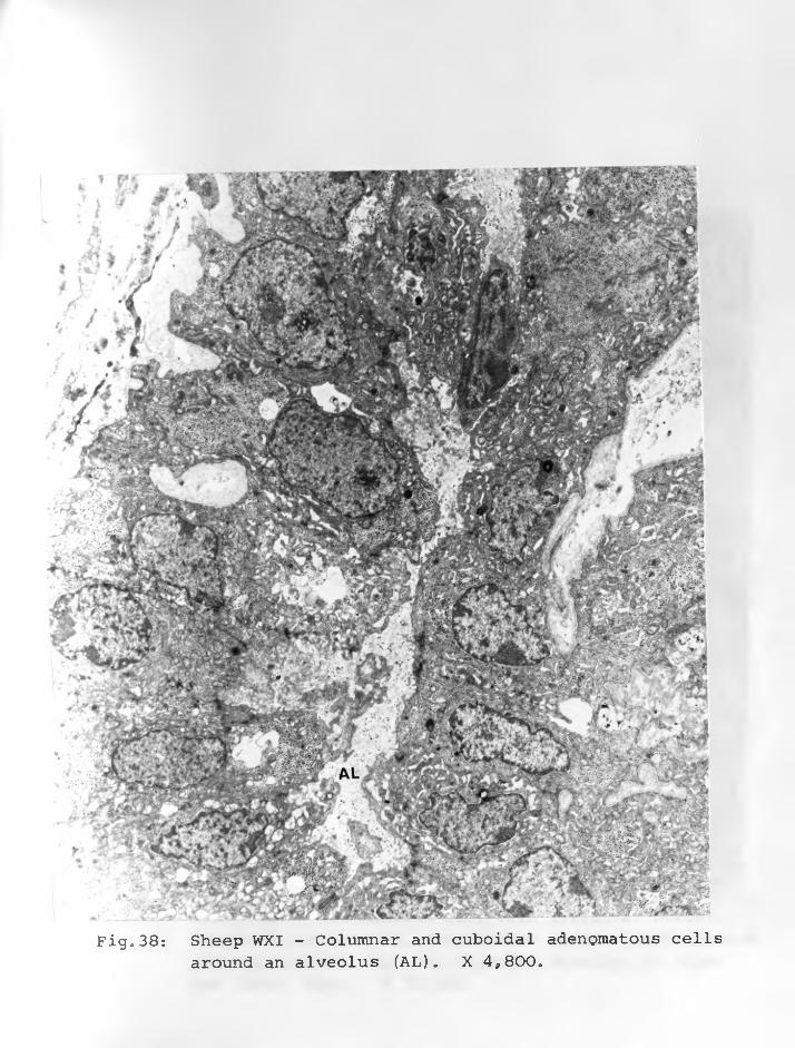
Fig.38: Sheep WXI - Columnar and cuboidal adenomatous cellsaround an alveolus (AL). X 4*800.
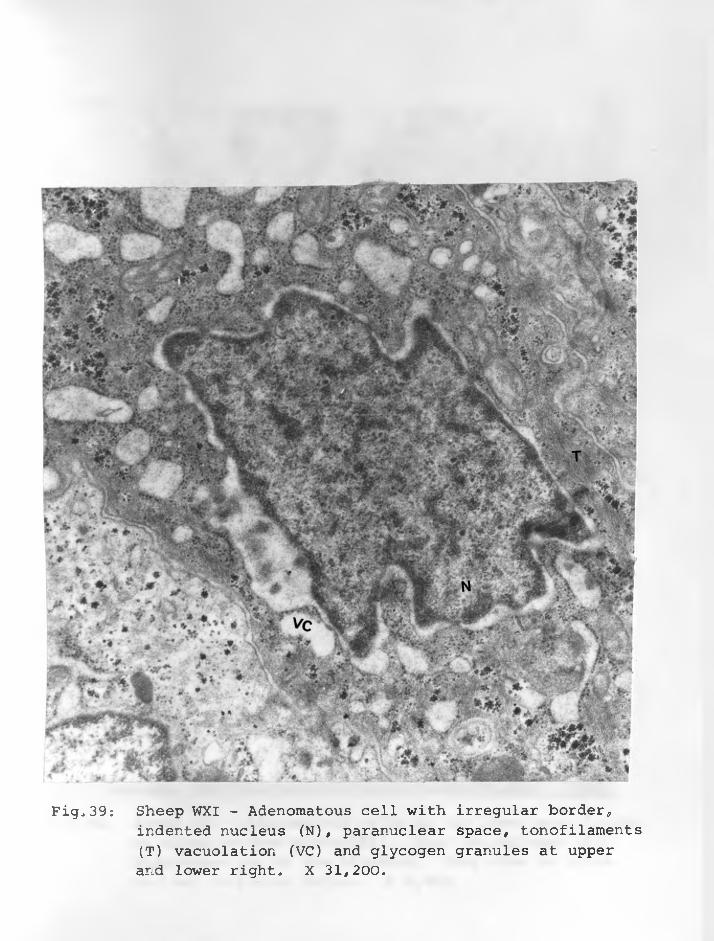
Fig,39: Sheep WXI - Adenomatous cell with irregular border*indented nucleus (N), paranuclear space, tonofilaments (T) vacuolation (VC) and glycogen granules at upper and lower right, X 31,200,
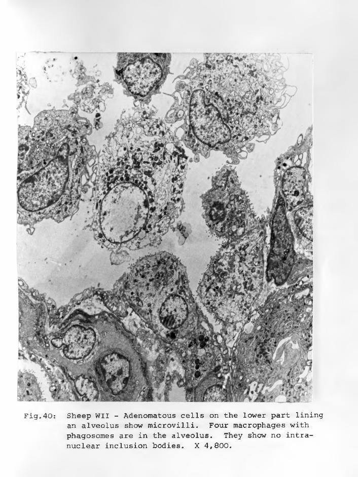
Fig.40: Sheep WII - Adenomatous cells on the lower part liningan alveolus show microvilli. Four macrophages with phagosomes are in the alveolus. They show no intranuclear inclusion bodies. X 4,800.
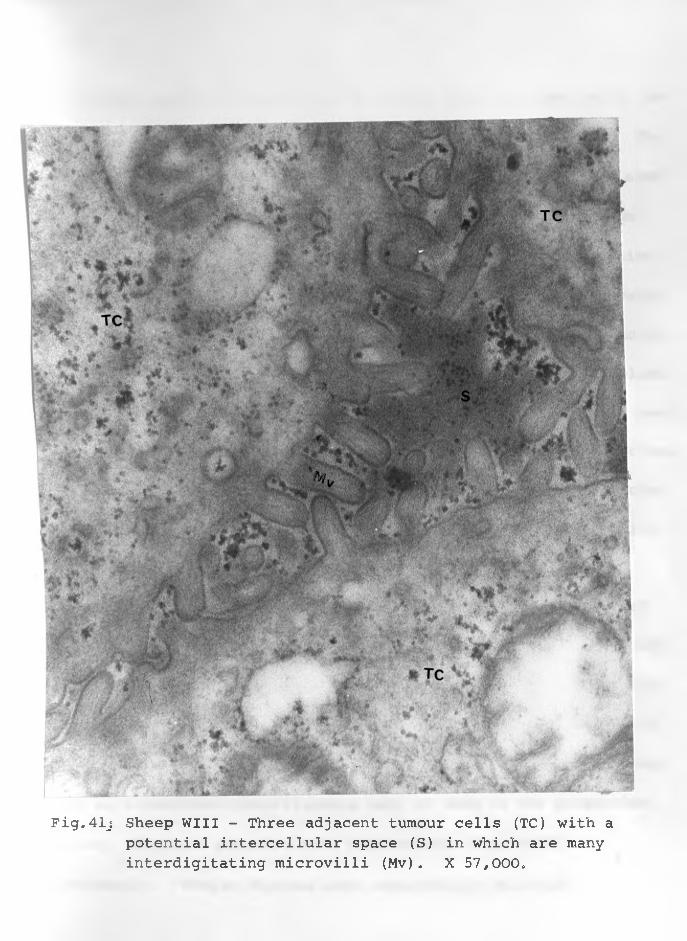
Fig.41j Sheep Will - Three adjacent tumour cells (TC) with a potential intercellular space (S) in which are many interdigitating microvilli (Mv). X 57,000e
-91-
Endoplasmic reticulum were of normal shape in some cells, de
pleted in others and morphologically abnormal in most cells. The
majority of endoplasmic reticulum were greatly dilated
into irregularly shaped vesicles (Figure 44) or vacuoles. Some
dilated ones were slightly regular and arranged in the direction
of the alveolar spaces, forming lacunae. In some cells the vesi
cles and vacuoles involved both the rough-surfaced and the smooth
surfaced ergastoplasm. Rarely were the two forms of ergastoplasm
prominent to the same extent in any one cell. Free ribosomes many
of them appearing as polyribosomes were scattered throughout these
cells but were more numerous in cytoplasm vacuolated by agranular
reticulum. Some vacuoles contained varying amounts of secretion
(Figures 45 & 46).
Most of the active adenomatous cells showed hyperplasia of
the Golgi apparatus (Figures 43, 47 & 48). These prominent Golgi
apparatus were often multiple and seen as convoluted and lamellar
membranes of smooth appearance. They were often associated with
numerous small vesicles and sometimes vacuoles. A few cells sho
wed well-developed tonofilaments many of them in the paranuclear
areas. Glycogen granules were encountered (Figure 48)
frequently. Myelin figures were occasionally observed
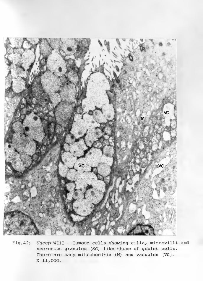
Fig.42: Sheep Will - Tumour cells showing cilia, microvilli andsecretion granules (SG) like those of goblet cells. There are many mitochondria (M) and vacuoles (VC).X 11*000.
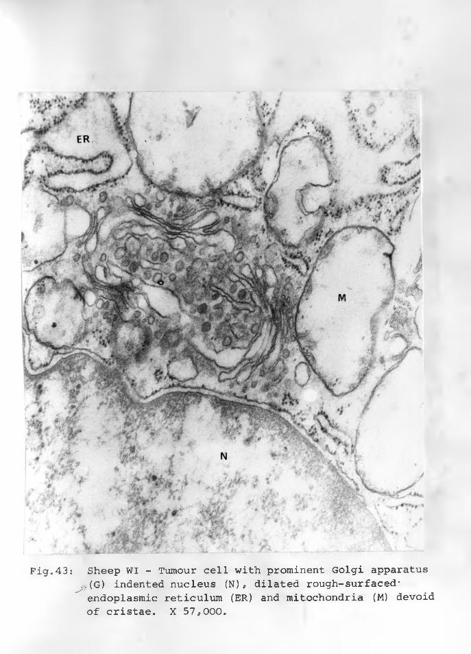
Fig.43: Sheep WI - Tumour cell with prominent Golgi apparatus^(G) indented nucleus (N) g dilated rough-surfaced*
endoplasmic reticulum (ER) and mitochondria (M) devoid of cristae. X 57,000.
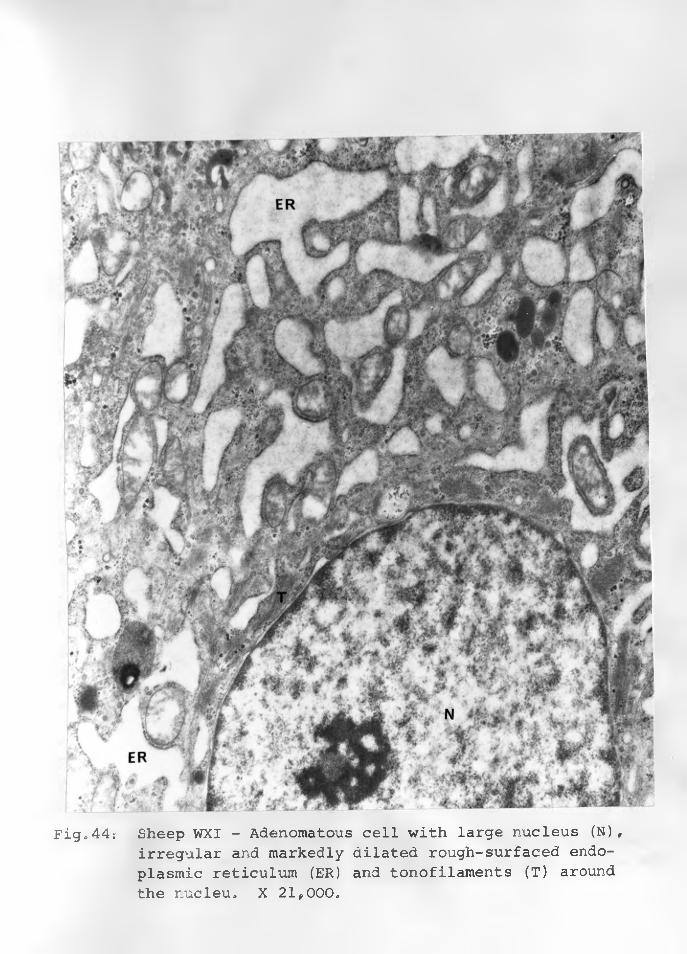
Fig»44: Sheep WXI - Adenomatous cell with large nucleus (N),irregular and markedly dilated rough-surfaced endoplasmic reticulum (ER) and tonofilaments (T) around the nucleu. X 21,000,
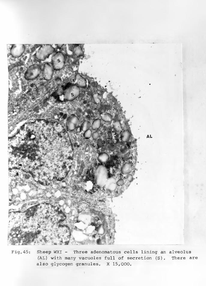
Fig.45: Sheep WXI - Three adenomatous cells lining an alveolus(AL) with many vacuoles full of secretion (S). There are also glycogen granules. X 15,000.
. \
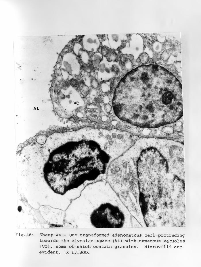
Fig.46: Sheep WV - One transformed adenomatous cell protrudingtowards the alveolar space (AL) with numerous vacuoles (VC), some of which contain granules. Microvilli are evident. X 13,800.
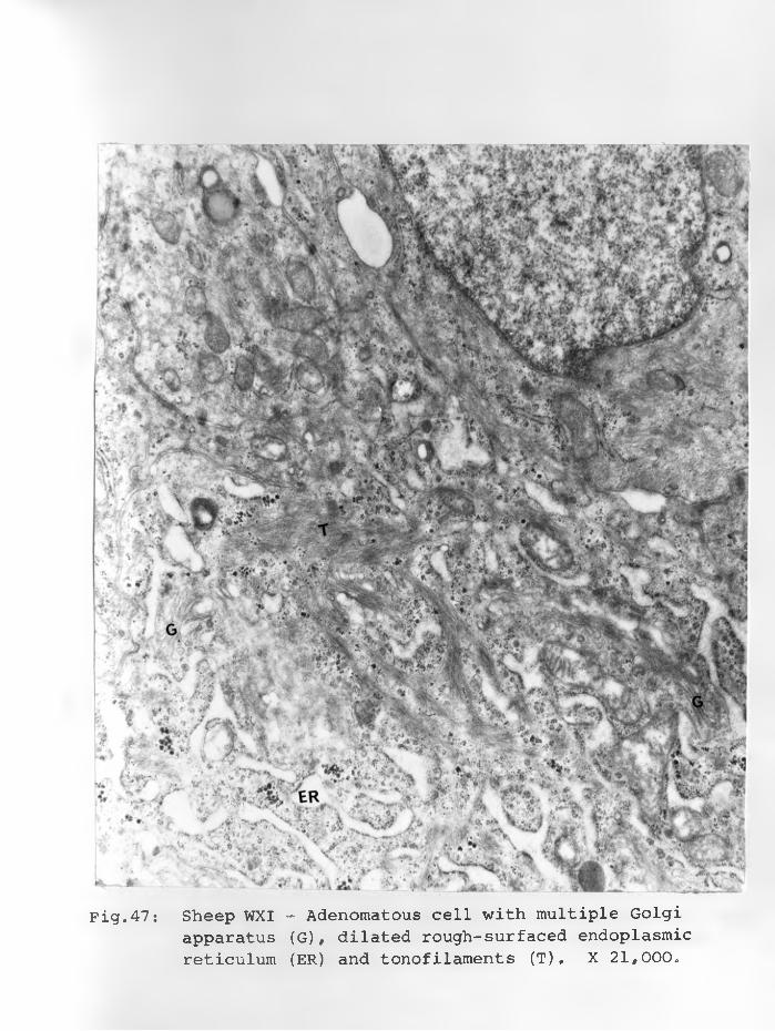
Fig.47: Sheep WXI - Adenomatous cell with multiple Golgiapparatus (G), dilated rough-surfaced endoplasmic reticulum (ER) and tonofilaments (T) . X 21,000.
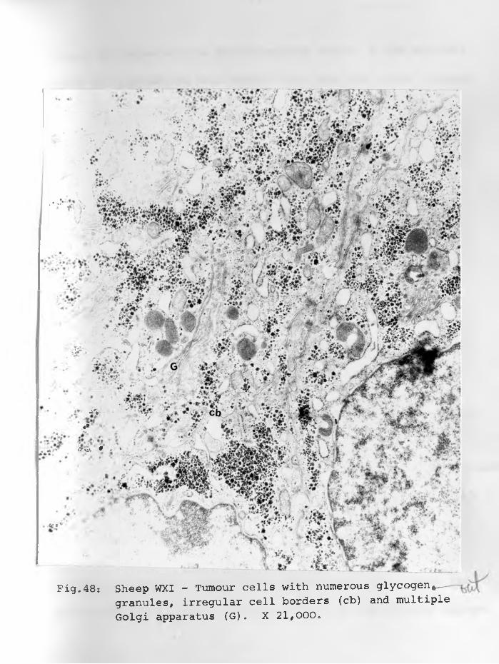
Fig.48: Sheep WXI - Tumour cells with numerous glycogen,— granules, irregular cell borders (cb) and multiple Golgi apparatus (G). X 21,000*
■ j'***
;i - « > * i v 9 . >
<e*'
■ 'Jr
-92-
(Figure 53) especially in disintegrating cells, A few sections
showed two adenomatous cell types, light and dark cells (Figures
36 & 37) with some of intermediate type, irrespective of the
presence of vacuoles. The dark cells contained more while the
light cells contained fewer cytoplasmic organelles particularly
ergastoplasm and ribosomes. Lysosomes were very rare. Where ^
present nucleoli were very prominent.
C
While some nuclei were eccentrically located the majority
were centrally placed. They were large , some ovoid and others
rounded often with scanty peripheral chromatin. The majority
had regular nuclear membrane^ but a number of cells had indenta
tions in their nuclei with a few having perinuclear spaces.
Chlamydiae were seen in one case (Will) in a sac-like
structure made by two adjacent tumour cells. The various develo-
bene<a jl
pmental forms were contained in a sac just beneath the plasma<membrane (Figure 49) some were oval, others spherical. They ranged
in size from 95 mu to 184 mu. They had some similarity to those
illustrated by LITWIN et al. (1961). In addition to these
Chlamydiae, there were virus-like particles (Figures 50-54)
observed in every/ grid made from lungs of sheep WXI. These par
ticles were found in the intercellular and in the alveolar spaces,
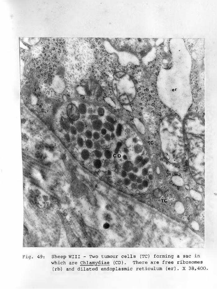
Fig. 49: Sheep Will - Two tumour cells (TC) forming a sac inwhich are Chlamydiae (CD). There are free ribosomes (rb) and dilated endoplasmic reticulum (er). X 38,400.
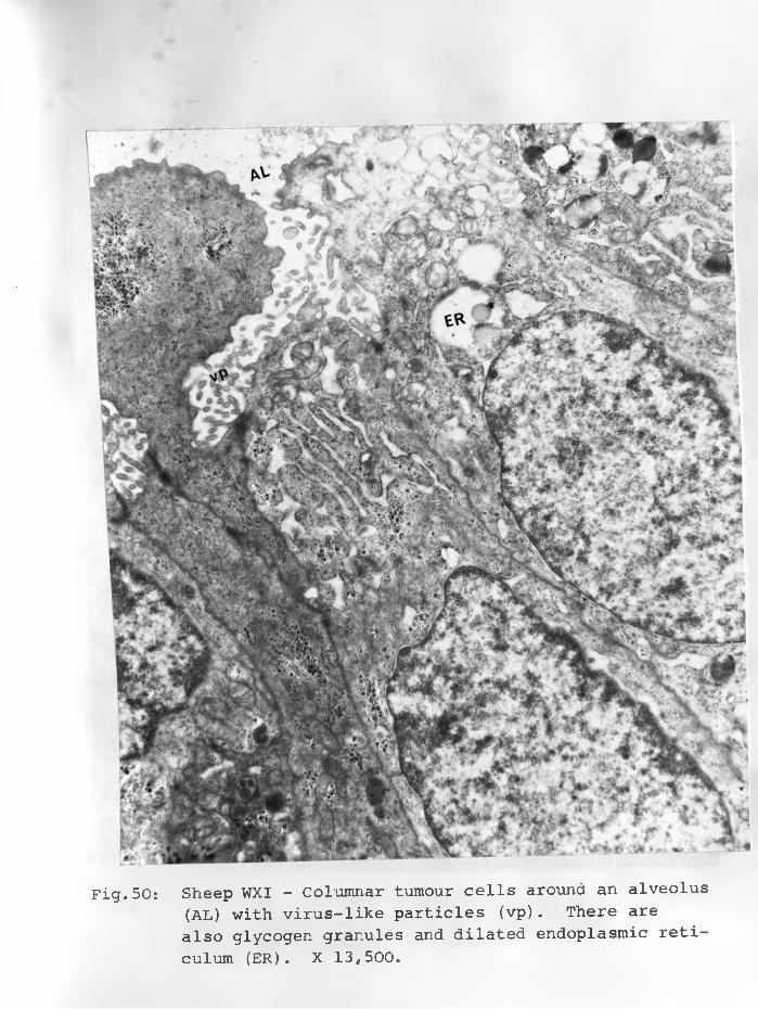
Fig.50: Sheep WXI - Columnar tumour cells around an alveolus(AL) with virus-like particles (vp) . There are also glycogen granules and dilated endoplasmic reticulum (ER). X 13,500.
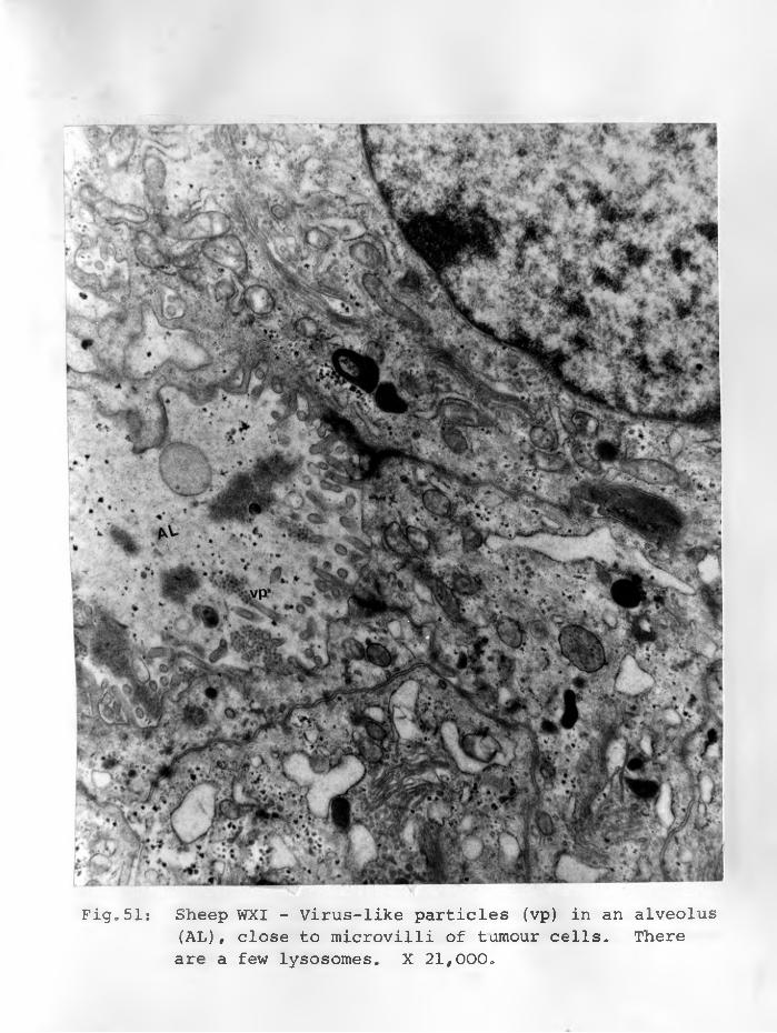
Fig*51: Sheep WXI - Virus-like particles (vp) in an alveolus(AL), close to microvilli of tumour cells. There are a few lysosomes. X 21, 000.

Fig.52: Sheep WXI - Aggregations of virus-like particles (Vp)in an alveolus (AL) and close to microvilli of tumourcells. X 26,700.
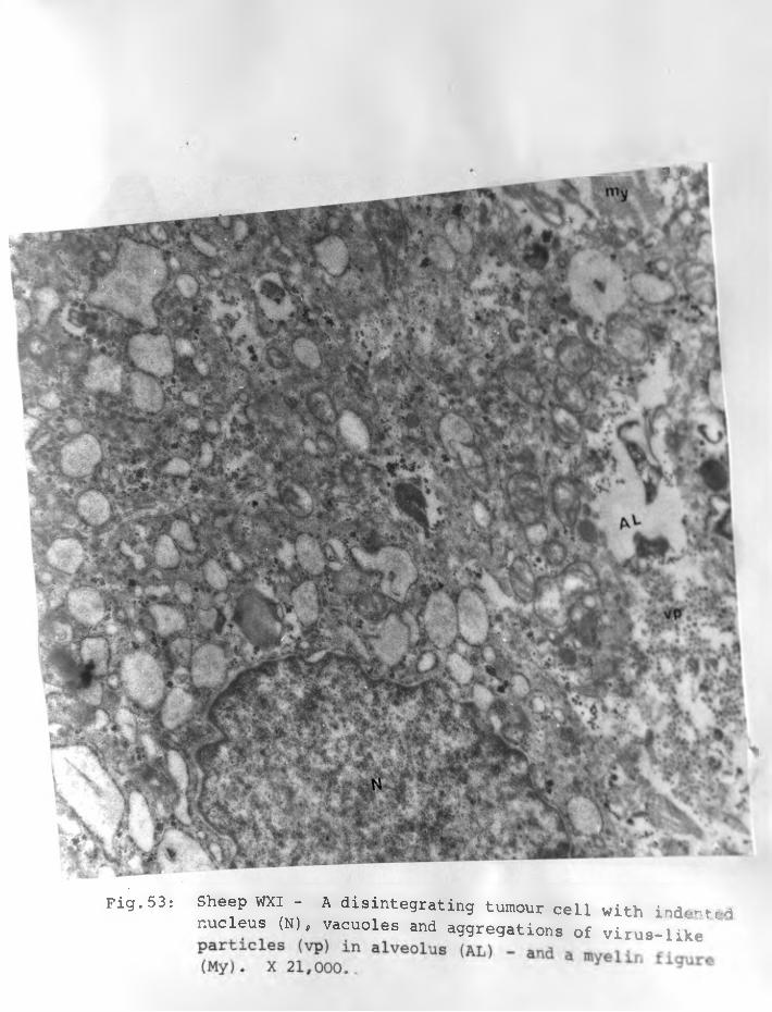
* *
Fig. 53: Sheep WXI - A disintegrating tumour cell with indentednucleus (N), vacuoles and aggregations of virus-like
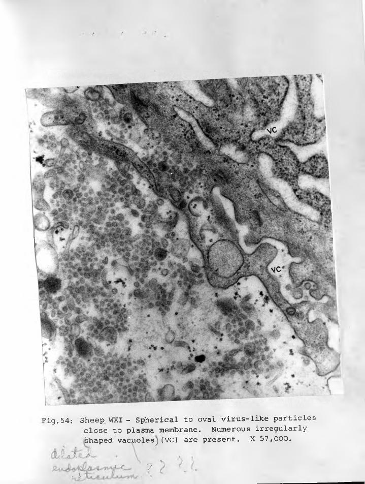
’ig.54: Sheep WXI - Spherical to oval virus-like particlesclose to plasma membrane. Numerous irregularly Shaped vacuoles)(VC) are present. X 57,000.
jJu jb iLL 1
JU<L 7
They were more numerous in the latter where they appeared as
aggregations. Many of the virus-like particles found in the al
veolar spaces were close to the surface of the plasma membrane.
Some of the particles appeared as if they had formed from the
tips of microvilli but not budding as such (Figures 51 & 52).
In some alveolar spaces the virus-like particles were mixed with
what appeared as secretion. These particles were not quite cubic.................................................................% != ■
as they appeared to vary in size (Figure 54) at high maginification,
ranging in diameter between 46-80 mu.
Fibroblasts and bands of collagen were found in two cases in
close association with tumour cells - the so-called chronic
"static" foci. In all the tumour cells examined none showed
any intracellular inclusion bodies nor Mycoplasma. No clear-cut
virus particles were seen intracellular!^. The latter was seer-
in the alveolar spaces of a few cases. Occasionally macrophages
were observed in the alveolar spaces,, with many vacuoles and
phagosomes. None showed any virus-like particles nor inclusions.
HI. DISCUSSION;
The submicroscopic observation of the morphology of the SPA
lungs shows ultrastructural cellular organization that tallies
in most respects with that of other carcinoma! cellsA

-94-
OBIDITSCH-MAYER and BREITFELLNER* 1968) . The hypertrophy of gra
nular and agranular ergastoplasm results into greatly distended
vesicles* These vesicles correspond to the vacuoles demonstrable
with light microscopic techniques and are evidently associated
with the P.A.S. positive watery mucinous fluid found in such lungs*
The hypertrophy of endoplasmic reticulum explains how the normally
attenuated alveolar epithelial cells swell when infected with
SPA agent.
The ergastoplasm and other organelles in SPA cells were mar^
kedly abnormal in the majority of cases. A few cells were almost
devoid of them. Well developed rough and smooth-surfaced endo
plasmic reticulum may indicate sites of slowest tumour growth.
Adenomatous cells showing this type of cytoplasm were mainly
those forming foci of tumour tissue surrounded by fibrous tissue -
the "static” ones.
The electron microscopic study does further confirm SPA as
a lung tumour with many morphological features similar to those
seen in squamous cell carcinoma of the human cervix (BHOWMICK and
MITRA, 1968), liver carcinoma (TOKER and TREVINO, 1966) and lung
carcinoma (GELLER and TOKER, 1969) in human beings. Apparently
cells of this tumour arise as a -result of transformation and stimulation by the SPA agent without showing any degenerative changes.
-95-
Virus-like particles in this investigation ranged
in size between 46-80 mp. This variation may dictate against
these particles being virus. It is not unusual to find
virus particles which do not have a cubic morphology showing
variation in their sizes as occurs in certain arboviruses
like Nairobi Sheep disease (TERPSTRA, 1969). Our particles
if they are virus - then the agent is not spherical in shape,
but different such as being oval. The virus-like particles
were observed outside the cells and hence may have no rela
tion to tumour growth.
Using histochemical means, ALIEV (1967) found a marked
increase in ribonucleic acid (RNA) content of tumour cells
of SPA compared to that of normal lung tissue. The fact
that only one case, sheep lung WXI showed presence of
virus-like particles is not remarkable as they are rarely
seen even in such virus caused tumours as Marek1s disease
and avian leukosis.
-96-
\Whether the observed particles are virus,, and if so whether
they are causative agents or only secondary in sheep lung tumour
is too early to tell. Herpes-like agent investigated in
Scotland (MACKAY, 1969? SMITH and MACKAY 1969) was not observed,
This problem requires more investigations, Chlamydiae occasio
nally found are unlikely to be the causative agents.
-97 -
CHAPTER 6
GENERAL DISCUSSION
I. AETIOLOGICAL STUDIES:
Agents involved in the causation of alveolar epithelial cell
transformation with progressive adenomatous process have not been
completely elucidated. The lungworm theory has been discarded.
In any case DUNGAL et al. (1938) could not find any correlation
between lungworm infestation and sheep pulmonary adenomatosis.
Several commonly encountered bacteria, especially Pasteurella
haemolytica are secondary invaders.
The role of Mycoplasma with respect to the SPA process is
doubtful. It is well-known that nearly all domesticated animals
harbour benign parasites in the upper respiratory tract, and
that some animals may harbour potentially pathogenic Mycoplasma
in the same site. Out of well over 50 known species of Mycoplasma
(POLAND, 1969) only a very small proportion are frankly pathoge
nic. The majority are of low pathogenicity or even saprophytes
like M. laidlawii. Indeed a number of them have been isolated
from cases of human leukemia (FALLON, 1966, HAYFLICK and K0PR0-
WSKI, 1965, LEACH and BUTLER, 1966), and found as contaminants
of continuous cell line tissue cultures (TULLY, 1966). Forty
-98-
isolates (KRAUSS, 1969 - unpublished data) had been made from__ _ 7 ? 7 ?
nasal discharges of healthy slaughter sheep originating from a
district in Kenya where pulmonary adenomatosis is unknown to
occur. Mycoplasma have been isolated from sheep lungs with |as-
teurella pneumonia (GREIG, 1955, WANDERA, 1968) and with Maedi
(WANDERA, 1970). The available evidence suggests that certain
disease conditions including respiratory infections and certain-
tumours, produce substances suitable for the rapid growth of
Mycoplasma. In the case of SPA, the mucinous fluid produced
by the adenomatous cells is utilized by these micro-organisms,
but Mycoplasma are not oncogenic agents. In turn, the presence
of these Mycoplasma appears to attract macrophage response.
Whether Chlamydiae fall in the same category as Mycoplasma
is too early to tell. But the fact that a number of sera from
sheep flocks in Kenya where sheep pulmonary adenomatosis has
not been reported had high antibody titres would indicate that
this organism is not the aetiological agent of SPA. Pathogenic
chlamydial agents cause disease (pneumonia., conjunctivitis, ^
polyarthritis) in young lambs apart from abortion in ewes and
cows. In a review, TAMARIN (1969) showed that Chlamydiae
cause about seven different conditions, and that the organisms
-99-
have been isolated from faeces of apparently normal sheep. Pneu
monia is one of the conditions caused, which occurs mainly in
calves and lambs. In calves (WILSON and THOMSON, 1968) the pneu
monia is proliferative and exudative. Lambs show a proliferative
type characterized by interstitial pneumonia often with lymphoid
hyperplasia (PAVLOV, 1967? STEVENSON, 1968). On the other hand
there is no age preference (CUBA-CAPARO, et al. 1961? WANDERA,
1968) with respect to pulmonary adenomatosis.
A clear aetiologic diagnosis through isolation, identifica
tion^ and transmission of SPA agent from this respiratory disease
cannot be made yet. The virus-like particles encountered in one
sheep are of little significance as they were not seen inside
tumour cells. However despite the failure to demonstrate causal
agents, the available evidence point^to a virus aetiologic agent.
This agent is not the same as the herpesvirus isolated (MACKAY,I1969? SMITH and MACKAY, 1969) in Scotland. Intranuclear inclu
sions of herpes type of virus were not seen in 8 cases studied
by electron microscopy either in the tumour cells or macrophages.
The causal agent of SPA persists at varying concentration
in transformed cells, as well as being continuously liberated
into the lung fluid (DUNGAL, 1946), and can pass through a
-100"
gradocol membrane with pores 0.9 u in diameter. It can remain
viable in affected lung tissue when stored at -20°C. for at
least 52 months (SIGURDSSON, 1958).
There is now an impressive list of malignant neoplasms in
which a virus entity plays a causal role, for example the leuke
mias and sarcomas of chicken and rodents and leukemias of cats.
Though no clear-cut virus particles were identified in this study
this does not mean that no virus is involved in the causation of
the disease. For while Marek's disease is known to be caused by
a virus (EIDSON, et al. 1969), virus-like particles were not seen
by WIGHT (1969). Also LINDBERG (1968) could only see sparse
virus-like particles in 3 of 5 Rous rat sarcoma tumours induced
by Rous virus strain Schmidt-Ruppin (SR-RSV).
More confirmatory evidence of virus aetiology in case of
SPA will add more information on a causal relationship of viruses
and certain cancers in large mammals including human beings.
II: CLINICAL RESPONSE OF TRANSMISSION EXPERIMENTS:
As sheep in Experiments 3 and 4 showed no distinct lesions
of the disease and those of Experiment 1 had no symptoms, only
results in Experiment 2 will be further discussed here.
- 1 0 1 -
Pyrexia was not a feature of the disease, Sheep that
showed temperature rise above the normal had secondary bacterial
pneumonia. This was confirmed by the fact that antibiotic
treatment of sheep that had fever reduced the temperature to
normal. Secondly, Pasteurella haemolytica was isolated from
such cases when they later died or were killed.
Symptoms of rapid breathing, dyspnoea, coughing, secretion
from the lungs and moist rales typical of the disease are only
manifest when the lung condition is advanced. They were shown
by 7 out of 10 sheep of Experiment 2. These clinical signs as
tabulated in APPENDIX "A", were observed over a period of monthsi/Increased respirations (40 and 43 respectively) were showed by
sheep 3616 and 3645 at day 107 after infection or just over
3 months. By 151 days after inoculation coughing could be dete
cted in sheep 3616, moist rales in sheep 3616 and 3645, and
watery discharges from the lungs of sheep 3645. Already by
this time 4 of the 10 sheep were showing signs of the clinical
disease. Sheep 3616 (at day 175) and sheep 3615 (at day 178)
were killed in extremis, suffering from severe effects of the
disease such as anoxia. Sheep 3615 had slight fibrinous pneu
monia, This may partially have worsened the effects of the ^
- 1 0 2 -
adenomatous process. Sheep 3616 had no fibrinous pneumonia,
and no bacteria were isolated from its lungs. Hence its sick
ness must have been essentially due to the disease of sheep
pulmonary adenomatosis.
The immediate cause of death in sheep 3608 appears to be
secondary fibrinous pneumonia, although the lung area covered
by adenomatous tissue was such that it would eventually have
killed it. In the 260 days of observation on sheep died
from the disease in absence of secondary infection. However
the four sheep (3615, 3616, 3617 and 3645) killed in extremis,
would have died before the experiment was terminated. Thus
in the experimental transmission, symptoms typical of SPA were
observed in 7/10 of the sheep in less than 9 months after
inoculation.
This had also been the experience of DUNGAL et al. (1938),
DUNGAL (1946) and ENCHEV (1966, 1968). From this the incubation
period could be regarded as being between 4-12 months. CUBA-
CAPARO et al. (1961) had observed clinical SPA in lambs 3-4
months old. This is an indication of an even shorter incubation
period.
-103-
III: PATHOLOGY AND PATHOGENESIS.
A proper appreciation of lung epithelial cell reaction to
certain irritants calls for a clear understanding of its fine
structure particularly of the pulmonary interalveolar walls.
These septa are made up of at least three cell-types forming
the barrier between air and blood. They are endothelial
cells of the capillaries, connective tissue cells and the septal
cells which have proliferative and phagocytic potency. The
third type is the epithelial cell lining the alveoli. The
longstanding controversy regarding the nature of the alveolar
lining has been conclusively clarified by electron microscopy.
It is composed of continuous extremely attenuated squamous
epithelium. This modified cell-lining has been identified
in albino rats (WATRACH and VATTER, 1959) and in most other
mammals (Schulz, quoted by EPLING, 1964). EPLING (1964)
demonstrated such a continuous layer in bovine lungs. The
present study has revealed its presence in ovine pulmonary alveoli.
Electron microscopy also reveals that not all pulmonary
alveoli are ventilated at any one time - some are artificially
atelectatic.
Pulmonary adenomatosis of sheep is a malignant lung
tumour brought about by the action of an infectious agent
to the alveolar epithelium which would appear to be par
ticularly susceptible to damage. The increase in cytoplasmic
volume may in part be caused by an increase in nucleic acids
and is indicative of enhanced protein synthesis.
With electron microscopy, it is apparent that the very
marked swelling of affected cells was due to changes in the
cytoplasmic organelles. One was the vesicular dilatation of
the endoplasmic reticulum - in both the smooth-surfaced and
rough-surfaced types. Such a change as WIGHT (1969) stated
is characteristic of irritated cells. The others were the
abundant ribosomes, prominent Golgi apparatus, and mito
chondria indicative of increased enzymatic activity and
protein synthesis for the internal requirements of the
cells and for secretion.

-105-
Epithelium of alveolar ducts, bronchioles and bronchi isainvolved to^varying extent. Metaplasia of alveolar epithelium
is preceded by a transient thickening of interalveolar walls.
There has been some confusion in the past as to the earliest
lesions. DE KOCK (1929a) was convinced the earliest changes
and hence primary lesions^ were the adenomatous proliferations
involving alveolar epithelium. He observed no interalveolar
wall thickening claimed by COWDRY (1925). MARKSON and TERLECKI
(1964) and TUSTIN (1969) had mistakenly taken the same line of
reasoning.
In support of Cowdry's view is the work of several authors
(COWDRY and MARSH, 1927; SHIRLAW, 1959; ENCHEV, 1966 and
WANDERA, 1968). The "slow pneumonia" - characterized by inter
stitial pneumonia found in 2/4 of SIGURDSSON's (1958) sheep
actual^ exhibited this earliest reaction. The same inoculum^ n ■
■ • - O t , ,had been used for all the sheep. The work discussed in this
thesis agrees with Cowdry's view.
In the present study, several multifocal ^iandulaXy foci
appeared to originate in "centres" of thickened interalveolar
septa. Even where the thickening had subsided, many adenoma
tous foci were located, as it were, at intersection of several
interalveolar septa. The latter appeared to be suspending the
adenomatous foci. Later metaplastic changes could be seen origi
nating from epithelial cells of alveoli not involved in atelec
tasis. The various adenomatous foci become confluent, forming
sheets of adenomatous tissue. Hence it is erroneous to think of
SPA in terms of "nodular" and "diffuse" forms as JAKOB and
KRAUSE (1965) and many other workers had tried to do. The various
descriptions of SPA demonstrate the progressive nature of this
disease^ and should not be used to classify it into forms, The
multicentric origin is followed by the enlargement and eventual
confluence of the various adenomatous foci. With the available
findings, it is no longer correct to equate the smallest adeno-
matous lesions with earliest Jaagsiekte change. Somesmallest
foci could be temporarily "static" and been like that for many
months.
There has been a belief that adenomatous cells lining the
alveoli had come from proliferating bronchiolar epithelium.
While this is possible the various illustrations in this thesis
clearly show an alveolar origin in most cases, There are in
fact some sections where adenomatous nodules are adjacent to
non-proliferative bronchioles. Proliferative bronchiolar epithe-
-107-
lium could be clearly distinguished from adenomatous nodules
originating from the alveolar epithelium.
Where intrabronchiolar and intrabronchial proliferation
occurs, the epithelial cells tend to form pseudo-stratified
and stratified columnar epithelium (WANDERA, 1967b) particularly
in advanced cases. Consequently there are varying degrees of
luminal occlusion. This reaction while present in some sheep's
lungs of the Transmission Experiment 2 was not very marked,
like that found in some natural cases.
SPA is a progressive and specific infectious disease,
and a special type of malignant lung tumour. Pulmonary
fibrosis if any is a secondary reaction and not a precondition
to the development of SPA as OMAR (1964) seems to have thought.
SPA should not be regarded simply as hyperplasia, and referring
to it as an "epithelialisation" ought to be discontinued.
"Epithelialisation" is used to refer to the metaplasia of
alveolar epithelium. But it is unsatisfactory as it conotes
lining by epithelial cells of a previously non-epithelial cell-
lined alveoli. What happens in SPA with respect to the extremely
attenuated squamous epithelium lining the alveoli, is metaplasia
first and followed by progressive proliferation. Besides SPA has a multicentric origin.
-108-
Masses of myxomatous tissue have been found in SPA lungs
by the author in 2 natural cases. They were located in more
advanced sections of cases and appeared to arise from where
fibroplasia had crowded out tumour cells. The significance
of this tissue is unknown. The tissue may be of secondary
significance and possibly fibrous tissue that has been trans
formed into soft fibroma. The myxomatous tissue is not
part of sheep pulmonary adenomatosis as thought NOBEL et al. (1969).
While no tumour metastases were found in the experimental
cases, such lesions have been encountered by other investigators
in regional lymph nodes (AYNAUD, 1 case, 1926; PAREDES 4 cases,
1953; CUBA-CAPARO et al. 3 cases, 1961; ENCHEV, 12 cases, 1963;
STAMP and NISBET, 2 cases, 1963; MARKSON and TERLECKI, 1 case,
1964; SANTIAGO-LUQUE, 4 cases, 1963; NOBEL, et al. , 6 cases, 1968;
WANDERA, 3 cases, 1967b and one additional case to date) and in
extrathoracic locations (ENCHEV, 1 case, 1963; NOBEL et al. 3
cases, 1968; TUSTIN, 2 cases, 1969). Calling it a "multiple"
papilliform cyst-adenomata" (DE KOCK, 1929a) is unfortunate and
misleading, first, because sheep pulmonary adenomatosis is certainly
a malignant tumour, and secondly there are no cyst formations as such
-109-
AYNAUD (1926) was the first to regard it as a lung carcinoma.
The classification into adenomatous, fibrous and mixed (scirrhous)
types attempted by NOBEL, et al. (1969) is incorrect. They
seem to have regarded the secondary fibroplasia and the apparent
s°ft fibroma formation as essential part of the SPA reaction.
It should be remembered that in case of metastases of carcinomas,
varying quantities of supportive tissue often are found.
Attempts to compare and classify it with certain lung
tumours in dogs and man, especially with the so-called alveolar
cell carcinoma or human pulmonary adenomatosis, have been made
(BONNE, 1939; SIMON, 1947; SPENCER et al. 1956; ENCHEV, 1963;
DAHME, 1966). Although some cases of bronchiolar adenocarcinoma
(NIELSEN, 1966) closely resemble histological appearances of SPA
this comparison is ill-advised for, in pulmonary adenomatosis
of sheep, not only is the alveolar epithelium involved but also
that of alveolar ducts, bronchioles and bronchi. In fact, this
disease tends to demonstrate the futility of permanent divisions
of lung tumours. The arbitrary divisions should be used for de
scriptive purposes only. For the histology of many of the lung
tumours in dogs and man is not conclusively indicative of their
site of origin.
- 1 1 0 -
IV. CONCLUSION.
The results of Transmission Experiment 2 reveal clearly
that pulmonary adenomatosis is a transmissible lung tumour.
Of the 7 sheep with clinical disease only 2 sheep survived to
260 days after inoculation before being killed. However they
had exhibited clinical signs characteristic of SPA much
earlier.
Because clinical cases have generally been recorded in
adult sheep, it had been regarded by some workers as a disease
with a very long incubation period. Clinical cases observed in
lambs 2-4 weeks old (DUNGAL, 1946; SHIRLAW, 1959) possibly
suggestive of intra-uterine infection. But as there is no
indications that histopathology was done, these reports could be
inaccurate. However, DUNGAL et al. (1938) produced clinical SPA
within 7 months by contact and after 4 months following intra-
pulmor.ary inoculation. The present work indicate that incubation
period particularly in susceptible animals should be 6 to 12 months
Attempts at the production of typical lung lesions, let
alone the clinical disease have not always been successful in
the past. While DE KOCK (1929b) was convinced that the disease
- 1 1 1 -
was contagious, his contact transmission experiments were incon
clusive as he only produced small lesions in the lungs of appare
ntly healthy sheep. Much later (DE KOCK, 1958), on the basis
of further observations and transmission experiments carried
out between 1929 and 1940, he suggested that susceptibility
to the disease might be inherited. While this is not proven,
inherited predisposition was shown in Iceland (DUNGAL, et al.
1938) where the Gottorp breed of sheep was the most susceptible,
with up to 90% losses (though it was the commonest breed) in
some flocks„ On the other hand the Adalbol breed was conspi
cuously resistant, and on some severely affected farms only about
10% were lost.
According to HUTT (1964) natural selection, has produced
stocks highly resistant to SPA in parts of the world where
the disease has been known for a long time. He believes
the high susceptibility of the Icelandic sheep could be
attributed to the fact that they had not previously been
exposed to Jaagsiekte. Indeed TUSTIN (1969) has observed
in South Africa that resistance of affected flocks increases
in proportion to the length of time that they had been in contact
w ^th infection. This tends to confirm what was observed in
- 1 1 2 -
Iceland (DUNGAL, et al. 1938) where mortality remained high in
many flocks for the first two years after which it decreased
gradually. This is in agreement with the situation in Kenya
where originally losses were as high as 30% (SHIRLAW, 1959)
in some flocks and have since dropped to between 1 and 5% in
endemic flocks.
DUNGAL (1946) observed a few clinical cases in transmission
experiments lasting 2-9 months, though SIGURDSSON (1958) in the
same country could not reproduce the clinical disease after 14
months of observation. Whether the reason lies in the fact
that he used few animals or the material from which the inoculum
was prepared had been preserved for a very long time with conse
quent drop in titre is not known. Or, it may be that sneep
used were more resistant to infection.
It would appear that despite a combination of various routes
used by ENCHEV (1966) the inoculum applied intratracheally was
responsible for the 5/16 of the animals with positive lesions in
*fee -sheep < & - lungs. Two of the animals, at days 87 9 and 348 after
inoculation respectively showed clinical signs of the disease./In his more recent work (ENCHEV, 1968), 5/37 lambs developed
lesions typical of SPA. Three of the positive cases died at
-113-
10, 280 and 322 days post-inoculation presumably from the disease,
and 2 were killed in a subclinical stage 412 days post-inocula
tion.
MARKSON and TERLECKI (1964) using contact, spraying and
intrapulmonary inoculation, were able to reproduce characteri
stic adenomatous lesions of SPA in 7 of 22 sheep observed for
between 201 and 1034 days. One of their sheep, showed the cli
nical disease, 619 days after inoculation. The remaining 6
had small isolated SPA nodules when killed 380, 771, 786, 794,
864 and 1034 days after infection. CUBA-CAPARO et al. (1967)
have shown that altitude is not a factor in the causation of
the disease unless it is associated with overcrowding.
In- the present study, no clinical disease developed in
3/8 sheep of Transmission Experiment 1 with SPA lesions nor any
lesions in Experiments 3 and 4. It can, however, be claimed that
for the first time, the clinical SPA disease has been reproduced
in 70% of the inoculated sheep (Transmission Experiment 2)
in less than 9 months. Breed susceptibility may have been a
contributing factor. The source (fresh SPA lungs) and the way
the inoculum was prepared (ultra-sonic treatment) its quantity
and the way adeministered (intratracheally) were other determining
-114-
factors. The facts that lambs used were not of SPF category,
that pneumonia was encountered in some infected animals and
that a few suffered from malnutrition, do not alter the
significance of the results.
In previous investigations (DUNGAL, et al. 1938;
SIGURDSSON, 1958; MARKSON and TERLECKI, 1964; ENCHEV, 1966;
WANDERA, 1968) the inocula were diluted to between 2-30%.
In the present work, the inoculum (Transmission Experiment 2
was diluted only up to 57 %$ and either 5 ml. inoculated at
this concentration or the volume doubled before inoculation.
DUNGAL, et al. (1938) using 2 ml. of a 30% preparation, via
the trachea, could only reproduce clinical disease in 1/3 of
the sheep after 10 months; while MARKSON and TERLECKI (1964)
using 8 ml. of a 10% preparation could only produce clinical
disease in 1/10 of inoculated sheep after 619 days; and ENCHEV
(1966) encountered clinical cases at over 300 days observation
following 10 ml. intratracheal inoculation of 5% preparation.
The sheep that they used may also have been less susceptible.
In this study, the intratracheal route being the natural one
for most respiratory infections was preferred for Transmission
-115-
Experiment 2. In this way the inoculum spread to the majority
of the lobes, their lobules and alveoli, and so a sufficient
number of viable infectious agents were in direct contact with
the majority of the most susceptible tissue - epithelium of
the alveoli and alveolar ducts. The fact that the adenomatous
cells were strongly P.A.S. positive indicates that they
produce the watery discharge which has a high mucin content.
That metaplasia of the alveolar epithelium can also be
caused by a variety of other factors, such as poisons
°f different plant origin, nematode infections, chronic
bacterial infections, influenza and many others were brought
out by COWDRY (1925) and GEEVER et al. (1943). This basic
reaction of the alveolar epithelium to an irritant, and not
infrequently follows prolonged pulmonary oedema, has been
referred to as "epithelialisation" by OMAR (1964) and several
other workers.
Unfortunately lesions caused by these irritants and
encountered in several mammals have been referred to as
"pulmonary adenomatosis". For when an interalveolar wall is
fibrotic (thickened), there are often accompanying alterations
-116-
in the alveolar epithelium, which become metaplastic and hyper
plastic. Although the histological appearances are adenomatoid,
the lesion being of a limited hyperplasia is not adenomatous.
Failure to recognise the distinction has resulted in confusing
several conditions especially interstitial fibrosis with alveo
lar metaplasia, with SPA, a specific sheep disease.
Experience with many natural and the experimental cases
under discussion clearly differentiates SPA from "pulmonary
adenomatosis" described in cattle (SEATON, 1958, associated
with feeding moldy feeds hence with pulmonary emphysema and
oedema), in Chinchilla (HELMBOLDT et al., 1958; TRAUTWEIN and
HELMBOLDT, 1967) and in many other mammals. The findings
in Chinchilla showed that chronic pneumonia was a prerequisite
to the development of adenomatoid lesions which is quite
dissimilar to the pathogenesis of SPA. The multicentric
origin, extent and distribution of SPA lesions and outpouring
of mucinous fluids from the affected lungs are very distinct.
Besides, the adenomatosis process is not a simple lining of
the alveoli with cuboidal/columnar cells.
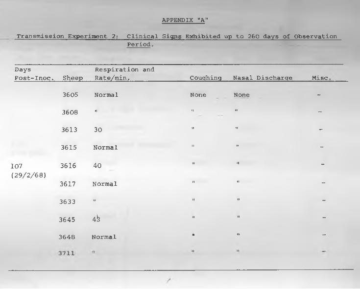
A P PE N D IX "A "
Transmission Experiment 2: Clinical Signs Exhibited up to 260 days of ObservationPeriod,
DaysPost-Inoc, Sheep
Respiration and Rate/min, Couqhinq Nasal Discharqe Mi sc
3605 Normal None None -
3608 II II II -
3613 30 II II -
3615 Normal II II -
107 3616 40 II II-
(29/2/68)3617 Normal II II -
3633 II II II -
3645 4b II II -
3648 Normal • II -
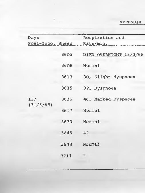
APPENDIX
DaysPost-Inoc,
137(30/3/68)
Respiration and Sheep Rate/min,
3605 DIEDi OVERNIGHT 13/3/68
3608 Normal
3613 30, Slight dyspnoea
3615 32, Dyspnoea
3616 46, Marked Dyspnoea
3617 Normal
3633 Normal
3645 42
3648 Normal
3711
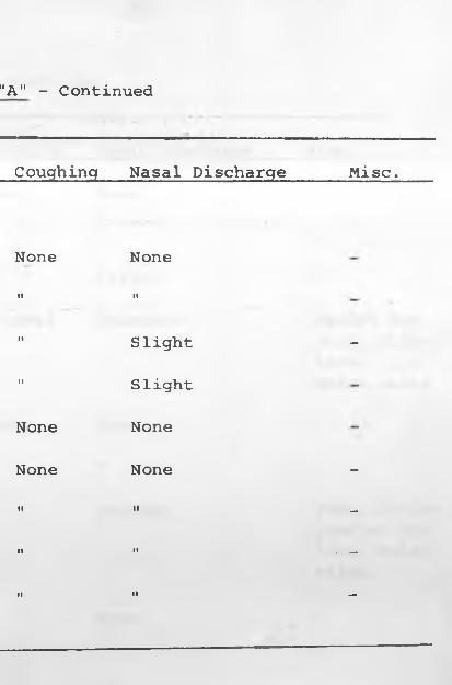
"A" - Continued
Couqhinq Nasal Discharqe Misc,
None NoneI I I t
I I S1 ig'ht -i t Slight
None None
None None -I I I I _
I I If . _
• l I I —
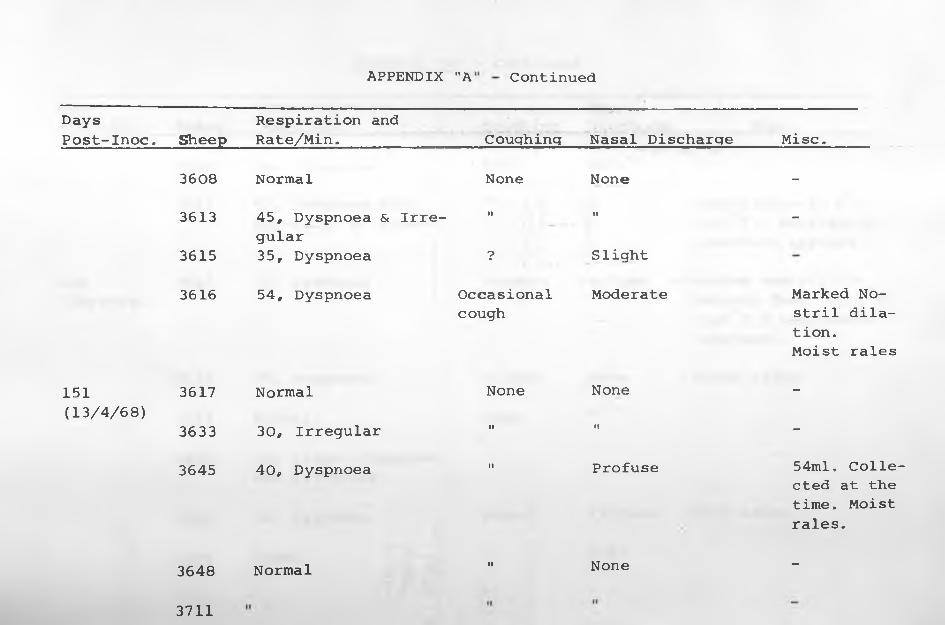
APPENDIX "A Continued
Days Respiration andPost-Inoc. Sheep Rate/Min. Coughinq Nasal Discharge Misc.
3608 Normal None None -
3613 45, Dyspnoea & Irregular
I t I I -
3615 35, Dyspnoea ? Slight
3616 54, Dyspnoea Occasional Moderate Marked No-cough stril dila
tion.Moist rales
151 3617 Normal None None -
(13/4/68)3633 30, Irregular I I I I -
3645 40, Dyspnoea I I Profuse 54ml. Collected at the time. Moist rales.
3648 Normal I I None -
3711
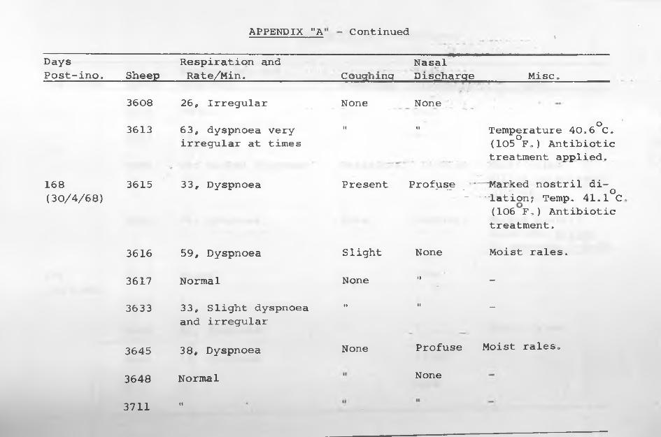
APPENDIX "A
DaysPost-ino, Sheep
Respiration and Rate/Min.
3608 26, Irregular
3613 63, dyspnoea very irregular at times
168 3615 (30/4/68)
33, Dyspnoea
3616 59, Dyspnoea
3617 Normal
3633 33, Slight dyspnoea and irregular
3645 38, Dyspnoea
3648 Normal
3711 II
Continued \
Coughinq------- *-------------------------
NasalDischarge Misc,
None None ;JII o" Temperature 40,6 C.
(105°F,) Antibiotic treatment applied,
Present Profuse * "Marked nostril di-•' - - o ~ "lation? Temp, 41,1 C(106 F,) Antibiotictreatment,
Slight None Moist rales,
None II __
•l II _
None Profuse Moist rales,
None
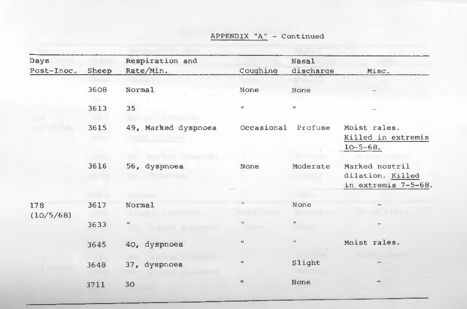
APPENDIX "A" - Continued
DaysPost-Inoc„ Sheep
Respiration and Rate/Min. Couqhinq
Nasaldischarqe Misc,
3608 Normal None None -
3613 35 I I I I -
3615 49, Marked dyspnoea Occasional Profuse Moist rales.
3616 56, dyspnoea None Moderate
Killed in extremis 10-5-68,
Marked nostril
178 3617 Normal I I None
dilation. Killed in extremis 7-5-68
(10/5/68)3633 I I I I I I -
3645 40, dyspnoea I t I I Moist rales,
3648 37, dyspnoea I I Slight -
3711 30 I I None -
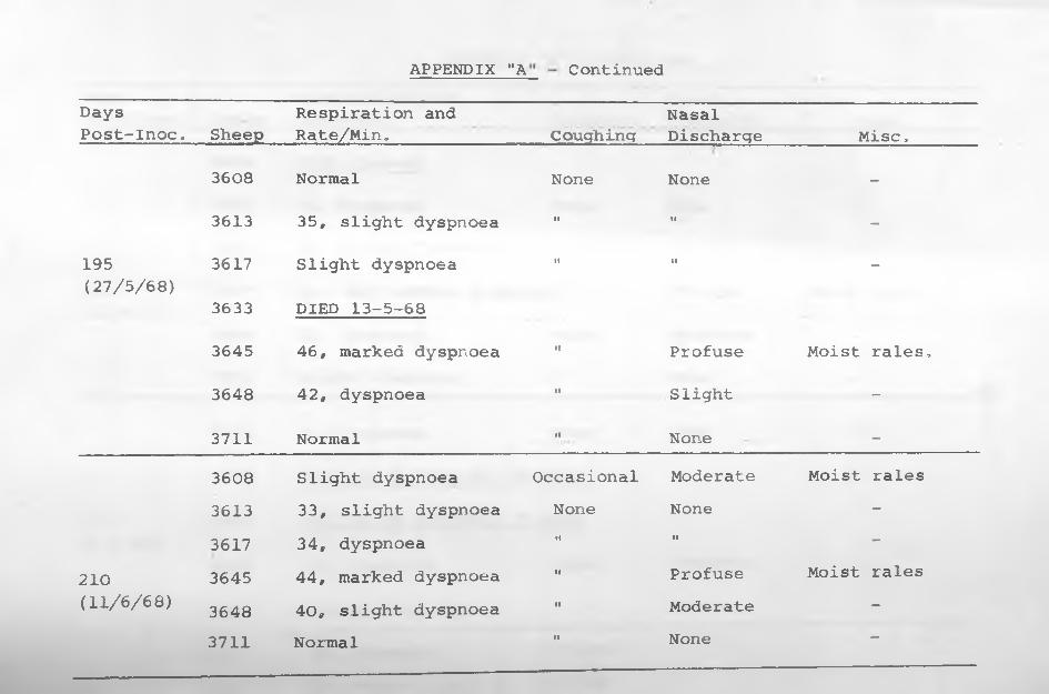
APPENDIX "A Continued
DaysPost-Inoc* Sheep
Respiration and Rate/Min» Couqhinq
NasalDischarqe Mi sc,
3608 Normal None None -
3613 35, slight dyspnoea I I I I -
195 3617 Slight dyspnoea I f I I —(27/5/68)
3633 DIED 13-5-68
3645 46, marked dyspnoea i e Profuse Moist rales,
3648 42, dyspnoea i t Slight -
3711 Normal 18 None -
3608 Slight dyspnoea Occasional Moderate Moist rales3613 33, slight dyspnoea None None -3617 34, dyspnoea I I i f -
210 3645 44, marked dyspnoea i i Profuse Moist rales(11/6/68) 3648 40, slight dyspnoea I I Moderate -
3711 Normal fl None -
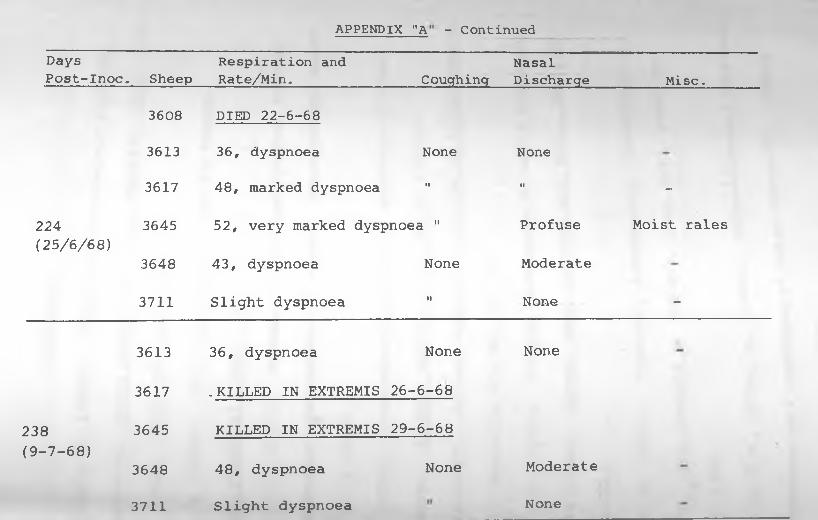
A P P E N D IX "A - C o n t i n u e d
Days Respiration and NasalPost-Inoc. Sheep Rate/Min.________________Coughing Discharge_________ Misc,
3608 DIED 22-6-68
3613 36, dyspnoea None
3617 48, marked dyspnoea i i
224 3645 52, very marked dyspnoea "(25/6/68)
3648 43, dyspnoea None
3711 Slight dyspnoea II
3613 36, dyspnoea None
3617 . KILLED IN EXTREMIS 26-6-68
238 3645 KILLED IN EXTREMIS 29-6-68(9-7-68)
3648 48, dyspnoea None
NoneII —
Profuse Moist rales
Moderate
None -
None
Moderate
3711 Slight dyspnoea None
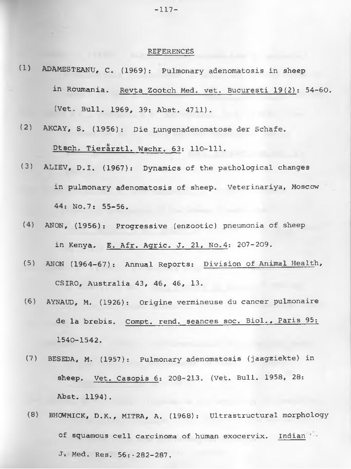
-117-
REFERENCES
(1) ADAMESTEANU, C. (1969): Pulmonary adenomatosis in sheep
in Roumania. Revta Zootch Med, vet. Bucuresti 19(2): 54-60.
(Vet, Bull. 1969, 39: Abst. 4711).
(2) AKCAY, S. (1956): Die Lungenadenomatose der Schafe.
Dtsch. Tierarztl. Wschr. 63: 110-111.
(3) ALIEV, D.I. (1967): Dynamics of the pathological changes
in pulmonary adenomatosis of sheep. Veterinariya, Moscow
44: No.7: 55-56.
(4) ANON, (1956): Progressive (enzootic) pneumonia of sheep
in Kenya. E. Afr. Agric. J. 21, No.4: 207-209.
(5) ANON (1964-67): Annual Reports: Division of Animal Health,
CSIRO, Australia 43, 46, 46, 13.
(6) AYNAUD, M. (1926): Origine vermineuse du cancer pulmonaire
de la brebis. Compt. rend, seances soc, Biol., Paris 95:
1540-1542.
(7) BESEDA, M. (1957): Pulmonary adenomatosis (jaagziekte) in
sheep. Vet. Casopis 6: 208-213. (Vet. Bull. 1958, 28:
Abst. 1194).
(8) BHOWMICK, D.K., MITRA, A. (1968): Ultrastructural morphology
of squamous cell carcinoma of human exocervix. Indian .'■*
J. Med. Res. 56:-282-287.
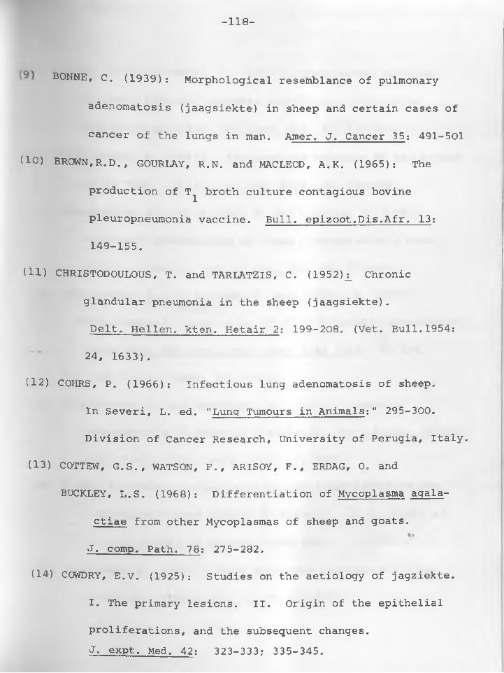
-118-
BONNE, C. (1939): Morphological resemblance of pulmonary
adenomatosis (jaagsiekte) in sheep and certain cases of
cancer of the lungs in man. Amer. J. Cancer 35: 491-501
(10) BROWN, R.D., GOURLAY, R.N. and MACLEOD, A.K. (1965): The
production of T^ broth culture contagious bovine
pleuropneumonia vaccine. Bull. epizoot.Dis.Afr. 13:
149-155.
(11) CHRISTODOULOUS, T. and TARLATZIS, C. (1952)j_ Chronic
glandular pneumonia in the sheep (jaagsiekte).
Pelt. Hellen. kten. Hetair 2: 199-208. (Vet. Bull.1954:
24, 1633).
(12) COHRS, P. (1966): Infectious lung adenomatosis of sheep.
In Severi, L. ed. "Lung Tumours in Animals:" 295-300.
Division of Cancer Research, University of Perugia, Italy.
(13) COTTEW, G.S., WATSON, F., ARISOY, F., ERDAG, 0. and
BUCKLEY, L.S. (1968): Differentiation of Mycoplasma agala-
ctiae from other Mycoplasmas of sheep and goats.%v
J. comp. Path. 78: 275-282.
(14) COWDRY, E.V. (1925): Studies on the aetiology of jagziekte.
I. The primary lesions. II. Origin of the epithelial
proliferations, and the subsequent changes.J. expt. Med. 42: 323-333? 335-345.
-119-
(15) CCWDRY, E.V. and MARSH, H. (1927): Comparative pathology
of South African jagziekte and Montana progressive
pneumonia of sheep. J. expt. Med. 45: 571-585.
(16) CUBA-CAPARO, A. (1961): Adenomatosis Pulmonar de los Ovinos
en el Peru. Bull. Off, int. Epiz. 56: 840-849.
(17) CUBA-CAPARO, A., DE LA VEGA, E. and COPAIRA, M. (1961):
Pulmonary adenomatosis of sheep - Metastasizing bron-
chiolar tumours. Am. J. vet. Res. 22: 673-682.
(18) CUBA-CAPARO, A. , DE LA VEGA, E. and DURAND, W. (1967):
Experimental transmission of ovine pulmonary adenomatosis.
Revta Fac, Med, vet. Univ. nac. Lima 18/20: 97-124.
(Vet. Bull., 1968, 38: 4670).
(19) CVJETANOVIC, V. and MARTINCIC, M. (1962): Pulmonary
adenomatosis in sheep in Yugoslavia. Vet. Arhiv. (Zagreb)
32: 77-82. (Vet. Bull. 1962: 32, 4269).
(20) DAHME, E. (1966): Classification and nomenclature of the
spontaneous lung tumours in Animals. In L. Severi ed.
"Lung Tumours in Animals." 143-150.
(21) DAMODARAN, S. (1960) : Ovine pulmonary adenomatosis (Jag
ziekte) . Indian vet. J. 37: 127-138.
-120-
122) DAVIES, G. and READ, W.C.S. (1968)j_ A modification of the
growth-inhibition test and its use for detecting
Mycoplasma mycoides var. mycoides. J. Hyg. Camb.66:
319-324.
(23) DAVIS, C.L. (1939): Primary carcinoma of the lung of sheep.
J. Amer, vet, med. Ass, 47: 237-240
(24) DEIANA, S. and CERETTO, F. (1967): Reperti istoimmunologici
nella adenomatosi polmonare enzootica degli ovini in
Sardegna (Histo-immunological results in sheep enzootic
pulmonary adenomatosis in Sardinia) Atti. Soc. ital.
Sci. vet. 21: 612-615.
(25) DE KOCK, G. (1929a): Are the lesions of jaagsiekte in sheep
of the nature of a neoplasm? 15th Ann. Rept. Dir. Vet.
Serv. U. South Africa: 611-641. .
(26) DE KOCK, G. (1929b): Further observations on the etiology
of jaagsiekte in sheep. 15th Ann. Rept. Dir. Vet. Serv.
U. South Afr.: 1169-1183.
(27) DE KOCK, G. (1958): The transformation of the lining of the
pulmonary alveoli with special reference to adenomatosis
in the lungs (jagsiekte) of sheep. Am. J. vet. Res. 19:
261-269.
-121-
(28) D HAND A, M.R., SHARMA, G.L. and BHALL, N.P. (1963): Possible
occurrence of jaagsiekte in indigenous sheep. Histo-
pathological evidence. Indian J. vet. Sci. Animal
Husbandry 33: 84-88.
(29) DUALDE PEREZ, D. (1963): Estudio General y Cauistico de la
Adenomatosis Pulmonar ovina en Espana. (Studies of
ovine pulmonary adenomatosis in Spain) . Proc. 17th
World Vet. Conqr. Hanover 1: 347-355.
(30) DUNGAL, N., GISLASON, G. and TAYLOR, E.L. (1938): Epizootic
adenomatosis in the lungs of sheep - comparisons with
jaagsiekte, verminous pneumonia and progressive pneu
monia. J. comp. Path. 51: 46-68.
(31) DUNGAL, N. (1946): Experiments with jaagsiekte.
Amer. J. Path. 22: 737-750.
(32) DYKES, J.R. and MeFADYEAN, J. (1888): Lung disease in sheep
caused by Strongylus rufescens. J. Comp. Path.l: 139-146.
(33) EBER, A. (1899): liber multiple Adenombildung in den Lungen
der Schafe. Z. Tiermed. 3: 161-172.
(34) EIDSON, C.S., RICHEY, D.J. and SCHMITTLE, S.C. (1969):
Studies on acute Marek's disease. XI: Propagation of
the GA isolate of Marek's disease in tissue culture.Avian Pis. 13: 636-653.
-122-
(35) ENCHEV, S, (1961): Studies of sheep's pulmonary adenomatosis
in Bulgaria. I. Epidemiology, symptoms and pathology.
II. Metastasis in pulmonary lymph nodes. Izv. vet, Inst,
Zaraz. Parazit. Bolesti, Sofia 1: 345-356 & 2: 177-186.
136) ENCHEV, S. (1962): Pulmonary adenomatosis of sheep in
Bulgaria. III. Histomorphology and Histogenesis.
Izv, vet. Inst. Zaraz. Parazit, Bolesti, Sofia 6: 78-85*
(Vet. Bull. 1964: 34, 658).
(37) ENCHEV, S. (1963): Concerning the malignant (metastasizing)
forms of so-called pulmonary adenomatosis (jaagsiekte)
in sheep. Izv, vet. Inst. Zaraz. Parazit. Bolesti,
Sofia 9: 19-31.
(38) ENCHEV, S. (1966): Experimentally induced pulmonary adenoma
tosis in sheep. Vet. Sci. Sofia 3: 947-954.
(39) ENCHEV, S. (1968): Reproduction of pulmonary adenomatosis
(jaagsiekte) in sheep II. Vet. Med. Nauki, Sofia 5(3):
13-24. (Vet. Bull. 1969: 39y 258).
(40) ENCHEV, S., TOMOV, T, and IVANOV, I. (1958): Pulmonary
adenomatosis of sheep in Bulgaria. Izv. Inst. Pat.
Zhivotni, Sofia 6: 365-367. (Vet. Bull. 1959: 29, 2621).
f a* 1 • *
. M9641I W * * * 1(41) ENCHEV. S. and HRISTOFOR . H
PMktokU ()•«•««•' **
1: 35-42.
42) spling, P. (1964)! Electron
The normal blood-air ba:: •
microscopy o* ll* I m 1
679-689.
(43) EYLAU, 0. (1953): Die inftctl&SS AdehOMtOS#
der Lungen des Schafe* (jag*ieHte)« ’ ■adenomatosis of the lung of • t£L£*
Wschr. 60: 184-189.
(44) FALLON, R.J. (1966): The relationship totv-ec
and human leukaemia. Vet. Rec. 79» *»■*
(«) FOTHEMHGHAM, M. (193S)l Sh..p pn. « r U k
hgric. Dept. II: 151-152.
OP
*
(46) FULTON, F. and DUMBELL, K.R. <1949^ Tfc. M ,
<i.«. « •«.». o, toB—. « •I- 97-111.
471 GEEVER* E-F" NEUBUERGBR. k .t . and .
pulmonary alveolar linin, urn*,
tions m man and animal, i„
-124-
(48) GELLER, S.A. and TOKER, C. (1969): Pulmonary adenomatosis
and peripheral adenocarcinoma of the lung: An Ultrastru-
ctural demonstration of common morphologic features.
Arch. Path. 88: 148-154.
(49) GREIG, A.S. (1955): The isolation of pleuropneumonia-like
organisms from the respiratory tracts of sheep.
Canad. J. comp. Med. 19: 265-271.
(50) HAYFLICK, L. and KOPROWSKI, H. (1965): Direct agar isolation
of Mycoplasmas from human leukemic bone marrow.
Nature 205: 713-714.
(51) HELMBOLDT, C.F., JUNGHERR, E.L. and CUBA-CAPARO, A.C. (1958):
Pulmonary adenomatosis in the Chinchilla. Am. J, vet.
Res. 19; 270-276.
(52) HODGES# R.T. (1969): The role of mycoplasma in some diseases
of pigs. Brit, vet. J, 125: 340-343.
(33) HUTT/ F.B. (1964): Resistance of jaagsiekte in Icelandic
Sheep. In: Animal Genetics:, 504-506.
(54) ITO/ S. and KARNOVSKY/ M.J. (1968): Formaldehyde-glutaral-
dehyde fixatives containing trinitro compounds.
J. Cell Biol. 30: 1-2. *
*
-125-
(55) JAKOB, W. and KRAUSE, H. (1965): Zum Auftreten der enzooti-
schen Lungenadenomatose in Schafbestanden. (Pulmonary
adenomatosis in sheep). Mh. Vet, med, 60: 217-233.
(56) JENNINGS, A.R. (1967): Cellular and tissue reactions,
In: Betts, A.O. and York, C.J. edited, "Viral and
Rickettsial Infections of Animals" Acad. Press.(N.Y.& Lon.)
(57) KENYA (1923): Ann. Rept. Dept. Agric. Kenya: 61.
(58) KENYA (1924): Ann, Rept. Dept, Agric. Kenya: 86.
(59) KOSTENKO, Yu. G. (1964): Adenomatosis of sheep. Veterinariya,
Moscow 41(4): 34-36. (Vet. Bull. 1965, 35: 2716).
(60) LEACH, R.H. and BUTLER, M. (1966): Comparison of mycoplasmas
associated with human tumours, leukemia, and tissue
cultures. J. Bact. 91: 934-941.
(61) LINDBERG, L.G. (1968): Virus-like particles in Rous rat
sarcoma. Acta path, et microbiol. Scand. 74: 189-198
(62) LITWIN, J., OFFICER, J.E., BROWN, A. and MOULDER, J.W. (1961)j_
A comparative study of the growth cycles of different
members of the psittacosis group of different host
cells. J. infect. Pis. 109: 251-279.
(63) LONGLEY, E.O. (1951): Contagious caprine pleuro-pneumonia.
A study of the disease in Nigeria. Colonial Publ. No.7: pp.27. H,M, Stationary Office, London.
-126-
(64) MACKAY, J.M.K. (1966): Association of pleuropneumonia-like
organisms (P.P.L.O.) with pulmonary adenomatosis in
sheep: Cultural and serological findings. In: Severi,
L. ed. "Lung Tumours in Animals." 403-412. Division
of Cancer Research, University of Perugia, Italy.
(65) MACKAY, J.M.K. (1969): Tissue culture studies of sheep
pulmonary adenomatosis (jaagsiekte). I. Direct cultures
of affected lungs. II. Transmission of cytopathic
effects to normal cultures. J.comp.Path. 79: 141-146;
147-154.
(66) MACKAY, J.M.K. and NISBET, D.I. (1966): Jaagsiekte - A ha
zard of intensified sheep husbandry. Vet.Rec. 78: 18-20.
(67) MADEIRA, A.C. (1949): Adenocarcinoma em focos multiplos do
Pulmao em um ovino. Rev. Med. Vet. Lisboa, 44: 95-100
(Vet. Bull. 1952: 22, 30).
(68) MARKSON; L.M. and TERLECKI, S. (1964): The experimental
transmission of ovine pulmonary adenomatosis.
Path, vet, 1: 269-288.
(69) MARSH, H. (1923a): Progressive pneumonia in sheep.
J. Amer. vet, med. Ass. 62: 458-473.
-127-
(70) MARSH, H. (1923b): The bacteriology of progressive
pneumonia of sheep. J. Amer. vet. Med. Ass. 64: 304-317.
(71) MARSH, H. (1966): Progressive pneumonia of sheep in the
United States and its relation to diseases of the
jaagsiekte complex. In: Severi, L. ed. "Lung Tumours
in Animals.11 285-294. Division of Cancer Research,
University of Perugia, Italy.
(72) MARTINCIC, M. and CVJETANOVIC, V. (1967): Sheep pulmonary
adenomatosis in SR Crna Gora, Yugoslavia. 18th World
Vet. Congr. I: 323.
(73) Me FAD YEAN, J. (1894): Verminous pneumonia in the sheep.
J. comp. Path, 7: 31-42.
(74) McFADYEAN, J. (1920) : Transformation of the alveolar epi
thelium in verminous pneumonia in the sheep.
J. comp. Path. 33: 1-10.
(75) McFADYEAN, J. (1938): Jaagsiekte. J. comp. Path. 51: 78-84.
(76) METTAM, R.W.M. (1927): Investigations into lung diseases of
lambs and sheep. Ann. Rept. Kenya Agric. Dept.: 163-179.
(77) METTAM, R.W.M. (1929): Laikipia lung disease. Ann. Rept.
Kenya Agric. Dept.: 333-362.
-128-
(78) MITCHELL, D.T. (1915): Investigations into jagziekte or
chronic catarrhal—pneumonia of sheep. 3rd & 4th Repts.
U. South Africa Dept, of Agric. 585-614.
(79) MITROFANOV, V.M. (1963): Histogenesis of pulmonary adenoma
tosis in sheep. Arkh. Patol. 25, No.10: 46-51.
(Vet. Bull. 1964: 34, 1409).
(80) MITROFANOV, V.M. (1964): Metastasizing pulmonary adenomato
sis. Arkh. Patol. 26, No.8: 68-74. (Vet. Bull. 1965:
35, 1003).
(81) MORAILLON, P. and YALCIN, N. (1967): Adenomatose pulmonaire
des ovin's en France. (Pulmonary adenomatosis of
sheep in France). Proc. 18th World Vet. Congr., Paris I:
323-324.
(82) NIELSEN, S.W. (1966) : Spontaneous canine pulmonary tumors.
In: Severi, L. ed. "Lung Tumours in Animals:" 151-162.
Division of Cancer Research, University of Perugia, Italy.
(83) NOBEL, T.A. (1958): Pulmonary adenomatosis (jaagsiekte) in
sheep with special reference to its occurrence in
Israel. Refuah Vet. 15: 1.
(84) NOBEL, T.A., NEUMANN, F. and KLOPFER, U. (1968): Metastases
in pulmonary adenomatosis of sheep. Refuah vet. 25: 57-52.
-129-
(85) NOBEL, T.A., NEUMANN, F. and KLOPFER, U. (1969): Histo
logical patterns of the metastases in pulmonary adeno
matosis of sheep (jaagsiekte). J. comp. Path. 79: 537-540.
(86) OBIDITSCH-MAYER, I. and BREITFELLNER, G. (1968): Electron
microscopy in Cancer of the lung. Cancer 21: 945-951.
(87) OMAR, A.R. (1964): The characteristic cells of the lung
and their reaction to injury. Vet. Bull. 34: 371-382,
431-443.
(88) PAGE, L.A. (1968): Proposal for the recognition of two species
in the Genus Chlamydia. Jones, Rake, and Stearns, 1945.
Int. J. Syst. Bact. 18: 51-66.
(89) PALLASKE, G. (1929): Zur Kenntnis der sognannten Adenomato-
se oder der raultiplen Adenome der Lunge des Schafes.
(On the so-called adenomatosis or multiple adenomas of
the lungs of sheep). Berl. Munch, tierarztl. Wschr. 45:
677-685,
(90) PALLASKE, G. (1954): Beitrag zur sogenannten Lungenadenoma-
tose der Schafe, (So-called adenomatosis of the lungs
in sheep). Berl. Munch. Tierarztl. Wschr. 67: 23-25.
(Vet. Bull. 1954: 24, 1884).!
-130-
(91) PAREDES, T. (1953): Contribucion al Estudio Clinico de la
Poliadenomatosis pulmonar del Carnero, B.V.M. Thesis.
Universidad de san Marcos, Lima, Peru. (Quoted by
Cuba-Caparo, A. et al. 1961).
(92) PAVLOV, N. (1967): Histopathology of neorickettsial pneu
monias in sheep. Zbl. vet, med. ReiheB. 12: 517-526.
(93) POLAND, JENNIFER (1969): A general description of mycoplasmas.
Br. vet. J. 125: 344-348.
(94) RAJYA, B.S. and SINGH, C.M. (1964): The pathology of pneu
monia and associated respiratory disease of sheep and
goats.' I. Occurrence of jaagsiekte and maedi in sheep
and goats in India. Am. J. vet. Res, 25: 61-67.
(95) ROBERTSON, Wm. (1904): Jagziekte or chronic catarrhal
pneumonia (sheep). J. comp. Path. 17: 221-224.
(96) ROMBOLI, B. (1959): Pulmonary diseases resembling jaagsiekte
in sheep in Italy. I. Lesions. Ann. Fac. Med, vet.
Pisa 12: 50-96. (Vet. Bull. 1960: 30, 4022).
(97) RYDER, M.L. and STEPHENSON, S.K. (1968): Wool Growth:
Academic Press (London).
(98) SANDERS, R.N. (1961): Contagious bovine pleuro-pneumonia.
Ann. Rept. Dept. Vet. Serv. & Animal Ind., Uganda,I960: P.4., Entebbe, Govt. Printers.
-131-
(99) SANTIAGO-LUQUE, J.M. (1963): Neuvos datos anatomo e
histopatologicos sobre la adenomatosis pulmonar ovina.
17th World Congr. Hanover, I; 357-360.
(100) SAVAGE, A. (1926): Pulmonary cancer in a sheep. J. Amer.
vet, med. Ass, 69: 86-88.
(101) SCHIEFER, B. (1966): Adenomatosis and epithelial trans
formation in the lungs of animals with special reference
to jaagsiekte. In: Severi, L. ed. "Lung Tumours in
Animals." 301-310. Division of Cancer Research,
University of Perugia, Italy.
(102) SCHIEFER, B. and KAST, A. (1969): Zum Vorkommen der Lunge-
nadenomatose der Schafe in Bayern II. Mitteilung
(Occurrence of pulmonary adenomatosis in sheep in
Bavaria. II): Berl. Munch, tierarztl. Wschr. 82: 1-2.
(103) SCHULZ, L. Cl., SOMOZA, A. and WEILAND, F. (1965): Zum Auft-
reten und zum Tumor--charakter der enzootischen Lungena-
denomatose des Schafes. Vergleichende Untersuchungen
in Chile und in der Bundesrepublik Deutschland. (Neo
plastic character of enzootic pulmonary adenomatosis
of sheep: comparative studies in Chile and Federal
Germany). Dtsch. tierarztl. Wschr. 72: 458-461.
-132-
(104) SCHULZ, L. Cl. and WEILAND, F. (1968): Lungenadenomatose
der Schafe in Norddeutschland. (Pulmonary adenomatosisI
of sheep in Northern Germany). Zbl. Vet. Med. 15: 132-139.
(105) SEATON, V.A. (1958): Pulmonary adenomatosis in Iowa cattle.
Am. J. vet. Res, 19: 600-609.
(106) SEDL^EIER, H., SCHIEFER, B. and JAKOB, H. (1966): Zum Vorko-
mmen der Lungenadenomatose bei Schafen in Bayern.
(Occurrence of pulmonary adenomatosis in Bavarian sheep).
Dtsch. tierarztl. Wschr. 73: 422-430.
(107) SHIRLAW, (1953): In "Sheep Lung Diseases "File" Vol.I -
Archives of Kenya Government Veterinary Department.
(108) SHIRLAW (1956): In "Sheep Lung Diseases "File" Vol.II. -
Archives of Kenya Government Veterinary Department.
(109) SHIRLAW, J.F. (1959): Studies on jaagsiekte in Kenya.
Bull, epizoot. Pis, Afr, 7: 287-302.
(110) SHIRLAW (1962): In "Sheep Lung Diseases "File". Vol.III -0
Archives of Kenya Government Veterinary Department.
(111) SIGURDSSON, B. (1958): Adenomatosis of sheep's lungs.
Experimental transmission. Arch, ges. Virusforsch, 8:
51-58.
-133-
(112) SIMON, M. A. (1947): So-called pulmonary adenomatosis and
"alveolar cell tumor." Amer. J. Path. 23: 413-427.
(113) SMITH, W. and MACKAY, J.M.K. (1969): Morphological obser
vations on a virus associated with sheep pulmonary
adenomatosis (jaagsiekte). J. comp. Path. 79: 421-424.
(114) SPENCER, H. and RAEBURN, G. (1956): Pulmonary (Bronchiolar)
adenomatosis. J. Path. Bact. 71: 145-154.
(115) STAMP, J.T. and NISBET, D.I. (1963): Pneumonia of sheep.
J. comp. Path. 73: 319-328.
(116) STEVENS, A.J. (1957): Respiratory diseases of sheep.
Vet. Rec. 69: 1249-1257.
(117) STEVENSON, R.G. (1968): Studies in Pneumonia of animals.
Ph.D. Thesis, University of Edinburgh.
(118) TAMARIN, RUTH (1969): Laboratory diagnosis of bedsonia
infections in sheep and cattle. Refuah Vet. 26(1):28-39.
(119) TERPSTRA, C. (1969): Nairobi sheep disease: Studies on virus
properties, epizootiology and vaccination in Uganda.
Thesis: From the Animal Health Research Centre, Entebbe,
(Uganda).
(120) TOKER, C. and TREVINO, N. (1966): Ultrastructure of human
primary hepatic carcinoma. Cancer 19: 1594-1606.
-134-
(121) TRAUTWEIN, G. and HELMBOLDT, C.F. (1967): Experimental
pulmonary talcum granuloma and epithelial hyperplasia
in the Chinchilla. Path, vet. 4: 254-267.
(122) TULLY, J.G. (1966): Mycoplasma granularum of swine origin
as a tissue culture contaminant. Proc. Soc. exp. Biol,
Med. 122: 565-568.1 - J - 1
(123) TUSTIN, R.C. (1969): Ovine jaagsiekte. J.S.Afr. vet, med. .
Ass. 4a(l)4 3-23.
(124) WANDERA, J.G. (1967a): Pneumonia of sheep in Kenya.
Part I: Bacterial and parasitic pneumonia.
Bull, epizoot. Pis, Afr. 15: 245-258.
(125) WAND ERA, J.G. (1967b): Pneumonia of sheep in Kenya.
Part II: Pathology of.chronic progressive pneumonia.
Bull, epizoot. Pis. Afr. 15: 393-407.
(126) WANDERA, J.G. (1968): Experimental transmission of sheep
pulmonary adenomatosis (jaagsiekte). Vet. Rec, 83:
478-482.
(127) WANDERA, J.G. (1970): Progressive interstitial pneumonia
(Maedi) of sheep in Kenya. Vet. Rec. 86:
(128) WATRACH, A.M. and VATTER, A.E. (1959): Electron Microscopy of
the pulmonary alveolar wall. Amer. J. vet. Res. 20: >723-735.
-134-
(121) TRAUTWEIN, G. and HELMBOLDT, C.F. (1967): Experimental
pulmonary talcum granuloma and epithelial hyperplasia
in the Chinchilla. Path, vet. 4; 254-267.
(122) TULLY, J.G. (1966): Mycoplasma granularum of swine origin
as a tissue culture contaminant. Proc. Soc, exp. Biol,
Med. 122: 565-568.
(123) TUSTIN, R.C. (1969): Ovine jaagsiekte. J.S.Afr. vet, med. .
Ass. 40(1)4 3-23.
(124) WANDERA, J.G. (1967a): Pneumonia of sheep in Kenya.
Part I: Bacterial and parasitic pneumonia.
Bull, epizoot. Pis. Afr. 15: 245-258.
(125) WANDERA, J.G. (1967b): Pneumonia of sheep in Kenya.
Part II: Pathology of^chronic progressive pneumonia.
Bull, epizoot. Pis. Afr. 15: 393-407.
(126) WANDERA, J.G. (1968): Experimental transmission of sheep
pulmonary adenomatosis (jaagsiekte). vet. Rec. 83:
478-482.
(127) WANDERA, J.G. (1970): Progressive interstitial pneumonia
(Maedi) of sheep in Kenya. Vet. Rec. 86:
(128) WATRACH, A.M. and VATTER, A.E. (1959): Electron Microscopy of
the pulmonary alveolar wall. Amer. J. vet. Res. 20: >723-735.
(129) WHITWORTH, S.H. (1926): Observations on a lung affection
of lambs and sheep. Ann. Rept, Kenya Agric. Dept.
(1926): 102-110.
(130) WILSON, M.R. and THOMSON, R.G. (1968): Chlamydia pneumonia
of calves. Res, vet. Sci. 9(5): 467-47 3.
(131) ZILBER, P.A., SHAPIRO, V.S., GARDASH' YAN, A.M. and
MITROFANOV, V.M. (1962): Cysts in mice inoculated with
the lung extract from sheep with pulmonary adenomatosis.
Voprosy Virusologii 7 : 288-291. (Vet. Bull., 1963/
33. Abs. 240).
(132) WIGHT, P.A.L. (1969): The ultrastructure of sciatic
nerves affected by fowl paralysis (Marek s disease).
J. comp. Path. 79: 563-570.
-135-
ONIV. **l.