Sharma, Kanika, Patrick W. Inglett, K. Ramesh Reddy, and ... · 2057 NOTES Limnol. Oceanogr.,...
6
2057 NOTES Limnol. Oceanogr., 50(6), 2005, 2057–2062 q 2005, by the American Society of Limnology and Oceanography, Inc. Microscopic examination of photoautotrophic and phosphatase-producing organisms in phosphorus-limited Everglades periphyton mats Abstract—Using a fluorescent-labeled enzyme substrate, we examined the location of in situ phosphatase activity in a pe- riphyton mat and explored the potential associations of phos- phatase-producing organisms (PPO) and cyanobacteria within these mats. Our results indicate that most PPOs are concen- trated in the lower section of the mat, and the phosphatase activity appears to be associated with heterotrophic organisms that are in close proximity to chlorophyll-containing cyano- bacteria. The lack of observed phosphatase by larger photo- synthetic cells and the close association of these cells with PPOs indicate a possible interaction whereby PPOs obtain photosynthetically fixed carbon from cyanobacteria and, in turn, provide inorganic phosphorus (P) and other compounds to the cyanobacteria. We believe these results may represent additional evidence for algal–bacterial symbiosis in aquatic systems and, in particular, the P-limited cyanobacterial mat communities. By supporting various endangered and threatened species and maintaining high genetic and ecological diversity, the Florida Everglades is a unique wetland ecosystem of global importance (Maltby and Dugan 1994). The Everglades eco- system also supports high levels of productivity despite its phosphorus (P)-limited nature. Much of this high productiv- ity is attributed to the growth and dominance of periphytic communities (McCormick and Stevenson 1998), which can cover much of the open water regions and serve as a base of the Everglades food web (Browder et al. 1994). Through its biotic activities (e.g., photosynthesis and nitrogen fixa- tion) the Everglades periphyton community has a pro- nounced effect on the biogeochemistry of the water column and the ecosystem as a whole. For this reason, periphyton communities and their associated functions are critical to the health and stability of the Everglades ecosystem. The Everglades periphyton are complex microbial assem- blages based on cyanobacterial filaments of Schizothrix sp. and Scytonema sp. (Gleason and Spackman 1974). These periphytic forms can occur in association with the benthos (benthic) or in association with submersed and emergent macrophytes (epiphytic) such as Typha, Cladium, and Utri- cularia purpurea. They may be either thin films (ca. 1–2 mm) or well-developed, thick (ca. 1–4-cm) growths referred to as floating and benthic periphyton ’mats’ or epiphytic ’sweaters.’ Both epiphytic and benthic mats can detach from the substrata via buoyancy from trapped gases and form floating periphyton mats at the water surface. Photosynthetic activity within the mats influences local pH and in hardwater Everglades regions can lead to precipitation of calcium car- bonate within the mat (Browder et al. 1994). In this regard, the Everglades periphyton mats are similar to other calci- fying cyanobacterial communities (Rejmankova and Komar- kova 2000). In thick cyanobacterial mats, vertical gradients of light, oxygen, pH, nutrients, and microbial metabolic products may exist (Jorgensen 1983; Stal et al. 1985). Mat organisms structure themselves in response to these physico-chemical gradients, leading to the formation of a biogeochemically distinct layer. With this structure, a cyanobacterial mat can simultaneously support diverse groups of microorganisms and their associated biogeochemical activities. This diversity of organisms and functions is a key factor in the ability of these mats to exist and thrive in extreme environments such as the extremely P-limited Florida Everglades (Cohen and Rosenberg 1989). Under conditions of P limitation, the availability of P is often regulated by activity of the enzyme phosphatase which hydrolyzes organic P (P o ) compounds to bioavailable inor- ganic phosphate (P i ) (Chrost 1991). Studies have shown in- creased phosphatase activity of bacterioplankton (Campbella et al. 1984) and cyanobacterial mats (Rejmankova and Ko- markova 2000) in P-limited environments. High phosphatase activity has also been observed in periphyton mats of the Everglades (Newman et al. 2003) and is likely a major factor contributing to the dominance of periphyton in P-limited systems such as the Everglades. In aquatic systems, it is often presumed that phosphatase activity of periphyton consortia is the simple summation of the activities of the component organisms, with the response of the community being linked to the overall conditions of P limitation in the system. Many aquatic organisms, includ- ing cyanobacteria (Grainger et al. 1989), diatoms, and green algae (Gonzalez-Gil et al. 1998), and eubacteria (Jansson et al. 1996) are known to produce phosphatase in pure cultures; however, several studies have also shown that the physio- logical and biochemical properties of bacteria in isolation do not reflect those of organisms growing in natural consortia (Deretic et al. 1994; Caldwell et al. 1997). For this reason, production of phosphatase in periphyton consortia may be the result of one or more specific groups of organisms. Iden- tifying active phosphatase-producing organisms (PPO) and investigating the potential associations with other mat or- ganisms serve as the initial steps to understanding phospha- tase expression in a periphyton community. Traditional methods of measuring phosphatase activity of natural periphyton primarily consist of assaying bulk sam- ples without attempting to separate the relative contribution
Transcript of Sharma, Kanika, Patrick W. Inglett, K. Ramesh Reddy, and ... · 2057 NOTES Limnol. Oceanogr.,...
2057
NOTES
Limnol. Oceanogr., 50(6), 2005, 2057–2062q 2005, by the American Society of Limnology and Oceanography, Inc.
Microscopic examination of photoautotrophic and phosphatase-producing organismsin phosphorus-limited Everglades periphyton mats
Abstract—Using a fluorescent-labeled enzyme substrate, weexamined the location of in situ phosphatase activity in a pe-riphyton mat and explored the potential associations of phos-phatase-producing organisms (PPO) and cyanobacteria withinthese mats. Our results indicate that most PPOs are concen-trated in the lower section of the mat, and the phosphataseactivity appears to be associated with heterotrophic organismsthat are in close proximity to chlorophyll-containing cyano-bacteria. The lack of observed phosphatase by larger photo-synthetic cells and the close association of these cells withPPOs indicate a possible interaction whereby PPOs obtainphotosynthetically fixed carbon from cyanobacteria and, inturn, provide inorganic phosphorus (P) and other compoundsto the cyanobacteria. We believe these results may representadditional evidence for algal–bacterial symbiosis in aquaticsystems and, in particular, the P-limited cyanobacterial matcommunities.
By supporting various endangered and threatened speciesand maintaining high genetic and ecological diversity, theFlorida Everglades is a unique wetland ecosystem of globalimportance (Maltby and Dugan 1994). The Everglades eco-system also supports high levels of productivity despite itsphosphorus (P)-limited nature. Much of this high productiv-ity is attributed to the growth and dominance of periphyticcommunities (McCormick and Stevenson 1998), which cancover much of the open water regions and serve as a baseof the Everglades food web (Browder et al. 1994). Throughits biotic activities (e.g., photosynthesis and nitrogen fixa-tion) the Everglades periphyton community has a pro-nounced effect on the biogeochemistry of the water columnand the ecosystem as a whole. For this reason, periphytoncommunities and their associated functions are critical to thehealth and stability of the Everglades ecosystem.
The Everglades periphyton are complex microbial assem-blages based on cyanobacterial filaments of Schizothrix sp.and Scytonema sp. (Gleason and Spackman 1974). Theseperiphytic forms can occur in association with the benthos(benthic) or in association with submersed and emergentmacrophytes (epiphytic) such as Typha, Cladium, and Utri-cularia purpurea. They may be either thin films (ca. 1–2mm) or well-developed, thick (ca. 1–4-cm) growths referredto as floating and benthic periphyton ’mats’ or epiphytic’sweaters.’ Both epiphytic and benthic mats can detach fromthe substrata via buoyancy from trapped gases and formfloating periphyton mats at the water surface. Photosyntheticactivity within the mats influences local pH and in hardwaterEverglades regions can lead to precipitation of calcium car-
bonate within the mat (Browder et al. 1994). In this regard,the Everglades periphyton mats are similar to other calci-fying cyanobacterial communities (Rejmankova and Komar-kova 2000).
In thick cyanobacterial mats, vertical gradients of light,oxygen, pH, nutrients, and microbial metabolic productsmay exist (Jorgensen 1983; Stal et al. 1985). Mat organismsstructure themselves in response to these physico-chemicalgradients, leading to the formation of a biogeochemicallydistinct layer. With this structure, a cyanobacterial mat cansimultaneously support diverse groups of microorganismsand their associated biogeochemical activities. This diversityof organisms and functions is a key factor in the ability ofthese mats to exist and thrive in extreme environments suchas the extremely P-limited Florida Everglades (Cohen andRosenberg 1989).
Under conditions of P limitation, the availability of P isoften regulated by activity of the enzyme phosphatase whichhydrolyzes organic P (Po) compounds to bioavailable inor-ganic phosphate (Pi) (Chrost 1991). Studies have shown in-creased phosphatase activity of bacterioplankton (Campbellaet al. 1984) and cyanobacterial mats (Rejmankova and Ko-markova 2000) in P-limited environments. High phosphataseactivity has also been observed in periphyton mats of theEverglades (Newman et al. 2003) and is likely a major factorcontributing to the dominance of periphyton in P-limitedsystems such as the Everglades.
In aquatic systems, it is often presumed that phosphataseactivity of periphyton consortia is the simple summation ofthe activities of the component organisms, with the responseof the community being linked to the overall conditions ofP limitation in the system. Many aquatic organisms, includ-ing cyanobacteria (Grainger et al. 1989), diatoms, and greenalgae (Gonzalez-Gil et al. 1998), and eubacteria (Jansson etal. 1996) are known to produce phosphatase in pure cultures;however, several studies have also shown that the physio-logical and biochemical properties of bacteria in isolation donot reflect those of organisms growing in natural consortia(Deretic et al. 1994; Caldwell et al. 1997). For this reason,production of phosphatase in periphyton consortia may bethe result of one or more specific groups of organisms. Iden-tifying active phosphatase-producing organisms (PPO) andinvestigating the potential associations with other mat or-ganisms serve as the initial steps to understanding phospha-tase expression in a periphyton community.
Traditional methods of measuring phosphatase activity ofnatural periphyton primarily consist of assaying bulk sam-ples without attempting to separate the relative contribution

2058 Notes
Fig. 1. Location of the site used for sampling P-limited floatingperiphyton mats in WCA-2A of the Florida Everglades.
of individual mat organisms. For this reason, we conductedthe following study to investigate phosphatase production byspecific groups of organisms (i.e., algae/bacteria, photo-trophs/heteretrophs) as they occur within an intact, calcare-ous periphyton mat of the Florida Everglades. Our goals inthis study were to determine the spatial distribution of phos-phatase activity within the mat and to examine the associa-tions between photoautotrophic and phosphatase-producingbacterial groups in a periphyton mat. To demonstrate thelocation of phosphatase activity in situ, we employed a mi-croscopic technique based on the fluorescent substrate ELFt97 (Molecular Probes). Studies such as this have importantimplications for understanding microbial cycling of P withinthese and similar periphyton mat communities.
Study site and sampling—Periphyton mats used in thisstudy were obtained from a site in the interior of Water Con-servation Area 2A (WCA-2A) of the Florida Everglades(Fig. 1). This area is typical of the P-limited regions (soiltotal phosphorus (TP), 0.6 g kg21; pore water P, 0.1 mg L21;and periphyton TP, 75 mg kg21) of the Northern Evergladesand is characterized by ridges and open slough areas. Typicalbiochemical parameters of this site are discussed by Inglettet al. (2004). Vegetation on the ridges is dominated by Clad-ium sp., while periphyton mats occur predominantly in openslough areas dominated by Nymphaea, Eleocharis, and Utri-cularia spp. Floating periphyton mats were sampled in No-
vember 2002. Several intact mats exhibiting well-developedlayers (Fig. 2a) were collected in site-water–filled polyeth-ylene containers and stored on ice while they were trans-ported to the Wetland Biogeochemistry Laboratory at theUniversity of Florida in Gainesville, Florida.
Cryoembedding and cryosectioning—Periphyton matswith intact layers in their vertical profiles were chosen andsectioned within 24 h of collection (Fig. 2). The sampleswere cryoembedded in Tissue-Tekt OCT compound (Miles;Yu et al. 1994). Embedded samples were sectioned (5 mmthick) with a cryostat (Leica), and orientation of the matswas adjusted such that the sections would contain all thelayers in the vertical profile of mats. Sections were mountedon glass slides and treated with formaldehyde (0.01%) be-fore storing them at 48C until further staining analysis.
Fluorescent staining—Sections of periphyton mats werestained with enzyme-labeled fluorescence substrate, ELFt97 [(59-chloro-29-phosphoryloxyphenyl)-6-chloro-4-(3H)-quinazolinone] and/or 49, 6-diamidino-2-phenyl-indole(DAPI). Enzymatic hydrolysis of water-soluble ELFt97phosphatase substrate (ELF-P) yields a water-insoluble, yel-low–green ELF–alcohol (ELF-A) precipitate that is extreme-ly photostable. The sites of phosphatase production were vi-sualized by epifluorescence microscopy. Fluorescent stainDAPI binds to double-stranded DNA, and the stained cellsfluoresce blue. Immediately prior to use in this study, ELF-P was diluted at ratio of 1 : 20 in ELF Detection Buffer andfiltered through 0.2-mm spin filters to remove substrate pre-cipitates. Each prefixed mat section was incubated with 30mL of ELF-P for 30 min in the dark at room temperature.Stained samples were washed with 10 mmol L21 phosphate-buffered saline to stop the reaction. Negative controls wereprepared by treating the sections of mats as described aboveexcept that they were incubated with ELF detection bufferwithout ELF-P substrate. Some randomly chosen ELF-P–stained cryosections were also stained with DAPI for 5 minin dark.
Microscopy and image analysis—A fluorescent morpho-metric microscope was used to examine prepared sections.The excitation spectrum of chlorophyll (Chl) a and b andELF-A are different; therefore, the yellow–green signals ofELF-A and the red fluorescence of chlorophyll were visu-alized sequentially with separate filter sets. ELF-P–treatedsamples were visualized with an Olympus type U filter. Fil-ters used for ELF-A detection were 360 6 40 nm for exci-tation and 530 6 25 nm for emission. The Texas red filterwas used for images of Chl a autofluorescence (CHL im-ages). DAPI-stained samples were observed under a fluores-cent microscope equipped with long-pass filter set (excita-tion 365 6 8 nm; emission . 420 nm). Images captured atthe same spot by the different filters were digitized with acooled color charge-coupled device camera. ELF-A, DAPI,and Chl images of the same field were stored in a single fileand later overlayed to show the location of the chlorophyll-containing organisms and the zones of phosphatase produc-tion.

2059Notes
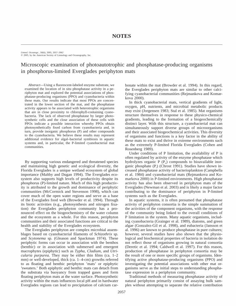
Fig. 2. Photo depicting the three layers in a typical P-limited Everglades periphyton mat similar to that used in this study. (a) Photo-micrographs of vertical cryosections (5-mm thickness) of the mat stained with fluorescent phosphate substrate. (b–f) Sites of alkalinephosphatase (yellow–green fluorescence) and chlorophyll (red fluorescence) activity are evident in (b) vertical mat cross section; (c, d)localized aggregate of filamentous cyanobacteria; (e) isolated colony of coccoid cyanobacteria; and (f) along remnant cyanobacterial sheaths/slime trails. AP, alkaline phosphatase; Chl, chlorophyll; CF, cyanobacterial filaments; CS, cyanobacterial sheath.
Results—Well-formed periphyton mats in P-limited areaswere between 2 and 2.5 cm thick, with three clearly definedlayers (Fig. 2a). The top mat layers were pale yellow towhite in color, probably the result of photobleaching at the
water surface and/or the presence of high concentrations ofthe pigment scytonemin (Dillon et al. 2003). Deeper matlayers were not exposed to high intensities of solar radiation,and as a result, they appear green from the dominance of
2060 Notes
photosynthetic cyanobacteria and green algae. Bottom matlayers were gray/black and likely contained remnants of soilfrom the benthic surface.
The presence of photoautotrophic organisms in the matsections was confirmed by high autofluorescence when ex-amined with the Texas red filter (Fig. 2c–f). Chlorophyll-containing filamentous and coccoidal cells were distributedthroughout the mat as expected. The most conspicuous pho-tosynthetic structures were numerous filaments ranging insize from 3 to 5 mm in diameter and .100 mm in length.This size is also in agreement with the reported dominanceof filamentous cyanobacteria (e.g., Schizothrix) in the Ev-erglades periphyton (Gleason and Spackman 1974).
Attempts to stain bacterial cells using DAPI were largelyunsuccessful in the mat sections, as observed in Fig. 2c–e.Isolated DAPI-stained cells were observed in some slides;however, application of DAPI predominantly resulted instaining large portions of observed field areas with blue fluo-rescence. This staining pattern cannot be attributed to sus-pected patterns of nucleic material and likely represents non-specific binding of DAPI with polysaccharide materialspresent in the mat sections. Alternately, it is also possiblethat extremely intense signal of ELF-A (which fluoresces atthe same wavelength as DAPI) may have overwhelmed theDAPI signal of the cells, making it difficult to separate thetwo signals. We did note in many cases that ELF-A precip-itation coincided with strong DAPI signals.
Sites of phosphatase activity were determined by ELF-Adeposition on mat sections. Within the mat profiles, phos-phatase activity was mainly present in the middle and thelower mat sections, while ELF-A precipitation was largelyabsent in the topmost mat layers of the mat that were ex-posed to air at the water surface (Fig. 2b). Within the middleand lower mat sections, ELF-A fluorescence was concen-trated in dense clusters. Closer examination of some of theseELF-A concentrations revealed higher phosphatase activityalong the edges of aggregates of chlorophyll-containing fil-aments and cells (Fig. 2c). When the same field images ofDAPI long-pass and Texas red filters were overlayed, denseaggregations of ELF-A precipitates were observed in closeassociation with chlorophyll-containing cells (Fig. 2e,f). Insome cases, ELF-A precipitation appeared with filamentous,sheathlike structures of the cyanobacteria. No red fluores-cence was observed with some of these structures, indicatingthat they may have been remnant sheaths of dead cyanobac-teria or mucilaginous slime trails (Fig. 2f).
Discussion—It is unclear whether the phosphatase activityobserved in this study using ELF was a result of surface-bound or free dissolved enzyme. The appearance of ELFfluorescence at localized, concentrated sites, however, indi-cates a dominance of surface-bound enzymes rather than freedissolved phosphatase, which would likely be randomly dis-tributed throughout the mat. Because phosphatase activitygenerates the bioavailable P required for basic cell growthand functions, the presence and activity of PPO (and sitesof phosphatase activity) are indicative of the location of Ptransformations within the mat. Because of the highly P-limited nature of the Everglades system, we expected to ob-serve a wide distribution of phosphatase activity in the pe-
riphyton mats. Contrary to this hypothesis, however, themicroscopic examination of ELF-stained periphyton matsections revealed an uneven distribution of phosphatase ac-tivity, with the majority of activity localized in the middleand lower sections of the mat (Fig. 2b). Appearance of en-zyme activity at specific sites within a periphyton mat in-dicates that not all mat-forming organisms are producingphosphatase and/or that this function may be limited to spe-cific sites of high growth/metabolism, where P demand ispresumably greatest.
One explanation for spatial segregation of PPO may beattributable to the influence of various biochemical factorsthat are known to determine the distribution of the groupsof bacteria in microbial mats (Jorgensen et al. 1983; Stal1994). Seasonal changes in light and temperature have beenshown to affect periphyton growth rates (McCormick et al.1998). These effects may also be observed through alteredspatial distribution of organismal groups in a mat. Ultravioletradiation at the mat surface may also contribute to structur-ing of mat PPO by causing the migration of diatoms andcyanobacteria to the lower mat layers (Janssen et al. 1996).In this manner, the lack of phosphatase activity in the upperregions of the periphyton mat of this study may be due tothe absence of these and other PPO from exposed layers.Whether or not the localization of phosphatase activity with-in the mat structure is advantageous for the functioning ofP-limited cyanobacterial mat is presently unclear. One ad-vantage may be that the localization of phosphatase maxi-mizes internal recycling, leading to increased P turnoverwithin the mat structure.
The absence of any phosphatase activity in the interior ofaggregated filaments indicates that the aggregate interior wasP sufficient relative to the exterior (Fig. 2c), which exhibitedhigh ELF-A fluorescence. The fact that the organisms on theaggregate exterior were nonphotosynthetic (i.e., lackingchlorophyll) indicates that heterotrophic bacteria may be thedominant producers of phosphatase in the mat. Another im-portant observation supporting this hypothesis is the pres-ence of phosphatase activity on the outer sheath of intactcyanobacterial cells, as well as the remnant sheaths of deadfilaments (Fig. 2f). Even though there is no quantitative ev-idence in this study to demonstrate that heterotrophic bac-teria are the dominant producers of phosphatase, it is sur-prising that in the low-P conditions of the Evergladesperiphyton, only bacteria should actively produce phospha-tase. Such an occurrence would likely indicate a significantP limitation of the periphyton bacterial populations andwould be contrary to the general observation that bacteriahave a higher uptake affinity for P relative to larger algae(Smith and Kalff 1981). By this reasoning, larger algal cellsshould be P limited (and exhibit higher phosphatase activi-ty), whereas adjacent bacteria should remain limited by an-other nutrient (e.g., nitrogen [N]) or by availability of carbon(C) substrates (Wynne and Rhee 1988).
The hydrolysis reaction of phosphatase enzymes liberatinginorganic P has also been shown to result in the release oflabile C compounds (Heath and Hanson pers. comm.). Forthis reason, bacterial expression of phosphatase is now beingconsidered as a possible mechanism to overcome C limita-tion (Benitez-Nelson and Buesseler 1999). Carbon limitation

2061Notes
Fig. 3. Schematic diagram of proposed association betweenphosphatase-producing organisms and cyanobacteria in periphytonmats. Phosphatase-producing organisms live in close associationwith the eukaryotic algae filaments and the cyanobacterial cells andfilaments, perhaps providing them with inorganic P through activityof cell-bound phosphatase.
may explain the dominance of bacterial phosphatase expres-sion in the current microscopic study. Conversely, the lackof observed algal phosphatase production in this study mayindicate there is a sufficient supply of P to the algal com-ponent of the Everglades periphyton. As there was a generallack of phosphatase expression by algal cells in the mats weexamined, it is possible that the P source to algal cells inthese mats was primarily derived from the bacterial phos-phatase activity. For this reason, we propose that there issome type of cooperative interaction between the algae andbacteria within the Everglades periphyton mat complex(summarized in Fig. 3).
Cooperative interactions between cyanobacteria and bac-teria have been discussed in the past, and they primarilyrevolve around the exchange of one or more nutrients orsubstrates (Marshall 1989). Chlorophyll-containing cyano-bacteria have the ability to photosynthesize and fix atmo-spheric N2. They are also known to maintain their colonialstructure by exudation of exopolysaccharides such as mu-cilage and/or firm sheaths (Browder et al. 1994). These ac-tive secretions, combined with products produced during celldeath and senescence, become an important source of C andN for the heterotrophic bacteria. Close proximity of bacteriamay be advantageous to algae because PPO generate bio-available P that is perhaps used by the algal cells. The highuptake affinity of bacteria for P would dictate that mostavailable P would enter the mat through the bacterial com-ponent. Once the bacterial stoichiometric needs of P are sat-isfied, additional P would become available for algal uptake.This available P would then support additional algal photo-synthesis to complete the exchange.
In conclusion, this study attempted to better establish theroles of various organismal groups in the production of phos-
phatase within a P-limited Everglades periphyton mat. In ourproposed model, algae may provide photosynthetically fixedC, while bacteria may increase levels of bioavailable P. Thecombined activities of these groups may thus facilitate theexistence of a periphyton mat community under conditionsof extreme P limitation. Because of the qualitative nature ofthe microscopic techniques in this work, however, we canonly speculate regarding this association.
Studies in the past have attributed the associations of au-totrophic and heterotrophic organisms to C and N exchange.We believe these current results may represent additionalsupport for algal–bacterial symbiosis involving P in aquaticsystems and, in particular, the P-limited cyanobacterial matcommunities. More information is required to definitivelydocument the role of the heterotrophs in cyanobacterial matphosphatase production. For this reason, the eventual fateand ecological importance of phosphatase produced by het-erotrophic bacteria within such mat communities representan exciting and potentially important area of new research.
Kanika Sharma1
Patrick W. InglettK. Ramesh ReddyAndrew V. Ogram
Wetland Biogeochemistry LaboratorySoil and Water Science DepartmentUniversity of Florida–IFASGainesville, Florida 32611
References
BENITEZ-NELSON, C. R., AND K. O. BUESSELER. 1999. Variabilityof inorganic and organic phosphorus turnover rates in thecoastal ocean. Nature 398: 502–505.
BROWDER, J. A., P. J. GLEASON, AND D. R. SWIFT. 1994. Periphytonin the Everglades: Spatial variation, environmental correlates,and ecological implications, p. 379–418. In S. M. Davis andJ. C. Ogden [eds.], Everglades: The ecosystem and its resto-ration. St. Lucie Press.
CALDWELL, D. E., G. M. WOLFAARDT, D. R. KORBER, AND J. R.LAWRENCE. 1997. Do bacterial communities transcend Dar-winism? Adv. Microb. Ecol. 15: 105–191.
CAMBELLA, A. D., N. J. ANTIA, AND P. J. HARRISON. 1984. Theutilization of inorganic and organic phosphorus compounds asnutrients by eukaryotic microalgae: A multidisciplinary per-spective. CRC Crit. Rev. Microbiol. 10: 317–391.
CHROST, R. J. 1991. Environmental control of the synthesis andactivity of aquatic microbial ectoenzymes, p. 29–59. In R. J.Chrost [ed.], Microbial enzymes in aquatic environments.Springer-Verlag.
COHEN, Y., AND E. ROSENBERG. 1989. Microbial mats: Physiolog-ical ecology of benthic microbial communities. American So-ciety for Microbiology.
1Corresponding author ([email protected]).
AcknowledgmentsThis study was supported in part by funds from the South Florida
Water Management District and the National Science Foundation(DEB-0078368). The authors thank Ed Phlips, Jim Sickman, andthe two anonymous referees for their comments on the manuscriptand Tim Vaught for help with microscopy. Florida Agricultural Ex-periment Station (FAES) Journal Series No. R-10905.
2062 Notes
DERETIC, V., M. J. SCHURR, J. C. BOUCHER, AND D. W. MARTIN.1994. Conversion of Pseudomonas aeruginosa to mucoidy incystic-fibrosis-environmental-stress and regulation of bacterialvirulence by alternative sigma-factors. J. Bacteriol. 176: 2773–2780.
DILLON, J. G., S. R. MILLER, AND R. W. CASTENHOLZ. 2003. UV-acclimation responses in natural populations of cyanobacteria(Calothrix sp.) Environ. Microbiol. 5: 473–483.
GLEASON, P. J., AND W. SPACKMAN, JR. 1974. Calcareous periphy-ton and water chemistry in the Everglades, p. 146–181. In P.J. Gleason [ed.], Environments of South Florida: Present andpast, Memoir No. 2. Miami Geological Society.
GONZALEZ-GIL, S., B. A. KEAFER, R. V. M. JOVINE, A. AGUILERA, S.H. LU, AND D. M. ANDERSON. 1998. Detection and quantificationof alkaline phosphatase in single cells of phosphorus-starved ma-rine phytoplankton. Mar. Ecol. Prog. Ser. 164: 21–35.
GRAINGER, S. L. J., A. PEAT, D. N. TIWARI, AND B. A. WHITTON.1989. Phosphomonoesterase activity of the cyanobacterium(blue-green-alga) Calothrix parietina. Microbios 59: 7–17.
INGLETT, P. W., K. R. REDDY, AND P. V. MCCORMICK. 2004. Pe-riphyton chemistry and nitrogenase activity in a northern Ev-erglades ecosystem. Biogeochemistry 67: 213–233.
JANSSON, M., P. BLOMQVIST, A. JONSSON, AND A. K. BERGSTROM.1996. Nutrient limitation of bacterioplankton, autotrophic andmixotrophic phytoplankton, and heterotrophic nanoflagellatesin Lake Ortrasket. Limnol. Oceanogr. 41: 1552–1559.
JORGENSEN, B. B., N. P. REVSBECH, AND Y. COHEN. 1983. Photo-synthesis and structure of benthic microbial mats—microelec-trode and SEM studies of 4 cyanobacterial communities. Lim-nol. Oceanogr. 28: 1075–1093.
MALTBY, E., AND P. J. DUGAN. 1994. Wetland ecosystem protection,management and restoration: An international perspective, p.
29–46. In S. M. Davis and J. C. Ogden [eds.], Everglades: Theecosystem and its restoration. St. Lucie Press.
MARSHALL, K. C. 1989. Cyanobacterial-heterotrophic bacterial in-teraction, p. 239–245. In Y. Cohen and E. Rosenberg [eds.],Microbial mats: Physiological ecology of benthic microbialcommunities. American Society for Microbiology.
MCCORMICK, P. V., AND R. J. STEVENSON. 1998. Periphyton as atool for ecological assessment and management in the FloridaEverglades. J. Phycol. 34: 726–733.
NEWMAN, S., P. V. MCCORMICK, AND J. G. BACKUS. 2003. Phospha-tase activity as an early warning indicator of wetland eutrophi-cation: Problems and prospects. J. Appl. Phycol. 15: 45–59.
REJMANKOVA, E., AND J. KOMARKOVA. 2000. A function of cyano-bacterial mats in phosphorus-limited tropical wetlands. Hydro-biologia 431: 135–153.
SMITH, R. E. H., AND J. KALFF. 1981. The effect of phosphoruslimitation on algal growth rates: Evidence from alkaline phos-phatase. Can. J. Fish. Aquat. Sci. 38: 1421–1427.
STAL, L. J. 1994. Microbial mats in coastal environments, p. 21–32. In L. J. Stal and P. Caumette [eds.], Microbial mats: Struc-ture, development and environmental significance. Springer-Verlag.
, H. VAN GEMERDEN, AND W. E. KRUMBIEN. 1985. Structureand development of a benthic marine microbial mat. FEMSMicrobiol. Ecol. 31: 111–125.
WYNNE, D., AND G. Y. RHEE. 1988. Changes in alkaline-phospha-tase activity and phosphate-uptake in P-limited phytoplankton,induced by light-intensity and spectral quality. Hydrobiologia160: 173–178.
YU, F. P., G. M. CALLIS, P. S. STEWART, T. GRIEBE, AND G. A.MCFETERS. 1994. Cryosectioning of biofilm for microscopicexamination. Biofouling 8: 85–91.