SH2017-0338 · •66 y.o. Cuban male with no history of malignancy. ... anemia, severe fatigue, and...
43
SH2017-0338 IGH sequencing identifies multiple EBV+ mucosal ulcers and may distinguish from recurrent malignant DLBCL Andrew Evans, MD, PhD Pathology & Laboratory Medicine University of Rochester Wilmot Cancer Institute
Transcript of SH2017-0338 · •66 y.o. Cuban male with no history of malignancy. ... anemia, severe fatigue, and...

SH2017-0338
IGH sequencing identifies multiple EBV+ mucosal ulcers and may distinguish from recurrent malignant DLBCL
Andrew Evans, MD, PhD
Pathology & Laboratory Medicine University of RochesterWilmot Cancer Institute

History and Presentation
•66 y.o. Cuban male with no history of malignancy.
•Lymphadenopathy 4 years prior, “atypical lymphoid proliferation” with increased EBV+ cells (reviewed at Mayo Clinic and Univ. Rochester)
•Began experiencing B. symptoms, anemia, severe fatigue, and rapid growth of lymph nodes (May 2015).
•PET scan demonstrated significant hypermetabolic LAD as well as uptake in the spleen, lungs, and bones.
•Axillary lymph node biopsy (June, 2015).


Flow cytometry: 93% T-cells, ~60% atypical Positive: CD2, CD4, CD5Negative: CD3, CD7, CD30.
PCR:Positive TCRg (clonal)Negative IGK (polyclonal)
No evidence of TFH derivation
Negative: PD-1, CD10, CD23Bcl-6: scattered cells.CD21: sparse FDC remnants.
CD2 CD3

CD30
CD15Pax5
EBER

Diagnosis
Axillary lymph node:
Peripheral T-cell lymphoma, not otherwise specified, with EBV positive Hodgkin/Reed-Sternberg-like cells.
EBV+ RS-like cells occur in PTCL- Quintanilla-Martinez et al. (1999) AJSP 23(10):1233-40.
Both EBV+ and negative HRS variants exist in PTCL (FHT-cell derivation).- Nicolae et al. (2013) AJSP 37(6):816-26.

Subsequent Course
•Rapid disease progression; within <1 month (July 2015) readmitted for upper GI bleed, prior to treatment.
•Endoscopy identified ulcerative gastric nodule, biopsied:

CD45


CD79a
EBER
MUM-1
Negative: CD20, Pax5, CD138, CD2, CD3, CD4, CD5
PCR:Positive IGK (clonal)Positive IGH (clonal)Negative TCRg (weak signal)
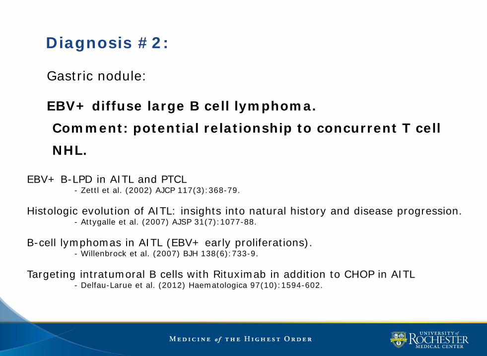
Diagnosis #2:
Gastric nodule:
EBV+ diffuse large B cell lymphoma.Comment: potential relationship to concurrent T cell NHL.
EBV+ B-LPD in AITL and PTCL- Zettl et al. (2002) AJCP 117(3):368-79.
Histologic evolution of AITL: insights into natural history and disease progression.- Attygalle et al. (2007) AJSP 31(7):1077-88.
B-cell lymphomas in AITL (EBV+ early proliferations).- Willenbrock et al. (2007) BJH 138(6):733-9.
Targeting intratumoral B cells with Rituximab in addition to CHOP in AITL - Delfau-Larue et al. (2012) Haematologica 97(10):1594-602.

Treatment
•Six (6) full cycles CHOP.
•Negative PET scan (Nov, 2015) = complete remission.
•One month later (Dec 2015), repeat upper endoscopy for surveillance - blind gastric biopsies (antrum and body):


CD20
EBER
CD79a
Negative: CD138
PCR:Positive IGH (clonal)Positive IGK (clonal) Negative TCRg

Diagnosis #3:
Gastric biopsy, antrum and body:
EBV+ B cell lymphoproliferative disorder
Comment: most consistent with recurrent DLBCL
•Short course of lenalidomide was poorly tolerated and stopped.
•Felt well with good performance status.
•Monitored for “active disease”.

Additional Course
•One year later (Dec 2016), with no additional treatment, lower GI bleeding recurred and colonoscopy revealed ulcerative lesion(s).
• Colonic biopsy performed:


EBER
PCR:IGH weak positive (polyclonal background)IGK positive oligoclonal/clonal
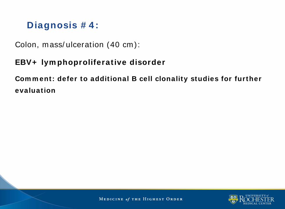
Diagnosis #4:
Colon, mass/ulceration (40 cm):
EBV+ lymphoproliferative disorder
Comment: defer to additional B cell clonality studies for further evaluation

Axillary lymph node (June 2015) – PTCL-NOS w/ EBV+ RS-like cells
- Polyclonal IGH pattern
NGS sequencing: IGH gene rearrangements

NGS sequencing: IGH gene rearrangements
Second gastric Bx (Dec 2015):• Monoclonal
• Single dominant VDJ recombination (two sequences differing by 1 base)
• DIFFERENT VH4-J4 rearrangement(V4-59, J4, D3-10)
• Total combined reads ~56%.
First gastric Bx (July 2015):• Monoclonal
• Single dominant VDJ recombination (two sequences differing by 1 base)
• VH3-J4 rearrangement (V3-73, J4, D4-23)
• Total combined reads ~55%.
Third colonic Bx (Dec 2016):• Oligoclonal
• At least 4 DIFFERENT VDJ clones
• Each ranging ~2-8% total reads
• Evidence of some specific VDJ rearrangements that were also found in earlier specimens at low level (≤1% of total combined reads above)

Proposed Diagnosis:
Multiple independent clonal EBV+ mucocutaneous ulcers, status-post PTCL-NOS with EBV+ RS-like cells.
Panel Diagnosis:
EBV+ lymphoproliferative disorder (mucocutaneous ulcer versus DLBCL)

Follow up:August/September 2017 (26 months).
Recent onset fatigue and weight loss
Persistent GI bleeding:
Negative upper endoscopic biopsy
Lower endoscopy identified TWO (2) distinct ulcerative lesions/masses
- Transverse colon
- Sigmoid colon (40cm – tattoo ink)

Mass/lesion #1 Mass/lesion #2 (40cm)

CD79a CD79a
Mass/lesion #1 Mass/lesion #2 (40cm)

Pax5 Pax5
Mass/lesion #1 Mass/lesion #2 (40cm)

CD20 CD20
Mass/lesion #1 Mass/lesion #2 (40cm)
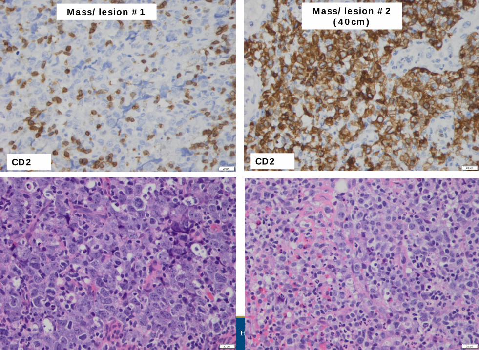
CD2 CD2
Mass/lesion #1 Mass/lesion #2 (40cm)

CD3 CD3
Mass/lesion #1 Mass/lesion #2 (40cm)

Diagnosis #5:
Morphologically c/w Diffuse large B cell lymphoma (DLBCL)
Diagnosis #6:
Morphologically c/w Peripheral T cell lymphoma (PTCL)

Diagnosis #5:
Morphologically c/w Diffuse large B cell lymphoma (DLBCL)
Diagnosis #6:
Morphologically c/w Peripheral T cell lymphoma (PTCL)
PCR:IGH: POSITIVEIGK: POSITIVETCRg: POSITIVE
PCR:IGH: POSITIVEIGK: POSITIVETCRg: POSITIVE

Diagnosis #5:
Morphologically c/w Diffuse large B cell lymphoma (DLBCL)
Diagnosis #6:
Morphologically c/w Peripheral T cell lymphoma (PTCL)
PCR:IGH: POSITIVEIGK: POSITIVETCRg: POSITIVE
PCR:IGH: POSITIVEIGK: POSITIVETCRg: POSITIVE
None are the same size rearrangement

Discussion:
Concurrent PTCL and EBV+ DLBCL at diagnosis and relapse.
Intervening multiple GI biopsies highly concerning for EBV+ DLBCL, but indolent/smoldering clinical course.
Positive PCR testing for clonality supported such an interpretation.
IGH sequencing, however, demonstrates entirely unique dominant B cell clone(s) (i.e. VDJ sequences) from each specimen.
Thus, we exclude “clonal evolution” of a single B cell line (i.e. single VDJ recombination event).
Next-gen deep sequencing of the IGH locus shows a complex relationship with varying dominant B cell clones over time.

Discussion (cont’d):Among the low level, or “subclonal” recombination sequences detected in each specimen, there is evidence that same clone (or clones) can be minimally detected at different points in time.
In a patient with abnormal immune system, the pathology within the GI tract, over >1 year, may represent multiple independent EBV+ lymphoproliferative disorders
Clonal dynamics may change or shift over time in response to, or as a cause of (?), recurrent T cell lymphoma .

Acknowledgements:
Yi Ding, MD, PhD
Todd Laughlin, PhD
Paul Rothberg, PhD
Richard Burack, MD, PhD
Carla Casulo, MD
Department of Pathology & Laboratory Medicine Hematology/Oncology, Wilmot Cancer Institute
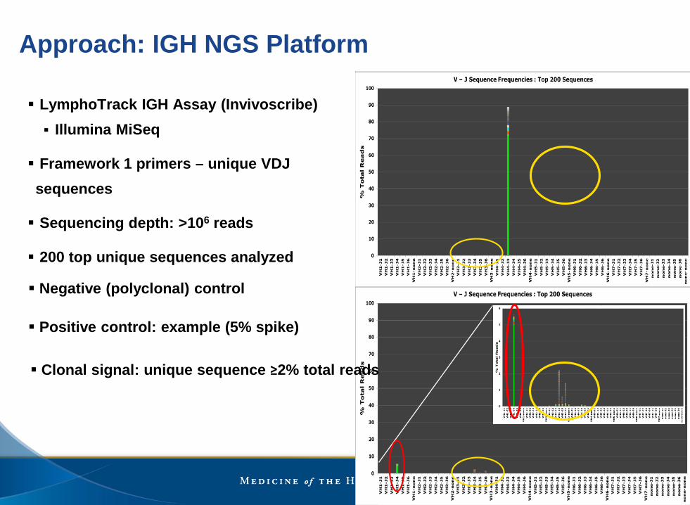
LymphoTrack IGH Assay (Invivoscribe) Illumina MiSeq
Framework 1 primers – unique VDJ sequences
Sequencing depth: >106 reads
200 top unique sequences analyzed
Negative (polyclonal) control
Positive control: example (5% spike)
Approach: IGH NGS Platform
Clonal signal: unique sequence ≥2% total reads

41

42