Sex-dependent behavioral deficits and neuropathology in a ...
12
Haida et al. Translational Psychiatry (2019)9:124 https://doi.org/10.1038/s41398-019-0457-y Translational Psychiatry ARTICLE Open Access Sex-dependent behavioral de fi cits and neuropathology in a maternal immune activation model of autism Obelia Haida 1 , Tareq Al Sagheer 1 , Anais Balbous 1,2 , Maureen Francheteau 1 , Emmanuel Matas 1 , Federico Soria 3 , Pierre Olivier Fernagut 1,3 and Mohamed Jaber 1,2 Abstract Infections during gestation and the consequent maternal immune activation (MIA) increase the risk of developing neuropsychiatric disorders in infants and throughout life, including autism spectrum disorders (ASD). ASD is a neurodevelopmental disorder that affects three times more males than females and is mainly characterized by deficits in social communication and restricted interests. Consistent findings also indicate that ASD patients suffer from movement disorders, although these symptoms are not yet considered as diagnosis criteria. Here we used the double- stranded RNA analog polyinosinic:polycytidylic acid (poly I:C) MIA animal model of ASD in mice and explored its effects in males and females on social and motor behavior. We then investigated brain areas implicated in controlling and coordinating movements, namely the nigro-striatal pathway, motor cortex and cerebellum. We show that male mice are more affected by this treatment than females as they show reduced social interactions as well as motor development and coordination deficits. Reduced numbers of Purkinje cells in the cerebellum was found more widespread and within distinct lobules in males than in females. Moreover, a reduced number of neurons was found in the motor cortex of males only. These results suggest that females are better protected against developmental insults leading to ASD symptoms in mice. They also point to brain areas that may be targeted to better manage social and motor consequences of ASD. Introduction Autism spectrum disorders (ASD) are a set of hetero- geneous neurodevelopmental disorders characterized by persistent difficulties in verbal and nonverbal commu- nication and restricted and repetitive patterns of beha- vior 1 . ASD can be diagnosed during childhood and affects 3 times more males than females 2 . In the absence of biological markers, ASD is currently diagnosed based on clinical scales and there is presently no cure but only symptomatic relief to some of its comorbidities such as anxiety, sleep disorders or seizures 3 . Infection during pregnancy is known to alter neurode- velopment either following direct infection of the fetus or, more often, through the immune response of the mother. Maternal immune activation (MIA) is known to induce several neurological and psychiatric disorders, that, although symptomatically different, share some over- lapping etiological and pathophysiological features 4 . These range from microcephaly, following the recently reported Zika virus infection for instance 5 , to schizo- phrenia and ASD. Indeed, bacterial, viral or parasite infections during pregnancy 6,7 are reported to increase risks of ASD in offspring 8 . The infectious agents do not cross the placental barrier but it is rather the maternal cytokines and immune reaction that impact the fetal brain © The Author(s) 2019 Open Access This article is licensed under a Creative Commons Attribution 4.0 International License, which permits use, sharing, adaptation, distribution and reproduction in any medium or format, as long as you give appropriate credit to the original author(s) and the source, provide a link to the Creative Commons license, and indicate if changes were made. The images or other third party material in this article are included in the article’ s Creative Commons license, unless indicated otherwise in a credit line to the material. If material is not included in the article’s Creative Commons license and your intended use is not permitted by statutory regulation or exceeds the permitted use, you will need to obtain permission directly from the copyright holder. To view a copy of this license, visit http://creativecommons.org/licenses/by/4.0/. Correspondence: Mohamed Jaber ([email protected]) 1 Université de Poitiers, INSERM, Laboratoire de Neurosciences Expérimentales et Cliniques, Poitiers, France 2 CHU Poitiers, Poitiers, France Full list of author information is available at the end of the article. These authors contributed equally: Haida Obelia, Al Sagheer Tareq, Fernagut Pierre Olivier, Jaber Mohamed 1234567890():,; 1234567890():,; 1234567890():,; 1234567890():,;
Transcript of Sex-dependent behavioral deficits and neuropathology in a ...

Haida et al. Translational Psychiatry (2019) 9:124
https://doi.org/10.1038/s41398-019-0457-y Translational Psychiatry
ART ICLE Open Ac ce s s
Sex-dependent behavioral deficits andneuropathology in a maternal immuneactivation model of autismObelia Haida1, Tareq Al Sagheer 1, Anais Balbous1,2, Maureen Francheteau1, Emmanuel Matas 1, Federico Soria3,Pierre Olivier Fernagut1,3 and Mohamed Jaber 1,2
AbstractInfections during gestation and the consequent maternal immune activation (MIA) increase the risk of developingneuropsychiatric disorders in infants and throughout life, including autism spectrum disorders (ASD). ASD is aneurodevelopmental disorder that affects three times more males than females and is mainly characterized by deficitsin social communication and restricted interests. Consistent findings also indicate that ASD patients suffer frommovement disorders, although these symptoms are not yet considered as diagnosis criteria. Here we used the double-stranded RNA analog polyinosinic:polycytidylic acid (poly I:C) MIA animal model of ASD in mice and explored its effectsin males and females on social and motor behavior. We then investigated brain areas implicated in controlling andcoordinating movements, namely the nigro-striatal pathway, motor cortex and cerebellum. We show that male miceare more affected by this treatment than females as they show reduced social interactions as well as motordevelopment and coordination deficits. Reduced numbers of Purkinje cells in the cerebellum was found morewidespread and within distinct lobules in males than in females. Moreover, a reduced number of neurons was foundin the motor cortex of males only. These results suggest that females are better protected against developmentalinsults leading to ASD symptoms in mice. They also point to brain areas that may be targeted to better manage socialand motor consequences of ASD.
IntroductionAutism spectrum disorders (ASD) are a set of hetero-
geneous neurodevelopmental disorders characterized bypersistent difficulties in verbal and nonverbal commu-nication and restricted and repetitive patterns of beha-vior1. ASD can be diagnosed during childhood and affects3 times more males than females2. In the absence ofbiological markers, ASD is currently diagnosed based onclinical scales and there is presently no cure but only
symptomatic relief to some of its comorbidities such asanxiety, sleep disorders or seizures3.Infection during pregnancy is known to alter neurode-
velopment either following direct infection of the fetus or,more often, through the immune response of the mother.Maternal immune activation (MIA) is known to induceseveral neurological and psychiatric disorders, that,although symptomatically different, share some over-lapping etiological and pathophysiological features4.These range from microcephaly, following the recentlyreported Zika virus infection for instance5, to schizo-phrenia and ASD. Indeed, bacterial, viral or parasiteinfections during pregnancy6,7 are reported to increaserisks of ASD in offspring8. The infectious agents do notcross the placental barrier but it is rather the maternalcytokines and immune reaction that impact the fetal brain
© The Author(s) 2019OpenAccessThis article is licensedunder aCreativeCommonsAttribution 4.0 International License,whichpermits use, sharing, adaptation, distribution and reproductionin any medium or format, as long as you give appropriate credit to the original author(s) and the source, provide a link to the Creative Commons license, and indicate if
changesweremade. The images or other third partymaterial in this article are included in the article’s Creative Commons license, unless indicated otherwise in a credit line to thematerial. Ifmaterial is not included in the article’s Creative Commons license and your intended use is not permitted by statutory regulation or exceeds the permitted use, you will need to obtainpermission directly from the copyright holder. To view a copy of this license, visit http://creativecommons.org/licenses/by/4.0/.
Correspondence: Mohamed Jaber ([email protected])1Université de Poitiers, INSERM, Laboratoire de Neurosciences Expérimentaleset Cliniques, Poitiers, France2CHU Poitiers, Poitiers, FranceFull list of author information is available at the end of the article.These authors contributed equally: Haida Obelia, Al Sagheer Tareq, FernagutPierre Olivier, Jaber Mohamed
1234
5678
90():,;
1234
5678
90():,;
1234567890():,;
1234
5678
90():,;

development following permeation of the placental com-partment9. A study of over 10 000 cases of ASD in theDanish medical Register reported a clear link betweenviral infection during the first trimester of pregnancy andASD; a weaker link was also established between bacterialinfection during the second semester and ASD10.MIA is modeled in animals mainly through injection of
bacterial lipopolysaccharide or double stranded RNAanalog polyinosinic:polycytidylic acid (poly I:C) that bothhave strong construct and face validity towards ASD andare preferred MIA paradigms compared to direct injec-tion of bacteria or viruses11,12. Poly I:C injection topregnant rodent females is considered to have a strongerconstruct validity than LPS9 and is generally performed atembryonic day 12.5 (E12.5), after the neural tube hasclosed and when progenitors are migrating, a time periodwhen neurodevelopment is highly vulnerable to environ-mental insults. This treatment regimen has been shown toinduce several cognitive an social features of ASD9 such asabnormal vocalization, deficits in social interaction andcommunication as well as altered cytokines levels withinthe cerebellum13. To this regard, a growing body of evi-dence is now pointing towards cerebellum abnormalitiesin ASD, where reduced numbers of Purkinje cells (PC) inpost-mortem ASD brains has been documented14,15.Here, we investigated the behavioral and cellular effects
of a poly I:C injection to pregnant female mice at E12.5.We explored behavioral developmental milestones inmales and females analyzed separately, followed by specialfocus on social interactions, gait and fine motor skills andcoordination. We then determined the consequence ofthis treatment on cell integrity in several brain areasimplicated in motor control and coordination, namely thenigro-striatal pathway, the motor cortex and the cere-bellum. We demonstrate that a single injection of poly I:Cto pregnant females induces not only long lastingabnormal social behavior in offspring but also develop-mental delays that were accompanied by reduced num-bers of PC within the cerebellum and neurons in themotor cortex. Interestingly, these deficits were observedmainly in males, as females seemed less vulnerable to MIAboth at the behavioral and cellular levels.
Materials and methodsAnimalsAnimal housing and experimental procedures were
performed in accordance with the European Uniondirective (2010/63/EU) and validated by the regionalethical committee (Approval # 2015020415093780).C57BL/6J Mice (Charles River Laboratories, France) werehoused in ventilated cages with access to food and waterad libitum. Room temperature was maintained at 23 °C ona 12 h light/dark cycle.
Pregnant mice received a single intraperitoneal injectionof either poly I:C (20 mg/kg, Sigma, P1530, n= 22) orNaCl 0.9% (n= 11) at E12.5 as previously described16.Because poly I:C considerably increases the risk ofresorption17,18, we used twice the number of matedfemales in the poly I:C group compared to control. Out ofthe 22 mated poly I:C females only 12 gave birth to pups(45.5% abortion) among which four litters died at neo-natal age. All 11 saline treated females gave birth amongwhich one litter died at neonatal age. Additionally, themean number of pups per litter was significantly lower inpoly I:C treated females compared to saline treatedfemales (2.2 vs 6.7 pups per litter, Mann-Whitney: p <0,001). At weaning (P21), pups from different litters wererandomly allocated to four experimental groups depend-ing on sex and prenatal treatment: saline males (n= 30),poly I:C males (n= 14), saline females (n= 30), poly I:Cfemales (n= 10). The experiment timeline is presented insupplementary Figure 1. Behavioral tests were performedin the least stressing and challenging order to avoidpotential training and learning effects as previouslydescribed with an ASD valproic acid (VPA) animalmodel19. Animals were tested during their light cycle andthe investigators were blind to treatment assignment.
Assessment of developmental milestonesDuring early postnatal life (1–3 weeks), pups were kept
in their home cage and righting reflex and eye openingwere assessed from P9 to P16. To assess righting reflex,mice were placed in the supine position and the timetaken to right was monitored three times with a five-minute inter-trial interval. Eye opening was assessed dailyat P12 to P16 and scored as either 0= Both Eyes Closed,1=One Eye Open, or 2= Both Eyes Open.
SHIRPA primary screenThe SHIRPA primary screen20 was performed in a
transparent Plexiglas arena (55 × 33 × 22 cm) with11 × 11 cm square grid on the bottom, and a 3-mm metalwire crossing diagonally on top. The transfer reactiontime was evaluated, followed by the wire maneuver test toassess motor coordination and muscle function. Negativegeotaxis, used to assess postural stability and coordinationin space, was evaluated as the time taken to turn andclimb a 45° inclined grid21.
Spontaneous activity in the cylinderSpontaneous activity in the cylinder was performed at
P30 as previously described22. Mice were put in a trans-parent Plexiglas cylinder, (diameter: 12 cm), and theiractivity was videotaped for 3 min. Number of rearing andtime spent grooming were quantified.
Haida et al. Translational Psychiatry (2019) 9:124 Page 2 of 12

Motor coordination on the challenging beamThe challenging beam test was performed at P33 as
previously described23. The beam consists of four Plex-iglas sections (25 cm each) starting with a width of 3.5 cmand gradually narrowed to 0.5 by 1 cm decrements. Ani-mals were first trained for 2 days to traverse the beamstarting at the widest section and ending at the narrowestsection that led into the home cage. On the test day, amesh grid (1 cm squares) was placed over the beam sur-face. Animals were videotaped while traversing the grid-surfaced beam for five trials. Time to traverse, errors,number of steps and errors per step made by each animalwere measured and averaged.
Spatial, temporal, and kinetic parameters of gaitGait was analyzed during spontaneous walk at P34 using
an automated gait analysis system (Gaitlab, Viewpoint,France) as previously described19. The following para-meters were analyzed: (i) stride length: distance betweentwo consecutive placements of the same paw, (ii) limbbase of support: distance between two pair prints atcontact during each step cycle and (iii) pair gap: gapbetween the placement of the two trailing feet, whichmeasures spatial coordination between the two pairs.
Sociability in the three chambers testSocial interaction was assessed between P35 and P45
using the three-chambers test as previously described19,24.The first phase (PHASE-I) comprises two identical non-social stimuli (inverted wire-cups) placed in the oppositechambers. The second phase (PHASE-II) comprises anon-social stimulus and a social stimulus (a naïve mousewith no previous contact with the tested animal). Eachphase was of 10 min during which time spent in eachchamber was recorded. Subsequently, a sociability index(SI) was calculated as follows: (time exploring socialchamber−time exploring non-social chamber)/(timeexploring social chamber+ time exploring non-socialchamber).
Tissue processing and immunohistochemistryAt the end of behavioral assays (P45), males and females
from each group were randomly selected for histopatho-logical analysis. Mice were deeply anesthetized withketamine-xylazine (120–20mg/kg) and transcardiallyperfused with 0.9% saline at 37 °C followed by 4% paraf-ormaldehyde (PFA) at 4 °C. Brains were post-fixed in 4%PFA at 4 °C for 24 h before cryoprotection in 30% sucrose.Serial 50 μm (cerebellum) and 40 μm (striatum, cortexand substantia nigra) free-floating sections were collectedand stored at −20 °C until use in an anti-freeze solution.PC within the cerebellum, dopaminergic neurons withinthe substantia nigra pars compacta (SNc) and neuronswithin the striatum and the motor cortex were quantified.
Every fourth cerebellar section was mounted on gelatin-coated slides for PC quantification or microglia mor-phology analyses. PC were identified based on theirmorphology on cresyl violet staining as previouslydescribed25 and their phenotype was further confirmedusing calbindin immunohistochemistry (1:2500; Swant,Cb-38a). Calbindin was not used for PC quantification asASD inducing treatments can lead to reduced calbindinprotein expression26. Microglia was revealed using arabbit anti-Iba1 primary antibody (1:500; Wako,019–19741) and a counterstaining with cresyl violet wasused to identify granular and molecular layers. Every sixthsection throughout the striatum, SNc and motor cortexwas selected and processed for either neuronal nucleiantigen (NeuN) to quantify neurons in the striatum andthe motor cortex, or for tyrosine hydroxylase (TH)immunoreactivity to quantify dopaminergic neurons inthe SNc. Sections were incubated for 1h30 in a blockingsolution (3% bovine serum albumin, 0.3% Triton X-100 inPBS 1M, pH 7.4). Rabbit anti-NeuN (1:500; abcam,Ab177487) or mouse anti-TH (1:5000; Immunostar,22941) antibodies were applied overnight at 4 °C. Bioti-nylated anti-rabbit or anti-mouse IgG was used as sec-ondary antibody (1:250; Vector laboratories, BA-1000 andBA-9200) for 1 h at room temperature. Signal wasamplified with an ABC Elite kit and revealed with dia-minobenzidine (Vector Laboratories). Sections weremounted on gelatin-coated slides and processed for cresylviolet counterstaining.
StereologyStereological estimates were performed using the opti-
cal fractionator method and systematic random samplingto obtain the total number of cerebellar PC, motor cortexneurons, striatal neurons and dopaminergic nigral neu-rons. Each region of interest was outlined based on theFranklin and Paxinos’s mouse brain atlas27 at ×2.5objective and neurons were counted at ×40 objectiveusing Mercator image analysis system (Explora Nova,France). Upper and lower guard zones of 1 μm were set atthe top and bottom of the section to exclude lost profilesand each neuron or visible nucleus was counted (seesupplementary Table 1 for stereology parameters).
Microglial morphologyZ-stack images of Iba1-stained cerebellar sections were
taken at ×40 objective in different parts of the sublobuleCrus II in males, and distinguishing between the granularand molecular layers. Images were then processed forsegmentation and fractal analysis by a semi-automatedmethod using ImageJ software as previously described28.Briefly, diaminobenzydine and cresyl violet stainings wereartificially separated by color deconvolution. Resulting z-stacks were converted to maximal projection images with
Haida et al. Translational Psychiatry (2019) 9:124 Page 3 of 12

the stack ‘sum’ function before thresholded binarization.Area and perimeter of cells and their convex hulls weremeasured with ImageJ default tools to calculate shapedescriptors. Fractal box counting dimension, form factor(4π x cell area/cell perimeter²), convexity (convex hullperimeter/cell perimeter) and solidity (cell area/convexhull area) were calculated for at least 20 cells per animal.
Statistical analysesData are expressed as mean ± Standard Error of the
Mean and analyzed using GraphPad Prism-7 software (LaJolla, California, USA). Normality and equality of groupvariances were tested by Shapiro-Wilk and Brown-Forsythe tests, respectively. Data having a Gaussian dis-tribution and equal variance were analyzed using two-wayanalysis of variance (ANOVAs) followed by Fisher’s LSDpost-hoc multiple comparisons test, except when com-paring only two groups where a Student’s T-test wasperformed. Kruskal-Wallis test followed by Dunn’s post-hoc test or Mann–Whitney test was applied when sam-ples did not follow a normal distribution or had unequalvariance. For all analyses, a p value < 0.05 was consideredsignificant.
ResultsPost-natal development delays in poly I:C miceTo determine the effect of prenatal exposure to poly I:C
on general postnatal development we investigated eyeopening delays and righting reflex at P9-P16. In the eyeopening measurements (Fig. 1a), Kruskal-Wallis testindicated a significant group effect at P13 (p < 0.05), P14(p < 0.01) and P15 (p < 0.01). A significant delay of eyeopening was observed in poly I:C females at P14 and P15(Dunn’s post-hoc test, p < 0.05 and p < 0.01, respectively).For the righting reflex, Kruskal-Wallis test revealed asignificant group effect at P9 (p < 0.01), P11 (p < 0.05) andP13 (p < 0.001). A significant increase of the time to rightwas observed in poly I:C males at P11 and P13 (Dunn’spost-hoc test, p < 0.05 and p < 0.01, respectively). Theseresults indicate that both males and females exposedprenatally to poly I:C manifest developmental delays,although different in nature.
Poly I:C males show altered spontaneous activity andmotor coordinationAt P30, poly I:C mice showed altered behavior in different
SHIRPA primary screen and cylinder test parameters (Fig.1c–h). Time spent immobile following transfer to theSHIRPA arena was assessed. Kruskal-Wallis test revealed asignificant group effect (p < 0.001) with poly I:C malesshowing ~65% increase in immobility compared with saline(Dunn’s multiple comparison test, p < 0.01, Fig. 1c). Loco-motor activity assessment (Fig. 1d) showed an effect oftreatment [F (1,74)= 18.72, p < 0.0001], and a treatment x
sex interaction [F (1,74)= 6.124, p < 0.05]. Fisher’s LSDpost- hoc analysis showed a significant (−24%) decrease inlocomotor activity only in poly I:C males (p < 0.0001). Wethen assessed postural stability and coordination in spaceusing negative geotaxis measured as the time needed toclimb a 45° inclined grid (Fig. 1e). We found an effect of sex[F (1,74)= 4.486, p < 0.05] and Fisher’s LSD post-hoc testindicated a significant increase of the time needed to climbthe grid in poly I:C males (p < 0.05) but not in females.Finally, we implemented the wire maneuver test to assessmotor coordination and muscle function (Fig. 1f). Kruskal-Wallis test showed a significant effect of group (p < 0.01)and Dunn’s multiple comparison test determined that onlypoly I:C males were statistically different from saline andspent more time to climb the metal wire (+17%, p < 0.05).Repetitive and restricted behaviors are among the core
symptoms and the earliest signs of ASD1. We assessedrepetitive grooming and rearing behaviors in a new envir-onment using the Plexiglas cylinder test for 3minutes.Kruskal-Wallis test indicated an effect of group in the timespent grooming (p < 0.05, Fig. 1g). However, Dunn’s mul-tiple comparison test indicated no significant change inboth poly I:C males and females compared to their salinecounterparts. Poly I:C treatment had a significant effect onrearing behavior (Kruskal-Wallis, p < 0.01) with a significant50% decrease of rearing in males (Dunn’s multiple com-parison test p < 0.05, Fig. 1h) while a trend was observed infemales (p= 0.1014). These results indicate that prenatalexposure to poly I:C leads to exploratory and motor coor-dination deficits mainly in males.
Poly I:C treatment did not affect gait nor walking skillsWe analyzed gait at P33–34 as several studies in ASD
patients clearly pointed to gait disturbances, includingreduced stride length and increased base of support29,30.Poly I:C treatment did not affect paw area [F (1,74)=0.03356, p= 0.8551], nor speed [F (1,74)= 3.451,p= 0.0672] or regularity of the run [F (1,74)= 5.328e-005, p= 0.9942], neither in male or female mice. Kruskal-Wallistest indicated no effect of treatment or sex on the pair gap ofwalking (Fig. 2a). Additionally, two-way ANOVA revealedno significant effect of group [F (3,148)= 0.7706, p=0.5122] on limbs base of support (Fig. 2b) or stride length [F(1, 74)= 0.1638, p= 0.6868] (Fig. 2c). Two-way ANOVAshowed an effect of sex [F (1, 74)= 4.801, p < 0.05] on stridelength but with no significant change in both poly I:C malesand females in comparison to saline controls (Fisher’s LSDpost-hoc analysis). On the challenging beam test, statisticaltests indicate no effect of treatment or sex on the timeneeded to traverse the beam sections (Fig. 2d). In addition,the number of errors per step (Fig. 2e) and the number oferrors at each beam section (Fig. 2f) were not affected by thetreatment. These results indicate that poly I:C prenatalexposure did not induce gait disturbances in mice.
Haida et al. Translational Psychiatry (2019) 9:124 Page 4 of 12

Fig. 1 (See legend on next page.)
Haida et al. Translational Psychiatry (2019) 9:124 Page 5 of 12

Reduced sociability in poly I:C malesOne of the hallmarks of ASD are deficits in social
interactions1. Social behavior of mice prenatally exposedto poly I:C was assessed at age of 5–6 weeks (P35-45)using the three-chambers test (Fig. 3) as previouslydescribed19. None of the treatment groups, regardless ofsex, showed a preference to any of the chambers during
the 10 min habituation (PHASE I) (Fig. 3a). In Phase II,Kruskal-Wallis test detected a significant effect of group(p < 0.0001). Indeed, while saline males spent more timein the social chamber than in the non-social one (69%versus 31%, p < 0.001), poly I:C males spent 58% of theirtime in the social chamber versus 42% in the non-socialone (p > 0.999) showing no significant preference towards
(see figure on previous page)Fig. 1 Prenatal exposure to poly I:C leads to developmental delays in males and females and altered spontaneous activity and motorcoordination in males. a Delay in eye opening was measured as the mean number of open eyes (0, 1, or 2) per group from P12 to P16 in males (left)and females (right). b Increased latency to right at P11 and P13 in poly I:C males. c Immobility time when transferred to the SHIRPA arena is increasedonly in poly I:C males. d Locomotor activity after the transfer to the SHIRPA arena. Only poly I:C males showed a decrease in this parameter. e Time toclimb on the grid was found increased in poly I:C mice compared to saline in males only. f Significant increase in time spent climbing the wire wasfound only in poly I:C males. g No significant change in the time spent grooming was found in poly I:C mice compared to saline. h Poly I:C prenatalexposure significantly decreased the number of vertical rears in the cylinder in poly I:C males only. Poly I:C males n= 11; saline males n= 27; poly I:Cfemales n= 10; saline females n= 30. Data expressed as mean ± SEM; two-way ANOVA followed by Fisher’s LSD post-hoc (d, e, and g), and Kruskal-Wallis test followed by Dunn’s multiple comparisons test (a, b, c, f, and h) (*p < 0.05; **p < 0.01; ****p < 0.0001)
Fig. 2 Prenatal exposure to poly I:C does not affect gait. Poly I:C mice show no significant change in pair gap (a), limbs base of support (b) orstride length (c). d Poly I:C treatment did not affect the time needed to traverse the beam nor the number of errors per steps crossing the beam (e).Poly I:C prenatally exposed males (black bars) and females (dark gray bars) showed no change in the number of errors per steps on different beamsections, in comparison to saline (f). Saline males n= 27; Poly I:C males n= 11; Saline females n= 30; Poly I:C females n= 10. Data expressed asmean ± SEM; two-way ANOVA followed by Fisher’s LSD post-hoc (b, c, d, and f), and Kruskal-Wallis test followed by Dunn’s multiple comparisons test(a, e)
Haida et al. Translational Psychiatry (2019) 9:124 Page 6 of 12
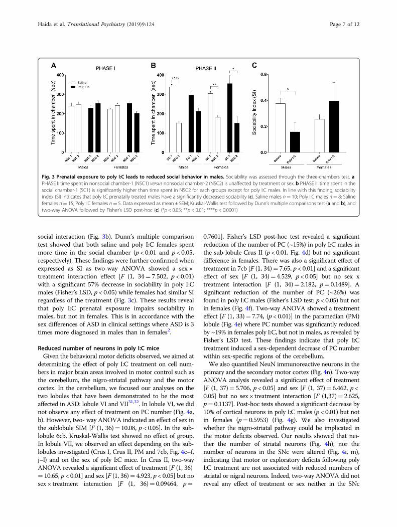
social interaction (Fig. 3b). Dunn’s multiple comparisontest showed that both saline and poly I:C females spentmore time in the social chamber (p < 0.01 and p < 0.05,respectively). These findings were further confirmed whenexpressed as SI as two-way ANOVA showed a sex ×treatment interaction effect [F (1, 34= 7.502, p < 0.01)with a significant 57% decrease in sociability in poly I:Cmales (Fisher’s LSD, p < 0.05) while females had similar SIregardless of the treatment (Fig. 3c). These results revealthat poly I:C prenatal exposure impairs sociability inmales, but not in females. This is in accordance with thesex differences of ASD in clinical settings where ASD is 3times more diagnosed in males than in females2.
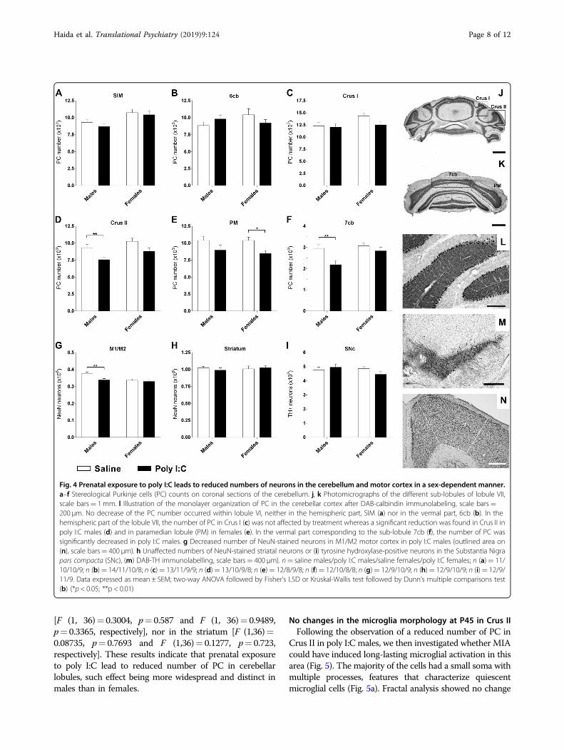
Reduced number of neurons in poly I:C miceGiven the behavioral motor deficits observed, we aimed at
determining the effect of poly I:C treatment on cell num-bers in major brain areas involved in motor control such asthe cerebellum, the nigro-striatal pathway and the motorcortex. In the cerebellum, we focused our analyses on thetwo lobules that have been demonstrated to be the mostaffected in ASD: lobule VI and VII31,32. In lobule VI, we didnot observe any effect of treatment on PC number (Fig. 4a,b). However, two- way ANOVA indicated an effect of sex inthe sublobule SIM [F (1, 36)= 10.08, p < 0.05]. In the sub-lobule 6cb, Kruskal-Wallis test showed no effect of group.In lobule VII, we observed an effect depending on the sub-lobules investigated (Crus I, Crus II, PM and 7cb, Fig. 4c–f,j–l) and on the sex of poly I:C mice. In Crus II, two-wayANOVA revealed a significant effect of treatment [F (1, 36)= 10.65, p < 0.01] and sex [F (1, 36)= 4.923, p < 0.05] but nosex × treatment interaction [F (1, 36)= 0.09464, p=
0.7601]. Fisher’s LSD post-hoc test revealed a significantreduction of the number of PC (~15%) in poly I:C males inthe sub-lobule Crus II (p < 0.01, Fig. 4d) but no significantdifference in females. There was also a significant effect oftreatment in 7cb [F (1, 34)= 7.65, p < 0.01] and a significanteffect of sex [F (1, 34)= 4.529, p < 0.05] but no sex xtreatment interaction [F (1, 34)= 2.182, p= 0.1489]. Asignificant reduction of the number of PC (~26%) wasfound in poly I:C males (Fisher’s LSD test: p < 0.05) but notin females (Fig. 4f). Two-way ANOVA showed a treatmenteffect [F (1, 33)= 7.74, (p < 0.01)] in the paramedian (PM)lobule (Fig. 4e) where PC number was significantly reducedby ~19% in females poly I:C, but not in males, as revealed byFisher’s LSD test. These findings indicate that poly I:Ctreatment induced a sex-dependent decrease of PC numberwithin sex-specific regions of the cerebellum.We also quantified NeuN immunoreactive neurons in the
primary and the secondary motor cortex (Fig. 4n). Two-wayANOVA analysis revealed a significant effect of treatment[F (1, 37)= 5.706, p < 0.05] and sex [F (1, 37)= 6.462, p <0.05] but no sex × treatment interaction [F (1,37)= 2.625,p= 0.1137]. Post-hoc tests showed a significant decrease by10% of cortical neurons in poly I:C males (p < 0.01) but notin females (p= 0.5953) (Fig. 4g). We also investigatedwhether the nigro-striatal pathway could be implicated inthe motor deficits observed. Our results showed that nei-ther the number of striatal neurons (Fig. 4h), nor thenumber of neurons in the SNc were altered (Fig. 4i, m),indicating that motor or exploratory deficits following polyI:C treatment are not associated with reduced numbers ofstriatal or nigral neurons. Indeed, two-way ANOVA did notreveal any effect of treatment or sex neither in the SNc
Fig. 3 Prenatal exposure to poly I:C leads to reduced social behavior in males. Sociability was assessed through the three-chambers test. aPHASE I: time spent in nonsocial chamber-1 (NSC1) versus nonsocial chamber-2 (NSC2) is unaffected by treatment or sex. b PHASE II: time spent in thesocial chamber-1 (SC1) is significantly higher than time spent in NSC2 for each groups except for poly I:C males. In line with this finding, sociabilityindex (SI) indicates that poly I:C prenatally treated males have a significantly decreased sociability (c). Saline males n= 10; Poly I:C males n= 8; Salinefemales n= 15; Poly I:C females n= 5. Data expressed as mean ± SEM; Kruskal-Wallis test followed by Dunn’s multiple comparisons test (a and b), andtwo-way ANOVA followed by Fisher’s LSD post-hoc (c) (*p < 0.05; **p < 0.01; ****p < 0.0001)
Haida et al. Translational Psychiatry (2019) 9:124 Page 7 of 12
[F (1, 36)= 0.3004, p= 0.587 and F (1, 36)= 0.9489,p= 0.3365, respectively], nor in the striatum [F (1,36)=0.08735, p= 0.7693 and F (1,36)= 0.1277, p= 0.723,respectively]. These results indicate that prenatal exposureto poly I:C lead to reduced number of PC in cerebellarlobules, such effect being more widespread and distinct inmales than in females.
No changes in the microglia morphology at P45 in Crus IIFollowing the observation of a reduced number of PC in
Crus II in poly I:C males, we then investigated whether MIAcould have induced long-lasting microglial activation in thisarea (Fig. 5). The majority of the cells had a small soma withmultiple processes, features that characterize quiescentmicroglial cells (Fig. 5a). Fractal analysis showed no change
Fig. 4 Prenatal exposure to poly I:C leads to reduced numbers of neurons in the cerebellum and motor cortex in a sex-dependent manner.a–f Stereological Purkinje cells (PC) counts on coronal sections of the cerebellum. j, k Photomicrographs of the different sub-lobules of lobule VII,scale bars= 1 mm. l Illustration of the monolayer organization of PC in the cerebellar cortex after DAB-calbindin immunolabeling, scale bars=200 µm. No decrease of the PC number occurred within lobule VI, neither in the hemispheric part, SIM (a) nor in the vermal part, 6cb (b). In thehemispheric part of the lobule VII, the number of PC in Crus I (c) was not affected by treatment whereas a significant reduction was found in Crus II inpoly I:C males (d) and in paramedian lobule (PM) in females (e). In the vermal part corresponding to the sub-lobule 7cb (f), the number of PC wassignificantly decreased in poly I:C males. g Decreased number of NeuN-stained neurons in M1/M2 motor cortex in poly I:C males (outlined area on(n), scale bars= 400 µm). h Unaffected numbers of NeuN-stained striatal neurons or (i) tyrosine hydroxylase-positive neurons in the Substantia Nigrapars compacta (SNc), (m) DAB-TH immunolabelling, scale bars= 400 µm). n= saline males/poly I:C males/saline females/poly I:C females; n (a)= 11/10/10/9; n (b)= 14/11/10/8; n (c)= 13/11/9/9; n (d)= 13/10/9/8; n (e)= 12/8/9/8; n (f)= 12/10/8/8; n (g)= 12/9/10/9; n (h)= 12/9/10/9; n (i)= 12/9/11/9. Data expressed as mean ± SEM; two-way ANOVA followed by Fisher’s LSD or Kruskal-Wallis test followed by Dunn’s multiple comparisons test(b) (*p < 0.05; **p < 0.01)
Haida et al. Translational Psychiatry (2019) 9:124 Page 8 of 12
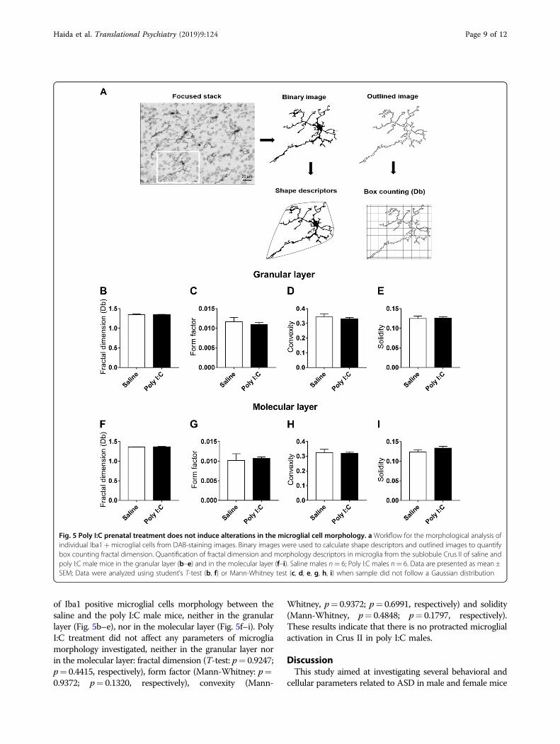
of Iba1 positive microglial cells morphology between thesaline and the poly I:C male mice, neither in the granularlayer (Fig. 5b–e), nor in the molecular layer (Fig. 5f–i). PolyI:C treatment did not affect any parameters of microgliamorphology investigated, neither in the granular layer norin the molecular layer: fractal dimension (T-test: p= 0.9247;p= 0.4415, respectively), form factor (Mann-Whitney: p=0.9372; p= 0.1320, respectively), convexity (Mann-
Whitney, p= 0.9372; p= 0.6991, respectively) and solidity(Mann-Whitney, p= 0.4848; p= 0.1797, respectively).These results indicate that there is no protracted microglialactivation in Crus II in poly I:C males.
DiscussionThis study aimed at investigating several behavioral and
cellular parameters related to ASD in male and female mice
Fig. 5 Poly I:C prenatal treatment does not induce alterations in the microglial cell morphology. a Workflow for the morphological analysis ofindividual Iba1+microglial cells from DAB-staining images. Binary images were used to calculate shape descriptors and outlined images to quantifybox counting fractal dimension. Quantification of fractal dimension and morphology descriptors in microglia from the sublobule Crus II of saline andpoly I:C male mice in the granular layer (b–e) and in the molecular layer (f–i). Saline males n= 6; Poly I:C males n= 6. Data are presented as mean ±SEM; Data were analyzed using student’s T-test (b, f) or Mann-Whitney test (c, d, e, g, h, i) when sample did not follow a Gaussian distribution
Haida et al. Translational Psychiatry (2019) 9:124 Page 9 of 12

that were exposed to a MIA procedure during their prenatallife. For this, we explored social interactions, motor and gaitperformances at different time points and determined theirhistological correlates in brain areas implicated in motorcontrol, regulation and coordination. Our results indicatethat while poly I:C prenatal exposure leads to delayeddevelopment in both male and female pups, only periado-lescent males, but not females, showed increased immobi-lity in a novel environment, decreased motor/exploratoryactivity and motor coordination deficits as attested byincreased time necessary to climb a grid or a wire. Socialbehavior in poly I:C young adult male was also significantlyreduced while poly I:C female sociability did not differ fromsaline. However, neither poly I:C males nor females showeddeficiencies in fine motor skills evaluated using the nar-rowing beam walking paradigm nor in gait. Interestingly,behavioral deficiencies observed mainly in poly I:C maleswere mirrored by restricted cell number reduction in thelateral parts of the cerebellum, its vermis, and in the motorcortex. Poly I:C females showed a reduced PC number onlyin the PM lobule of the cerebellum, and that was differentfrom the affected lobules in poly I:C males. In line withthese findings, we also recently reported similar resultsusing the VPA ASD animal model where males were moreaffected by the prenatal treatment than females whether atthe behavioral or cellular levels19.Most previous studies using the poly I:C prenatal
exposure rodent model have focused only on males33 orhave pooled together males and females16 rendering itdifficult to determine potential sex differences followingMIA. This is of relevance given that ASD ratio is 3:1 inmales2 and being a male constitutes a major risk factor indeveloping neurological or psychiatric disorders34. Poly I:C-induced MIA is a common procedure used to generateanimal models of ASD, but also depression and schizo-phrenia35–37. These psychiatric pathologies, althoughdifferent in their expression, seem to share to some extentoverlapping clinical and therapeutic features and commonbiological mechanisms such as epigenetic modificationsinvolving histone acetylation and promoter methylation38.The link between MIA and ASD is potent and has beenconfirmed in human10,39 and animal models33,35,40. Thepoly I:C animal model of ASD has been repeatedly vali-dated and is recognized to provide strong construct andface validity16,35,41. While this manuscript was in thesubmission process a review article proposing guidelinesfor the use of MIA models was published and that aims tolimit discrepancies in the MIA procedures that mayaccount for variations between findings42. The poly I:Cprocedure used here has been widely documented andconsistently showed induction of MIA (as evidenced forexample by elevation of cytokine blood levels in treatedpregnant females) and behavioral consequences inoffspring43,44.
Previous reports have shown that poly I:C prenataltreatment induces decreased social interaction with nor-mal grooming behavior35 and decreased motor perfor-mance in a rotarod task only in males33. Additionally,prior research using similar dose and time of exposure ofpoly I:C have found that this treatment induces in theoffspring males decreased exploration in open-field aswell as reduced preference for the social chamber in threechambers test41. However, and to the best of our knowl-edge, this is the first study exploring in detail motorbehavior, fine motor skills and gait in both sexes in thepoly I:C mouse model. This is also the first study thatfocuses on cerebellar sub-regions, motor cortex and thenigrostriatal pathway in these animal models andreporting restricted reduction of neurons in distinctregions of the cerebellum and the motor cortex and thatwere mainly observed in poly I:C males.The finding that gestational MIA can induce ASD-like
postnatal and durable behavior abnormalities underlines theroles played by the prenatal environment in shaping braindevelopment and in provoking lasting complex behaviors.Alterations in neuronal development occurring around E12in rodents can result in severe abnormalities as this is a keyperiod of neuronal proliferation, migration, differentiation,synaptogenesis, apoptosis and myelination45.Cerebellar lobules VI and VII play a major role in
movement regulation, exploratory behavior, stereotypedand repetitive behaviors, and oculomotor activity46–48 butalso in cognitive functions such as speech49. PC are theintegrating center of the cerebellum and the sole outputfrom the cerebellar cortex. Several studies have shown adecrease number of theses neurons in ASD14,15,50,51,55 andin ASDmodels16,19,33. In this study, we have demonstrated areduction of the PC number in the cerebellum of miceprenatally exposed to poly I:C and that were different intheir extend and regional specificity in males and females.Most previous studies with animal modes of ASD usedeither only males33 or combined males and females withinthe same group16. In this latter reference, the authorsshowed a 26% reduced number of PC in the vermal lobuleVII, but not in lobule VI. This finding was later confirmedby Naviaux et al. in 2013 who reported a decrease of the PCnumber by 63% at the age of 16 weeks33. Our results arethus in accordance with these studies indicating no changein the number of PC in the lobule VI and a significantdecrease in the lobule VII, of 15 to 26% magnitude at theage of 45 days. Moreover, we extend these findings andshow here that this reduction appears to be restricted to thesub-lobules Crus II and 7cb in males and in PM in females,cerebellar regions that are easier to delineate with coronalsections as used here compared to sagittal sections used inprior studies. In human, Crus II plays an important role inspeech, social cognition, stereotyped and repetitive beha-viors, movement regulation and oculomotor activity49,52,53
Haida et al. Translational Psychiatry (2019) 9:124 Page 10 of 12
all reported to be affected in ASD54. The PC of this regionare inter-connected with the area 46 of the dorsolateralprefrontal cortex, a region involved in working memory,attention, movement regulation and organization. Deficitsin PC in this cerebellar area could underlie some ASDsymptoms such as language/communication disturbances,stereotyped and repetitive behaviors, social interactiondeficits and sensorimotor impairments.Additionally, we report here a deficit in PC in the vermis
(sub-lobule 7cb) of male animals, in line with previousreports in this animal model16,33. These findings are inrelevance with clinical data that report vermal hypoplasiain ASD patients. The role of the vermal part of the cer-ebellum is still not clearly determined. Transcranialmagnetic stimulation of this cerebellar area in humanprovoked impaired visual motion discrimination sug-gesting a role of the cerebellar vermis in visual motionprocessing55. In non-human primate it has been demon-strated that the cerebellar vermis receives projectionsfrom motor areas including the primary motor cortex56.In relation, we show here a modest but significantdecrease of the neuronal number in the M1/M2 motorcortex in poly I:C males. These findings could be indica-tive of a connectivity dysfunction between the motorcortex and the cerebellum occurring only in male animals.Vargas et al.57, have reported a microglial activation within
the cerebellum of ASD patients. This was further corrobo-rated by a later study showing microglial activation mediatedby TLR3-poly I:C binding58. Given these clinical data, wehave examined microglial status in poly I:C animals butfound no morphological alterations, in accordance with Huiet al. using the poly I:C mouse model59.Together, theseresults indicate that behavioral deficits displayed by malemice exposed prenatally to poly I:C occur in the absence ofprotracted neuroinflammation in the cerebellum.In a previous study, we recently reported that VPA mouse
models of ASD also exhibit autism-like behavioral pheno-type and less PC19. The main differences between the twostudies are that VPA treatment affected similarly bothmales and females, except for the social interaction para-digm that was not alerted in VPA females. Here, poly I:Ctreated females seemed better protected against MIA asthey showed mild and specific behavioral and cellular def-icits. These results highlight the heterogeneity of symptomsfound in ASD and indicate that males are differentiallyvulnerable to various environmental insults occurring dur-ing pregnancy. The basis for this male bias is unknown withtheories including the “extreme male brain”, hormonaldifferences, and genetic influences (for a review see60.
ConclusionWe report here that a single poly I:C injection on E12.5
negatively affects behavioral neurodevelopmental para-meters, exploratory behavior and sociability and that these
deficiencies are somewhat more pronounced and of dif-ferent nature in males than in females. These findings arereminiscent of clinical and epidemiological observationsshowing higher incidence of ASD in males. This stressesthe need to investigate how gender may protect againstthe effects of MIA in ASD and emphasize the importanceof identifying the underlying biological parameters in bothsexes in animal models relevant to neurodevelopmentaldisorders such as ASD.
AcknowledgementsThis work was supported by grants from the Fondation pour la RechercheMédicale (FRM). TAS was awarded a scholarship from the Association ofSpecialization and Scientific Guidance (ASSG- Lebanon). OH was supported bya fellowship from the Institut National de la Santé et de la Recherche Médicale(INSERM) and the region Nouvelle-Aquitaine. The University of Poitiers andINSERM provided infrastructural support. The funders had no role in studydesign, data collection and analysis, decision to publish or preparation of themanuscript. We thank the staff of the PREBIOS animal facility (University ofPoitiers-France), Afsaneh Gaillard and Marcello Solinas for kind advice inbehavioral studies, Erwan Bezard for support with the glial studies and DenisCouratin for technical support.
Author details1Université de Poitiers, INSERM, Laboratoire de Neurosciences Expérimentaleset Cliniques, Poitiers, France. 2CHU Poitiers, Poitiers, France. 3Université deBordeaux, CNRS, Institut des Maladies Neurodégénératives, Bordeaux, France
Conflict of interestThe authors declare that they have no conflict of interest.
Publisher’s noteSpringer Nature remains neutral with regard to jurisdictional claims inpublished maps and institutional affiliations.
Supplementary Information accompanies this paper at (https://doi.org/10.1038/s41398-019-0457-y).
Received: 3 October 2018 Revised: 14 February 2019 Accepted: 12 March2019
References1. American Psychiatric Association. Diagnostic and Statistical Manual of Mental
Disorders (DSM-5®). (American Psychiatric Association, Arlington, VA, 2013).2. Loomes, R., Hull, L. & Mandy, W. P. L. What is the male-to-female ratio in autism
spectrum disorder? A systematic review and meta-analysis. J. Am. Acad. ChildAdolesc. Psychiatry 56, 466–474 (2017).
3. Masi, A., DeMayo, M. M., Glozier, N. & Guastella, A. J. An overview of autismspectrum disorder, heterogeneity and treatment options. Neurosci. Bull. 33,183–193 (2017).
4. Buckley, P. F., Miller, B. J., Lehrer, D. S. & Castle, D. J. Psychiatric comorbiditiesand schizophrenia. Schizophr. Bull. 35, 383–402 (2009).
5. Rasmussen, S. A., Jamieson, D. J., Honein, M. A. & Petersen, L. R. Zika virus andbirth defects—reviewing the evidence for causality. N. Engl. J. Med. 374,1981–1987 (2016).
6. Atladóttir, H. O. et al. Association of hospitalization for infection in childhoodwith diagnosis of autism spectrum disorders: a Danish cohort study. Arch.Pediatr. Adolesc. Med. 164, 470–477 (2010).
7. Spann, M. N., Sourander, A., Surcel, H.-M., Hinkka-Yli-Salomäki, S. & Brown, A. S.Prenatal toxoplasmosis antibody and childhood autism. Autism Res. J. Int Soc.Autism Res. 10, 769–777 (2017).
8. Patterson, P. H. Immune involvement in schizophrenia and autism: Etiology,pathology and animal models. Behav. Brain. Res. 204, 313–321 (2009).
Haida et al. Translational Psychiatry (2019) 9:124 Page 11 of 12

9. Reisinger, S. et al. The Poly(I:C)-induced maternal immune activation model inpreclinical neuropsychiatric drug discovery. Pharmacol. Ther. 149, 213–226(2015).
10. Atladóttir, H. O. et al. Maternal infection requiring hospitalization duringpregnancy and autism spectrum disorders. J. Autism Dev. Disord. 40,1423–1430 (2010).
11. Boksa, P. Effects of prenatal infection on brain development and behavior: areview of findings from animal models. Brain Behav. Immun. 24, 881–897(2010).
12. Meyer, U. Prenatal poly(i:C) exposure and other developmental immuneactivation models in rodent systems. Biol. Psychiatry 75, 307–315 (2014).
13. Pendyala, G. et al. Maternal immune activation causes behavioral impairmentsand altered cerebellar cytokine and synaptic protein expression. Neu-ropsychopharmacology 42, 1435–1446 (2017).
14. Bailey, A. et al. A clinicopathological study of autism. Brain J. Neurol. 121(Pt 5),889–905 (1998).
15. Whitney, E. R., Kemper, T. L., Bauman, M. L., Rosene, D. L. & Blatt, G. J. CerebellarPurkinje cells are reduced in a subpopulation of autistic brains: a stereologicalexperiment using calbindin-D28k. Cerebellum Lond. Engl. 7, 406–416 (2008).
16. Shi, L. et al. Activation of the maternal immune system alters cerebellardevelopment in the offspring. Brain Behav. Immun. 23, 116–123 (2009).
17. Thaxton, J. E. et al. NKG2D blockade inhibits poly(I:C)-triggered fetal loss in wildtype but not in IL-10-/- mice. J. Immunol. Balt. 190, 3639–3647 (2013).
18. Wang, J. Potential effects of interferon regulatory factor 4 in a murine modelof polyinosinic-polycytidylic acid-induced embryo resorption. Reprod. Fertil.Dev. 28, 1443–1478 (2015).
19. Al Sagheer, T. et al. Motor impairments correlate with social deficits andrestricted neuronal loss in an environmental model of autism. Int. J. Neu-ropsychopharmacol. 21, 871–882 (2018).
20. Brooks, S. P. & Dunnett, S. B. Tests to assess motor phenotype in mice: a user’sguide. Nat. Rev. Neurosci. 10, 519–529 (2009).
21. Rogers, D. C. et al. Behavioral and functional analysis of mouse phenotype:SHIRPA, a proposed protocol for comprehensive phenotype assessment.Mamm. Genome J. Int Mamm. Genome Soc. 8, 711–713 (1997).
22. Fleming, S. M., Ekhator, O. R. & Ghisays, V. Assessment of sensorimotorfunction in mouse models of Parkinson's disease. J. Vis. Exp. JoVE 76, e5030(2013).
23. Fleming, S. M. et al. Early and progressive sensorimotor anomalies in miceoverexpressing wild- type human alpha-synuclein. J. Neurosci. J. Soc. Neurosci.24, 9434–9440 (2004).
24. Moy, S. S. et al. Sociability and preference for social novelty in five inbredstrains: an approach to assess autistic-like behavior in mice. Genes. Brain. Behav.3, 287–302 (2004).
25. Woodruff-Pak, D. S. Stereological estimation of Purkinje neuron number inC57BL/6 mice and its relation to associative learning. Neuroscience 141,233–243 (2006).
26. Main, S. L. & Kulesza, R. J. Repeated prenatal exposure to valproic acid results incerebellar hypoplasia and ataxia. Neuroscience 340, 34–47 (2017).
27. Franklin K. B. J., Paxinos G. The Mouse Brain in Stereotaxic Coordinates. (Aca-demic Press, Cambridge, MA, 2008).
28. Soria, F. N. et al. Glucocerebrosidase deficiency in dopaminergic neuronsinduces microglial activation without neurodegeneration. Hum. Mol. Genet. 26,2603–2615 (2017).
29. Vilensky, J. A., Damasio, A. R. & Maurer, R. G. Gait disturbances in patients withautistic behavior: a preliminary study. Arch. Neurol. 38, 646–649 (1981).
30. Weiss, M. J., Moran, M. F., Parker, M. E. & Foley, J. T. Gait analysis of teenagersand young adults diagnosed with autism and severe verbal communicationdisorders. Front. Integr. Neurosci. 7, 33 (2013).
31. Courchesne, E. et al. Abnormality of cerebellar vermian lobules VI and VII inpatients with infantile autism: identification of hypoplastic and hyperplasticsubgroups with MR imaging. AJR Am. J. Roentgenol. 162, 123–130 (1994).
32. Skefos, J. et al. Regional alterations in purkinje cell density in patients withautism. PLoS ONE 9, e81255 (2014).
33. Naviaux, R. K. et al. Antipurinergic therapy corrects the autism-like features inthe poly(IC) mouse model. PLoS ONE. http://www.ncbi.nlm.nih.gov/pmc/articles/PMC3596371/. (2013).
34. McCarthy, M. M. Sex differences in the developing brain as a source ofinherent risk. Dialog. Clin. Neurosci. 18, 361–372 (2016).
35. Schwartzer, J. J. et al. Maternal immune activation and strain specific inter-actions in the development of autism-like behaviors in mice. Transl. Psychiatry3, e240 (2013).
36. Labouesse, M. A., Langhans, W. & Meyer, U. Abnormal context-reward asso-ciations in an immune-mediated neurodevelopmental mouse model withrelevance to schizophrenia. Transl. Psychiatry 5, e637 (2015).
37. Giovanoli, S. et al. Preventive effects of minocycline in a neurodevelopmentaltwo-hit model with relevance to schizophrenia. Transl. Psychiatry 6, e772(2016).
38. Tang, B., Jia, H., Kast, R. J. & Thomas, E. A. Epigenetic changes at gene pro-moters in response to immune activation in utero. Brain Behav. Immun. 30,168–175 (2013).
39. Fox, E., Amaral, D., Van & de Water, J. Maternal and fetal anti-brain antibodies indevelopment and disease. Dev. Neurobiol. 72, 1327–1334 (2012).
40. Hsiao, E. Y. & Patterson, P. H. Placental regulation of maternal-fetal interactionsand brain development. Dev. Neurobiol. 72, 1317–1326 (2012).
41. Smith, S. E. P., Li, J., Garbett, K., Mirnics, K. & Patterson, P. H. Maternal immuneactivation alters fetal brain development through interleukin-6. J. Neurosci. J.Soc. Neurosci. 27, 10695 (2007).
42. Kentner, A. C. et al. Maternal immune activation: reporting guidelines toimprove the rigor, reproducibility, and transparency of the model. Neu-ropsychopharmacology. 44, 245–258 (2019).
43. Meyer, U. et al. The time of prenatal immune challenge determines thespecificity of inflammation-mediated brain and behavioral pathology. J.Neurosci. 26, 4752–4762 (2006).
44. Cunningham, C., Campion, S., Teeling, J., Felton, L. & Perry, V. H. The sicknessbehaviour and CNS inflammatory mediator profile induced by systemicchallenge of mice with synthetic double- stranded RNA (poly I:C). Brain Behav.Immun. 21, 490–502 (2007).
45. Rice, D. & Barone, S. Critical periods of vulnerability for the developing nervoussystem: evidence from humans and animal models. Environ. Health Perspect.108(Suppl 3), 511–533 (2000).
46. Pierce, K. & Courchesne, E. Evidence for a cerebellar role in reducedexploration and stereotyped behavior in autism. Biol. Psychiatry 49, 655–664(2001).
47. Takagi, M., Zee, D. S. & Tamargo, R. J. Effects of lesions of the oculomotorvermis on eye movements in primate: saccades. J. Neurophysiol. 80,1911–1931 (1998).
48. Voogd, J., Schraa-Tam, C. K. L., van der Geest, J. N. & De Zeeuw, C. I. Visuomotorcerebellum in human and nonhuman primates. Cerebellum. 11, 392–410 (2012).
49. Stoodley, C. J., Valera, E. M. & Schmahmann, J. D. Functional topography of thecerebellum for motor and cognitive tasks: an fMRI study. Neuroimage 59,1560–1570 (2012).
50. Bauman, M. & Kemper, T. L. Histoanatomic observations of the brain in earlyinfantile autism. Neurology 35, 866–874 (1985).
51. Wegiel J. et al. Stereological study of the neuronal number and volume of38 brain subdivisions of subjects diagnosed with autism reveals significantalterations restricted to the striatum, amygdala and cerebellum. ActaNeuropathol. Commun. 2014. http://www.ncbi.nlm.nih.gov/pmc/articles/PMC4177256/.
52. D’Mello, A. M., Crocetti, D., Mostofsky, S. H. & Stoodley, C. J. Cerebellar graymatter and lobular volumes correlate with core autism symptoms. Neuro-image Clin. 7, 631–639 (2015).
53. Jack, A., Keifer, C. M. & Pelphrey, K. A. Cerebellar contributions to biologicalmotion perception in autism and typical development. Hum. Brain. Mapp. 38,1914–1932 (2017).
54. Fatemi, S. H. et al. Consensus paper: pathological role of the cerebellum inautism. Cerebellum. 11, 777–807 (2012).
55. Cattaneo, Z. et al. Cerebellar vermis plays a causal role in visual motiondiscrimination. Cortex 58, 272–280 (2014).
56. Coffman, K. A., Dum, R. P. & Strick, P. L. Cerebellar vermis is a target ofprojections from the motor areas in the cerebral cortex. Proc. Natl Acad. Sci.USA. 108, 16068 (2011).
57. Vargas, D. L., Nascimbene, C., Krishnan, C., Zimmerman, A. W. & Pardo, C. A.Neuroglial activation and neuroinflammation in the brain of patients withautism. Ann. Neurol. 57, 67–81 (2005).
58. Town, T., Jeng, D., Alexopoulou, L., Tan, J. & Flavell, R. A. Microglia recognizedouble-stranded RNA via TLR3. J. Immunol. 176, 3804–3812 (2006).
59. Hui C. W. et al. Prenatal immune challenge in mice leads to partly sex-dependent behavioral, microglial, and molecular abnormalities associatedwith Schizophrenia. Front. Mol. Neurosci. 2018 https://www.ncbi.nlm.nih.gov/pmc/articles/PMC5809492/.
60. Becker, K. G. Male gender bias in autism and pediatric autoimmunity. AutismRes. 5, 77–83 (2012).
Haida et al. Translational Psychiatry (2019) 9:124 Page 12 of 12