Serotonin 5-Hydroxytryptamine2C Receptor Signaling in...
12
Serotonin 5-Hydroxytryptamine 2C Receptor Signaling in Hypothalamic Proopiomelanocortin Neurons: Role in Energy Homeostasis in Females Jian Qiu, Changhui Xue, Martha A. Bosch, Jonathan G. Murphy, Wei Fan, Oline K. Rønnekleiv, and Martin J. Kelly Department of Physiology and Pharmacology (J.Q., C.X., M.A.B., O.K.R., M.J.K.), Department of Anesthesiology and Perioperative Medicine (O.K.R.), Center for the Study of Weight Regulation and Associated Disorders (J.G.M., W.F.), Division of Neuroscience, Oregon National Primate Research Center (M.A.B., O.K.R.), Oregon Health & Science University, Portland, Oregon Received May 22, 2007; accepted July 10, 2007 ABSTRACT Hypothalamic proopiomelanocortin (POMC) neurons play a critical role in the regulation of energy balance, and there is a convergence of critical synaptic input including GABA and se- rotonin on POMC neurons to regulate their output. We found previously that 17-estradiol (E 2 ) reduced the potency of the GABA B receptor agonist baclofen to activate G protein-coupled inwardly rectifying potassium (GIRK) channels in hypothalamic POMC neurons through a membrane estrogen receptor (mER) via a G q phospholipase C (PLC)-protein kinase C-protein kinase A pathway. We hypothesized that the mER and neuro- transmitter receptor signaling pathways converge to control energy homeostasis. Because 5-HT 2C receptors mediate many of the effects of serotonin in POMC neurons, we elucidated the common signaling pathways of E 2 and 5-HT in guinea pigs using single-cell reverse transcription-polymerase chain reac- tion (RT-PCR), real time RT-PCR, and whole-cell patch record- ing. Both 5-hydroxytryptamine 2C (5-HT 2C ) and 5-HT 2A recep- tors were coexpressed in POMC neurons. The 5-HT 2A/C agonist ()-1-(2,5-dimethoxy-4-iodophenyl)-2-aminopropane (DOI) desensitized the GABA B response in a dose-dependent manner, which was antagonized by the selective 5-HT 2C recep- tor antagonists 8-[5-(2,4-dimethoxy-5-(4-trifluoromethyl- phenylsulphonamido) phenyl-5-oxopentyl]1,3,8-triazaspiro[4.5] decane-2,4-dione hydrochloride (RS102221) and 1,2,3, 4,10,14b-hexahydro-2-methyldibenzo [ c,f ]pyrazino[1,2-a]- azepine hydrochloride (ORG 3363). The 5-HT 2C receptor was G q -coupled to PLC activation and hydrolysis of plasma membrane phosphatidylinositol bisphosphate to directly inhibit GIRK channel activity. Coapplication of the two agonists at their EC 50 concentrations (DOI, 20 M, and E 2 , 50 nM) produced additive effects. Although there was a significant gender difference in the effects of E 2 on baclofen responses, there was no gender difference in 5-HT 2C receptor-mediated effects. Fin- ally, both DOI and estrogen (intracerebroventricular) inhibited feeding in ovariectomized female mice. Therefore, the G q signaling pathways of the mER and 5-HT 2C receptors may converge to enhance synaptic efficacy in brain circuits that are critical for maintaining homeostatic functions. The hypothalamus is a key central nervous system center for controlling many homeostatic processes, and hypotha- lamic POMC neurons are critical neurons in these hypotha- lamic circuits (Elmquist et al., 1999; Cone, 2005; Gropp et al., This study was supported by United States Public Health Service grants NS43330, NS38809, DK68098, and DK62179. Article, publication date, and citation information can be found at http://molpharm.aspetjournals.org. doi:10.1124/mol.107.038083. ABBREVIATIONS: POMC, proopiomelanocortin; E 2 , 17-estradiol; GABA B , -amino butyric acid B receptor; GIRK, G protein-coupled inwardly rectifying potassium; mER, membrane estrogen receptor; PLC, phospholipase C; PKC, protein kinase C ; PCR, polymerase chain reaction; PKA, protein kinase A; 5-HT, 5-hydroxytryptamine, serotonin; RT-PCR, reverse transcription-polymerase chain reaction; scRT-PCR, single-cell reverse transcription-polymerase chain reaction; qPCR, quantitative polymerase chain reaction; DOI, ()-1-(2,5-dimethoxy-4-iodophenyl)-2-aminopro- pane; RS102221, 8-[5-(2,4-dimethoxy-5-(4-trifluoromethylphenylsulphonamido) phenyl-5-oxopentyl] 1,3,8-triazaspiro[4.5] decane-2,4-dione hy- drochloride; ORG 3363, 1,2,3,4,10,14b-hexahydro-2-methyldibenzo [c,f]pyrazino[1,2-a]azepine hydrochloride, R-enantiomer; PIP 2 , phosphatidyl- inositol 4,5 bisphosphate; icv, intracerebroventricular; PVN, paraventricular nucleus; GDX, gonadectomized; BIS, bisindolymaleimide I hydrochloride; DHT, dihydrotestosterone; wortmannin, (1S,6br,9aS,11R,11bR) 11-(acetyloxy)-1,6b,7,8,9a,10,11,11b-octahydro-1-(methoxy- methyl)-9a,11b-dimethyl-3H-furo [4,3,2-de]indeno[4,5,-h]-2-h-2-benzopyran-3,6,9-trione; spiperone, 8-[4-(4-fluorophenyl)-4-oxobutyl]-1-phenyl- 1,3,8-triazaspiro[4,5]decan-4-one hydrochloride; m-CPP, 1-(3-chlorophenyl)piperazine hydrochloride; MK212, 6-chloro-2-(1-piperazinyl) pyrazine hydrochloride; aCSF, artificial cerebral spinal fluid; DTT, dithiothreitol; DEPC, diethylpyrocarbonate; MLVRT, murine leukemia virus reverse transcriptase; C T , cycle threshold; PI, phosphatidylinositol; GAPDH, glyceraldehyde-3-phosphate dehydrogenase; ANOVA, analysis of variance; T, testosterone; IP 3 , inositol 1,4,5 triphosphate; DAG, diacylglycerol; U73122, 1-[6-[[17-methoxyestra-1,3,5(10)-trien-17-yl]amino]hexyl]-1H- pyrrole-2,5-dione; U73343, 1-[6-[[17-3-methoxyestra-1,3,5(10)-trien-17-yl]amino]hexyl]-2,5- pyrrolidine-dione. 0026-895X/07/7204-885–896$20.00 MOLECULAR PHARMACOLOGY Vol. 72, No. 4 Copyright © 2007 The American Society for Pharmacology and Experimental Therapeutics 38083/3254655 Mol Pharmacol 72:885–896, 2007 Printed in U.S.A. 885 at ASPET Journals on June 14, 2018 molpharm.aspetjournals.org Downloaded from
Transcript of Serotonin 5-Hydroxytryptamine2C Receptor Signaling in...
Serotonin 5-Hydroxytryptamine2C Receptor Signaling inHypothalamic Proopiomelanocortin Neurons: Role in EnergyHomeostasis in FemalesJian Qiu, Changhui Xue, Martha A. Bosch, Jonathan G. Murphy, Wei Fan,Oline K. Rønnekleiv, and Martin J. KellyDepartment of Physiology and Pharmacology (J.Q., C.X., M.A.B., O.K.R., M.J.K.), Department of Anesthesiology andPerioperative Medicine (O.K.R.), Center for the Study of Weight Regulation and Associated Disorders (J.G.M., W.F.),Division of Neuroscience, Oregon National Primate Research Center (M.A.B., O.K.R.), Oregon Health & Science University,Portland, Oregon
Received May 22, 2007; accepted July 10, 2007
ABSTRACTHypothalamic proopiomelanocortin (POMC) neurons play acritical role in the regulation of energy balance, and there is aconvergence of critical synaptic input including GABA and se-rotonin on POMC neurons to regulate their output. We foundpreviously that 17�-estradiol (E2) reduced the potency of theGABAB receptor agonist baclofen to activate G protein-coupledinwardly rectifying potassium (GIRK) channels in hypothalamicPOMC neurons through a membrane estrogen receptor (mER)via a G�q phospholipase C (PLC)-protein kinase C�-proteinkinase A pathway. We hypothesized that the mER and neuro-transmitter receptor signaling pathways converge to controlenergy homeostasis. Because 5-HT2C receptors mediate manyof the effects of serotonin in POMC neurons, we elucidated thecommon signaling pathways of E2 and 5-HT in guinea pigsusing single-cell reverse transcription-polymerase chain reac-tion (RT-PCR), real time RT-PCR, and whole-cell patch record-ing. Both 5-hydroxytryptamine2C (5-HT2C) and 5-HT2A recep-tors were coexpressed in POMC neurons. The 5-HT2A/Cagonist (�)-1-(2,5-dimethoxy-4-iodophenyl)-2-aminopropane
(DOI) desensitized the GABAB response in a dose-dependentmanner, which was antagonized by the selective 5-HT2C recep-tor antagonists 8-[5-(2,4-dimethoxy-5-(4-trifluoromethyl-phenylsulphonamido) phenyl-5-oxopentyl]1,3,8-triazaspiro[4.5]decane-2,4-dione hydrochloride (RS102221) and 1,2,3,4,10,14b-hexahydro-2-methyldibenzo [c,f]pyrazino[1,2-a]-azepine hydrochloride (ORG 3363). The 5-HT2C receptor wasG�q-coupled to PLC activation and hydrolysis of plasmamembrane phosphatidylinositol bisphosphate to directly inhibitGIRK channel activity. Coapplication of the two agonists at theirEC50 concentrations (DOI, 20 �M, and E2, 50 nM) producedadditive effects. Although there was a significant genderdifference in the effects of E2 on baclofen responses, there wasno gender difference in 5-HT2C receptor-mediated effects. Fin-ally, both DOI and estrogen (intracerebroventricular) inhibitedfeeding in ovariectomized female mice. Therefore, the G�qsignaling pathways of the mER and 5-HT2C receptors mayconverge to enhance synaptic efficacy in brain circuits that arecritical for maintaining homeostatic functions.
The hypothalamus is a key central nervous system centerfor controlling many homeostatic processes, and hypotha-lamic POMC neurons are critical neurons in these hypotha-lamic circuits (Elmquist et al., 1999; Cone, 2005; Gropp et al.,
This study was supported by United States Public Health Service grantsNS43330, NS38809, DK68098, and DK62179.
Article, publication date, and citation information can be found athttp://molpharm.aspetjournals.org.
doi:10.1124/mol.107.038083.
ABBREVIATIONS: POMC, proopiomelanocortin; E2, 17�-estradiol; GABAB, �-amino butyric acid B receptor; GIRK, G protein-coupled inwardlyrectifying potassium; mER, membrane estrogen receptor; PLC, phospholipase C; PKC�, protein kinase C �; PCR, polymerase chain reaction; PKA,protein kinase A; 5-HT, 5-hydroxytryptamine, serotonin; RT-PCR, reverse transcription-polymerase chain reaction; scRT-PCR, single-cell reversetranscription-polymerase chain reaction; qPCR, quantitative polymerase chain reaction; DOI, (�)-1-(2,5-dimethoxy-4-iodophenyl)-2-aminopro-pane; RS102221, 8-[5-(2,4-dimethoxy-5-(4-trifluoromethylphenylsulphonamido) phenyl-5-oxopentyl] 1,3,8-triazaspiro[4.5] decane-2,4-dione hy-drochloride; ORG 3363, 1,2,3,4,10,14b-hexahydro-2-methyldibenzo [c,f]pyrazino[1,2-a]azepine hydrochloride, R-enantiomer; PIP2, phosphatidyl-inositol 4,5 bisphosphate; icv, intracerebroventricular; PVN, paraventricular nucleus; GDX, gonadectomized; BIS, bisindolymaleimide Ihydrochloride; DHT, dihydrotestosterone; wortmannin, (1S,6br,9aS,11R,11bR) 11-(acetyloxy)-1,6b,7,8,9a,10,11,11b-octahydro-1-(methoxy-methyl)-9a,11b-dimethyl-3H-furo [4,3,2-de]indeno[4,5,-h]-2-h-2-benzopyran-3,6,9-trione; spiperone, 8-[4-(4-fluorophenyl)-4-oxobutyl]-1-phenyl-1,3,8-triazaspiro[4,5]decan-4-one hydrochloride; m-CPP, 1-(3-chlorophenyl)piperazine hydrochloride; MK212, 6-chloro-2-(1-piperazinyl) pyrazinehydrochloride; aCSF, artificial cerebral spinal fluid; DTT, dithiothreitol; DEPC, diethylpyrocarbonate; MLVRT, murine leukemia virus reversetranscriptase; CT, cycle threshold; PI, phosphatidylinositol; GAPDH, glyceraldehyde-3-phosphate dehydrogenase; ANOVA, analysis of variance;T, testosterone; IP3, inositol 1,4,5 triphosphate; DAG, diacylglycerol; U73122, 1-[6-[[17�-methoxyestra-1,3,5(10)-trien-17-yl]amino]hexyl]-1H-pyrrole-2,5-dione; U73343, 1-[6-[[17�-3-methoxyestra-1,3,5(10)-trien-17-yl]amino]hexyl]-2,5- pyrrolidine-dione.
0026-895X/07/7204-885–896$20.00MOLECULAR PHARMACOLOGY Vol. 72, No. 4Copyright © 2007 The American Society for Pharmacology and Experimental Therapeutics 38083/3254655Mol Pharmacol 72:885–896, 2007 Printed in U.S.A.
885
at ASPE
T Journals on June 14, 2018
molpharm
.aspetjournals.orgD
ownloaded from
2005; Luquet et al., 2005). POMC neurons modulate theexcitability of hypothalamic neurons that control reproduc-tion, stress responses, fluid balance, temperature, and appe-tite through direct synaptic contacts. In addition, POMCneurons project to other brain areas (e.g., midbrain) to con-trol motivated behaviors such as sexual and maternal behav-ior. There is a heavy projection of serotonin fibers from thedorsal and median raphe nuclei to the arcuate and PVN, andthere is a dense expression of 5-HT2A receptors in the PVNand 5-HT2C receptors in the arcuate (Gundlah et al., 1999).Serotonin analogs and drugs that increase the activity ofcentral serotonergic pathways have been developed andwidely used as appetite suppressants (Breisch et al., 1976;Bickerdike, 2003). Indeed, the 5-HT reuptake inhibitor/5-HTreleaser d-fenfluramine and selective 5-HT2C receptor ago-nists activate �-melanocyte-stimulating hormone-containingneurons (Heisler et al., 2002, 2006), but the cellular mecha-nism(s) of the 5-HT2C receptor agonist action have not beenelucidated.
It is interesting that serotonin neurons are targets of ovar-ian and testicular steroids (Bethea, 1993). These steroid ef-fects have traditionally been attributed to activation of thenuclear receptors (McEwen, 2001). In nonhuman primates,estrogens up-regulate tryptophan hydroxylase in midbrain5-HT neurons (Bethea et al., 1998), decrease serotonin trans-porter mRNA expression in midbrain raphe (Bethea et al.,1998), and decrease expression of the 5-HT2C receptor in anumber of hypothalamic nuclei (Gundlah et al., 1999). E2
modulates many of the homeostatic functions through thetranscription factors estrogen receptor-� and -� (Couse andKorach, 1999). However, in contrast to the relatively slowgenomic effects of E2, we have identified a putative mER thatis G�q-coupled to a PLC-PKC-PKA pathway (Qiu et al.,2003). E2 reduces the potency of the GABAB receptor agonistbaclofen to activate G-protein-coupled inwardly rectifying K�
channels in hypothalamic neurons, and this membrane-de-limited signaling pathway also plays a critical role in thecontrol of energy homeostasis (Qiu et al., 2006).
It is noteworthy that serotoninergic drugs (i.e., selectiveserotonin reuptake inhibitors) and E2 are effective in allevi-ating postmenopausal symptoms in women (Stearns et al.,2002). It may be that E2 and serotonin, via mER and 5-HT2C
receptors, respectively, synergize to regulate energy metab-olism in postpubertal female patients. Therefore, under-standing the actions of E2 and serotonin on POMC neuronsmay provide insight into fundamental differences betweenfemale and male patients in the hypothalamic control offeeding and energy homeostasis. In the present study, wesought to elucidate the cellular cascades activated by 5-HTcompared with E2 in hypothalamic POMC neurons usingwhole-cell recording, scRT-PCR, and real-time RT-PCR tech-niques and their functional consequences at the whole ani-mal level. Our findings delineate the 5-HT2C signaling path-way in male and female patients and determine itsconvergence with the mER signaling pathway in arcuatePOMC neurons to control energy homeostasis.
Materials and MethodsAnimals and Treatments. All animal procedures described in
this study are in accordance with institutional guidelines based onthe National Institutes of Health standards. Male and female To-
peka guinea pigs (400–600 g), bred in our institutional breedingfacility, and female multicolor guinea pigs (400–500 g; Elm HillBreeding Labs, Chelmsford, MA) were used in these experiments.The guinea pigs were maintained under constant temperature (26°C)and light (on between 6:30 AM and 8:30 PM). Animals were housedindividually with food and water provided ad libitum. They weregonadectomized under ketamine-xylazine anesthesia (33 and 6 mg/kg, respectively, s.c.) 5 to 7 days before experimentation and weregiven sesame oil vehicle (0.1 ml, s.c.) 24 h before experimentation.Serum estrogen concentrations were measured in the GDX femaleguinea pigs by radioimmunoassay (Oregon National Primate Re-search Center Radioimmunoassay Core, Beaverton, OR) from trunkblood collected on the day of experimentation and were �10 pg/ml.An additional group of female 11- to 12-week-old C57BL/6J mice(The Jackson Laboratory, Bar Harbor, ME) used for feeding studywere housed upon arrival on a 12-h light/dark cycle (lights off at 6:00PM) with free access to standard pellet diet and water.
Drugs. All drugs were purchased from Calbiochem (La Jolla, CA)unless otherwise specified. tetrodotoxin (Alomone Labs, Jerusalem,Israel) was dissolved in Milli-Q H2O and further diluted with 0.1%acetic acid (final concentration, 1 mM), pH 4 to 5. E2 was purchasedfrom Steraloids (Wilton, NH), recrystallized to ensure purity, anddissolved in 100% ethanol to a stock concentration of 1 mM. T andDHT (Steraloids) was also dissolved in 100% ethanol. The PKCinhibitors BIS (100 �M) and rottlerin (10 mM), the PLC inhibitorU73122 (20 mM), the less active analog U73343 (20 mM), the PI-4-kinase inhibitor wortmannin (10 mM), RS102221 hydrochloride (10mM), and spiperone hydrochloride (20 mM; Tocris, Ellisville, MO)were dissolved in dimethyl sulfoxide. Phosphatidylinositol bisphos-phate (PIP2) was dissolved in the pipette solution at concentration of5 �M. The solution was sonicated intermittently on ice for 30 min.Sonication was repeated each time before filling a new pipette. DOI(20 mM), m-CPP (10 mM), MK212 (10 mM), and ORG 3363 (20 mM;Organon NV, Oss, The Netherlands) were dissolved in H2O. The G�q
binding protein designed to mimic the C terminus of the G�q subunitand the G�s binding protein designed to mimic the C terminus of theG�s subunit were synthesized by PeptidoGenic Research (Livermore,CA). The peptide sequence for G�q peptide was Ac-LGLNLKEYNLV-OH, and the peptide sequence for G�s peptide was CRMHLRQYELL.The peptides were also dissolved in H2O. Aliquots of the stock solu-tions were stored at �20°C until needed.
Electrophysiology. Adult Topeka guinea pigs were gonadecto-mized 6 to 10 days before each experiment. Each animal was quicklykilled by decapitation, the brain rapidly removed from the skull, anda block containing the hypothalamus immediately dissected. Thehypothalamic block was submerged in cold (4°C) oxygenated (95%O2/5% CO2) aCSF containing the following constituents: 124 mMNaCl, 5 mM KCl, 26 mM NaHCO3, 2.6 mM NaH2PO4, 10 mMdextrose, 10 mM HEPES, 2 mM MgSO4, and 2 mM CaCl2 at 4°C.Coronal slices (300–350 �m) through the caudal-rostral extent of thearcuate nucleus were cut with the aid of a vibrating microtome. Theslices were transferred to a multiwell auxiliary chamber containingoxygenated aCSF and kept there until electrophysiological recordingafter �2 h. During recording, slices were maintained in a chamberperfused via a peristaltic pump with warmed (35°C) oxygenated aCSFat a rate of 1.5 ml/min. Microelectrodes (resistances of 3–6 M�) werefabricated from borosilicate glass pipettes (1.5 mm outer diameter) andfilled with an internal solution, pH 7.30, containing the following con-stituents: 128 mM potassium gluconate, 10 mM NaCl, 2 mM MgCl2, 11mM EGTA, 10 mM HEPES, 1 mM ATP, 0.25 mM GTP, and 0.25%biocytin. Standard whole-cell, voltage-clamp procedures were followedusing an Axopatch 200A amplifier (Molecular Devices, Sunnyvale, CA).Signals were digitized with a Digidata 1200 and analyzed usingpClamp 7.0 software (Molecular Devices). The liquid junctional poten-tial of �10 mV was corrected in the data analysis. Current and voltagetraces were also recorded on an analog chart recorder (Gould Instru-ment Systems, Cleveland, OH). After the formation of a �1 G� seal,intracellular access was achieved by suction, and only those cells that
886 Qiu et al.
at ASPE
T Journals on June 14, 2018
molpharm
.aspetjournals.orgD
ownloaded from
showed less than 10% change in access resistance throughout the re-cording were included in this study. All of the responses to baclofenwere measured in voltage clamp as outward currents (Vhold � �60 mV).For the electrophysiology analysis, only cells with gigaohm or betterseals were included in this study.
The protocol for drug administration in the whole-cell patch volt-age-clamp experiments (Vhold, �60 mV) was followed as described ina previous publication (Qiu et al., 2003). After seals were formed andthe whole-cell configuration was obtained, slices were perfused withtetrodotoxin (1 �M) for 5 min. The first GABAB receptor-mediatedresponse was generated by perfusing baclofen (at EC50 concentrationof 5 �M) until a steady-state outward current was obtained (R1).After drug washout, the current returned to its predrug resting level.The cells were then treated with serotonin receptor agonist drugsDOI and/or other drugs for 15 min, baclofen (5 �M) was perfusedagain, and R2 was measured. The effects of serotonin receptor ago-nist drugs or other drugs on the baclofen response are expressed asa percentage of R2 over R1.
Composite dose-response curves were generated from the follow-ing logistic equation fitted by computer (Sigma Plot 8.0; SPSS Inc.,Chicago, IL) to the data: Imax � 100 ([agonist]nH/([agonist]nH �EC50
nH)), where Imax is the maximum outward current for a givenagonist, EC50 represents the agonist potency, and nH is the Hillslope.
Immunocytochemistry. After electrical recording, the sliceswere prepared for fluorescence immunocytochemistry as describedpreviously (Qiu et al., 2003). In brief, the slices were fixed with 4%paraformaldehyde in Sorensen’s phosphate buffer, pH 7.4, for 120min, immersed overnight in 20% sucrose dissolved in Sorensen’sbuffer, and frozen in OCT embedding medium (Sakura Finetek,Torrance, CA) and prepared for immunocytochemistry as describedpreviously (Kelly and Rønnekleiv, 1994). In brief, coronal sections(20 �m) were cut on a cryostat (model 1720 Digital Cryostat; Leitz,Wetzlar, Germany) and mounted on Fisher SuperFrost Plus slides(Fisher Scientific Co., Pittsburgh, PA). Sections were washed for 5min with 0.1 M sodium phosphate buffer, pH 7.4, and then strepta-vidin-Cy2 (1:7500–1:10,000; Jackson ImmunoResearch LaboratoriesInc., West Grove, PA) was applied for 2 h. The reaction was termi-nated by washing with buffer. The slices were scanned for the in-jected neuron with a Nikon (Melville, NY) Eclipse 800 fluorescencemicroscope. After localization of the biocytin-filled neurons, theslides containing the appropriate sections were processed for thepresence of �-endorphin using fluorescence immunohistochemistry
as described previously (Kelly and Rønnekleiv, 1994). In brief, thesections with the biocytin-identified neurons were incubated over-night with a polyclonal �-endorphin antibody (kindly provided by Dr.Robert Eskay, National Institutes of Health, Bethesda, MD) at1:5000 and washed in 0.1 M phosphate buffer followed by incubationwith a donkey anti-rabbit IgG-Cy3 at 1:500 (Jackson ImmunoRe-search Laboratories Inc.). The sections were washed with sodiumphosphate buffer, and coverslips were applied using a glycerolglycinebuffer (2:1), pH 8.6, containing 5% N-propylgallate (Sigma-Aldrich,St. Louis, MO) to reduce photobleaching. Immunostained cells wereanalyzed and photographed using a Nikon E800 microscope.
Dispersed Single-Cell RT-PCR. Arcuate single-cell harvestfrom guinea pig hypothalamic slices was performed as describedpreviously (Qiu et al., 2003). In brief, coronal hypothalamic slices(350 �m) were cut on a vibrating microtome and placed in an auxil-iary chamber containing oxygenated aCSF. The slices were allowedto recover for 1 to 2 h in the chamber before dispersion. The arcuatenucleus of the hypothalamus was microdissected and incubated in 2to 3 ml of aCSF containing 1 mg/ml protease XIV (Sigma-Aldrich) for�15 min at 37°C. The tissue was then washed three times in 1volume of low-calcium aCSF and two times in normal aCSF. Thecells were isolated by trituration with flame-polished Pasteur pi-pettes, dispersed on a dish, and perfused continuously with aCSF ata rate of 1.5 ml/min. Cells were visualized using a Nikon invertedmicroscope, and individual neurons were patched and harvested intothe patch pipette by applying negative pressure. The content of thepipette was expelled into a siliconized microcentrifuge tube contain-ing 5 �l of the following solution: 0.5 �l of 10 buffer (100 mMTris-HCl, 500 mM KCl, and 1% Triton X-100; Promega, Madison,WI), 15 U of RNasin (Promega), 0.5 �l of 100 mM DTT, and DEPC-treated water (Ambion, Austin, TX). In addition, hypothalamic tis-sue was homogenized, and total RNA was extracted using theRNeasy kit (QIAGEN, Valencia, CA) according to the protocol of themanufacturer. The harvested cell solution and 25 ng of hypothalamictotal RNA in 1 �l were denatured for 5 min at 65°C and cooled on icefor 5 min, and then single-stranded cDNA was synthesized fromcellular RNA by adding 50 U of MLVRT (Applied Biosystems, FosterCity, CA), 1.5 �l of 10 buffer, 2 mM MgCl2, 0.2 �l of dNTPs, 15 Uof RNasin, 10 mM DTT, 100 ng of random hexamers (Promega), andDEPC-treated water to a final volume of 20 �l. Cells and tissue RNAused as negative controls were processed as described above butwithout MLVRT. The reaction mixtures were incubated at 42°C for60 min, denatured at 99°C for 5 min, and cooled on ice for 5 min.
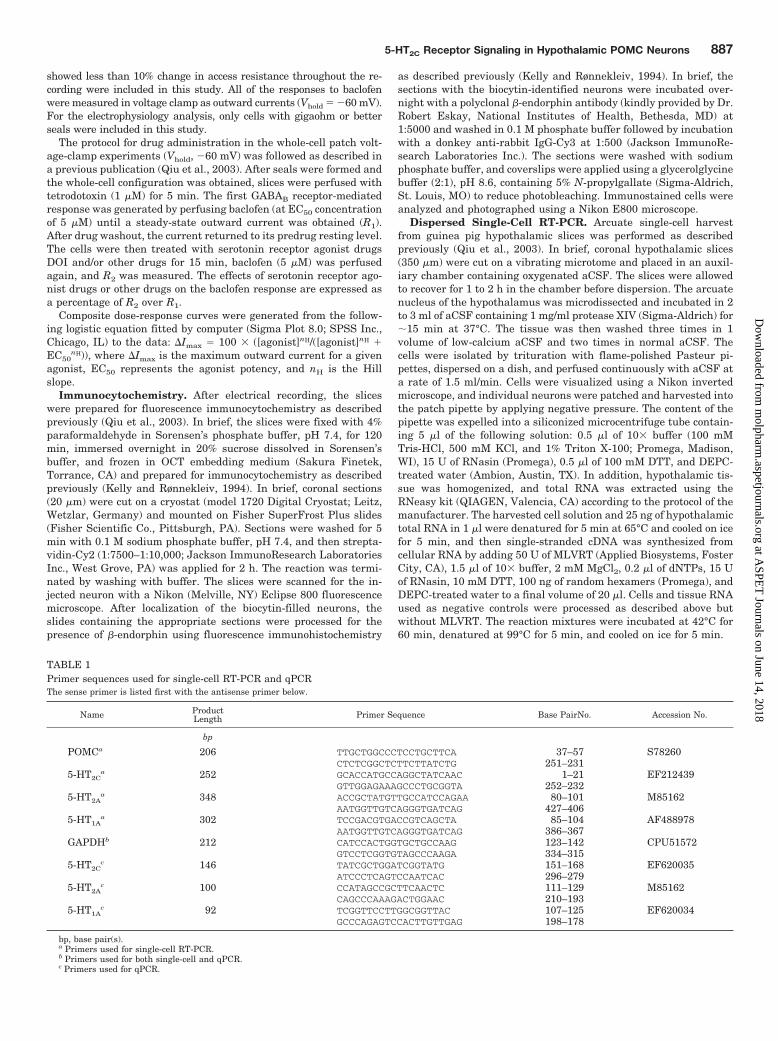
TABLE 1Primer sequences used for single-cell RT-PCR and qPCRThe sense primer is listed first with the antisense primer below.
Name ProductLength Primer Sequence Base PairNo. Accession No.
bp
POMCa 206 TTGCTGGCCCTCCTGCTTCA 37–57 S78260CTCTCGGCTCTTCTTATCTG 251–231
5-HT2Ca 252 GCACCATGCCAGGCTATCAAC 1–21 EF212439
GTTGGAGAAAGCCCTGCGGTA 252–2325-HT2A
a 348 ACCGCTATGTTGCCATCCAGAA 80–101 M85162AATGGTTGTCAGGGTGATCAG 427–406
5-HT1Aa 302 TCCGACGTGACCGTCAGCTA 85–104 AF488978
AATGGTTGTCAGGGTGATCAG 386–367GAPDHb 212 CATCCACTGGTGCTGCCAAG 123–142 CPU51572
GTCCTCGGTGTAGCCCAAGA 334–3155-HT2C
c 146 TATCGCTGGATCGGTATG 151–168 EF620035ATCCCTCAGTCCAATCAC 296–279
5-HT2Ac 100 CCATAGCCGCTTCAACTC 111–129 M85162
CAGCCCAAAGACTGGAAC 210–1935-HT1A
c 92 TCGGTTCCTTGGCGGTTAC 107–125 EF620034GCCCAGAGTCCACTTGTTGAG 198–178
bp, base pair(s).a Primers used for single-cell RT-PCR.b Primers used for both single-cell and qPCR.c Primers used for qPCR.
5-HT2C Receptor Signaling in Hypothalamic POMC Neurons 887
at ASPE
T Journals on June 14, 2018
molpharm
.aspetjournals.orgD
ownloaded from
Primers listed in Table 1 were designed using the Clone ManagerSoftware (Scientific and Educational Software, Cary, NC) and syn-thesized by Invitrogen (Carlsbad, CA). PCR was performed using 3�l of cDNA template (2 �l for GAPDH) from each RT reaction in a 30�l of PCR reaction volume containing the following: 3 �l of 10buffer, 2.4 �l of 25 mM MgCl2 (2 mM final concentration), 0.2 mMdNTPs, 0.2 �M forward and reverse primers, 2 U of TaqDNA poly-merase (Promega), and 0.22 �g of TaqStart antibody (Clontech,Mountain View, CA). TaqDNA polymerase and TaqStart antibodywere combined and incubated at room temperature for 5 min, andthe remainder of the reaction contents was added to the tube andincubated at 94°C for 2 min. PCR reactions for 5-HT1A, 5-HT2A, and5-HT2C went through 42 to 47 cycles of amplification according to thefollowing protocols: 20-s denaturation (94°C), 30-s annealing (59–62°C), 30-s elongation (72°C), with a final 72°C extension for 5 min.POMC and GAPDH PCR went through 45 and 37 cycles of amplifi-cation, respectively, in two steps: 30-s denaturation (94°C), 45-sannealing (65–67°C), with a final 72°C extension for 5 min. Tenmicroliters of the PCR products were visualized with ethidium bro-mide on a 1.5% agarose gel.
Real-Time RT-PCR. Total RNA was extracted from the micro-dissected arcuate nucleus of GDX female guinea pigs (n � 6) usingthe RNAqueous-Micro kit (Ambion). The RNA was treated withDNase I using the DNA-free kit (Ambion) according to manufactur-er’s instructions and quantified using the NanoDrop spectrophotom-eter (NanoDrop Technologies, Wilmington, DE). Reverse transcrip-tion was carried out with 200 ng of total RNA using 50 U RT (AppliedBiosystems), 1.5 �l of 10 buffer, 2 mM MgCl2, 0.2 mM dNTPs, 15 Uof RNasin, 10 mM DTT, 100 ng of random hexamers, and DEPC-treated water to a final volume of 20 �l. As a negative control, RNAwas processed as described above but without MLVRT. The reactionmixtures were incubated at 42°C for 60 min, denatured at 99°C for 5min, and cooled on ice for 5 min.
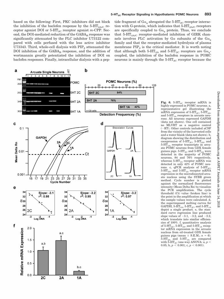
qPCR was performed using an equivalent of 1.5 ng of total RNA (3�l of a 1:20 dilution of cDNA template), 0.5 �M forward and reverseprimers, and the Power SybrGreen PCR Master Mix (Applied Bio-systems) in a 20-�l reaction volume. The qPCR reaction for 5-HT2C,5-HT2A, 5-HT1A, and GAPDH contained 10 �l of 2 Master Mix, 0.5�M forward and reverse primers, 3 �l of cDNA, and nuclease-freewater to a 20 �l final volume. qPCR was performed on samples in theABI Prism 7500 Fast machine in triplicate under the followingconditions: 95°C, 10 min; 40 cycles of amplification at 95°C, 15 s, and60°C, 1 min followed by a dissociation step for melting point analysiswith 35 cycles of 95°C for 15 s, 60°C to 95°C in increments of 1°C for1 min, and 95°C for 15 s. Standard curves using diluted cDNA fromguinea pig hypothalamus (1:5, 1:10, 1:50, 1:100, 1:500) (Fig. 6e) wereprepared to determine the efficiency of the primers. The slopes of thestandard curves for 5-HT2C, 5-HT2A, 5-HT1A, and GAPDH were�3.1, �3.2, �3.2, and �3.3, respectively (Fig. 6e). The efficiency wascalculated for each primer pair using the following formula: E �10(�1/m) � 1, where m � slope (Livak and Schmittgen, 2001; Pfaffl,2001). The efficiencies were 100% for all transcripts. The similarefficiencies between the primer pairs allowed us to make quantita-tive estimates between 5-HT2C, 5-HT2A, and 5-HT1A mRNA expres-sion. The amplification data were analyzed by the ABI 7500 Systemversion 1.3.0 software and calculated using the CT method (Livakand Schmittgen, 2001).
For quantification of 5-HT2C, 5-HT2A, and 5-HT1A receptor mRNA,expression differences were analyzed using qPCR. The CT methodwas used to calculate relative mRNA expression (Livak and Schmit-tgen, 2001). 5-HT2C, 5-HT2A, and 5-HT1A receptor mRNA valueswere normalized to the endogenous control gene GAPDH. 5-HT2C
was used as the calibrator and as a reference to which 5-HT2A and5-HT1A were compared. The relative target gene expression wascalculated using 2�CT, where CT � target CT � control CT,CT � CT target � CT calibrator. Mean and S.E.M. were calcu-lated using Prism 4 software (GraphPad Software Inc., San Di-ego, CA).
Feeding Study. Eleven- to twelve-week-old female mice anesthe-tized by intraperitoneal injection of 0.15 ml of mouse cocktail (ket-amine-xylazine-saline, 1:1:8) were ovariectomized and then keptanesthetized with isoflurane during the icv cannulation procedure.The mice were placed into a stereotaxic instrument (Cartesian In-struments, Bend, OR), the cranial surface cleaned, and a cannulaplaced into the third ventricle as described previously (Cepoi et al.,2004). In brief, a small hole was drilled, and a sterile stainless steelguide cannula (25 gauge, 1.1 cm long, with an obturator stylet placedwithin) was implanted at midline, 0.825 mm posterior to bregma and4.8 mm below bregma based on Franklin and Paxinos (1997). Micewere housed individually and allowed to recover for 10 days. There-after, the animals were adapted repeatedly for at least 3 weeks to theexperimental procedure, which included a brief restraint in a proce-dure bag during which time the icv injection was performed. To testthe effects of the compounds on feeding after an overnight fast, micewere placed in clean cages with bedding material and free access towater but without food for 16 h (5:00 PM to 9:00 AM). At the end ofthe fast, each mouse was lightly restrained, the obturator stylet wasremoved from the guide cannulae, and saline (0.9% NaCl), E2 (0.012nmol) or DOI (110 nmol) in 2-�l total volume was infused over a1-min period. Another 1-min period was allowed for diffusion of thedrugs before removing the injection needle. The mice were put backinto their cages with a preweighed food pellet (Purina Mouse Chow,5144; Purina, St. Louis, MO). Body weight and pellet weights weredetermined at 1, 2, 6, and 24 h after injection. The correct cannulaeplacement was confirmed by injecting methylene blue dye at the endof the study and visualizing the location of the dye in brain slices.
Statistical Analysis. Comparisons between groups were per-formed using a one-way or two-way ANOVA and Bonferroni post testfor the tissue analysis and whole-animal experiments and a one-wayANOVA for the electrophysiological experiments with post hoc New-man-Keuls paired analysis. Differences were considered statisticallysignificant if the probability of error was �5%.
ResultsGender Differences in the Estrogen-Mediated De-
sensitization of the GABAB Response in HypothalamicNeurons. Whole-cell recordings were made in arcuate neu-rons (n � 153) from GDX male and female guinea pigs. Asubgroup of these neurons (n � 56) was identified usingdual-labeling immunocytochemistry (Fig. 1), and 40% of theneurons were �-endorphin-positive (i.e., POMC neurons). Forthe electrophysiology analysis, only cells with gigaohm orbetter seals were included in this study. There was no differ-ence in the mean resting membrane potential (male guineapigs: �55.5 � 1.3 mV (n � 21) versus female guinea pigs:�55.0 � 0.5 mV (n � 102), Ihold � 0 pA) or the mean inputresistance (male guinea pigs: 0.96 � 0.3 G� versus femaleguinea pigs: 0.92 � 0.1 G�) between the two groups. Similarto our previous findings in mice (Qiu et al., 2006), there wereno gender differences in the mean outward current inducedby 5 �M baclofen (male mice: 37.7 � 3.8 pA, n � 43; femalemice: 34.7 � 2.4 pA, n � 88). However, there was a signifi-cant gender difference in the effects of E2 on the baclofenresponse in GDX guinea pigs. POMC neurons from GDXmale guinea pigs were less sensitive to the effects of estrogenby approximately 15% (Fig. 1). T at a higher concentration (1�M) could attenuate the baclofen response (Fig. 1, a and b) inGDX male guinea pigs but still was not as efficacious as E2.However, DHT (1 �M), which cannot be aromatized to E2,had no effect on the baclofen response (Fig. 1, a and b),suggesting an estrogenic response.
888 Qiu et al.
at ASPE
T Journals on June 14, 2018
molpharm
.aspetjournals.orgD
ownloaded from
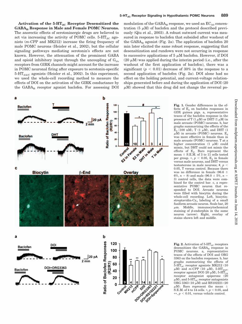
Activation of the 5-HT2C Receptor Desensitized theGABAB Response in Male and Female POMC Neurons.The anorectic effects of serotoninergic drugs are believed toact via increasing the activity of POMC cells. 5-HT2C ago-nists (m-CPP and MK212) increase the firing frequency ofmale POMC neurons (Heisler et al., 2002), but the cellularsignaling pathways mediating serotonin’s effects are notknown. However, the attenuation of the prominent GABAand opioid inhibitory input through the uncoupling of Gi/o
receptors from GIRK channels might account for the increasein POMC neuronal firing after exposure to serotonin-specific5-HT2A/C agonists (Heisler et al., 2002). In this experiment,we used the whole-cell recording method to measure theeffects of DOI on the activation of the GIRK conductance bythe GABAB receptor agonist baclofen. For assessing DOI
modulation of the GABAB response, we used an EC50 concen-tration (5 �M) of baclofen and the protocol described previ-ously (Qiu et al., 2003). A robust outward current was mea-sured in response to baclofen that subsided after washout ofthe GABAB agonist (Fig. 2a). The application of baclofen 20min later elicited the same robust response, suggesting thatdesensitization and rundown were not occurring in responseto successive applications of 5 �M baclofen. However, if DOI(20 �M) was applied during the interim period (i.e., after thewashout of the first application of baclofen), there was asignificant (p � 0.01) decrease of 30% in the response to asecond application of baclofen (Fig. 2a). DOI alone had noeffect on the holding potential, and current-voltage relation-ships generated before and during the application of DOI (20�M) showed that this drug did not change the reversal po-
Fig. 1. Gender differences in the ef-fects of E2 on baclofen responses inGDX guinea pigs. a, representativetraces of the baclofen response in thepresence of T (1 �M) or DHT (1 �M) inmale arcuate (POMC) neurons. b, bargraphs summarizing the effects of theE2 (100 nM), T (1 �M), and DHT (1�M) in arcuate (POMC) neurons. E2was more effective in female than inmale arcuate (POMC) neurons. T at ahigher concentration (1 �M) couldmimic, but DHT could not mimic theeffects of E2. Bars represent themean � S.E.M. of 3 to 11 cells testedper group. �, p � 0.05, E2 in femaleversus male neurons, and DHT versustestosterone in male neurons; #, p �0.05, T versus control. Because therewas no difference in female (96.6 �6%, n � 8) and male (96.0 � 5%, n �3) control cells, the data were com-bined for the control bar. c, a repre-sentative POMC neuron that re-sponded to DOI. Arcuate neuronswere filled with biocytin during thewhole-cell recording. Left, biocytin-streptavidin-Cy2 labeling of a smallfusiform arcuate neuron. Scale bar, 20�m. Middle, immunocytochemicalstaining of �-endorphin in the sameneuron (arrow). Right, overlay ofstains shown left and middle.
Fig. 2. Activation of 5-HT2C receptorsdesensitizes the GABAB response inPOMC neurons. a, representativetraces of the effects of DOI and ORG3363 on the baclofen responses. b, bargraphs summarizing the effects of5-HT2C receptor agonists MK212 (10�M) and m-CPP (10 �M), 5-HT2A/Creceptor agonist DOI (20 �M), 5-HT2Areceptor antagonist spiperone (20�M), and 5-HT2C receptor antagonistsORG 3363 (10 �M) and RS102221 (20�M). Bars represent the mean �S.E.M. of 4 to 14 cells. �, p � 0.05, and��, p � 0.01, versus vehicle control.
5-HT2C Receptor Signaling in Hypothalamic POMC Neurons 889
at ASPE
T Journals on June 14, 2018
molpharm
.aspetjournals.orgD
ownloaded from
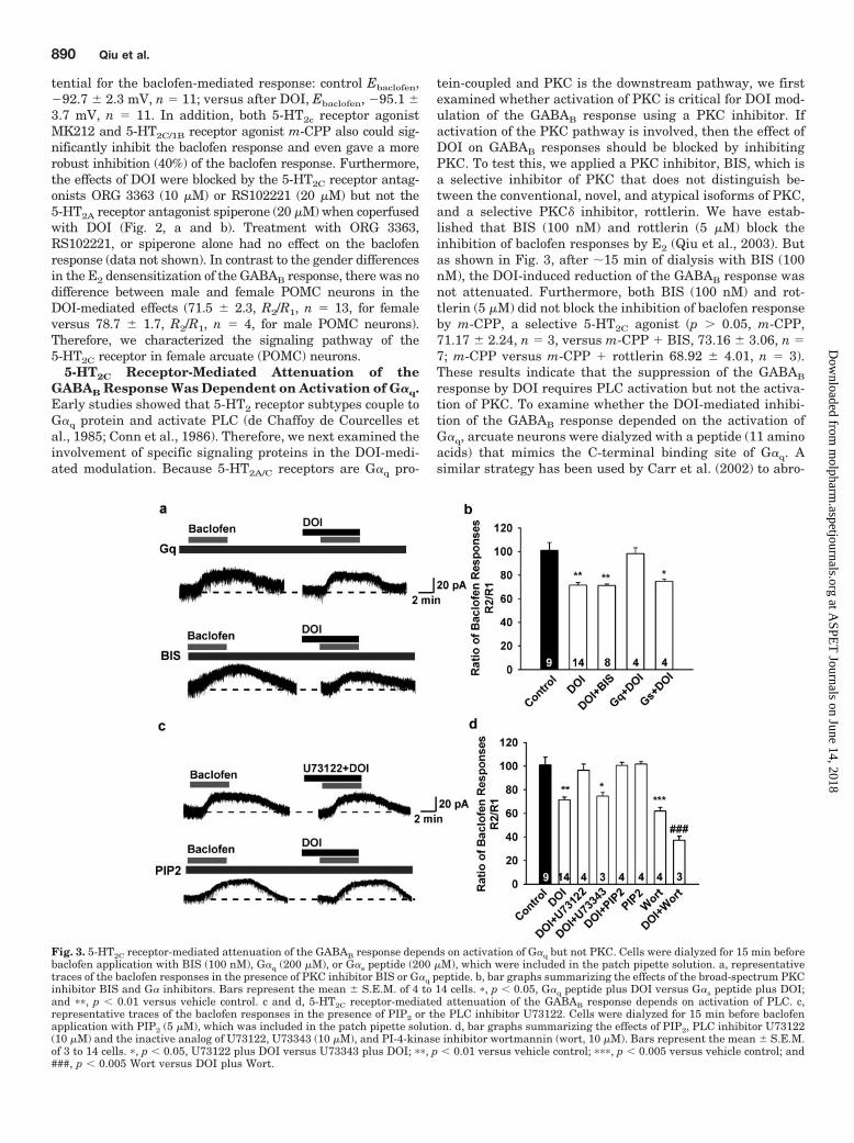
tential for the baclofen-mediated response: control Ebaclofen,�92.7 � 2.3 mV, n � 11; versus after DOI, Ebaclofen, �95.1 �3.7 mV, n � 11. In addition, both 5-HT2c receptor agonistMK212 and 5-HT2C/1B receptor agonist m-CPP also could sig-nificantly inhibit the baclofen response and even gave a morerobust inhibition (40%) of the baclofen response. Furthermore,the effects of DOI were blocked by the 5-HT2C receptor antag-onists ORG 3363 (10 �M) or RS102221 (20 �M) but not the5-HT2A receptor antagonist spiperone (20 �M) when coperfusedwith DOI (Fig. 2, a and b). Treatment with ORG 3363,RS102221, or spiperone alone had no effect on the baclofenresponse (data not shown). In contrast to the gender differencesin the E2 densensitization of the GABAB response, there was nodifference between male and female POMC neurons in theDOI-mediated effects (71.5 � 2.3, R2/R1, n � 13, for femaleversus 78.7 � 1.7, R2/R1, n � 4, for male POMC neurons).Therefore, we characterized the signaling pathway of the5-HT2C receptor in female arcuate (POMC) neurons.
5-HT2C Receptor-Mediated Attenuation of theGABAB Response Was Dependent on Activation of G�q.Early studies showed that 5-HT2 receptor subtypes couple toG�q protein and activate PLC (de Chaffoy de Courcelles etal., 1985; Conn et al., 1986). Therefore, we next examined theinvolvement of specific signaling proteins in the DOI-medi-ated modulation. Because 5-HT2A/C receptors are G�q pro-
tein-coupled and PKC is the downstream pathway, we firstexamined whether activation of PKC is critical for DOI mod-ulation of the GABAB response using a PKC inhibitor. Ifactivation of the PKC pathway is involved, then the effect ofDOI on GABAB responses should be blocked by inhibitingPKC. To test this, we applied a PKC inhibitor, BIS, which isa selective inhibitor of PKC that does not distinguish be-tween the conventional, novel, and atypical isoforms of PKC,and a selective PKC� inhibitor, rottlerin. We have estab-lished that BIS (100 nM) and rottlerin (5 �M) block theinhibition of baclofen responses by E2 (Qiu et al., 2003). Butas shown in Fig. 3, after �15 min of dialysis with BIS (100nM), the DOI-induced reduction of the GABAB response wasnot attenuated. Furthermore, both BIS (100 nM) and rot-tlerin (5 �M) did not block the inhibition of baclofen responseby m-CPP, a selective 5-HT2C agonist (p � 0.05, m-CPP,71.17 � 2.24, n � 3, versus m-CPP � BIS, 73.16 � 3.06, n �7; m-CPP versus m-CPP � rottlerin 68.92 � 4.01, n � 3).These results indicate that the suppression of the GABAB
response by DOI requires PLC activation but not the activa-tion of PKC. To examine whether the DOI-mediated inhibi-tion of the GABAB response depended on the activation ofG�q, arcuate neurons were dialyzed with a peptide (11 aminoacids) that mimics the C-terminal binding site of G�q. Asimilar strategy has been used by Carr et al. (2002) to abro-
Fig. 3. 5-HT2C receptor-mediated attenuation of the GABAB response depends on activation of G�q but not PKC. Cells were dialyzed for 15 min beforebaclofen application with BIS (100 nM), G�q (200 �M), or G�s peptide (200 �M), which were included in the patch pipette solution. a, representativetraces of the baclofen responses in the presence of PKC inhibitor BIS or G�q peptide. b, bar graphs summarizing the effects of the broad-spectrum PKCinhibitor BIS and G� inhibitors. Bars represent the mean � S.E.M. of 4 to 14 cells. �, p � 0.05, G�q peptide plus DOI versus G�s peptide plus DOI;and ��, p � 0.01 versus vehicle control. c and d, 5-HT2C receptor-mediated attenuation of the GABAB response depends on activation of PLC. c,representative traces of the baclofen responses in the presence of PIP2 or the PLC inhibitor U73122. Cells were dialyzed for 15 min before baclofenapplication with PIP2 (5 �M), which was included in the patch pipette solution. d, bar graphs summarizing the effects of PIP2, PLC inhibitor U73122(10 �M) and the inactive analog of U73122, U73343 (10 �M), and PI-4-kinase inhibitor wortmannin (wort, 10 �M). Bars represent the mean � S.E.M.of 3 to 14 cells. �, p � 0.05, U73122 plus DOI versus U73343 plus DOI; ��, p � 0.01 versus vehicle control; ���, p � 0.005 versus vehicle control; and###, p � 0.005 Wort versus DOI plus Wort.
890 Qiu et al.
at ASPE
T Journals on June 14, 2018
molpharm
.aspetjournals.orgD
ownloaded from
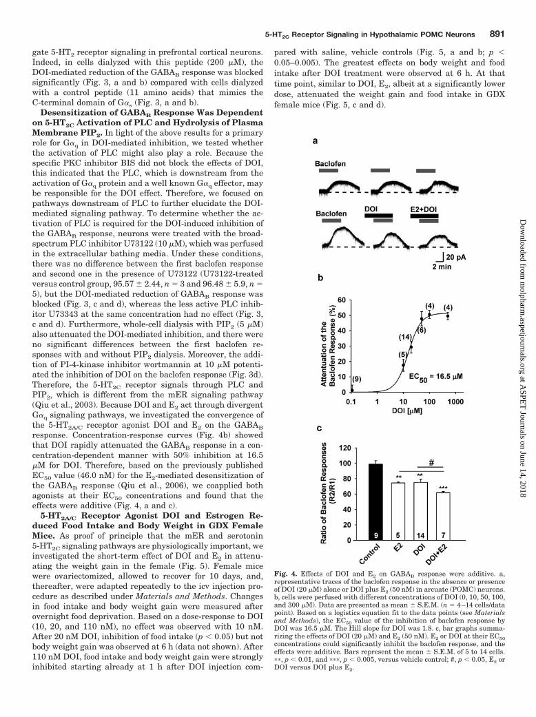
gate 5-HT2 receptor signaling in prefrontal cortical neurons.Indeed, in cells dialyzed with this peptide (200 �M), theDOI-mediated reduction of the GABAB response was blockedsignificantly (Fig. 3, a and b) compared with cells dialyzedwith a control peptide (11 amino acids) that mimics theC-terminal domain of G�s (Fig. 3, a and b).
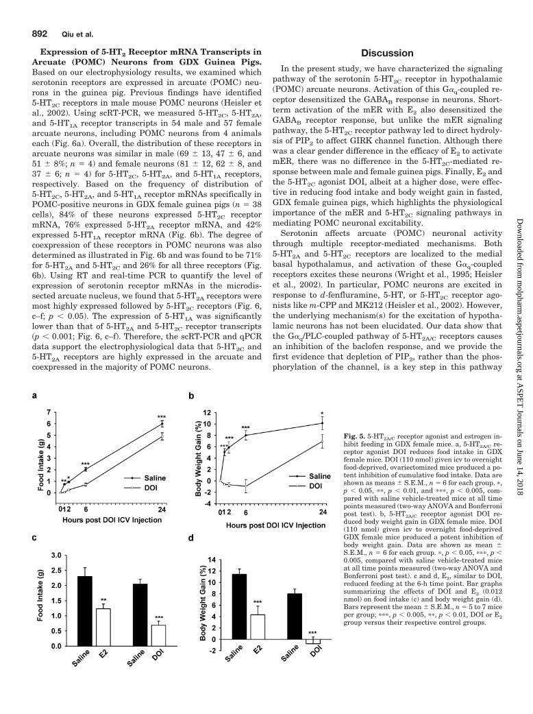
Desensitization of GABAB Response Was Dependenton 5-HT2C Activation of PLC and Hydrolysis of PlasmaMembrane PIP2. In light of the above results for a primaryrole for G�q in DOI-mediated inhibition, we tested whetherthe activation of PLC might also play a role. Because thespecific PKC inhibitor BIS did not block the effects of DOI,this indicated that the PLC, which is downstream from theactivation of G�q protein and a well known G�q effector, maybe responsible for the DOI effect. Therefore, we focused onpathways downstream of PLC to further elucidate the DOI-mediated signaling pathway. To determine whether the ac-tivation of PLC is required for the DOI-induced inhibition ofthe GABAB response, neurons were treated with the broad-spectrum PLC inhibitor U73122 (10 �M), which was perfusedin the extracellular bathing media. Under these conditions,there was no difference between the first baclofen responseand second one in the presence of U73122 (U73122-treatedversus control group, 95.57 � 2.44, n � 3 and 96.48 � 5.9, n �5), but the DOI-mediated reduction of GABAB response wasblocked (Fig. 3, c and d), whereas the less active PLC inhib-itor U73343 at the same concentration had no effect (Fig. 3,c and d). Furthermore, whole-cell dialysis with PIP2 (5 �M)also attenuated the DOI-mediated inhibition, and there wereno significant differences between the first baclofen re-sponses with and without PIP2 dialysis. Moreover, the addi-tion of PI-4-kinase inhibitor wortmannin at 10 �M potenti-ated the inhibition of DOI on the baclofen response (Fig. 3d).Therefore, the 5-HT2C receptor signals through PLC andPIP2, which is different from the mER signaling pathway(Qiu et al., 2003). Because DOI and E2 act through divergentG�q signaling pathways, we investigated the convergence ofthe 5-HT2A/C receptor agonist DOI and E2 on the GABAB
response. Concentration-response curves (Fig. 4b) showedthat DOI rapidly attenuated the GABAB response in a con-centration-dependent manner with 50% inhibition at 16.5�M for DOI. Therefore, based on the previously publishedEC50 value (46.0 nM) for the E2-mediated desensitization ofthe GABAB response (Qiu et al., 2006), we coapplied bothagonists at their EC50 concentrations and found that theeffects were additive (Fig. 4, a and c).
5-HT2A/C Receptor Agonist DOI and Estrogen Re-duced Food Intake and Body Weight in GDX FemaleMice. As proof of principle that the mER and serotonin5-HT2C signaling pathways are physiologically important, weinvestigated the short-term effect of DOI and E2 in attenu-ating the weight gain in the female (Fig. 5). Female micewere ovariectomized, allowed to recover for 10 days, and,thereafter, were adapted repeatedly to the icv injection pro-cedure as described under Materials and Methods. Changesin food intake and body weight gain were measured afterovernight food deprivation. Based on a dose-response to DOI(10, 20, and 110 nM), no effect was observed with 10 nM.After 20 nM DOI, inhibition of food intake (p � 0.05) but notbody weight gain was observed at 6 h (data not shown). After110 nM DOI, food intake and body weight gain were stronglyinhibited starting already at 1 h after DOI injection com-
pared with saline, vehicle controls (Fig. 5, a and b; p �0.05–0.005). The greatest effects on body weight and foodintake after DOI treatment were observed at 6 h. At thattime point, similar to DOI, E2, albeit at a significantly lowerdose, attenuated the weight gain and food intake in GDXfemale mice (Fig. 5, c and d).
Fig. 4. Effects of DOI and E2 on GABAB response were additive. a,representative traces of the baclofen response in the absence or presenceof DOI (20 �M) alone or DOI plus E2 (50 nM) in arcuate (POMC) neurons.b, cells were perfused with different concentrations of DOI (0, 10, 50, 100,and 300 �M). Data are presented as mean � S.E.M. (n � 4–14 cells/datapoint). Based on a logistics equation fit to the data points (see Materialsand Methods), the EC50 value of the inhibition of baclofen response byDOI was 16.5 �M. The Hill slope for DOI was 1.8. c, bar graphs summa-rizing the effects of DOI (20 �M) and E2 (50 nM). E2 or DOI at their EC50concentrations could significantly inhibit the baclofen response, and theeffects were additive. Bars represent the mean � S.E.M. of 5 to 14 cells.��, p � 0.01, and ���, p � 0.005, versus vehicle control; #, p � 0.05, E2 orDOI versus DOI plus E2.
5-HT2C Receptor Signaling in Hypothalamic POMC Neurons 891
at ASPE
T Journals on June 14, 2018
molpharm
.aspetjournals.orgD
ownloaded from
Expression of 5-HT2 Receptor mRNA Transcripts inArcuate (POMC) Neurons from GDX Guinea Pigs.Based on our electrophysiology results, we examined whichserotonin receptors are expressed in arcuate (POMC) neu-rons in the guinea pig. Previous findings have identified5-HT2C receptors in male mouse POMC neurons (Heisler etal., 2002). Using scRT-PCR, we measured 5-HT2C, 5-HT2A,and 5-HT1A receptor transcripts in 54 male and 57 femalearcuate neurons, including POMC neurons from 4 animalseach (Fig. 6a). Overall, the distribution of these receptors inarcuate neurons was similar in male (69 � 13, 47 � 6, and51 � 8%; n � 4) and female neurons (81 � 12, 62 � 8, and37 � 6; n � 4) for 5-HT2C, 5-HT2A, and 5-HT1A receptors,respectively. Based on the frequency of distribution of5-HT2C, 5-HT2A, and 5-HT1A receptor mRNAs specifically inPOMC-positive neurons in GDX female guinea pigs (n � 38cells), 84% of these neurons expressed 5-HT2C receptormRNA, 76% expressed 5-HT2A receptor mRNA, and 42%expressed 5-HT1A receptor mRNA (Fig. 6b). The degree ofcoexpression of these receptors in POMC neurons was alsodetermined as illustrated in Fig. 6b and was found to be 71%for 5-HT2A and 5-HT2C and 26% for all three receptors (Fig.6b). Using RT and real-time PCR to quantify the level ofexpression of serotonin receptor mRNAs in the microdis-sected arcuate nucleus, we found that 5-HT2A receptors weremost highly expressed followed by 5-HT2C receptors (Fig. 6,c–f; p � 0.05). The expression of 5-HT1A was significantlylower than that of 5-HT2A and 5-HT2C receptor transcripts(p � 0.001; Fig. 6, c–f). Therefore, the scRT-PCR and qPCRdata support the electrophysiological data that 5-HT2C and5-HT2A receptors are highly expressed in the arcuate andcoexpressed in the majority of POMC neurons.
DiscussionIn the present study, we have characterized the signaling
pathway of the serotonin 5-HT2C receptor in hypothalamic(POMC) arcuate neurons. Activation of this G�q-coupled re-ceptor desensitized the GABAB response in neurons. Short-term activation of the mER with E2 also desensitized theGABAB receptor response, but unlike the mER signalingpathway, the 5-HT2C receptor pathway led to direct hydroly-sis of PIP2 to affect GIRK channel function. Although therewas a clear gender difference in the efficacy of E2 to activatemER, there was no difference in the 5-HT2C-mediated re-sponse between male and female guinea pigs. Finally, E2 andthe 5-HT2C agonist DOI, albeit at a higher dose, were effec-tive in reducing food intake and body weight gain in fasted,GDX female guinea pigs, which highlights the physiologicalimportance of the mER and 5-HT2C signaling pathways inmediating POMC neuronal excitability.
Serotonin affects arcuate (POMC) neuronal activitythrough multiple receptor-mediated mechanisms. Both5-HT2A and 5-HT2C receptors are localized to the medialbasal hypothalamus, and activation of these G�q-coupledreceptors excites these neurons (Wright et al., 1995; Heisleret al., 2002). In particular, POMC neurons are excited inresponse to d-fenfluramine, 5-HT, or 5-HT2C receptor ago-nists like m-CPP and MK212 (Heisler et al., 2002). However,the underlying mechanism(s) for the excitation of hypotha-lamic neurons has not been elucidated. Our data show thatthe G�q/PLC-coupled pathway of 5-HT2A/C receptors causesan inhibition of the baclofen response, and we provide thefirst evidence that depletion of PIP2, rather than the phos-phorylation of the channel, is a key step in this pathway
Fig. 5. 5-HT2A/C receptor agonist and estrogen in-hibit feeding in GDX female mice. a, 5-HT2A/C re-ceptor agonist DOI reduces food intake in GDXfemale mice. DOI (110 nmol) given icv to overnightfood-deprived, ovariectomized mice produced a po-tent inhibition of cumulative food intake. Data areshown as means � S.E.M., n � 6 for each group. �,p � 0.05, ��, p � 0.01, and ���, p � 0.005, com-pared with saline vehicle-treated mice at all timepoints measured (two-way ANOVA and Bonferronipost test). b, 5-HT2A/C receptor agonist DOI re-duced body weight gain in GDX female mice. DOI(110 nmol) given icv to overnight food-deprivedGDX female mice produced a potent inhibition ofbody weight gain. Data are shown as mean �S.E.M., n � 6 for each group. �, p � 0.05, ���, p �0.005, compared with saline vehicle-treated miceat all time points measured (two-way ANOVA andBonferroni post test). c and d, E2, similar to DOI,reduced feeding at the 6-h time point. Bar graphssummarizing the effects of DOI and E2 (0.012nmol) on food intake (c) and body weight gain (d).Bars represent the mean � S.E.M., n � 5 to 7 miceper group; ���, p � 0.005, ��, p � 0.01, DOI or E2group versus their respective control groups.
892 Qiu et al.
at ASPE
T Journals on June 14, 2018
molpharm
.aspetjournals.orgD
ownloaded from
based on the following: First, PKC inhibitors did not blockthe inhibition of the baclofen response by the 5-HT2A/C re-ceptor agonist DOI or 5-HT2C receptor agonist m-CPP. Sec-ond, the DOI-mediated reduction of the GABAB response wassignificantly attenuated by the PLC inhibitor U73122 com-pared with cells perfused with the less active inhibitorU73343. Third, whole-cell dialysis with PIP2 attenuated theDOI inhibition of the GABAB response, and the addition ofwortmannin greatly potentiated the inhibition of DOI onbaclofen responses. Finally, intracellular dialysis with a pep-
tide fragment of G�q abrogated the 5-HT2C receptor interac-tion with G-protein, which indicates that 5-HT2A/C receptorsare specifically coupled to G�q protein. Thus, we concludethat 5-HT2A/C receptor-mediated inhibition of GIRK chan-nels involves PLC activation by G� subunits of the G�q
family and that the receptor-mediated hydrolysis of plasmamembrane PIP2 is the critical mediator. It is worth notingthat although both 5-HT2A and 5-HT2C receptors are G�q-coupled, the inhibition of the baclofen response in POMCneurons is mainly through the 5-HT2C receptor because the
Fig. 6. 5-HT2C receptor mRNA ishighly expressed in POMC neurons. a,representative gel illustrating themRNA expression of 5-HT2C, 5-HT2A,and 5-HT1A receptors in arcuate neu-rons. All neurons expressed GAPDH(data not shown). One cell containedno MLVRT as a negative control(�RT). Other controls included aCSFfrom the vicinity of the harvested cellsand a water blank (data not shown). b,diagram showing the distribution andcoexpression of 5-HT2C, 5-HT2A, and5-HT1A receptor transcripts in arcu-ate POMC neurons from GDX femaleguinea pigs. 5-HT2C and 5-HT2A weredetected in the majority of POMCneurons, 84 and 76% respectively,whereas 5-HT1A receptor mRNA wasdetected in only 42% of POMC neu-rons. c, qPCR analysis of 5-HT2C,5-HT2A, and 5-HT1A receptor mRNAexpression in the microdissected arcu-ate nucleus using the SYBR greenmethod. Cycle number is plottedagainst the normalized fluorescenceintensity (Mean Delta Rn) to visualizethe PCR amplification. The cyclethreshold (Ct) value (broken line) isthe point in the amplification at whichthe sample values were calculated. d,the superimposed melting curves forGAPDH, 5-HT2A, 5-HT2C, and 5-HT1Adepict a single product. e, the stan-dard curve regression line producedslope values of �3.1, �3.2, and �3.2,which translate into similar efficien-cies of 100%. f, quantitative analysisof 5-HT2C, 5-HT2A, and 5-HT1A recep-tor mRNA expression in the arcuatenucleus from oil-treated GDX femaleguinea pigs (mean � S.E.M.; n � 6).5-HT2A and 5-HT1A are comparedwith 5-HT2C (one-way ANOVA: a, p �0.05; b, p � 0.001; c, p � 0.001).
5-HT2C Receptor Signaling in Hypothalamic POMC Neurons 893
at ASPE
T Journals on June 14, 2018
molpharm
.aspetjournals.orgD
ownloaded from
5-HT2C-selective agonists m-CPP and MK212 attenuated ba-clofen responses in POMC neurons. Moreover, the selective5-HT2C antagonists ORG 3363 and RS102221 but not the5-HT2A antagonist spiperone potently blocked the actions ofDOI in guinea pig arcuate (POMC) neurons, indicating thatthe inhibition of the GABAB response is through the 5-HT2C
receptor. This is compatible with previous findings that the5-HT2C receptor agonists show greater efficacy for activatingthe PLC pathway, whereas 5-HT2A receptor agonists haverelatively greater efficacy for activating the phospholipase A2
pathway (Berg et al., 1998; Kurrasch-Orbaugh et al., 2003).The PLC hydrolysis of PIP2 and inhibition of GIRK channelsis not unique to 5-HT2C receptors because other G�q-coupledreceptors have different propensities for activating this path-way depending on the subcellular localizations of the G-protein-coupled receptor relative to the proximity of PLC andGIRK channels (Cho et al., 2005).
It is interesting that GABAB, �-opioid, and 5-HT1A recep-tors are all expressed in POMC neurons (Kelly et al., 1992;Lagrange et al., 1994; Qiu et al., 2003; present findings). Allof these receptors are G�i/o-coupled to activation of GIRKchannels, which uniformly inhibit POMC neuronal activity.Similar to our findings with the 5-HT2C receptor-mediateddesensitization of the GABAB response in POMC neurons,activation of 5-HT2A receptors can desensitize 5-HT1A recep-tors and increase the excitability of CRH neurons, as mea-sured by ACTH release (Zhang et al., 2001). The heterologous
desensitization in these paraventricular nucleus neurons hasnot been characterized but may be via PKC-mediated phos-phorylation of GIRK channels (Brown et al., 2005) or byPLC-mediated PIP2 depletion (Brown et al., 2005; Cho et al.,2005). Therefore, the PLC-mediated PIP2 depletion maybe acommon signaling pathway for 5-HT2A/C receptors in hypo-thalamic neurons.
Evidence from several species indicates that food intakeand body weight are influenced both by changes in endoge-nous estrogens and by exogenous estrogenic treatments(Czaja and Goy, 1975). Butera and Czaja (1984) have shownthat the anorexigenic effects of E2 are attributable to thedirect actions of the steroid in the arcuate-ventromedial hy-pothalamic nuclei, and recently, we have shown that E2 andSTX, a selective ligand for the mER, attenuated the weightgain in female guinea pigs after ovariectomy (Qiu et al.,2006). The present findings with icv administration of E2 inGDX female mice corroborate the findings in the guinea pigfor a central action of steroid to regulate energy homeostasis.Therefore, this membrane-delimited signaling pathway mayplay a vital role in the control of energy homeostasis. It isinteresting that eating disorders are much more prevalent(95%) in young women compared with men, but the reasonsfor this difference are not clear (Sodersten et al., 2006). Inanimal models, there are clear gender differences in foodintake and energy homeostasis with hormone treatment. Es-trogen reduces food intake and body weight in female and
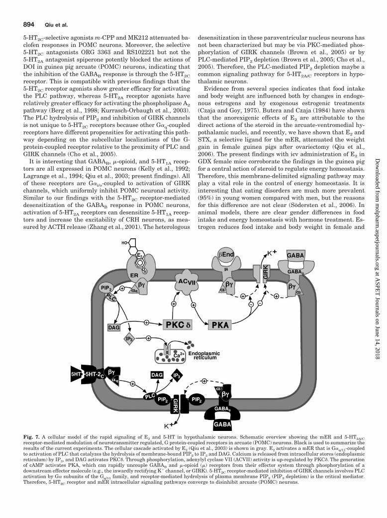
Fig. 7. A cellular model of the rapid signaling of E2 and 5-HT in hypothalamic neurons. Schematic overview showing the mER and 5-HT2A/Creceptor-mediated modulation of neurotransmitter regulated, G protein-coupled receptors in arcuate (POMC) neurons. Black is used to summarize theresults of the current experiments. The cellular cascade activated by E2 (Qiu et al., 2003) is shown in gray. E2 activates a mER that is G�q/11-coupledto activation of PLC that catalyzes the hydrolysis of membrane-bound PIP2 to IP3 and DAG. Calcium is released from intracellular stores (endoplasmicreticulum) by IP3, and DAG activates PKC�. Through phosphorylation, adenylyl cyclase VII (ACVII) activity is up-regulated by PKC�. The generationof cAMP activates PKA, which can rapidly uncouple GABAB and �-opioid (�) receptors from their effector system through phosphorylation of adownstream effector molecule (e.g., the inwardly rectifying K� channel, or GIRK). 5-HT2C receptor-mediated inhibition of GIRK channels involves PLCactivation by G� subunits of the Gq/11 family, and receptor-mediated hydrolysis of plasma membrane PIP2 (PIP2 depletion) is the critical mediator.Therefore, 5-HT2C receptor and mER intracellular signaling pathways converge to disinhibit arcuate (POMC) neurons.
894 Qiu et al.
at ASPE
T Journals on June 14, 2018
molpharm
.aspetjournals.orgD
ownloaded from
male subjects, although E2 is more effective in female sub-jects and T in male subjects (Czaja and Goy, 1975; Czaja,1984). We found that T at a higher concentration could mimicbut the nonhydrolyzable androgen DHT could not mimic theeffects of E2. Moreover, E2 was more efficacious in femalethan in male arcuate (POMC) neurons, but there was nogender difference in the actions of 5-HT2A/C agonists. Takentogether, the gender differences in the control of feeding andenergy homeostasis may be due in part to a greater efficacy ofE2 in female versus male guinea pigs to disinhibit POMCneurons via the mER signaling pathway.
Serotonin 5-HT2C receptors have also been strongly impli-cated in inhibiting feeding. For example, the selective 5-HT2C
antagonist RS102221 increases food intake and body weightwhen injected intraperitoneally (Bonhaus et al., 2006), and5-HT2C receptor-deficient mice are hyperphagic, obese, andrefractory to threshold anorexic doses of d-fenfluramine(Tecott et al., 1995; Vickers et al., 1999), a drug that blocksthe reuptake of 5-HT and stimulates its release (Heal et al.,1998). In contrast, 5-HT2A receptor knockout mice do notexhibit an obesity phenotype, so one may assume that5-HT2A receptors are not critically involved in energy ho-meostasis (Zhou et al., 2005). We found that 5-HT2C mRNAwas highly expressed and colocalized with 5-HT2A receptor inPOMC neurons. This would disagree with in situ hybridiza-tion results in rhesus (female) monkeys showing that 5-HT2C
receptor mRNA is highly expressed in the arcuate region,whereas the 5-HT2A receptor mRNA is more localized to thePVN (Gundlah et al., 1999). In addition, Heisler et al. (2002)found that up to 80% of POMC neurons express 5-HT2C
receptor mRNA. However, the role of the 5-HT2C and 5-HT2A
receptors in different physiological processes may lie in theircoupling to distinctive signaling pathways.
Based on our cellular electrophysiological data, we haveproposed a model for the convergence of the mER and 5-HT2C
signaling pathways in arcuate (POMC) neurons (Fig. 7). It isnoteworthy that the downstream signaling pathway of ba-clofen inhibition by 5-HT2C receptor agonists in arcuate(POMC) neurons is different from mER in which E2 desen-sitizes the GABAB response via a G�q-PLC-PKC�-PKA path-way (Qiu et al., 2003). These differences may be due to thedifferent compartmentalization of the receptors. It has beenreported that the signaling components for G-protein activa-tion in neurons are compartmentalized or preassembled(Lober et al., 2006). G-protein-coupled receptors, G-proteins,and other effector molecules can preassemble into stablesignaling complexes (Rebois and Hebert, 2003). Therefore,the 5-HT2C receptor and mER may be preassembled intocomplexes with different downstream effectors that convergeon GIRK channels. For example, Brown and colleagues(2005) have shown that although protein kinases (i.e., PKC�)mediate the inhibition of GIRK1/2 channels by the muscarinicM3 receptor, resynthesis of PIP2 is required for completerecovery from inhibition. Therefore, PIP2 turnover is criticalfor GIRK channel function. Our data support this idea be-cause 5-HT2C receptors can inhibit the GIRK channels by anindependent pathway from mER, but the two pathways con-verge on the same population of GIRK channels as shown bythe additive effects of the two agonists given together. Asproof of principle, we have found that both E2 and DOI areeffective to inhibit feeding in GDX female mice, and as pre-dicted from the cellular findings, E2 was more potent than
DOI to inhibit food intake and weight gain. Therefore, theG�q signaling pathways of mER and 5-HT2C receptors mayconverge to enhance synaptic efficacy in brain circuits thatare critical for maintaining homeostatic functions.
Acknowledgments
We recognize the skilled technical assistance of Rebecka Amodeiand Scott Kuhn. We recognize Dr. Jan Kelder (Organon NV) for hisgenerous gift of ORG 3363.
ReferencesBerg KA, Maayani S, Goldfarb J, and Clarke WP (1998) Pleiotropic behavior of
5-HT2A and 5-HT2C receptor agonists. Ann N Y Acad Sci 861:104–110.Bethea CL (1993) Colocaliztion of progestin receptors with serotonin in Raphe
neurons of Macaque. Neuroendocrinology 57:1–6.Bethea CL, Pecins-Thompson M, Schutzer WE, Gundlah C, and Lu ZN (1998)
Ovarian steroids and serotonin neural function. Mol Neurobiol 18:87–123.Bickerdike MJ (2003) 5-HT2C receptor agonists as potential drugs for the treatment
of obesity. Curr Top Med Chem 3:885–897.Bonhaus SJ, Weinhardt KK, Taylor M, DeSouza A, McNeeley PM, Szczepanski K,
Fontan DJ, Trinh J, Rocha CL, and Dawson MW (2006) RS-102221: a novel highaffinity and selective, 5-HT2C receptor antagonist. Neuropharmacology 36:621–629.
Breisch ST, Zemlan FP, and Hoebel BG (1976) Hyperphagia and obesity followingserotonin depletion by intraventricular P-chlorophenylalanine. Science 192:382–385.
Brown SG, Thomas A, Dekker LV, Tinker A, and Leaney JL (2005) PKC-� sensitizesKir3.1/3.2 channels to changes in membrane phospholipid levels after M3 receptoractivation in HEK-293 cells. Am J Physiol Cell Physiol 289:C543–C556.
Butera PC and Czaja JA (1984) Intracranial estradiol in ovariectomized guinea pigs:Effects on ingestive behaviors and body weight. Brain Res 322:41–48.
Carr DB, Cooper DC, Ulrich SL, Spruston N, and Surmeier DJ (2002) Serotoninreceptor activation inhibits sodium current and dendritic excitability in prefrontalcortex via a protein kinase C-dependent mechanism. J Neurosci 22:6846–6855.
Cepoi D, Phillips T, Cismowski M, Goodfellow VS, Ling N, Cone RD, and Fan W(2004) Assessment of a small molecule melancortin-4 receptor-specific agonist onenergy homeostasis. Brain Res 1000:64–71.
Cho H, Lee D, Lee SH, and Ho WK (2005) Receptor-induced depletion of phospha-tidylinositol 4,5-bisphosphate inhibits inwardly rectifying K� channels in a recep-tor-specific manner. Proc Natl Acad Sci U S A 102:4643–4648.
Cone RD (2005) Anatomy and regulation of the central melanocortin system. NatNeurosci 8:571–578.
Conn PJ, Sanders-Bush E, Hoffman B, and Hartig PR (1986) A unique serotoninreceptor in choroid plexus is linked to phosphatidylinositol turnover. Proc NatlAcad Sci U S A 83:4086–4088.
Couse JF and Korach KS (1999) Estrogen receptor null mice: What have we learnedand where will they lead us? Endocr Rev 20:358–417.
Czaja JA (1984) Sex differences in the activational effects of gonadal hormones onfood intake and body weight. Physiol Behav 33:553–558.
Czaja JA and Goy RW (1975) Ovarian hormones and food intake in female guineapigs and Rhesus monkeys. Horm Behav 6:329–349.
de Chaffoy de Courcelles D, Leysen JE, de Clerck F, Van Belle H, and Janssen PA(1985) Evidence that phospholipid turnover is the signal transducing systemcoupled to serotonin-S2 receptor sites. J Biol Chem 260:7603–7608.
Elmquist JK, Elias CF, and Saper CB (1999) From lesions to leptin: hypothalamiccontrol of food intake and body weight. Neuron 22:221–232.
Franklin KBJ and Paxinos G (1997) Atlas of the Mouse Brain. Academic Press, SanDiego, CA.
Gropp E, Shanabrough M, Borok E, Xu AW, Janoschek R, Buch T, Plum L, BalthasarN, Hampel B, Waisman A, et al. (2005) Agouti-related peptide-expressing neuronsare mandatory for feeding. Nat Neurosci 8:1289–1291.
Gundlah C, Pecins-Thompson M, Schutzer WE, and Bethea CL (1999) Ovariansteroid effects on serotonin 1A, 2A and 2C receptor mRNA in Macaque hypothal-amus. Mol Brain Res 63:325–339.
Heal DJ, Cheetham SC, Prow MR, Martin KF, and Buckett WR (1998) A comparisonof the effects on central 5-HT function of sibutramine hydrochloride and otherweight-modifying agents. Br J Pharmacol 125:301–308.
Heisler LK, Cowley MA, Tecott LH, Fan W, Low MJ, Smart JL, Rubinstein M, TatroJB, Marcus JN, Holstege H, et al. (2002) Activation of central melanocortinpathways by fenfluramine. Science 297:609–611.
Heisler LK, Jobst EE, Sutton GM, Zhou L, Borok E, Thornton-Jones Z, Liu HY,Zigman JM, Balthasar N, Kishi T, et al. (2006) Serotonin reciprocally regulatesmelanocortin neurons to modulate food intake. Neuron 51:239–249.
Kelly MJ, Loose MD, and Rønnekleiv OK (1992) Estrogen suppresses �-opioid andGABAB-mediated hyperpolarization of hypothalamic arcuate neurons. J Neurosci12:2745–2750.
Kelly MJ and Rønnekleiv OK (1994) Electrophysiological analysis of neuroendocrineneuronal activity in hypothalamic slices, in Methods in Neurosciences: Pulsatilityin Neuroendocrine Systems (Levine JE ed) pp 47–67, Academic Press, Inc., SanDiego.
Kurrasch-Orbaugh DM, Watts VJ, Barker EL, and Nichols DE (2003) Serotonin5-hydroxytryptamine 2A receptor-coupled phospholipase C and phospholipase A2signaling pathways have different receptor reserves. J Pharmacol Exp Ther 304:229–237.
Lagrange AH, Rønnekleiv OK, and Kelly MJ (1994) The potency of �-opioid hyper-
5-HT2C Receptor Signaling in Hypothalamic POMC Neurons 895
at ASPE
T Journals on June 14, 2018
molpharm
.aspetjournals.orgD
ownloaded from
polarization of hypothalamic arcuate neurons is rapidly attenuated by 17�-estradiol. J Neurosci 14:6196–6204.
Livak KJ and Schmittgen TD (2001) Analysis of relative gene expression data usingreal-time quantitative PCR and the 2�CT method. Methods 25:402–408.
Lober RM, Pereira MA, and Lambert NA (2006) Rapid activation of inwardly recti-fying potassium channels by immobile G-protein-coupled receptors. J Neurosci26:12602–12608.
Luquet S, Perez FA, Hnasko TS, and Palmiter RD (2005) NPY/AgRP neurons areessential for feeding in adult mice but can be ablated in neonates. Science 310:683–685.
McEwen BS (2001) Estrogens effects on the brain: Multiple sites and molecularmechanisms. J Appl Physiol 91:2785–2801.
Pfaffl MW (2001) A new mathematical model for relative quantification in real-timeRT-PCR. Nucleic Acids Res 29:e45.
Qiu J, Bosch MA, Tobias SC, Grandy DK, Scanlan TS, Rønnekleiv OK, and Kelly MJ(2003) Rapid signaling of estrogen in hypothalamic neurons involves a novel Gprotein-coupled estrogen receptor that activates protein kinase C. J Neurosci23:9529–9540.
Qiu J, Bosch MA, Tobias SC, Krust A, Graham S, Murphy S, Korach KS, ChambonP, Scanlan TS, Rønnekleiv OK, et al. (2006) A G protein-coupled estrogen receptoris involved in hypothalamic control of energy homeostasis. J Neurosci 26:5649–5655.
Rebois RV and Hebert TE (2003) Protein complexes involved in heptahelical recep-tor-mediated signal transduction. Receptors Channels 9:169–194.
Sodersten P, Bergh C, and Zandian M (2006) Understanding eating disorders. HormBehav 50:572–578.
Stearns V, Ullmer L, Lopez JF, Smith Y, Isaacs C, and Hayes DF (2002) Hot flushes.Lancet 360:1851–1861.
Tecott LH, Sun LM, Akana SF, Strack AM, Lowenstein DH, Dallman MF, and JuliusD (1995) Eating disorder and epilepsy in mice lacking 5-HT2C serotonin receptors.Nature 374:542–546.
Vickers SP, Clifton PG, Dourish CT, and Tecott LH (1999) Reduced satiating effectof d-fenfluramine in serotonin 5-HT2C receptor mutant mice. Psychopharmacology(Berl) 143:309–314.
Wright DE, Seroogy KB, Lungren KH, Davis BM, and Jennes L (1995) Comparativelocalization of serotonin 1A, 1C, and 2 receptor subtype mRNAs in rat brain.J Comp Neurol 351:357–373.
Zhang Y, D’Souza D, Raap DK, Garcia F, Battaglia G, Muma NA, and Van de Kar LD(2001) Characterization of the functional heterologous desensitization of hypotha-lamic 5-HT1A receptors after 5-HT2A receptor activation. J Neurosci 21:7919–7927.
Zhou L, Williams T, Lachey JL, Kishi T, Cowley MA, and Heisler LK (2005) Sero-tonergic pathways converge upon central melanocortin systems to regulate energybalance. Peptides 26:1728–1732.
Address correspondence to: Dr. Jian Qiu, Department of Physiology andPharmacology, L334, Oregon Health and Science University, Portland, OR97239-3098. E-mail: [email protected]
896 Qiu et al.
at ASPE
T Journals on June 14, 2018
molpharm
.aspetjournals.orgD
ownloaded from