Seroprevalence of parvovirus B19 NS1-specific IgG in B19-infected and uninfected individuals and in...
8
Seroprevalence of Parvovirus B19 NS1-Specific IgG in B19-Infected and Uninfected Individuals and in Infected Pregnant Women Andrea Hemauer, 1 Andreas Gigler, 1 Karen Searle, 2 Karin Beckenlehner, 1 Ulla Raab, 1 Kristina Broliden, 3 Hans Wolf, 1 Gisela Enders, 2 and Susanne Modrow 1 * 1 Institut fu ¨ r Medizinische Mikrobiologie, Universita ¨ t Regensburg, Regensburg, Germany 2 Medizinisch-diagnostisches Gemeinschaftslabor, Stuttgart, Germany 3 Department of Clinical Virology, Swedish Institute for Infectious Disease Control, Stockholm, Sweden Parvovirus B19 is the causative agent of erythe- ma infectiosum in children, but the virus is as- sociated with an increasing range of different diseases. These include acute and chronic arthri- tis, hydrops fetalis in pregnant women, aplastic anemia, and thrombocytopenia. The host’s im- mune response is directed against the viral structural proteins VP1 and VP2. This study in- vestigated the presence of IgG against the viral nonstructural protein NS1 using Western blot. Serum panels from healthy individuals, B19- infected pregnant women, and various disease groups were tested. The disease groups in- cluded patients with symptoms that may be linked to parvovirus B19 infection. The results showed that IgG against the NS1 protein was present in 22% of healthy individuals with past B19 infection. In cases of persistent or prolonged B19 infections, the prevalence of NS1-specific antibodies was as high as 80%. It is concluded that NS1-specific IgG may be used as an indica- tor of chronic or more severe courses of parvo- virus B19 infections. J. Med. Virol. 60:48–55, 2000. © 2000 Wiley-Liss, Inc. KEY WORDS: parvovirus B19; NS1-specific antibodies; antibody preva- lence; hydrops fetalis INTRODUCTION Parvovirus B19 was discovered by chance in sera of healthy blood donors [Cossart et al., 1975]. Several years later, B19 infections were linked to erythema in- fectiosum (fifth disease), a common childhood exan- thema [Anderson et al., 1983]. To date, B19 is the only member of the parvoviridae known to cause disease in humans. Infections occur in childhood and throughout adult life and about 70–80% of the population are se- ropositive by the age of 40 years [Cohen and Buckley, 1988]. Progress in the field of molecular biology led to the development of assays for the detection of viral nucleic acids in sera and biopsies, which allowed the association of B19 infections with more severe symp- toms. Among these are hydrops fetalis [Anderson et al., 1988], acute and chronic arthritis [Reid et al., 1985; Naides et al., 1990; Biasi et al., 1996], acute and per- sistent aplastic anemia, thrombocytopenia, pancytope- nia [Chorba et al., 1986; Young, 1988; Srivastava et al., 1990] and persistent infections in immunocompro- mised patients [Flunker et al., 1998] and also rarely in immunocompetent individuals [Cassinotti et al., 1993]. Recent case reports on B19-associated hepatitis [Hill- ingso et al., 1998; Pardi et al., 1998], myocarditis [Orth et al., 1997; Enders et al., 1998], and encephalitis [Umene and Nuone, 1995] have shown the increasing diversity of symptoms that can arise during B19 infec- tions. Because the symptoms of B19 infection are nonspe- cific and can therefore be confused with various other infectious and noninfectious agents, diagnosis relies on immunodiagnostic or DNA testing methods. Labora- tory diagnosis is made routinely by enzyme-linked im- munosorbent assay (ELISA), Western blot, or similar test formats using either or both of the structural pro- teins VP1 and VP2. In general, IgM antibodies against the structural proteins VP1 and VP2 are the first se- rological markers of an acute B19 infection. They may be detected 6–10 days after the first contact, whereas IgG antibodies can be detected about 12 days after in- fection. During the following weeks the concentration of IgM antibodies falls to an undetectable level, whereas IgG persists lifelong and may be used as a Grant sponsor: Deutsche Forschungsgemeinschaft DFG; Grant number: Mo620/5-1. *Correspondence to: Dr. Susanne Modrow, Institut fu ¨ r Medi- zinische Mikrobiologie, Universita ¨ t Regensburg, Franz-Josef- Strauss-Allee 11, D-93053 Regensburg, Germany. E-mail: [email protected] Accepted 24 May 1999 Journal of Medical Virology 60:48–55 (2000) © 2000 WILEY-LISS, INC.
Transcript of Seroprevalence of parvovirus B19 NS1-specific IgG in B19-infected and uninfected individuals and in...
Seroprevalence of Parvovirus B19 NS1-Specific IgGin B19-Infected and Uninfected Individuals and inInfected Pregnant Women
Andrea Hemauer,1 Andreas Gigler,1 Karen Searle,2 Karin Beckenlehner,1 Ulla Raab,1Kristina Broliden,3 Hans Wolf,1 Gisela Enders,2 and Susanne Modrow1*1Institut fur Medizinische Mikrobiologie, Universitat Regensburg, Regensburg, Germany2Medizinisch-diagnostisches Gemeinschaftslabor, Stuttgart, Germany3Department of Clinical Virology, Swedish Institute for Infectious Disease Control, Stockholm, Sweden
Parvovirus B19 is the causative agent of erythe-ma infectiosum in children, but the virus is as-sociated with an increasing range of differentdiseases. These include acute and chronic arthri-tis, hydrops fetalis in pregnant women, aplasticanemia, and thrombocytopenia. The host’s im-mune response is directed against the viralstructural proteins VP1 and VP2. This study in-vestigated the presence of IgG against the viralnonstructural protein NS1 using Western blot.Serum panels from healthy individuals, B19-infected pregnant women, and various diseasegroups were tested. The disease groups in-cluded patients with symptoms that may belinked to parvovirus B19 infection. The resultsshowed that IgG against the NS1 protein waspresent in 22% of healthy individuals with pastB19 infection. In cases of persistent or prolongedB19 infections, the prevalence of NS1-specificantibodies was as high as 80%. It is concludedthat NS1-specific IgG may be used as an indica-tor of chronic or more severe courses of parvo-virus B19 infections. J. Med. Virol. 60:48–55,2000. © 2000 Wiley-Liss, Inc.
KEY WORDS: parvovirus B19; NS1-specificantibodies; antibody preva-lence; hydrops fetalis
INTRODUCTION
Parvovirus B19 was discovered by chance in sera ofhealthy blood donors [Cossart et al., 1975]. Severalyears later, B19 infections were linked to erythema in-fectiosum (fifth disease), a common childhood exan-thema [Anderson et al., 1983]. To date, B19 is the onlymember of the parvoviridae known to cause disease inhumans. Infections occur in childhood and throughoutadult life and about 70–80% of the population are se-ropositive by the age of 40 years [Cohen and Buckley,
1988]. Progress in the field of molecular biology led tothe development of assays for the detection of viralnucleic acids in sera and biopsies, which allowed theassociation of B19 infections with more severe symp-toms. Among these are hydrops fetalis [Anderson et al.,1988], acute and chronic arthritis [Reid et al., 1985;Naides et al., 1990; Biasi et al., 1996], acute and per-sistent aplastic anemia, thrombocytopenia, pancytope-nia [Chorba et al., 1986; Young, 1988; Srivastava et al.,1990] and persistent infections in immunocompro-mised patients [Flunker et al., 1998] and also rarely inimmunocompetent individuals [Cassinotti et al., 1993].Recent case reports on B19-associated hepatitis [Hill-ingso et al., 1998; Pardi et al., 1998], myocarditis [Orthet al., 1997; Enders et al., 1998], and encephalitis[Umene and Nuone, 1995] have shown the increasingdiversity of symptoms that can arise during B19 infec-tions.
Because the symptoms of B19 infection are nonspe-cific and can therefore be confused with various otherinfectious and noninfectious agents, diagnosis relies onimmunodiagnostic or DNA testing methods. Labora-tory diagnosis is made routinely by enzyme-linked im-munosorbent assay (ELISA), Western blot, or similartest formats using either or both of the structural pro-teins VP1 and VP2. In general, IgM antibodies againstthe structural proteins VP1 and VP2 are the first se-rological markers of an acute B19 infection. They maybe detected 6–10 days after the first contact, whereasIgG antibodies can be detected about 12 days after in-fection. During the following weeks the concentrationof IgM antibodies falls to an undetectable level,whereas IgG persists lifelong and may be used as a
Grant sponsor: Deutsche Forschungsgemeinschaft DFG; Grantnumber: Mo620/5-1.
*Correspondence to: Dr. Susanne Modrow, Institut fur Medi-zinische Mikrobiologie, Universitat Regensburg, Franz-Josef-Strauss-Allee 11, D-93053 Regensburg, Germany.E-mail: [email protected]
Accepted 24 May 1999
Journal of Medical Virology 60:48–55 (2000)
© 2000 WILEY-LISS, INC.

marker for past B19 infection. In addition to immuno-globulins against the capsid proteins VP1 and VP2,patients with persistent or chronic B19 infections mayalso develop immune reactions to the nonstructuralprotein NS1. NS1-specific IgG has been detected insera from patients with chronic arthritis and other per-sistent B19 infections [von Poblotzki et al., 1995a,1995b]. We tested a large panel of sera from patientswith diverse symptoms associated with parvovirus B19infections for the presence of NS1-specific antibodies.The seroprevalence of NS1-specific antibodies was in-vestigated with respect to B19-associated symptoms.
METHODSSerum Samples
A total of 250 serum samples from patients with dif-ferent symptoms known to be associated with B19 in-fections were tested for the presence of NS1-specificantibodies. Samples were divided into groups, eachgroup representing a distinct pattern of symptoms. Allpatients were grouped only once, even if symptoms al-lowed classification into more than one group. A total of153 samples from healthy individuals were used ascontrols and were screened for the presence of B19-specific antibodies. Serum samples were generouslyprovided by the Institute for Medical Microbiology,University of Regensburg, Germany; by the SwedishInstitute for Infectious Disease Control, Stockholm,Sweden; by the Medizinisch-diagnostisches Gemein-schaftslabor, Stuttgart, Germany; by Dr. Louwen,Westfalische Wilhelms-Universitat Munster, Munster,Germany; and by Prof. Bernhard Lang, Innere MedizinI, Universitatsklinik Regensburg, Germany.
Group 1: 153 healthy individuals with or withoutprior B19 infection. The healthly status of donors wasverified by means of a 33-item questionaire developedby the clinical Department for Internal Medicine at theUniversity of Regensburg.
Group 2: 43 acutely B19-infected, immunocompetentindividuals, either with asymptomatic B19 infectionsor suffering from erythema infectiosum or flu-likesymptoms. Acute infections were diagnosed with re-spect to positive IgM values.
Group 3: 39 pregnant women with B19 infectionswho were diagnosed following contact with infectedpersons or following the occurrence of typical B19-associated symptoms in the mother or fetus.
Group 4: 22 patients suffering from hemapoietic dis-orders such anemia, aplastic crisis, thrombocytopenia,and pancytopenia. Seven of these patients presentedwith liver dysfunction in combination with severeaplastic anemia.
Group 5: 22 patients with diverse joint symptomssuch as acute or chronic arthritis, arthralgias, polyar-thritis, or synovitis of unknown origin.
Group 6: 66 patients with rheumatoid arthritis (RA)or juvenile RA. RA was classified in accordance withthe criteria of the American Society of Rheumatology(http://www. rheumatology.org/classifi/classifi.html).
Group 7: 42 patients with systemic lupus erythema-
tosus (SLE) characterized by the presence of anti-nuclear antibodies (ANA).
Group 8: 11 B19-infected patients with immunosup-pression due to non-Hodgkin lymphoma, acute myeloidleukemia (AML), or acute lymphoid leukemia (ALL).B19 infections were verified by positive IgG and IgMvalues and by the detection of B19 genomes in the serausing polymerase chain reaction (PCR).
Group 9: 5 chronically B19-infected, immunocompe-tent patients, with diverse symptoms such as long-lasting joint symptoms, hemapoietic disorders, or re-current exanthema. The persistence of infection wasdocumented by the presence of B19 genomes in sera orbone marrow samples taken over a period of at least 6months.
Detection of B19-Specific Antibodies and DNA
IgM and IgG against VP1, VP2, and NS1 proteins ofparvovirus B19 were detected in sera by Western blotassays according to the manufacturer’s instructions(Recomblot, Mikrogen GmbH; Munich, Germany). Thetest system is based on viral proteins expressed inEscherichia coli. The purified protein preparationshave been shown to react specifically and do not exhibitcross-reactivities with bacterial proteins. Additionally,the B19 IgM and IgG status of these sera were con-firmed using a VP2-based ELISA (Biotrin, Dublin, Ire-land).
Detection of B19 genomes was made using nested-PCR amplification of the region from nt 2956 to 3448 ofthe B19 genome (nucleotide positions refer to the iso-late pYT103 [Shade et al., 1986]). PCR was carried outas described previously [Hemauer et al., 1996].
RESULTSPrevalence of NS1-Specific IgG in Healthy
Individuals With Past B19 Infection
To examine whether NS1-specific antibodies mayalso be used as a marker for other courses of B19 in-fection, 250 sera were tested from patients with vari-ous symptoms for the presence of NS1-specific IgG.These patients displayed symptoms known to be asso-ciated frequently or rarely with B19 infections. Thepatients were divided into nine groups with respect tosymptoms to analyze the prevalence of NS1-specific an-tibodies in relation to the various B19-associated dis-eases.
Sera derived from 153 healthy individuals (age range18–75 years) with or without past B19 infection werealso tested. All the sera were selected from persons whoanswered a 33-item questionnaire confirming theirhealthy status. None exhibited or could remember hav-ing had B19-associated symptoms. The results of theseroprevalence for NS1-specific IgG in healthy indi-viduals are shown in Table I. All the sera were IgMnegative by ELISA. When tested for IgG against theviral capsid proteins by ELISA and Western blot, 80%(123 sera) were positive. Of these, 27 (22%) were alsopositive for NS1-specific IgG by Western blots (seeTable I). None of the sera in this group were found to
NS1-Specific IgG in B19 Infections 49

contain B19 DNA when tested by nested PCR. Of the153 individuals, 30 were negative for VP1/VP2-specificIgG and correspondingly none of them had IgG or IgMagainst the NS1 protein.
Prevalence of NS1-Specific IgG in Patients WithSymptoms Associated With B19 Infection
Sera derived from 43 patients infected acutely withparvovirus B19 (group 2) were tested for the presenceof NS1-specific IgG and IgM. All sera displayed IgMantibodies against the structural proteins VP1 andVP2 when analyzed by Western blots and VP2-basedELISA verifying the acute nature of the infection; inaddition, detectable amounts of VP1/VP2-specific IgGwere demonstrated in all sera. This finding probablyindicates that the patients had been infected at least 12days before the serum samples were obtained. The in-fections proceeded either as asymptomatic or with theclassical B19-associated manifestations such as erythe-ma infectiosum or flu-like illness. None of the sera werepositive for NS1-specific IgM, five of these patients(11%) showed NS1-specific IgG. A correlation of theNS1-specific IgG with either asymptomatic infectionsor with erythema infectiosum was not observed. Theprevalence of NS1-specific IgG in acute infection wasonly half of that found in healthy persons (group 1, seeTable I).
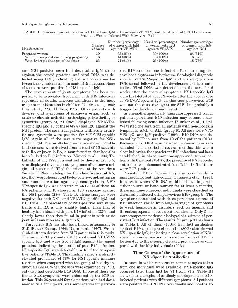
Parvovirus B19 is known to cause severe complica-tions during pregnancy due to intrauterine infection ofthe fetus, particularly during the second and third tri-mester of pregnancy [de Krijger et al., 1998; Essary etal., 1998]. Fetal infection may be followed by hydropicchanges due to the destruction of erythroid precursorcells in the fetal liver. We tested the sera of 40 preg-nant women (group 3) in whom diagnosis was madefollowing contact with infected persons or who pre-sented with typical symptoms associated with B19 in-
fections (exanthema, fever) or with sonographic abnor-malities of the fetus. In addition, other viral infectionssuch as cytomegalovirus (CMV), varicella zoster virus,and rubella, which are known to cause hydropicchanges, had been excluded by routine diagnosis exceptfor one woman who was acutely infected with CMV andnegative for parvovirus B19. VP1/VP2-specific IgG wasdetected in 39 sera (97%), 33 of the patients were posi-tive for VP1/VP2-specific IgM and 24 of the pregnantwomen displayed IgG against the NS1 protein (61%;Table I). NS1-specific IgM could not be detected in anyof the sera. In 23 women, hydropic changes of the fetuswere observed, e.g., hydrops fetalis, fetal ascites, peri-cardial effusion, or fetal encephalitis. All women withsonographic abnormalities had serological markers foran acute B19 infection, as either IgM or PCR or bothwere positive. NS1-specific IgG could be detected in 18of the 23 women with B19-associated hydropic changesin the fetus (78%). In 16 uncomplicated pregnanciesthe prevalence was significantly lower (37%), with only6 of these women displaying IgG against NS1 (TableII).
Infections with parvovirus B19 are frequently accom-panied by hemapoietic disorders such as anemia oraplastic anemia due to the destruction of erythroid pre-cursors in the bone marrow [Kurtzman et al., 1987].Severe thrombocytopenia and pancytopenia have alsobeen reported to be caused by B19 infection [Young,1988; Brown and Young, 1995]. We therefore includeda panel of 22 sera from patients with anemia, throm-bocytopenia, or pancytopenia in this study (group 4).Seven of these patients developed anemia-associatedliver dysfunctions due to the severe aplastic crisis [Yotoet al., 1994; Langnas et al., 1995]. The IgG results areshown in Table I. Of 21 patients (95%) who were foundto be positive for VP1/VP2-specific IgG, 10 (47%) hadan IgG response to NS1 protein. Five of the VP1/VP2-
TABLE I. Seroprevalence of Parvovirus B19 IgG in Healthy, Pregnant and Disease Groups
ManifestationNumber
of patients
Number (percentage)of patients with IgG
against VP1/VP2
Number (percentage)of patients with IgG
against NS1Group 1: Healthy population 153 123 (80%) 27 (22%)Group 2: Acute infection in immunocompetent
(asymptomatic, erythema infectiosum, flu-likesymptoms) 43 43 (100%) 5 (11%)
Group 3: Pregnant women with contact with B19-infectedpersons or displaying B19 associated symptoms(exanthema, flu-like, hydrops fetalis) 40 39 (97%) 24 (61%)
Group 4: Hematopoeitic disorders (anemia, aplastic crisis,thrombocytopenia, pancytopenia) 22 21 (95%) 10 (47%)
Group 5: Joint symptoms (acute and chronic arthritis,arthralgias, polyarthritis, synovitis) 22 21 (95%) 10 (47%)
Group 6: Rheumatoid arthritis, juvenile rheumatoidarthritis 66 46 (70%) 13 (28%)
Group 7: Systemic lupus erythematosus 42 34 (81%) 13 (38%)Group 8: Chronic infection in immunocompromised with
non-Hodgkin lymphoma, AML, or ALL 11 11 (100%) 6 (54%)Group 9: Chronic infection in immunocompetent (at least 6
months PCR positive) 5 5 (100%) 4 (80%)
AML, acute myeloid leukemia; ALL, acute lymphoid leukemia; PCR, polymerase chain reaction.Antibody reactivity was tested against viral structural (VP1/VP2) and nonstructural (NS1) proteins.
50 Hemauer et al.
and NS1-positive sera had detectable IgM titersagainst the capsid proteins, and viral DNA was de-tected using PCR, indicating a direct correlation be-tween the symptoms and an acute B19 infection. Noneof the sera were positive for NS1-specific IgM.
The involvement of joint symptoms has been re-ported to be associated frequently with B19 infectionsespecially in adults, whereas exanthema is the mostfrequent manifestation in children [Naides et al., 1990;Biasi et al., 1996; Phillips, 1997]. Of 22 patients withdiverse joint symptoms of unknown origin such asacute or chronic arthritis, arthralgia, polyarthritis, orsynovitis (group 5), 21 (95%) displayed VP1/VP2-specific IgG and 10 of these (47%) had IgG against theNS1 protein. The sera from patients with acute arthri-tis and synovitis were positive for VP1/VP2-specificIgM. Again all of the sera were negative for NS1-specific IgM. The results for group 6 are shown in TableI. These sera were derived from a total of 66 patientswith RA or juvenile RA, a manifestation that has alsobeen linked to B19 infection [Mimori et al., 1994; Ta-kahashi et al., 1998]. In contrast to those in group 5,who displayed diverse joint symptoms of unknown ori-gin, all patients fulfilled the criteria of the AmericanSociety of Rheumatology for the classification of RA,i.e., they were rheumatoid factor positive, indicating anautoimmune reaction against gamma globulin. VP1/VP2-specific IgG was detected in 46 (70%) of these 66RA patients and 13 showed an IgG response againstthe NS1 protein (28%; Table I). These samples werenegative for both NS1- and VP1/VP2-specific IgM andB19 DNA. The percentage of NS1-positive sera in pa-tients with RA is only slightly higher than that ofhealthy individuals with past B19 infection (22%) andclearly lower than that found in patients with acutejoint inflammation (47%, group 5).
Parvovirus B19 has also been linked occasionally toSLE [Fawaz-Estrup, 1996; Nigro et al., 1997]. We in-cluded 42 sera derived from SLE patients in this study.The sera of 34 patients (81%) contained VP1/VP2-specific IgG and were free of IgM against the capsidproteins, indicating the status of past B19 infection.NS1-specific IgG was detectable in 13 of the seroposi-tive patients (Table I). This finding reflects a slightlyelevated prevalence of 38% for NS1-specific immunereaction when compared with the group of healthy in-dividuals (22%). When the sera were examined by PCR,only two had detectable B19 DNA. In one of these pa-tients, SLE symptoms were enhanced by the B19 in-fection. This 26-year-old female patient, who had docu-mented SLE for 3 years, was seronegative for parvovi-
rus B19 and became infected after her daughterdeveloped erythema infectiosum. Serological diagnosisshowed VP1/VP2-specific IgM and a strong positivePCR signal followed by the development of IgG anti-bodies. Viral DNA was detectable in the sera for 6weeks after the onset of symptoms. NS1-specific IgGwere first detected about 3 weeks after the appearanceof VP1/VP2-specific IgG. In this case parvovirus B19was not the causative agent for SLE, but probably atrigger for the clinical manifestation.
In chemotherapeutically immunosuppressed tumorpatients, persistent B19 infection may become estab-lished following acute infection [Flunker et al., 1998].We tested the sera from 11 patients with non-Hodgkinlymphoma, AML, or ALL (group 8). All sera were VP1/VP2-IgG- and IgM-positive (100%). B19 DNA was de-tected by PCR in sera from 10 of the patients (91%).Because viral DNA was detected in consecutive serasampled over a period of several months, this was aclear indication that persistent B19 infections had beenestablished in these immunosuppressed tumor pa-tients. In 6 patients (54%), the presence of NS1-specificantibodies was demonstrated (see Table I); 5 of thesewere PCR positive.
Persistent B19 infections may also occur rarely inimmunocompetent individuals [Cassinotti et al., 1993].In cases in which B19 DNA could be shown to persisteither in sera or bone marrow for at least 6 months,these immunocompetent individuals were classified aschronically infected with parvovirus B19 (group 9). Thesymptoms associated with these persistent courses ofB19 infection varied from long-lasting joint symptomsto weak hemapoietic disorders such as anemia andthrombocytopenia or recurrent exanthema. Only 5 im-munocompetent patients displayed the criteria of per-sistent B19 infection. The results for group 9 are shownin Table I. All of them (100%) had IgG antibodiesagainst B19-capsid proteins and 4 (80%) also showedNS1-specific IgG, indicating a close correlation of NS1-specific immune reaction with chronic forms of B19 in-fection due to the strongly elevated prevalence as com-pared with healthy individuals (22%).
Time Course of the Appearance ofNS1-Specific Antibodies
In cases in which consecutive serum samples takenfrom one individual were available, NS1-specific IgGoccurred later than IgG for VP1 and VP2. Table IIIshows four examples of antibody development in B19-infected patients with different symptoms. All patientswere positive for B19 DNA over weeks and months af-
TABLE II. Seroprevalence of Parvovirus B19 IgG and IgM to Structural (VP1/VP2) and Nonstructural (NS1) Proteins inPregnant Women Infected With Parvovirus B19
ManifestationNumberof cases
Number (percentage)of women with IgM
against VP1/VP2
Number (percentage)of women with IgGagainst VP1/VP2
Number (percentage)of women with IgG
against NS1Pregnant women 39 33 (85%) 39 (100%) 24 (61%)
Without complications during pregnancy 16 14 (88%) 16 (100%) 6 (37%)With hydropic changes of the fetus 23 21 (91%) 23 (100%) 18 (78%)
NS1-Specific IgG in B19 Infections 51

ter infection as shown by PCR. Cases I and III areimmunocompetent subjects with no known underlyingdisease. Case II is an individual immunosuppresseddue to chemotherapy for ALL and case IV is a B19-infected women with hydropic complications duringpregnancy. In all cases for which consecutive sera wereavailable, NS1-specific IgG became detectable 4–6weeks after the first contact with the virus and cantherefore be detected about 2 weeks after the onset ofIgG against the capsid proteins. We were not able todemonstrate IgM against the nonstructural proteinand NS1-specific IgG was detectable only in combina-tion with IgG against the structural proteins VP1 andVP2. This clear connection to B19 infection demon-strates that the detection of NS1 antibodies is a specificreaction (Table III).
DISCUSSION
Parvovirus B19 may still be considered to belong tothe group of emerging viruses. During the past fewyears it has been shown that parvovirus B19 infectionsmay be associated with an increasingly varied panel ofrare, rather untypical symptoms in addition to themore commonly observed manifestations such as ery-thema infectiosum, hydrops fetalis, and aplastic ane-mia. Due to this diversity of symptoms related to in-fections with parvovirus B19, a reliable differential di-agnosis has become increasingly important. Earlierreports had shown that the presence of antibodiesagainst the nonstructural protein NS1 of parvovirusB19 could be linked to chronic or persistent forms ofinfection [von Poblotzki et al., 1995a, 1995b]. To exam-ine whether NS1-specific antibodies may also be usedas a marker for other courses of B19 infection, 250 serawere tested from patients with various symptoms forthe presence of NS1-specific IgG. These patients dis-played symptoms that are known to be associated fre-
quently or rarely with B19 infections. With respect tothe symptoms the patients were placed into ninegroups to analyze the prevalence of NS1-specific anti-bodies in relation to the various B19-associated dis-eases.
Before attempting to demonstrate an association be-tween the NS1-specific immune reaction and particu-lar B19-correlated symptoms in pregnancy and variousdisease states, a panel of sera derived from healthyindividuals was tested for the presence of NS1-specificIgG. The IgG seroprevalence for NS1 in the healthypopulation was 22%, which is in accordance with recentdata obtained by studying healthy blood donors usingWestern blots [Searle et al., 1998; Venturoli et al.,1998]. Von Poblotzki and et al. [1995a, 1995b] foundthe seroprevalence in the general population to belower. This finding may have been due to the largerserum panels tested in the more recent studies and tothe use of Western blots instead of a less sensitiveELISA. In addition it should be considered that thehealthy individuals tested in the current study weretested retrospectively and therefore the values can re-flect only the status of past B19 infection and not thecourse and the symptoms the patients actually had.Most of the healthy individuals were not able to recallthe event of the previous B19 infection or any of themanifestations of the disease. In comparison, the sero-prevalence of NS1-specific IgG was found to be elevatedin the groups with prolonged or persistent parvovirusB19 infections. These patients presented with arthritisand various joint symptoms, or hematopoietic disor-ders such as anemia with or without liver failure,thrombocytopenia, or pancytopenia. In all cases thesymptoms could be correlated with a previous B19 in-fection.
Prolonged viremia in infected individuals may leadto the infection of cells other than the usual erythroid
TABLE III. Occurrence of Antibodies to Parvovirus B19 Structural (VP1/VP2) and Nonstructural (NS1) Proteins FromFour Patients
Patient Symptoms
Serumdonation
dateVP2-specific
IgMVP1/VP2-specific
IgGNS1-specific
IgG PCRI Aplastic crisis associated with January 25 − − − +
liver dysfunction February 28 + + − +March 3 + + + +
II Immunosuppressed April 9 − − − −Acute lymphoblastic leukemia June 6 + + − +
July 5 − + (+) +July 26 − + + +
III Aplastic anemia January 11 + + − +January 25 + + − +February 2 + + (+) +March 3 + + + +April 24 + + + +October 7 − + + +December 13 − + + +
IV Pregnant women with April 24 + − − +hydrops fetalis May 8 + + − +
May 13 + + (+) +July 10 − + + −
Sera were donated on different dates and were also tested for B19 DNA by polymerase chain reaction (PCR).
52 Hemauer et al.

precursors. Such cells are unable to support a produc-tive infection cycle and therefore do not allow the pro-duction of viral particles. Gene expression in nonper-missive cells is shifted toward the preferential tran-scription of the NS1 gene without production of thecapsid proteins VP1 and VP2 [Liu et al., 1992; Pallieret al., 1997]. The NS1 protein has been shown to becytotoxic [Ozawa et al., 1988] and is able to the stimu-late apoptotic processes [Moffatt et al., 1998], resultingin cell lysis and the release of NS1 protein, a processthat may render this nonstructural viral componentaccessible to the immune response of the host. The in-fection of thrombocytes, reticulocytes, neutrophils, andother white blood cells is associated with symptomssuch as thrombocytopenia, neutropenia, or pancytope-nia due to the continuous destruction of these cells.That prolonged viral persistence in the individual maybe a precondition for the formation of NS1-specific an-tibodies as supported in this study by the fact thatthere is reduced incidence of IgG in acutely infectedpersons with erythema infectiosum compared with thehealthy population. In patients with acute B19 infec-tion, the seroprevalence of NS1-specific immune reac-tions (11%) is only half that of the group of healthyindividuals (22%) and exceptionally lower than thatobserved in patients with persistent B19 infections(Table I). This finding may be explained by the factthat the virus is not present long enough to stimulatethe production of NS1-specific antibodies. When test-ing consecutive sera derived from patients with chronicB19 infections it was shown that NS1-specific IgG oc-cur several weeks after IgG produced against the viralcapsid proteins (Table III). This finding supports thehypothesis that viral persistence or at least inefficientvirus elimination has to be established after acute in-fection before NS1 proteins are synthesized in elevatedconcentration and become accessible to the host’s im-mune system.
NS1-specific immune reactions were observed in 61%of the pregnant women infected with parvovirus B19.This observation may be due to a low level immuno-suppression generally found during pregnancy, whichmay lead consequently to ineffective virus elimination.With respect to the cases that simultaneously dis-played hydropic manifestations in the fetus, NS1-specific IgG was detectable in 78% of the women’s sera(Table II). This may also be used as an indication thatprolonged B19 persistence during pregnancy may en-hance the risk of fetal infection and disease. Becausefetal complications occur 2–4 weeks after maternal in-fection and antibodies against the NS1 protein firstbecome detectable around 4–6 weeks postinfection, wecannot exclude the possibility that this may contributeto the higher frequency of NS1 IgG detected in serafrom women with fetal complications. This hypothesisis supported by Searle et al. [1998], who did not findsignificant differences in the prevalence of NS1 IgG inpregnant women with and without fetal complications,but also detected a higher prevalence of NS1-specificantibodies in B19 infections during pregnancy.
Further investigations with a larger number of pa-tients will help to further elucidate this point.
Virus infections are associated commonly with ane-mia, but also with thrombocytopenia, neutropenia, andpancytopenia. NS1-specific antibodies were found in47% of patients with hematological disorders, which isabout double that observed in healthy controls (seeTable I). In addition to VP1/VP2- and NS1-specific IgG,five of the patients displayed IgM against the viral cap-sid proteins with simultaneous presence of B19 DNA.It may be concluded that these patients had been in-fected recently with parvovirus B19 and that the virushad not been eliminated from the organism althoughelevated IgG levels were found. As thrombocytes, re-ticulocytes, and neutrophils do not represent targetcells for productive B19 infection, it can also be con-cluded that in these cases nonpermissive cells had beeninfected due to prolonged viral presence in the hostfollowed by the synthesis of NS1 protein.
Of the patients with joint symptoms of unknown ori-gin, 47% showed an antibody reaction against the NS1protein, which is a significantly higher percentage thanreactions observed in healthy individuals or with ery-thema infectiosum (see Table I). Because not all of thepatients showed IgM or viral DNA in their sera, thesymptoms can be mediated either immunologically orthrough direct cytotoxic viral action in cases in whichthe virus is still present. B19 infections have beenlinked to a variety of autoimmune reactions [Soloninkaet al., 1989; Vigeant et al., 1994; Kerr and Boyd, 1996].The joint symptoms may be caused by immune com-plexes between antibodies and virus proteins or par-ticles present in the synovial fluid. These complexesmay evoke autoimmune reactions via complement ac-tivation or cytokine secretion by immunologically ac-tive cells. Furthermore, it cannot be excluded that dueto genomic variation B19 mutants exist that displayepitopes mimicking autoantigens and therefore inducean autoimmunological process [Lunardi et al., 1998].Alternatively, the joint destruction may also be causeddirectly by the B19 infection. Recent data indicate thatparvovirus B19 may persist in synovial membranes ofpersons with arthritic symptoms [Cassinotti et al.,1998; Stahl et al., 1998]. Therefore, it seems reasonablethat low levels of virus replication in this nonpermis-sive tissue associated with the production of NS1 pro-teins may contribute to an continuous cell destructionfollowed by inflammatory reactions.
In contrast, the prevalence of NS1-specific antibodiesin RA patients is only slightly elevated (28%) whencompared with healthy controls (22%) (see Table I). Inaddition, these patients did not display any serologicalmarkers indicating an acute B19 infection, becauseneither VP1/VP2-specific IgM nor viral DNA could bedetected. This result confirms recent findings that RAis not associated with acute or persistent parvovirusB19 infections [Kerr et al., 1996], although there arereports of individual cases in which an association be-tween RA and B19 infection has been described [Mi-mori et al., 1994; Takahashi et al., 1998]. Because
NS1-Specific IgG in B19 Infections 53
symptoms similar or identical to rheumatic manifesta-tions are common side effects of B19 infections, RA-enhancing mechanisms cannot be excluded. Similarmechanisms have been proposed for the developmentof SLE. Data describing an association of B19 infectionand SLE have been published [Fawaz-Estrup, 1996;Nigro et al., 1997] along with data that discount it[Nesher et al., 1995]. When testing sera derived fromSLE patients relative to the control groups, we de-tected a slightly increased prevalence of 38% of NS1-specific IgG (see Table I). In one case of SLE, however,the symptoms were enhanced when the patient wasinfected with parvovirus B19 [Hemauer et al., 1999].Viral DNA could be detected in the consecutive sera ofthis patient over 6 weeks, reflecting incomplete viruselimination, and IgM remained positive over the com-plete follow up time (10 months), indicating an ineffec-tive Ig-class switch.
The highest incidence of NS1-IgG-positive individu-als was found in the persistent B19 infections in im-munosuppressed (54%) and immunocompetent (80%)individuals (Table I). In all cases the presence of thevirus over several months was confirmed using PCReither in serum or bone marrow samples, indicatingthe clear correlation between the prolonged persistenceof parvovirus B19 and the formation of NS1-specificantibodies. These patients show a variety of B19-correlated symptoms such as chronic arthritis, chronicor recurring arthralgias, recurring exanthema, chronicanemia, thrombocytopenia, or pancytopenia. Further-more, we observed a strongly elevated prevalence ofNS1-specific IgG in pregnant women with complica-tions during pregnancy (see Table II).
In conclusion, elevated levels of NS1-specific anti-bodies were found in patients with distinct B19-correlated manifestations. This phenomenon was par-ticularly pronounced in persistent courses of B19 infec-tion, independent of the host’s immune status, inpatients with joint symptoms or hemapoietic disorders(see Table I), and in pregnant women with B19-associated manifestations of the fetus (see Table II).Although there is no correlation between the appear-ance of NS1-specific antibodies and particular clinicalmanifestations, NS1-specific immune reactions may beused as a serological marker for persistent B19 infec-tions. Because the number of patients tested in somegroups was relatively small due to the rarity of themanifestation, the data may not be significant in somecases.
ACKNOWLEDGMENTS
Andrea Hemauer has been supported by the Stud-ienstiftung des Deutschen Volkes, Ulla Raab is sup-ported by an HSP-III stipendium. The authors thankDr. Louwen, Universitatsklinik, Munster, Germany,for B19-positive sera from pregnant women and Dr. M.Motz (Mikrogen GmbH, Munich, Germany) for the gen-erous donation of Recomblots.
REFERENCES
Anderson MJ, Jones SE, Fisher-Hoch S-P, Lewis E, Hall SM, BartlettCL, Cohen BJ, Mortimer PP, Pereira MS. 1983. Human parvovi-rus the cause of erythema infectiosum (fifth disease)? Lancet 1:1378.
Anderson MJ, Khousam MN, Maxwell DJ, Gould SJ, Happerfield LC,Smith WJ. 1988. Human parvovirus B19 and hydrops fetalis. Lan-cet 1:535.
Biasi D, Zeminian S, Caramaschi P, Carletto A, Manzo T, BambaraLM. 1996. A case of parvovirus B19 adult acute arthritis withsome allergic disease clinical features. Clin Rheumatol 15:508–510.
Brown KE, Young NS. 1995. Parvovirus B19 infection and hemato-poeisis. Blood Rev 9:176–182.
Cassinotti P, Schultze D, Schlageter P, Chevili S, Siegl G. 1993. Per-sistent parvovirus B19 infection following an acute infection withmeningitis in an immunocompetent patient. Eur J Clin MicrobiolInfect Dis 12:701–704.
Cassinotti P, Siegl G, Michel BA, Bruhlmann P. 1998. Presence andsignificance of human parvovirus B19 DNA in synovial mem-branes and bone marrow from patients with arthritis of unknownorigin. J Med Virol 56:199–204.
Chorba T, Coccia P, Holman RC, Tattersall P, Anderson LJ, SudmanJ, Young NS, Kurcynski E, Saarinen UM, Moir R, Lawrence DN,Jason JM, Evatt B. 1986. The role of parvovirus B19 in aplasticcrisis and erythema infectiosum (fifth disease). J Infect Dis 154:383–393.
Cohen BJ, Buckley MM. 1988. The prevalence of antibody to humanparvovirus B19 in England and Wales. J Med Microbiol 25:151–153.
Cossart YE, Field AM, Cant B, Widdows D. 1975. Parvovirus likeparticles in human sera. Lancet 1:72–73.
de Krijger RR, van Elsacker-Niele AM, Mulder-Stapel A, SalimansMM, Dreef E, Weiland HT, van Krieken JH, Vermeij-Keers C.1998. Detection of parvovirus B19 infection in first and secondtrimester fetal loss. Pediatr Pathol Lab Med 18:23–24.
Enders G, Dotsch J, Bauer W, Nutzenadel W, Hengel H, Haffner D,Schalasta G, Searle K, Brown KE. 1998. Life-threatening parvo-virus B19-associated myocarditis and cardiac transplantation aspossible therapy: two case reports. Clin Infect Dis 26:355–358.
Essary LR, Vnencak-Jones CL, Manning SS, Olson SJ, Johnson JE.1998. Frequency of parvovirus B19 infection in nonimmune hy-drops fetalis and utility of three diagnostic methods. Hum Pathol29:696–701.
Fawaz-Estrup F. 1996. Human parvovirus infection: Rheumaticmanifestations, angioedema, C1 esterase inhibitor deficiency,ANA positivity and possible onset of systemic lupus erythemato-sus. J Rheumatol 23:1180–1185.
Flunker G, Peters A, Wiersbitzky S, Modrow S, Seidel W. 1998. Per-sistent parvovirus B19 infection in immunocompromised children.Med Microbiol Immunol Berlin 186:189–194.
Hemauer A, Beckenlehner K, Wolf H, Lang B, Modrow S. 1999. Acuteparvovirus B19 infection in connection with a flare of systemiclupus erythematodes in a female patient. J Clin Virol 14:73–77.
Hemauer A, von Poblotzki A, Gigler A, Cassinotti P, Siegl G, Wolf H,Modrow S. 1996. Sequence variability among different parvovirusB19 isolates. J Gen Virol 77:1781–1785.
Hillingso JG, Jensen IP, Tom-Petersen L. 1998. Parvovirus B19 andacute hepatitis in adults. Lancet 351:955–956.
Kerr JR, Boyd N. 1996. Autoantibodies following parvovirus B19 in-fection. J Infect 32:41–47.
Kerr JR, Cartron JP, Curran MD, Moore JE, Elliott JR, Mollan RA.1996. A study of the role of parvovirus B19 in rheumatoid arthri-tis. Br J Rheumatol 35:494.
Kurtzman GJ, Ozawa K, Cohen B, Hanson G, Oseas R, Young NS.1987. Chronic bone marrow failure due to persistent B19 parvo-virus infection. N Engl J Med 317:287–294.
Langnas AN, Markin RS, Cattral MS, Naides SJ. 1995. ParvovirusB19 as a possible causative agent of fulminant liver failure andassociated aplastic anemia. Hepatology 22:1661–1665.
Liu JM, Green SW, Shimada T, Young NS. 1992. A block in full lengthtranscript maturation in cells nonpermissive for B19 parvovirus. JVirol 66:4686–4692.
Lunardi C, Tiso M, Borgato L, Nanni L, Millo R, da Sandre G,Bargellesi-Severi A, Puccetti A. 1998. Chronic parvovirus B19 in-
54 Hemauer et al.

fection induces the production of anti-virus antibodies with auto-antigen binding properties. Eur J Immunol 28:936–948.
Mimori A, Misaki Y, Hachiya T, Ito K, Kano S. 1994. Prevalence ofantihuman parvovirus B19 IgG antibodies in patients with refrac-tory rheumatoid arthritis and polyarticular juvenile rheumatoidarthritis. Rheumatol Int 14:87–90.
Moffatt S, Yaegashi N, Tada K, Tanaka N, Sugamura K. 1998. Hu-man parvovirus B19 nonstructural protein induces apoptosis inerythroid lineage cells. J Virol 72:3018–3028.
Naides SJ, Scharosch LL, Foto F, Howard EJ. 1990. Rheumatologicalmanifestations of human Parvovirus B19 infection in adults. Ar-thritis Rheum 33:1297–1309.
Nesher G, Osborn TG, Moore TL. 1995. Parvovirus infection mimick-ing systemic lupus erythematosus. Semin Arthritis Rheum 24:297–303.
Nigro G, Piazze J, Taliani G, Mazzocco M, Cassinotti P, Cosmi EV.1997. Postpartum lupus erythematosus associated with parvovi-rus B19 infection. J Rheumatol 24:968–970.
Orth T, Herr W, Spahn T, Voigtlander T, Michel D, Mertens T, MayetWJ, Dippold W, Meyer zum Buschenfelde, K-H. 1997. Human par-vovirus B19 infection associated with severe acute perimyocarditisin a 34-year old man. Eur Heart J 18:524–525.
Ozawa K, Ayub J, Kajigaya S, Shimada T, Young NS. 1988. The geneencoding the nonstructural protein of B19 (human) parvovirusmay be lethal in transfected cells. J Virol 62:2884–2889.
Pallier C, Greco A, Le Junter J, Saib A, Vassias I, Morinet F. 1997.The 38 untranslated region of the B19 parvovirus capsid proteinmRNAs inhibits its own mRNA translation in nonpermissive cells.J Virol 71:9482–9489.
Pardi DS, Romero Y, Mertz LE, Douglas DD. 1998. Hepatitis-associated aplastic anemia and acute parvovirus B19 infection: areport of two cases and a review of the literature. Am J Gastro-enterol 93:468–470.
Phillips PE. 1997. Viral arthritis. Curr Opin Rheumatol 9:337–344.Reid DM, Reid TM, Brown T, Rennie JA, Eastmond C. 1985. Human
parvovirus-associated arthritis: a clinical and laboratory descrip-tion. Lancet 1:422–425.
Searle K, Schalasta G, Enders G. 1998. Development of antibodies tothe nonstructural protein NS1 of Parvovirus B19 during acutesymptomatic and subclinical infection in pregnancy: implicationsfor pathogenesis doubtful. J Med Virol 56:192–198.
Shade RO, Blundell MC, Cotmore SF, Tattersall P, Astell CR. 1986.Nucleotide sequence and genome organisation of human parvovi-rus B19 isolated from the serum of a child during aplastic crisis. JVirol 58:921–936.
Soloninka CA, Anderson MJ, Laskin CA. 1989. Anti-DNA and anti-lymphocyte antibodies during acute infection with human parvo-virus B19. J Rheumatol 16:777–781.
Srivastava A, Bruno E, Bridell R, Cooper R, Srivastave C, Van BesienK, Hoffman R. 1990. Parvovirus B19-induced perturbation of hu-man megakaryocytopoeisis in vitro. Blood 76:1997–2004.
Stahl HD, Seidel B, Hubner B, Altrichter S, Pfeiffer R, Pustoweit B,Jungmichel D, Emmrich F. 1998. High incidence of parvovirusB19 in synovial membranes of patients with undifferentiatedmono- and oligoarthritis. Arthritis Rheum 41S:1657.
Takahashi Y, Murai C, Shibata S, Munakata Y, Ishii T, Ishii K, SaitohT, Sawai T, Sugamura K, Sasaki T. 1998. Human parvovirus B19as a causative agent for rheumatoid arthritis. Proc Natl Acad SciUSA 95:8227–8232.
Umene K, Nuone T. 1995. A new genome type of human parvovirusB19 present in sera of patients with encephalopathy. J Gen Virol76:2645–2651.
Venturoli, S, Gallinella G, Manatesi E, Gentilomi G, Musiani M, Zer-bini M. 1998. IgG response to the immunoreactive region of par-vovirus B19 nonstructural protein by immunoblot assay with arecombinant antigen. J Infect Dis 178:1826–1829.
Vigeant P, Menard H, Boire G. 1994. Chronic modulation of the au-toimmune response following parvovirus B19 infection. J Rheu-matol 21:1165–1167.
von Poblotzki A, Gigler A, Lang B, Wolf H, Modrow S. 1995a. Anti-bodies to parvovirus B19 NS-1 protein in infected individuals. JGen Virol 76:519–527.
von Poblotzki A, Hemauer A, Gigler A, Puchhammer-Stockl E, HeinzFX, Pont J, Wolf H, Modrow S. 1995b. Antibodies to the nonstruc-tural protein of parvovirus B19 in persistently infected patients:implications for pathogenesis. J Infect Dis 172:1356–1359.
Yoto Y, Kudoh T, Asanuma H, Numazaki K, Tsutsumi Y, Nakata S,Chiba S. 1994. Transient disturbance of consciousness and hepaticdysfunction associated with human parvovirus B19 infection. Lan-cet 344:624–625.
Young N. 1988. Hematological and hematopoeitic consequences ofB19 parvovirus infection. Semin Hematol 25:159–172.
NS1-Specific IgG in B19 Infections 55