
SERCA pump activity is physiologically regulated by ...parkerlab.bio.uci.edu/Ian S...
10
THE JOURNAL OF CELL BIOLOGY JCB: ARTICLE © 2008 Green et al. The Rockefeller University Press $30.00 J. Cell Biol. Vol. 181 No. 7 1107–1116 www.jcb.org/cgi/doi/10.1083/jcb.200706171 JCB 1107 K.N. Green and A. Demuro contributed equally to this paper. Correspondence to Frank M. LaFerla: [email protected] Abbreviations used in this paper: A , amyloid ; AD, Alzheimer’s disease; AICD, APP intracellular domain; APP, amyloid precursor protein; CCE, capacita- tive Ca 2+ entrance; FAD, familial AD; IP 3 , inositol 1,4,5-trisphosphate; MEF, mouse embryonic fibroblast; nAChR, nicotinic acetylcholine receptor; pcDNA, pseudo-cDNA; PSDKO, presenilin double knockout; SERCA, sarco ER Ca 2+ -ATPase. The online version of this paper contains supplemental material. Introduction PS1 and PS2 are highly conserved integral membranous pro- teins that localize predominantly to the ER. Mutations in the PS1 and PS2 genes that cause autosomal-dominant early- onset Alzheimer’ s disease (AD) disrupt several cellular path- ways, including altered -secretase–mediated cleavage of the amyloid precursor protein (APP) to form amyloid (A ) pep- tides (Duff et al., 1996) and disruption of intracellular Ca 2+ homeostasis (LaFerla, 2002; Demuro et al., 2005). Ca 2+ sig- naling disruptions manifest as enhanced filling of ER Ca 2+ stores (Leissring et al., 1999b), attenuation of capacitive Ca 2+ entry stores (Leissring et al., 2000; Yoo et al., 2000; Smith et al., 2002; Herms et al., 2003), and by exaggerated liberation of Ca 2+ from the ER by the second messenger inositol 1,4,5-tris- phosphate (IP 3 ; Leissring et al., 1999b; Yoo et al., 2000; Smith et al., 2002; Stutzmann et al., 2004). Given that mutations in presenilin disrupt intracellular Ca 2+ signaling, we set out to determine whether presenilins may serve a physiological role in intracellular Ca 2+ homeostasis. In support of a role in Ca 2+ homeostasis, overexpression of wild-type PS1 or PS2 in Xenopus laevis oocytes causes en- hanced IP 3 -mediated Ca 2+ release, an effect that is exacerbated by mutations in both genes (Leissring et al., 1999b). However, it remains unclear whether the exaggerated IP 3 -evoked responses result from modulation of the IP 3 signaling pathway, such as sensitization of IP 3 receptors by presenilins, or as a consequence of overfilling of ER stores. Recently, the presenilins have been reported to be able to form ER leak channels, and it has been re- ported that mutations in the presenilins disrupt this function (Tu et al., 2006). However, it is unclear how leak channel formation could account for the numerous reports of wild-type presenilin overexpression increasing IP 3 -mediated calcium release. Ca 2+ pumps, along with Ca 2+ release channels, are the key components of Ca 2+ regulatory systems in neuronal and non- neuronal cells (Berridge et al., 2000). The sarco ER Ca 2+ - ATPase (SERCA) pumps have the highest affinity for Ca 2+ removal from the cytosol and, together with plasma membrane Ca 2+ -ATPases and transporters, determine the resting cytosolic Ca 2+ concentration. Three differentially expressed genes encode at least five isoforms of the SERCA pump. SERCA1a and -1b are expressed in skeletal muscle, whereas SERCA2a is ex- pressed in cardiac muscle (Aubier and Viires, 1998). SERCA2b, which has a C-terminal extension, is ubiquitously expressed in smooth muscle tissues and nonmuscle tissues including neurons (Baba-Aissa et al., 1998). SERCA3 has limited expression in various nonmuscle tissues (Baba-Aissa et al., 1998). I n addition to disrupting the regulated intramembraneous proteolysis of key substrates, mutations in the presenil- ins also alter calcium homeostasis, but the mechanism linking presenilins and calcium regulation is unresolved. At rest, cytosolic Ca 2+ is maintained at low levels by pump- ing Ca 2+ into stores in the endoplasmic reticulum (ER) via the sarco ER Ca 2+ -ATPase (SERCA) pumps. We show that SERCA activity is diminished in fibroblasts lacking both PS1 and PS2 genes, despite elevated SERCA2b steady- state levels, and we show that presenilins and SERCA physically interact. Enhancing presenilin levels in Xenopus laevis oocytes accelerates clearance of cytosolic Ca 2+ , whereas higher levels of SERCA2b phenocopy PS1 over- expression, accelerating Ca 2+ clearance and exaggerat- ing inositol 1,4,5-trisphosphate–mediated Ca 2+ liberation. The critical role that SERCA2b plays in the pathogenesis of Alzheimer’s disease is underscored by our findings that modulating SERCA activity alters amyloid production. Our results point to a physiological role for the presenilins in Ca 2+ signaling via regulation of the SERCA pump. SERCA pump activity is physiologically regulated by presenilin and regulates amyloid production Kim N. Green, Angelo Demuro, Yama Akbari, Brian D. Hitt, Ian F. Smith, Ian Parker , and Frank M. LaFerla Department of Neurobiology and Behavior, University of California, Irvine, Irvine, CA 92697 on July 1, 2008 www.jcb.org Downloaded from Published June 30, 2008 http://www.jcb.org/cgi/content/full/jcb.200706171/DC1 Supplemental Material can be found at:
Transcript of SERCA pump activity is physiologically regulated by ...parkerlab.bio.uci.edu/Ian S...

TH
EJ
OU
RN
AL
OF
CE
LL
BIO
LO
GY
JCB: ARTICLE
© 2008 Green et al. The Rockefeller University Press $30.00J. Cell Biol. Vol. 181 No. 7 1107–1116www.jcb.org/cgi/doi/10.1083/jcb.200706171 JCB 1107
K.N. Green and A. Demuro contributed equally to this paper.
Correspondence to Frank M. LaFerla: [email protected]
Abbreviations used in this paper: A � , amyloid � ; AD, Alzheimer ’ s disease; AICD, APP intracellular domain; APP, amyloid precursor protein; CCE, capacita-tive Ca 2+ entrance; FAD, familial AD; IP 3 , inositol 1,4,5-trisphosphate; MEF, mouse embryonic fi broblast; nAChR, nicotinic acetylcholine receptor; pcDNA, pseudo-cDNA; PSDKO, presenilin double knockout; SERCA, sarco ER Ca 2+ -ATPase.
The online version of this paper contains supplemental material.
Introduction PS1 and PS2 are highly conserved integral membranous pro-
teins that localize predominantly to the ER. Mutations in the
PS1 and PS2 genes that cause autosomal-dominant early-
onset Alzheimer ’ s disease (AD) disrupt several cellular path-
ways, including altered � -secretase – mediated cleavage of the
amyloid precursor protein (APP) to form amyloid � (A � ) pep-
tides ( Duff et al., 1996 ) and disruption of intracellular Ca 2+
homeostasis ( LaFerla, 2002 ; Demuro et al., 2005 ). Ca 2+ sig-
naling disruptions manifest as enhanced fi lling of ER Ca 2+
stores ( Leissring et al., 1999b ), attenuation of capacitive Ca 2+
entry stores ( Leissring et al., 2000 ; Yoo et al., 2000 ; Smith
et al., 2002 ; Herms et al., 2003 ), and by exaggerated liberation of
Ca 2+ from the ER by the second messenger inositol 1,4,5-tris-
phosphate (IP 3 ; Leissring et al., 1999b ; Yoo et al., 2000 ; Smith
et al., 2002 ; Stutzmann et al., 2004 ). Given that mutations in
presenilin disrupt intracellular Ca 2+ signaling, we set out to
determine whether presenilins may serve a physiological role
in intracellular Ca 2+ homeostasis.
In support of a role in Ca 2+ homeostasis, overexpression of
wild-type PS1 or PS2 in Xenopus laevis oocytes causes en-
hanced IP 3 -mediated Ca 2+ release, an effect that is exacerbated
by mutations in both genes ( Leissring et al., 1999b ). However, it
remains unclear whether the exaggerated IP 3 -evoked responses
result from modulation of the IP 3 signaling pathway, such as
sensitization of IP 3 receptors by presenilins, or as a consequence
of overfi lling of ER stores. Recently, the presenilins have been
reported to be able to form ER leak channels, and it has been re-
ported that mutations in the presenilins disrupt this function ( Tu
et al., 2006 ). However, it is unclear how leak channel formation
could account for the numerous reports of wild-type presenilin
overexpression increasing IP 3 -mediated calcium release.
Ca 2+ pumps, along with Ca 2+ release channels, are the key
components of Ca 2+ regulatory systems in neuronal and non-
neuronal cells ( Berridge et al., 2000 ). The sarco ER Ca 2+ -
ATPase (SERCA) pumps have the highest affi nity for Ca 2+
removal from the cytosol and, together with plasma membrane
Ca 2+ -ATPases and transporters, determine the resting cytosolic
Ca 2+ concentration. Three differentially expressed genes encode
at least fi ve isoforms of the SERCA pump. SERCA1a and -1b
are expressed in skeletal muscle, whereas SERCA2a is ex-
pressed in cardiac muscle ( Aubier and Viires, 1998 ). SERCA2b,
which has a C-terminal extension, is ubiquitously expressed in
smooth muscle tissues and nonmuscle tissues including neurons
( Baba-Aissa et al., 1998 ). SERCA3 has limited expression in
various nonmuscle tissues ( Baba-Aissa et al., 1998 ).
In addition to disrupting the regulated intramembraneous
proteolysis of key substrates, mutations in the presenil-
ins also alter calcium homeostasis, but the mechanism
linking presenilins and calcium regulation is unresolved.
At rest, cytosolic Ca 2+ is maintained at low levels by pump-
ing Ca 2+ into stores in the endoplasmic reticulum (ER) via
the sarco ER Ca 2+ -ATPase (SERCA) pumps. We show that
SERCA activity is diminished in fi broblasts lacking both
PS1 and PS2 genes, despite elevated SERCA2b steady-
state levels, and we show that presenilins and SERCA
physically interact. Enhancing presenilin levels in Xenopus
laevis oocytes accelerates clearance of cytosolic Ca 2+ ,
whereas higher levels of SERCA2b phenocopy PS1 over-
expression, accelerating Ca 2+ clearance and exaggerat-
ing inositol 1,4,5-trisphosphate – mediated Ca 2+ liberation.
The critical role that SERCA2b plays in the pathogenesis
of Alzheimer ’ s disease is underscored by our fi ndings that
modulating SERCA activity alters amyloid � production.
Our results point to a physiological role for the presenilins
in Ca 2+ signaling via regulation of the SERCA pump.
SERCA pump activity is physiologically regulated by presenilin and regulates amyloid � production
Kim N. Green , Angelo Demuro , Yama Akbari , Brian D. Hitt , Ian F. Smith , Ian Parker , and Frank M. LaFerla
Department of Neurobiology and Behavior, University of California, Irvine, Irvine, CA 92697
on July 1, 2008 w
ww
.jcb.orgD
ownloaded from
Published June 30, 2008
http://www.jcb.org/cgi/content/full/jcb.200706171/DC1Supplemental Material can be found at:
JCB • VOLUME 181 • NUMBER 7 • 2008 1108
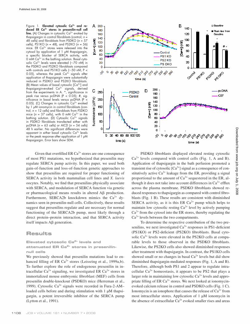
PSDKO fi broblasts displayed elevated resting cytosolic
Ca 2+ levels compared with control cells ( Fig. 1, A and B ).
Application of thapsigargin in the bath perfusion promoted a
transient rise of cytosolic [Ca 2+ ] signal as a consequence of con-
stitutively active Ca 2+ leakage from the ER, providing a signal
proportional to the amount of Ca 2+ sequestered in the ER, al-
though it does not take into account differences in Ca 2+ effl ux
across the plasma membrane. PSDKO fi broblasts showed re-
duced responses to thapsigargin as compared with control fi bro-
blasts ( Fig. 1 B ). These results are consistent with diminished
SERCA activity, as it is this ER Ca 2+ pump which helps to
maintain low cytosolic resting Ca 2+ level by actively pumping
Ca 2+ from the cytosol into the ER stores, thereby regulating the
Ca 2+ levels between the two compartments.
To determine the respective contribution of the two pre-
senilins, we next investigated Ca 2+ responses in PS1-defi cient
(PS1KO) or PS2-defi cient (PS2KO) fi broblasts. Basal cyto-
solic Ca 2+ levels were elevated in the PS2KO cells at compa-
rable levels to those observed in the PSDKO fi broblasts.
Likewise, the PS2KO cells also showed diminished responses
after treatment with thapsigargin. In contrast, the PS1KO cells
showed small or no changes in basal Ca 2+ levels but did show
diminished thapsigargin-mediated responses ( Fig. 1, A and B ).
Therefore, although both PS1 and 2 appear to regulate intra-
cellular Ca 2+ homeostasis, it appears to be PS2 that plays a
larger role in maintaining low cytosolic Ca 2+ levels and appro-
priate fi lling of ER Ca 2+ stores. We next looked at ionomycin-
evoked calcium release in control and PSDKO cells ( Fig. 1 C ).
Ionomycin is an ionophore that causes the release of Ca 2+ from
most intracellular stores. Application of 1 μ M ionomycin in
the absence of extracellular Ca 2+ evoked smaller rises and areas
Given that overfi lled ER Ca 2+ stores are one consequence
of most PS1 mutations, we hypothesized that presenilin may
regulate SERCA pump activity. In this paper, we used both
gain-of-function and loss-of-function genetic approaches to
show that presenilins are required for proper functioning of
SERCA activity in both mammalian cell lines and X. laevis
oocytes. Notably, we fi nd that presenilins physically associate
with SERCA, and modulation of SERCA function via genetic
or pharmacological means results in altered A � production.
Furthermore, SERCA2b knockdown mimics the Ca 2+ dy-
namics seen in presenilin-null cells. Collectively, these results
suggest that presenilins regulate and are necessary for normal
functioning of the SERCA2b pump, most likely through a
direct protein – protein interaction, and that SERCA activity
itself impacts A � generation.
Results Elevated cytosolic Ca 2+ levels and attenuated ER Ca 2+ stores in presenilin-null cells We previously showed that presenilin mutations lead to en-
hanced fi lling of ER Ca 2+ stores ( Leissring et al., 1999a , b ).
To further explore the role of endogenous presenilin in in-
tracellular Ca 2+ signaling, we investigated ER Ca 2+ stores in
immortalized mouse embryonic fi broblast (MEF) cells from
presenilin double-knockout (PSDKO) mice ( Herreman et al.,
1999 ). Cytosolic Ca 2+ signals were recorded in Fura-2-AM –
loaded cells before and during stimulation with 1 μ M thapsi-
gargin, a potent irreversible inhibitor of the SERCA pump
( Lytton et al., 1991 ).
Figure 1. Elevated cytosolic Ca 2+ and re-duced ER Ca 2+ stores in presenilin - null cell line. (A) Changes in cytosolic Ca 2+ evoked by thapsigargin in control fi broblasts (control; n = 48 cells) and fi broblasts from PSDKO ( n = 57 cells), PS1KO ( n = 48), and PS2KO ( n = 56) mice. ER Ca 2+ stores were released into the cytosol by application of 1 μ M thapsigargin, a specifi c blocker of SERCA activity, with 0 mM Ca 2+ in the bathing solution. Basal cyto-solic Ca 2+ levels were elevated ( � 70 nM) in the PSDKO and PS2KO fi broblasts compared with controls and PS1KO cells ( � 50 nM; P < 0.05), whereas the peak Ca 2+ signals after application of thapsigargin were substantially reduced in PSDKO and PS2KO fi broblasts. (B) Mean values of basal cytosolic [Ca 2+ ] and thapsigargin-evoked Ca 2+ signals, derived from the experiments in A. *, signifi cance in peak rise versus pcDNA (P < 0.05); #, sig-nifi cance in basal levels versus pcDNA (P < 0.05). (C) Changes in cytosolic Ca 2+ evoked by 1 μ M ionomycin in control fi broblasts (con-trol; n = 12 cells) and fi broblasts from PSDKO mice ( n = 37 cells), with 0 mM Ca 2+ in the bathing solution. (D) Cytosolic Ca 2+ signals in PSDKO fi broblasts transfected either with pcDNA ( n = 63 cells) or AICD ( n = 54 cells) 48 h earlier. No signifi cant differences were apparent in either basal cytosolic Ca 2+ levels or the peak response after application of 1 μ M thapsigargin. Error bars show SEM.
on July 1, 2008 w
ww
.jcb.orgD
ownloaded from
Published June 30, 2008
1109 PRESENILINS REGULATE SERCA PUMP ACTIVITY • Green et al.
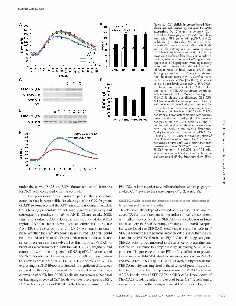
PS1, PS2, or both together rescued both the basal and thapsigargin-
evoked Ca 2+ levels to the same degree ( Fig. 2, A and B ).
SERCA2b steady-state levels are elevated in presenilin-null cells The observed phenotype of elevated basal cytosolic Ca 2+ and re-
duced ER Ca 2+ store content in presenilin-null cells is consistent
with either reduced levels of SERCA2b or a reduction in func-
tional activity of SERCA pumps ( Zhang et al., 2006 ). Surpris-
ingly, we found that SERCA2b steady-state levels, the isoform of
SERCA found in brain neurons, were elevated, rather than dimin-
ished, in the PSDKO fi broblasts ( Fig. 2, C and E ), suggesting that
SERCA activity was impaired in the absence of presenilins and
that the cells attempt to compensate by increasing SERCA ex-
pression. The presence of either PS1 or 2 is suffi cient to prevent
this increase in SERCA2b steady-state levels as shown in PS1KO
and PS2KO cell lines ( Fig. 2, D and E ). Given our hypothesis that
SERCA activity was impaired in the absence of presenilin, we at-
tempted to mimic the Ca 2+ phenotype seen in PSDKO cells via
siRNA knockdown of SERCA2b in CHO cells. Knockdown of
SERCA2b levels resulted in elevated basal Ca 2+ levels, and a
marked decrease in thapsigargin evoked Ca 2+ release ( Fig. 2 F ).
under the curve (9,425 vs. 7,394 fl uorescent units) from the
PSDKO cells compared with the controls.
The presenilins are an integral part of the � -secretase
complex that is responsible for cleavage of the C99 fragment
of APP to form A � and the APP intracellular domain (AICD).
Cells lacking presenilins do not have � -secretase activity and
consequently produce no A � or AICD ( Zhang et al., 2000 ;
Hass and Yankner, 2005 ). Because the absence of the AICD
region of APP has been shown to cause defi cits in Ca 2+ release
from ER stores ( Leissring et al., 2002 ), we sought to deter-
mine whether the Ca 2+ dyshomeostasis in PSDKO cells could
be attributed to lack of AICD production rather than to the ab-
sence of presenilins themselves. For this purpose, PSDKO fi -
broblasts were transfected with the AICD (C57) fragment and
compared with control pseudo-cDNA (pcDNA) – transfected
PSDKO fi broblasts. However, even after 48 h of incubation
to allow expression of AICD ( Fig. 1 D ), control and AICD-
expressing PSDKO fi broblasts showed no signifi cant differences
in basal or thapsigargin-evoked Ca 2+ levels. Given that over-
expression of AICD into PSDKO cells did not recover either basal
or thapsigargin-evoked Ca 2+ levels, we then overexpressed PS1,
PS2, or both together in PSDKO cells. Overexpression of either
Figure 2. Ca 2+ defi cits in presenilin-null fi bro-blasts are not caused by reduced SERCA2b expression. (A) Changes in cytosolic Ca 2+ evoked by thapsigargin in PSDKO fi broblasts transfected 48 h earlier with pcDNA ( n = 25 cells), PS1 ( n = 40 cells), PS2 ( n = 48 cells), or both PS1 and 2 ( n = 47 cells), with 0 mM Ca 2+ in the bathing solution. Basal cytosolic Ca 2+ levels were reduced ( � 20 nM) in the presenilin-transfected fi broblasts compared with controls, whereas the peak Ca 2+ signals after application of thapsigargin were signifi cantly increased in presenilin-transfected fi broblasts. (B) Mean values of basal cytosolic Ca 2+ and thapsigargin-evoked Ca 2+ signals, derived from the experiments in A. *, signifi cance in peak rise versus pcDNA (P < 0.05); #, signifi -cance in basal levels versus pcDNA (P < 0.05). (C) Steady-state levels of SERCA2b protein are higher in PSDKO fi broblasts compared with controls based on Western blotting. The PSDKO fi broblasts also displayed C83 – C99 APP fragments that were not present in the con-trols because of the lack of � -secretase activity. � -Actin levels are shown as a loading control. (D) Steady-state levels of SERCA2b in PS1KO and PS2KO fi broblasts compared with controls based on Western blotting. (E) Densitometric analysis of the SERCA2b levels in C and D, normalized to � -actin, showing elevation of SERCA2b levels in the PSDKO fi broblasts. *, signifi cance in peak rise versus pcDNA (P < 0.05; n = 3). (F) Genetic down-regulation of SERCA2b expression lowers ER Ca 2+ stores and elevates basal Ca 2+ levels. siRNA-mediated down-regulation of SERCA2b leads to lower ER Ca 2+ stores (*, P < 0.001; n = 105 cells) when compared with cells treated with a con-trol (scrambled) siRNA. Error bars show SEM.
on July 1, 2008 w
ww
.jcb.orgD
ownloaded from
Published June 30, 2008
JCB • VOLUME 181 • NUMBER 7 • 2008 1110
expression system to monitor the clearance of Ca 2+ ions from the
cytosol after a transient infl ux across the plasma membrane. For
this purpose, oocytes were induced to express the Ca 2+ -permeable
nicotinic acetylcholine receptor (nAChR) that served as a “ Ca 2+
switch, ” allowing precisely controlled cytosolic Ca 2+ transients to
be evoked by pulsing the membrane potential to strongly negative
voltages to increase the electrochemical driving force for Ca 2+
entry. Oocytes were loaded with the Ca 2+ -sensitive dye Oregon
Green BAPTA 1 and were voltage-clamped at a holding potential
of 0 mV to minimize Ca 2+ infl ux. In the presence of 100 – 500 nM
acetylcholine, a brief (300 ms) hyperpolarizing pulse to � 150 mV
produced a transient Ca 2+ fl uorescence signal because of Ca 2+
This phenotype was remarkably similar to that seen in the PS-
DKO cell line, giving further credence to presenilin regulation of
SERCA function. It should be noted that it is unlikely that changes
in SERCA function directly modulate basal cytosolic Ca 2+ levels
but that they probably do so through changes in other Ca 2+ infl ux
pathways such as store-operated Ca 2+ entry.
PS1 and PS2 mimic SERCA2b-accelerated cytosolic Ca 2+ clearance Based on these data from presenilin-null cell lines, we moved to a
more regulatable system to directly establish whether presenilins
modulate SERCA pump activity. We used the X. laevis oocyte
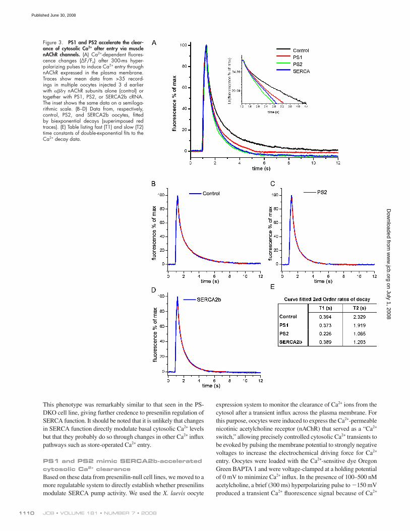
Figure 3. PS1 and PS2 accelerate the clear-ance of cytosolic Ca 2+ after entry via muscle nAChR channels. (A) Ca 2+ -dependent fl uores-cence changes ( Δ F/F o ) after 300-ms hyper-polarizing pulses to induce Ca 2+ entry through nAChR expressed in the plasma membrane. Traces show mean data from > 35 record-ings in multiple oocytes injected 3 d earlier with � � � � nAChR subunits alone (control) or together with PS1, PS2, or SERCA2b cRNA. The inset shows the same data on a semiloga-rithmic scale. (B – D) Data from, respectively, control, PS2, and SERCA2b oocytes, fi tted by biexponential decays (superimposed red traces). (E) Table listing fast (T1) and slow (T2) time constants of double-exponential fi ts to the Ca 2+ decay data.
on July 1, 2008 w
ww
.jcb.orgD
ownloaded from
Published June 30, 2008

1111 PRESENILINS REGULATE SERCA PUMP ACTIVITY • Green et al.
Ca 2+ infl ux across the plasma membrane, with PS2 having the
most robust effect ( Fig. 3 ). To prove that overexpression of
presenilin was accelerating Ca 2+ clearance from the cytosol by
increasing SERCA activity, we measured Ca 2+ clearance in the
presence of thapsigargin, a specifi c inhibitor of SERCA. If our
hypothesis was true, then presenilin should no longer acceler-
ate clearance of Ca 2+ from the cytosol in the presence of thap-
sigargin, compared with control oocytes also in the presence of
thapsigargin. We incubated control and PS2-expressing oo-
cytes in 30 μ M thapsigargin in the bathing solution for 30 min.
After a 30-min incubation, we applied a 300-ms hyperpolariza-
tion pulse, as before, to allow a controlled infl ux of Ca 2+ across
the plasma membrane into the cytosol and then tracked the
clearance of this Ca 2+ into the intracellular stores. In both con-
trol and PS2-expressing oocytes, thapsigargin reduced the
speed of the Ca 2+ fl uorescence decay, with PS2-expressing
oocytes showing the strongest effect, which is consistent with
impairment of PS2 modulation of SERCA activity ( Fig. 4, A
and B ). Double-exponential curve fi tting revealed that both
control and PS2-expressing oocytes had similar � 1 and � 2
values, despite PS2-overexpressing oocytes having markedly
faster � 2 values compared with control oocytes in the absence
of thapsigargin ( Fig. 4 C ).
PS1 familial AD (FAD) mutation M146V accelerates cytosolic Ca 2+ sequestration Mutations in the presenilins have been associated with en-
hanced IP 3 -mediated Ca 2+ release from oocytes ( Leissring et al.,
1999a , b ) and a variety of mammalian cell types ( Smith et al.,
2002 ). We have showed that wild-type presenilins accelerate
Ca 2+ sequestration from the cytosol in a thapsigargin-sensitive
pathway and that overexpression of SERCA phenocopies this.
We expressed the presenilin FAD mutant PS1M146V in X. laevis
infl ux through open nicotinic channels. The decay rate of the
fl uorescence signals after termination of the voltage pulse was
then used to quantify the rate of Ca 2+ sequestration from the cy-
tosol. For video footage of Ca 2+ entry and subsequent clearance
from the cytosol in this system please see Video 1 (available at
http://www.jcb.org/cgi/content/full/jcb.200706171/DC1).
Fig. 3 A shows mean traces of Oregon Green fl uorescence,
illustrating differences in the decay rate of the Ca 2+ signal in
control, PS1-, PS2- and SERCA2b-expressing oocytes. The de-
cay presumably refl ects a summation of several factors (e.g.,
diffusion of Ca 2+ ions into the interior of the oocyte, mito-
chondrial uptake, and extrusion across the plasma membrane)
in addition to sequestration into the ER by SERCA pumps.
Consistent with this, decay kinetics were best fi t by double-
exponential processes ( Fig. 2, B – D ), with time constants of a
few hundred milliseconds and a few seconds ( Fig. 2 E ). Seques-
tration by SERCA pumps is expected to be refl ected primarily
in the slower component ( � 2) and, in agreement with this inter-
pretation, � 2 was accelerated almost twofold in SERCA2b-
overexpressing oocytes as compared with control cells.
The acceleration of Ca 2+ sequestration was even greater in
oocytes expressing PS2 than in those overexpressing SERCA2b,
and a small acceleration was also evident with overexpression
of PS1 ( Fig. 3 E ). These results confi rm our fi nding that PS2
plays a more important role in regulating ER store Ca 2+ refi lling
than does PS1 ( Fig. 1 ), and they point to regulatory roles in both
mammalian cells and X. laevis oocytes.
Pharmacological inhibition of SERCA prevents presenilin-mediated acceleration of cytosolic Ca 2+ sequestration Overexpression of either PS1, 2, or SERCA resulted in an ac-
celerated sequestration of the cytosolic Ca 2+ after a controlled
Figure 4. Thapsigargin-sensitive cytosolic Ca 2+ sequestration is bigger in PS2-expressing oocytes as compared with control oocytes. (A) Traces show Ca 2+ -dependent fl uorescent signals obtained in control and PS2-express-ing oocytes in the absence of (control) and during application of 30 μ M thapsigargin (Tg) in the bathing solution. (B) Comparison of thapsigargin-sensitive component of cytosolic Ca 2+ clearance in control oocytes versus PS2-expressing oocytes. Traces were obtained by direct subtraction of records obtained in the presence of 30 μ M thapsigargin minus rec-ords obtained in the absence of thapsigargin. (C) Table listing fast (T1) and slow (T2) time constants of double-exponential fi ts to the Ca 2+ decay data.
on July 1, 2008 w
ww
.jcb.orgD
ownloaded from
Published June 30, 2008

JCB • VOLUME 181 • NUMBER 7 • 2008 1112
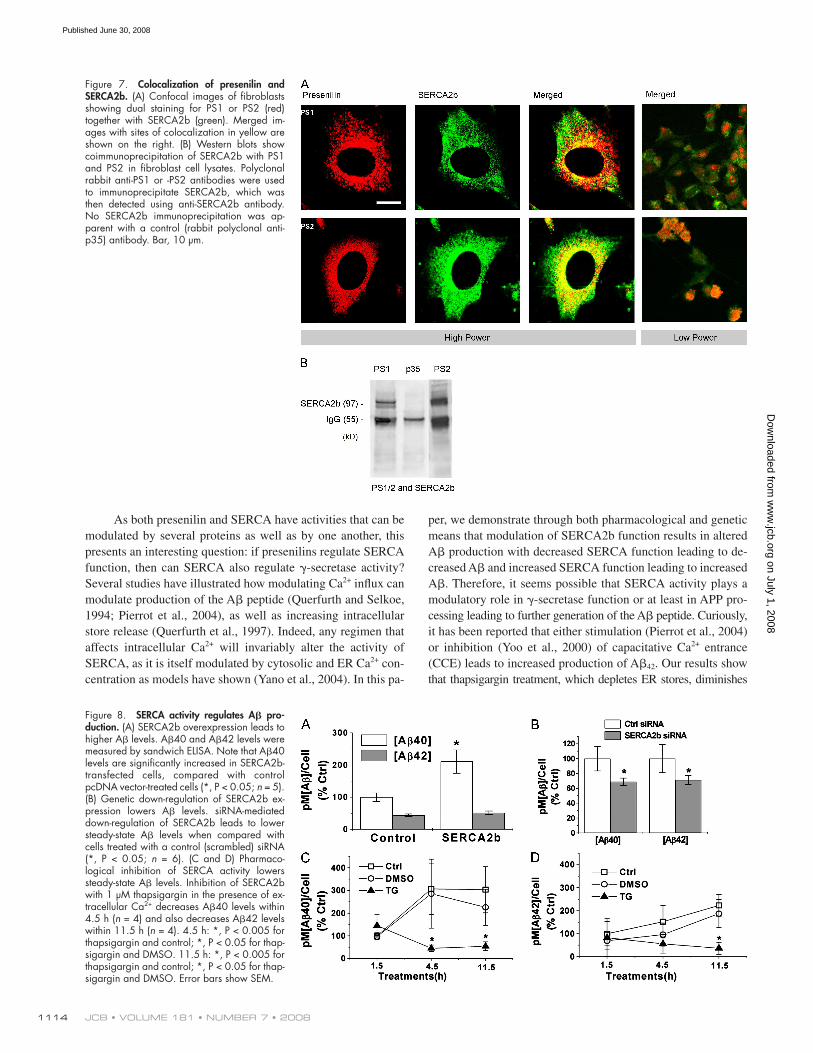
SERCA2b and PS1/PS2 are colocalized and interact Although both PS1 and PS2 have been localized to ER in sev-
eral cell types ( Walter et al., 1996 ) and SERCA is an ER Ca 2+
pump, it is not known if they colocalize within specifi c regions
of the ER. To address this question, we used confocal dual-label
imaging of wild-type fi broblasts using a polyclonal antiserum
against SERCA2b and a polyclonal antiserum against either
PS1 or PS2 and found that SERCA2b and PS2 do colocalize,
whereas colocalization between SERCA2b and PS1 was pre-
sent but not as prevalent ( Fig. 7 A ).
To biochemically determine whether presenilins physi-
cally associate with SERCA2b, we conducted immunoprecipita-
tion experiments. Fibroblast cell lysates were immunoprecipitated
with either a PS1- or PS2-specifi c antibody or an irrelevant con-
trol antibody (anti-p35). The resultant pellets were fractionated
on a 4 – 12% Bis/Tris gel and immunoblotted for SERCA2b.
Both PS1 and PS2 were found to specifi cally bind to SERCA2b,
whereas SERCA2b did not coprecipitate with the control anti-
body ( Fig. 7 B ). Conversely, immunoprecipitating with an anti-
SERCA2b antibody followed by immunoblotting for PS1 or
PS2 was not feasible because the weights of the IgG heavy and
light chains ( � 55 and 25 kD) are the same as of presenilin
holoprotein ( � 55 kD) or carboxy fragment ( � 22 kD), and al-
though using different species of polyclonal antibodies revealed
bands at the correct weights, we could not absolutely determine
whether they were presenilin or cross-reactivity with intraspe-
cies IgG chains.
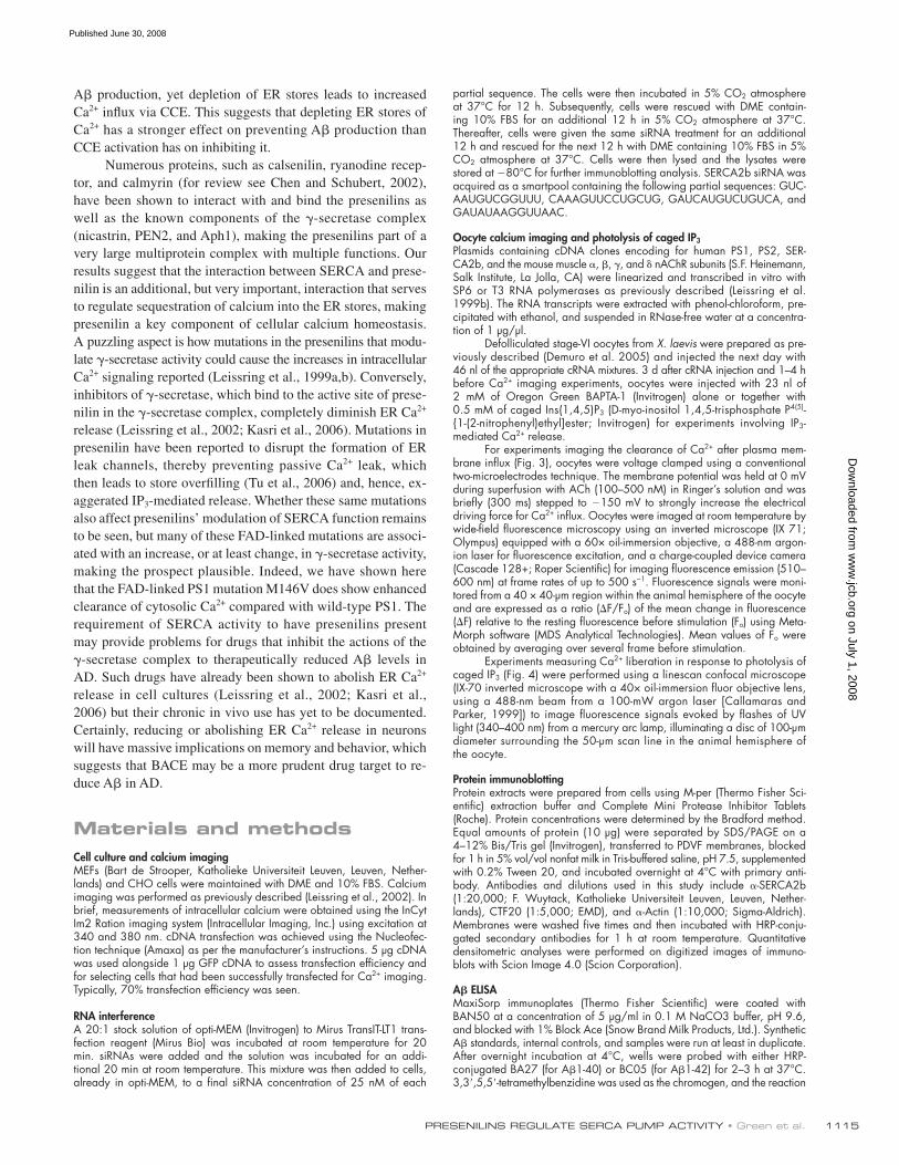
SERCA activity regulates A � production Our data indicate that presenilins are required for normal SERCA
function and physiological maintenance of cellular calcium ho-
meostasis. Moreover, given that presenilins are integral for the
production of A � , we investigated whether SERCA function
infl uenced A � production. To address this issue, we used phar-
macological and gain-of-function and loss-of-function genetic
approaches. First, we considered the consequences of over-
expressing SERCA2b in CHO cells stably expressing APP. After
48 h, we found that higher SERCA2b levels caused a marked
increase in A � 40 production ( Fig. 8 A ). Conversely, reducing
SERCA2b via siRNA-mediated knockdown caused a signifi cant
decrease in both A � 40 and A � 42 levels ( Fig. 8 B ). Pharmaco-
logical inhibition of SERCA2b with thapsigargin rapidly reduced
A � 40 and A � 42 production ( Fig. 8, C and D ), which is in agree-
ment with SERCA2b knockdown. The sum of these fi ndings in-
dicates that SERCA pump activity impacts A � production.
Discussion Cellular Ca 2+ dyshomeostasis has been consistently observed in
numerous experimental systems harboring presenilin mutations,
including X. laevis oocytes, transfected cell lines, genetically al-
tered mice, and human fi broblasts from FAD patients ( LaFerla,
2002 ). These pathological disruptions in Ca 2+ signaling suggest
that the presenilins may play a physiological role in cellular Ca 2+
regulation, although this has not been fi rmly established or charac-
terized. In this paper, we examined the role of wild-type presenilins
oocyte and recorded cytosolic Ca 2+ decay after a 300-ms Ca 2+
infl ux through the nAChR as before. The Ca 2+ decay was
substantially accelerated in oocytes expressing PS1M146V
compared with control ( Fig. 5 A ). Double-exponential curve fi t-
ting revealed substantial acceleration in both � 1 and � 2 compo-
nents with PS1M146V over control ( Fig. 5 B ). Rates of decay
were faster than wild-type PS1 expression alone ( � 1, 0.373 vs.
0.276; � 2, 1.919 vs. 1.114), suggesting that this mutation im-
pacts Ca 2+ cytosolic sequestration more effectively than wild-
type PS1 protein.
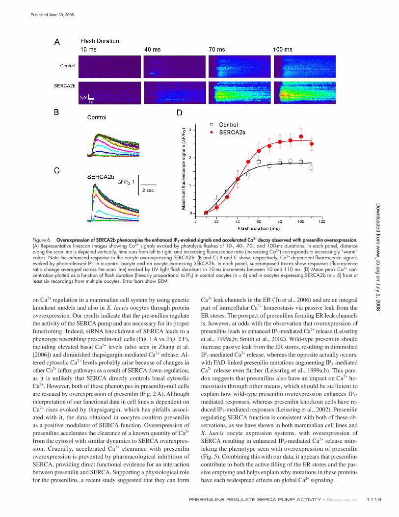
SERCA2b overexpression increases IP 3 -mediated calcium release We previously showed that overexpression of either PS1 or PS2
leads to an increase in IP 3 -mediated Ca 2+ release in X. laevis oo-
cytes ( Leissring et al., 1999a , b ), but it was unclear whether this
arose through increased activation or conductance of IP 3 recep-
tor/channels or from enhanced store fi lling. To discriminate be-
tween these possibilities, we examined whether enhanced SERCA
pump activity could replicate the increased IP 3 response. We
monitored Ca 2+ liberation by linescan confocal microscopy in
response to photorelease of IP 3 from a caged precursor evoked
by UV fl ashes of varying duration as previously described
( Leissring et al., 1999a , b ). Similar to the effects of presenilin
overexpression, mean Ca 2+ responses evoked by high IP 3 were
enhanced by � 50% ( Fig. 6 D ), and the decay of IP 3 -evoked
Ca 2+ transients was accelerated ( Figs. 6, B and C ). These fi nd-
ings show that an increase in SERCA pump activity mimics the
phenotype previously seen with presenilin overexpression.
Figure 5. PS1 FAD mutation M146V accelerates cytosolic Ca 2+ sequestra-tion. (A) Traces show Ca 2+ -dependent fl uorescence changes ( Δ F/F o ) after 300-ms hyperpolarizing pulses to induce Ca 2+ entry through nAChR ex-pressed in the plasma membrane obtained from control and PS1 M146V expressing oocytes. (B) Table listing fast (T1) and slow (T2) time constants of double-exponential fi ts to the Ca 2+ decay data.
on July 1, 2008 w
ww
.jcb.orgD
ownloaded from
Published June 30, 2008
1113 PRESENILINS REGULATE SERCA PUMP ACTIVITY • Green et al.
Ca 2+ leak channels in the ER ( Tu et al., 2006 ) and are an integral
part of intracellular Ca 2+ homeostasis via passive leak from the
ER stores. The prospect of presenilins forming ER leak channels
is, however, at odds with the observation that overexpression of
presenilins leads to enhanced IP 3 -mediated Ca 2+ release ( Leissring
et al., 1999a , b ; Smith et al., 2002 ). Wild-type presenilin should
increase passive leak from the ER stores, resulting in diminished
IP 3 -mediated Ca 2+ release, whereas the opposite actually occurs,
with FAD-linked presenilin mutations augmenting IP 3 -mediated
Ca 2+ release even further ( Leissring et al., 1999a , b ). This para-
dox suggests that presenilins also have an impact on Ca 2+ ho-
meostasis through other means, which should be suffi cient to
explain how wild-type presenilin overexpression enhances IP 3 -
mediated responses, whereas presenilin knockout cells have re-
duced IP 3 -mediated responses ( Leissring et al., 2002 ). Presenilin
regulating SERCA function is consistent with both of these ob-
servations, as we have shown in both mammalian cell lines and
X. laevis oocyte expression systems, with overexpression of
SERCA resulting in enhanced IP 3 -mediated Ca 2+ release mim-
icking the phenotype seen with overexpression of presenilin
( Fig. 5 ). Combining this with our data, it appears that presenilins
contribute to both the active fi lling of the ER stores and the pas-
sive emptying and helps explain why mutations in these proteins
have such widespread effects on global Ca 2+ signaling.
on Ca 2+ regulation in a mammalian cell system by using genetic
knockout models and also in X. laevis oocytes through protein
overexpression. Our results indicate that the presenilins regulate
the activity of the SERCA pump and are necessary for its proper
functioning. Indeed, siRNA knockdown of SERCA leads to a
phenotype resembling presenilin-null cells ( Fig. 1 A vs. Fig. 2 F ),
including elevated basal Ca 2+ levels (also seen in Zhang et al.
[2006] ) and diminished thapsigargin-mediated Ca 2+ release. Al-
tered cytosolic Ca 2+ levels probably arise because of changes in
other Ca 2+ infl ux pathways as a result of SERCA down-regulation,
as it is unlikely that SERCA directly controls basal cytosolic
Ca 2+ . However, both of these phenotypes in presenilin-null cells
are rescued by overexpression of presenilin ( Fig. 2 A ). Although
interpretation of our functional data in cell lines is dependent on
Ca 2+ rises evoked by thapsigargin, which has pitfalls associ-
ated with it, the data obtained in oocytes confi rm presenilin
as a positive modulator of SERCA function. Overexpression of
presenilins accelerates the clearance of a known quantity of Ca 2+
from the cytosol with similar dynamics to SERCA overexpres-
sion. Crucially, accelerated Ca 2+ clearance with presenilin
overexpression is prevented by pharmacological inhibition of
SERCA, providing direct functional evidence for an interaction
between presenilin and SERCA. Supporting a physiological role
for the presenilins, a recent study suggested that they can form
Figure 6. Overexpression of SERCA2b phenocopies the enhanced IP 3 -evoked signals and accelerated Ca 2+ decay observed with presenilin overexpression. (A) Representative linescan images showing Ca 2+ signals evoked by photolysis fl ashes of 10-, 40-, 70-, and 100-ms durations. In each panel, distance along the scan line is depicted vertically, time runs from left to right, and increasing fl uorescence ratio (increasing Ca 2+ ) corresponds to increasingly “ warm ” colors. Note the enhanced response in the oocyte overexpressing SERCA2b. (B and C) B and C show, respectively, Ca 2+ -dependent fl uorescence signals evoked by photoreleased IP 3 in a control oocyte and an oocyte expressing SERCA2b. In each panel, superimposed traces show responses (fl uorescence ratio change averaged across the scan line) evoked by UV light fl ash durations in 10-ms increments between 10 and 110 ms. (D) Mean peak Ca 2+ con-centration plotted as a function of fl ash duration (linearly proportional to IP 3 ) in control oocytes ( n = 6) and in oocytes expressing SERCA2b ( n = 5) from at least six recordings from multiple oocytes. Error bars show SEM.
on July 1, 2008 w
ww
.jcb.orgD
ownloaded from
Published June 30, 2008
JCB • VOLUME 181 • NUMBER 7 • 2008 1114
per, we demonstrate through both pharmacological and genetic
means that modulation of SERCA2b function results in altered
A � production with decreased SERCA function leading to de-
creased A � and increased SERCA function leading to increased
A � . Therefore, it seems possible that SERCA activity plays a
modulatory role in � -secretase function or at least in APP pro-
cessing leading to further generation of the A � peptide. Curiously,
it has been reported that either stimulation ( Pierrot et al., 2004 )
or inhibition ( Yoo et al., 2000 ) of capacitative Ca 2+ entrance
(CCE) leads to increased production of A � 42 . Our results show
that thapsigargin treatment, which depletes ER stores, diminishes
As both presenilin and SERCA have activities that can be
modulated by several proteins as well as by one another, this
presents an interesting question: if presenilins regulate SERCA
function, then can SERCA also regulate � -secretase activity?
Several studies have illustrated how modulating Ca 2+ infl ux can
modulate production of the A � peptide ( Querfurth and Selkoe,
1994 ; Pierrot et al., 2004 ), as well as increasing intracellular
store release ( Querfurth et al., 1997 ). Indeed, any regimen that
affects intracellular Ca 2+ will invariably alter the activity of
SERCA, as it is itself modulated by cytosolic and ER Ca 2+ con-
centration as models have shown ( Yano et al., 2004 ). In this pa-
Figure 7. Colocalization of presenilin and SERCA2b. (A) Confocal images of fi broblasts showing dual staining for PS1 or PS2 (red) together with SERCA2b (green). Merged im-ages with sites of colocalization in yellow are shown on the right. (B) Western blots show coimmunoprecipitation of SERCA2b with PS1 and PS2 in fi broblast cell lysates. Polyclonal rabbit anti-PS1 or -PS2 antibodies were used to immunoprecipitate SERCA2b, which was then detected using anti-SERCA2b antibody. No SERCA2b immunoprecipitation was ap-parent with a control (rabbit polyclonal anti-p35) antibody. Bar, 10 μ m.
Figure 8. SERCA activity regulates A � pro-duction. (A) SERCA2b overexpression leads to higher A � levels. A � 40 and A � 42 levels were measured by sandwich ELISA. Note that A � 40 levels are signifi cantly increased in SERCA2b-transfected cells, compared with control pcDNA vector-treated cells (*, P < 0.05; n = 5). (B) Genetic down-regulation of SERCA2b ex-pression lowers A � levels. siRNA-mediated down-regulation of SERCA2b leads to lower steady-state A � levels when compared with cells treated with a control (scrambled) siRNA (*, P < 0.05; n = 6). (C and D) Pharmaco-logical inhibition of SERCA activity lowers steady-state A � levels. Inhibition of SERCA2b with 1 μ M thapsigargin in the presence of ex-tracellular Ca 2+ decreases A � 40 levels within 4.5 h ( n = 4) and also decreases A � 42 levels within 11.5 h ( n = 4). 4.5 h: *, P < 0.005 for thapsigargin and control; *, P < 0.05 for thap-sigargin and DMSO. 11.5 h: *, P < 0.005 for thapsigargin and control; *, P < 0.05 for thap-sigargin and DMSO. Error bars show SEM.
on July 1, 2008 w
ww
.jcb.orgD
ownloaded from
Published June 30, 2008
1115 PRESENILINS REGULATE SERCA PUMP ACTIVITY • Green et al.
partial sequence. The cells were then incubated in 5% CO 2 atmosphere at 37 ° C for 12 h. Subsequently, cells were rescued with DME contain-ing 10% FBS for an additional 12 h in 5% CO 2 atmosphere at 37 ° C. Thereafter, cells were given the same siRNA treatment for an additional 12 h and rescued for the next 12 h with DME containing 10% FBS in 5% CO 2 atmosphere at 37 ° C. Cells were then lysed and the lysates were stored at � 80 ° C for further immunoblotting analysis. SERCA2b siRNA was acquired as a smartpool containing the following partial sequences: GUC-AAUGUCGGUUU, CAAAGUUCCUGCUG, GAUCAUGUCUGUCA, and GAUAUAAGGUUAAC.
Oocyte calcium imaging and photolysis of caged IP 3 Plasmids containing cDNA clones encoding for human PS1, PS2, SER-CA2b, and the mouse muscle � , � , � , and � nAChR subunits (S.F. Heinemann, Salk Institute, La Jolla, CA) were linearized and transcribed in vitro with SP6 or T3 RNA polymerases as previously described ( Leissring et al. 1999b ). The RNA transcripts were extracted with phenol-chloroform, pre-cipitated with ethanol, and suspended in RNase-free water at a concentra-tion of 1 μ g/ μ l.
Defolliculated stage-VI oocytes from X. laevis were prepared as pre-viously described ( Demuro et al. 2005 ) and injected the next day with 46 nl of the appropriate cRNA mixtures. 3 d after cRNA injection and 1 – 4 h before Ca 2+ imaging experiments, oocytes were injected with 23 nl of 2 mM of Oregon Green BAPTA-1 (Invitrogen) alone or together with 0.5 mM of caged Ins(1,4,5)P 3 (D-myo-inositol 1,4,5-trisphosphate P 4(5) - { 1-(2-nitrophenyl)ethyl]ester; Invitrogen) for experiments involving IP 3 -mediated Ca 2+ release.
For experiments imaging the clearance of Ca 2+ after plasma mem-brane infl ux ( Fig. 3 ), oocytes were voltage clamped using a conventional two-microelectrodes technique. The membrane potential was held at 0 mV during superfusion with ACh (100 – 500 nM) in Ringer ’ s solution and was briefl y (300 ms) stepped to � 150 mV to strongly increase the electrical driving force for Ca 2+ infl ux. Oocytes were imaged at room temperature by wide-fi eld fl uorescence microscopy using an inverted microscope (IX 71; Olympus) equipped with a 60 × oil-immersion objective, a 488-nm argon-ion laser for fl uorescence excitation, and a charge-coupled device camera (Cascade 128+; Roper Scientifi c) for imaging fl uorescence emission (510 – 600 nm) at frame rates of up to 500 s −1 . Fluorescence signals were moni-tored from a 40 × 40- μ m region within the animal hemisphere of the oocyte and are expressed as a ratio ( � F/F o ) of the mean change in fl uorescence ( � F) relative to the resting fl uorescence before stimulation (F o ) using Meta-Morph software (MDS Analytical Technologies). Mean values of F o were obtained by averaging over several frame before stimulation.
Experiments measuring Ca 2+ liberation in response to photolysis of caged IP 3 ( Fig. 4 ) were performed using a linescan confocal microscope (IX-70 inverted microscope with a 40 × oil-immersion fl uor objective lens, using a 488-nm beam from a 100-mW argon laser [ Callamaras and Parker, 1999 ]) to image fl uorescence signals evoked by fl ashes of UV light (340 – 400 nm) from a mercury arc lamp, illuminating a disc of 100- μ m diameter surrounding the 50- μ m scan line in the animal hemisphere of the oocyte.
Protein immunoblotting Protein extracts were prepared from cells using M-per (Thermo Fisher Sci-entifi c) extraction buffer and Complete Mini Protease Inhibitor Tablets (Roche). Protein concentrations were determined by the Bradford method. Equal amounts of protein (10 μ g) were separated by SDS/PAGE on a 4 – 12% Bis/Tris gel (Invitrogen), transferred to PDVF membranes, blocked for 1 h in 5% vol/vol nonfat milk in Tris-buffered saline, pH 7.5, supplemented with 0.2% Tween 20, and incubated overnight at 4 ° C with primary anti-body. Antibodies and dilutions used in this study include � -SERCA2b (1:20,000; F. Wuytack, Katholieke Universiteit Leuven, Leuven, Nether-lands), CTF20 (1:5,000; EMD), and � -Actin (1:10,000; Sigma-Aldrich). Membranes were washed fi ve times and then incubated with HRP-conju-gated secondary antibodies for 1 h at room temperature. Quantitative densitometric analyses were performed on digitized images of immuno-blots with Scion Image 4.0 (Scion Corporation).
A � ELISA MaxiSorp immunoplates (Thermo Fisher Scientifi c) were coated with BAN50 at a concentration of 5 μ g/ml in 0.1 M NaCO3 buffer, pH 9.6, and blocked with 1% Block Ace (Snow Brand Milk Products, Ltd.). Synthetic A � standards, internal controls, and samples were run at least in duplicate. After overnight incubation at 4 ° C, wells were probed with either HRP-conjugated BA27 (for A � 1-40) or BC05 (for A � 1-42) for 2 – 3 h at 37 ° C. 3,3 ,5,5 -tetramethylbenzidine was used as the chromogen, and the reaction
A � production, yet depletion of ER stores leads to increased
Ca 2+ infl ux via CCE. This suggests that depleting ER stores of
Ca 2+ has a stronger effect on preventing A � production than
CCE activation has on inhibiting it.
Numerous proteins, such as calsenilin, ryanodine recep-
tor, and calmyrin (for review see Chen and Schubert, 2002 ),
have been shown to interact with and bind the presenilins as
well as the known components of the � -secretase complex
(nicastrin, PEN2, and Aph1), making the presenilins part of a
very large multiprotein complex with multiple functions. Our
results suggest that the interaction between SERCA and prese-
nilin is an additional, but very important, interaction that serves
to regulate sequestration of calcium into the ER stores, making
presenilin a key component of cellular calcium homeostasis.
A puzzling aspect is how mutations in the presenilins that modu-
late � -secretase activity could cause the increases in intracellular
Ca 2+ signaling reported ( Leissring et al., 1999a , b ). Conversely,
inhibitors of � -secretase, which bind to the active site of prese-
nilin in the � -secretase complex, completely diminish ER Ca 2+
release ( Leissring et al., 2002 ; Kasri et al., 2006 ). Mutations in
presenilin have been reported to disrupt the formation of ER
leak channels, thereby preventing passive Ca 2+ leak, which
then leads to store overfi lling ( Tu et al., 2006 ) and, hence, ex-
aggerated IP 3 -mediated release. Whether these same mutations
also affect presenilins ’ modulation of SERCA function remains
to be seen, but many of these FAD-linked mutations are associ-
ated with an increase, or at least change, in � -secretase activity,
making the prospect plausible. Indeed, we have shown here
that the FAD-linked PS1 mutation M146V does show enhanced
clearance of cytosolic Ca 2+ compared with wild-type PS1. The
requirement of SERCA activity to have presenilins present
may provide problems for drugs that inhibit the actions of the
� -secretase complex to therapeutically reduced A � levels in
AD. Such drugs have already been shown to abolish ER Ca 2+
release in cell cultures ( Leissring et al., 2002 ; Kasri et al.,
2006 ) but their chronic in vivo use has yet to be documented.
Certainly, reducing or abolishing ER Ca 2+ release in neurons
will have massive implications on memory and behavior, which
suggests that BACE may be a more prudent drug target to re-
duce A � in AD.
Materials and methods Cell culture and calcium imaging MEFs (Bart de Strooper, Katholieke Universiteit Leuven, Leuven, Nether-lands) and CHO cells were maintained with DME and 10% FBS. Calcium imaging was performed as previously described ( Leissring et al., 2002 ). In brief, measurements of intracellular calcium were obtained using the InCyt Im2 Ration imaging system (Intracellular Imaging, Inc.) using excitation at 340 and 380 nm. cDNA transfection was achieved using the Nucleofec-tion technique (Amaxa) as per the manufacturer ’ s instructions. 5 μ g cDNA was used alongside 1 μ g GFP cDNA to assess transfection effi ciency and for selecting cells that had been successfully transfected for Ca 2+ imaging. Typically, 70% transfection effi ciency was seen.
RNA interference A 20:1 stock solution of opti-MEM (Invitrogen) to Mirus TransIT-LT1 trans-fection reagent (Mirus Bio) was incubated at room temperature for 20 min. siRNAs were added and the solution was incubated for an addi-tional 20 min at room temperature. This mixture was then added to cells, already in opti-MEM, to a fi nal siRNA concentration of 25 nM of each
on July 1, 2008 w
ww
.jcb.orgD
ownloaded from
Published June 30, 2008
JCB • VOLUME 181 • NUMBER 7 • 2008 1116
Herms , J. , I. Schneider , I. Dewachter , N. Caluwaerts , H. Kretzschmar , and F. Van Leuven . 2003 . Capacitive calcium entry is directly attenuated by mutant presenilin-1, independent of the expression of the amyloid precursor pro-tein. J. Biol. Chem. 278 : 2484 – 2489 .
Herreman , A. , D. Hartmann , W. Annaert , P. Saftig , K. Craessaerts , L. Serneels , L. Umans , V. Schrijvers , F. Checler , H. Vanderstichele , et al . 1999 . Presenilin 2 defi ciency causes a mild pulmonary phenotype and no changes in amyloid precursor protein processing but enhances the em-bryonic lethal phenotype of presenilin 1 defi ciency. Proc. Natl. Acad. Sci. USA . 96 : 11872 – 11877 .
Kasri , N.N. , S.L. Kocks , L. Verbert , S.S. Hebert , G. Callewaert , J.B. Parys , L. Missiaen , and H. De Smedt . 2006 . Up-regulation of inositol 1,4,5-trisphosphate receptor type 1 is responsible for a decreased endoplasmic-reticulum Ca2+ content in presenilin double knock-out cells. Cell Calcium . 40 : 41 – 51 .
LaFerla , F.M. 2002 . Calcium dyshomeostasis and intracellular signalling in Alzheimer ’ s disease. Nat. Rev. Neurosci. 3 : 862 – 872 .
Leissring , M.A. , I. Parker , and F.M. LaFerla . 1999a . Presenilin-2 mutations modulate amplitude and kinetics of inositol 1,4,5-trisphosphate-mediated calcium signals. J. Biol. Chem. 274 : 32535 – 32538 .
Leissring , M.A. , B.A. Paul , I. Parker , C.W. Cotman , and F.M. LaFerla . 1999b . Alzheimer ’ s presenilin-1 mutation potentiates inositol 1,4,5-trisphos-phate-mediated calcium signaling in Xenopus oocytes. J. Neurochem. 72 : 1061 – 1068 .
Leissring , M.A. , Y. Akbari , C.M. Fanger , M.D. Cahalan , M.P. Mattson , and F.M. LaFerla . 2000 . Capacitative calcium entry defi cits and elevated lumi-nal calcium content in mutant presenilin-1 knockin mice. J. Cell Biol. 149 : 793 – 798 .
Leissring , M.A. , M.P. Murphy , T.R. Mead , Y. Akbari , M.C. Sugarman , M. Jannatipour , B. Anliker , U. Muller , P. Saftig , B. De Strooper , et al . 2002 . A physiologic signaling role for the gamma -secretase-derived intra-cellular fragment of APP. Proc. Natl. Acad. Sci. USA . 99 : 4697 – 4702 .
Lytton , J. , M. Westlin , and M.R. Hanley . 1991 . Thapsigargin inhibits the sarco-plasmic or endoplasmic reticulum Ca-ATPase family of calcium pumps. J. Biol. Chem. 266 : 17067 – 17071 .
Pierrot , N. , P. Ghisdal , A.S. Caumont , and J.N. Octave . 2004 . Intraneuronal amyloid-beta1-42 production triggered by sustained increase of cy-tosolic calcium concentration induces neuronal death. J. Neurochem. 88 : 1140 – 1150 .
Querfurth , H.W. , and D.J. Selkoe . 1994 . Calcium ionophore increases amyloid beta peptide production by cultured cells. Biochemistry . 33 : 4550 – 4561 .
Querfurth , H.W. , J. Jiang , J.D. Geiger , and D.J. Selkoe . 1997 . Caffeine stimu-lates amyloid beta-peptide release from beta-amyloid precursor protein-transfected HEK293 cells. J. Neurochem. 69 : 1580 – 1591 .
Smith , I.F. , J.P. Boyle , P.F. Vaughan , H.A. Pearson , R.F. Cowburn , and C.S. Peers . 2002 . Ca(2+) stores and capacitative Ca(2+) entry in human neu-roblastoma (SH-SY5Y) cells expressing a familial Alzheimer ’ s disease presenilin-1 mutation. Brain Res. 949 : 105 – 111 .
Stutzmann , G.E. , A. Caccamo , F.M. LaFerla , and I. Parker . 2004 . Dysregulated IP3 signaling in cortical neurons of knock-in mice expressing an Alzheimer ’ s-linked mutation in presenilin1 results in exaggerated Ca2+ signals and altered membrane excitability. J. Neurosci. 24 : 508 – 513 .
Tu , H. , O. Nelson , A. Bezprozvanny , Z. Wang , S.F. Lee , Y.H. Hao , L. Serneels , B. De Strooper , G. Yu , and I. Bezprozvanny . 2006 . Presenilins form ER Ca2+ leak channels, a function disrupted by familial Alzheimer ’ s disease-linked mutations. Cell . 126 : 981 – 993 .
Walter , J. , A. Capell , J. Grunberg , B. Pesold , A. Schindzielorz , R. Prior , M.B. Podlisny , P. Fraser , P.S. Hyslop , D.J. Selkoe , and C. Haass . 1996 . The Alzheimer ’ s disease-associated presenilins are differentially phosphory-lated proteins located predominantly within the endoplasmic reticulum. Mol. Med. 2 : 673 – 691 .
Yano , K. , O.H. Petersen , and A.V. Tepikin . 2004 . Dual sensitivity of sarco-plasmic/endoplasmic Ca2+-ATPase to cytosolic and endoplasmic reticu-lum Ca2+ as a mechanism of modulating cytosolic Ca2+ oscillations. Biochem. J. 383 : 353 – 360 .
Yoo , A.S. , I. Cheng , S. Chung , T.Z. Grenfell , H. Lee , E. Pack-Chung , M. Handler , J. Shen , W. Xia , G. Tesco , et al . 2000 . Presenilin-mediated modulation of capacitative calcium entry. Neuron . 27 : 561 – 572 .
Zhang , S.L. , A.V. Yeromin , X.H. Zhang , Y. Yu , O. Safrina , A. Penna , J. Roos , K.A. Stauderman , and M.D. Cahalan . 2006 . Genome-wide RNAi screen of Ca(2+) infl ux identifi es genes that regulate Ca(2+) release-activated Ca(2+) channel activity. Proc. Natl. Acad. Sci. USA . 103 : 9357 – 9362 .
Zhang , Z. , P. Nadeau , W. Song , D. Donoviel , M. Yuan , A. Bernstein , and B.A. Yankner . 2000 . Presenilins are required for gamma-secretase cleavage of beta-APP and transmembrane cleavage of Notch-1. Nat. Cell Biol. 2 : 463 – 465 .
was stopped by 6% O -phosphoric acid and read at 450 nm on a plate reader (Molecular Dynamics). Data are reported as mean per live cell + SEM, and statistical signifi cance was evaluated using Student ’ s t test.
Immunoprecipitation 50 μ g MEF cell lysate was incubated with 40 μ l of Protein A Sepharose beads (Sigma-Aldrich) for 1 h and centrifuged, and the supernatant was recovered. A further 40 μ l of beads was added along with anti-PS1 (Cell Signaling Technology), PS2 (G. Thinakaran, University of Chicago, Chicago, IL), or p35 (Santa Cruz Biotechnology, Inc.) as a control (1:100), and the volume was made up to 1 ml with water and incubated overnight at 4 ° C overnight. After pelleting the beads, the supernatant was discarded and the beads were washed with STEN buffer (0.15 M NaCl, 0.05 M Tris HCl, 0.002 M EDTA, and 2% NP-40, pH 7.6) and then STEN containing 0.1% SDS. The beads were then pelleted and 4 × loading buffer was added (Invitrogen). The samples were boiled for 10 min and spun down again, and the supernatant was run on a 4 – 12% Bis/Tris gel. SERCA2b was probed using � SERCA2b (1:20,000; F. Wuytack).
Confocal microscopy Fluorescent immunolabeling followed a standard two-way technique (pri-mary antibody followed by fl uorescent secondary antibody). Free-fl oating sections were rinsed in TBS, pH 7.4, and then blocked (0.25% Triton X-100 and 5% normal goat serum in TBS) for 1 h. Sections were incubated in primary antibody overnight at 4 ° C, rinsed in PBS, and incubated for 1 h in either fl uorescently labeled anti – rabbit or anti – mouse secondary an-tibodies (Alexa 488, 1:200; Invitrogen). Confocal images were captured on a confocal system (Radiance 2100;Bio-Rad Laboratories). All double-labeled specimens were imaged using the -strobing function to prevent nonspecifi c cross-excitation of fl uorophores.
Statistics Data are presented as mean ± 1 SEM, with n = number of cells examined. An unpaired Student ’ s t test was used to determine statistical signifi cance (P < 0.05).
O nline supplemental material Video 1 shows representative calcium clearance from oocyte cytosol after a 300-ms infl ux through nAChR. Online supplemental material is available at http://www.jcb.org/cgi/content/full/jcb.200706171/DC1.
We thank Dr. Bart de Strooper for the PS knockout fi broblasts, Dr. Frank Wuytack for the generous gift of the anti-SERCA2b antibody, and Dr. Gopal Thinakaran for providing us with the anti-PS2 antibody. We are also grateful to Dr. Stephen F. Heinemann for providing the muscle nAChRs subunits cDNAs.
This work was supported by the National Institutes of Health (grants AG17968, AG16573, and GM48071).
Submitted: 26 June 2007 Accepted: 28 May 2008
References Aubier , M. , and N. Viires . 1998 . Calcium ATPase and respiratory muscle func-
tion. Eur. Respir. J. 11 : 758 – 766 .
Baba-Aissa , F. , L. Raeymaekers , F. Wuytack , L. Dode , and R. Casteels . 1998 . Distribution and isoform diversity of the organellar Ca2+ pumps in the brain. Mol. Chem. Neuropathol. 33 : 199 – 208 .
Berridge , M.J. , P. Lipp , and M.D. Bootman . 2000 . The versatility and universal-ity of calcium signalling. Nat. Rev. Mol. Cell Biol. 1 : 11 – 21 .
Callamaras , N. , and I. Parker . 1999 . Radial localization of inositol 1,4,5-trisphos-phate-sensitive Ca2+ release sites in Xenopus oocytes resolved by axial confocal linescan imaging. J. Gen. Physiol. 113 : 199 – 213 .
Chen , Q. , and D. Schubert . 2002 . Presenilin-interacting proteins. Expert Rev. Mol. Med. 4 : 1 – 18 .
Demuro , A. , E. Mina , R. Kayed , S.C. Milton , I. Parker , and C.G. Glabe . 2005 . Calcium dysregulation and membrane disruption as a ubiquitous neurotoxic mechanism of soluble amyloid oligomers. J. Biol. Chem. 280 : 17294 – 17300 .
Duff , K. , C. Eckman , C. Zehr , X. Yu , C.M. Prada , J. Perez-tur , M. Hutton , L. Buee , Y. Harigaya , D. Yager , et al . 1996 . Increased amyloid-beta42(43) in brains of mice expressing mutant presenilin 1. Nature . 383 : 710 – 713 .
Hass , M.R. , and B.A. Yankner . 2005 . A { gamma } -secretase-independent mech-anism of signal transduction by the amyloid precursor protein. J. Biol. Chem. 280 : 36895 – 36904 .
on July 1, 2008 w
ww
.jcb.orgD
ownloaded from
Published June 30, 2008