Sequence control of Aggregation Some Examples. Domain Swapping.
14
Sequence control of Aggregation Some Examples
-
date post
21-Dec-2015 -
Category
Documents
-
view
225 -
download
0
Transcript of Sequence control of Aggregation Some Examples. Domain Swapping.
Sequence control of Aggregation
Some Examples

Domain Swapping

Domain Swapping of RNase A
Yanshun & Eisenberg (2002) Protein Sci. 6:1285-1299.
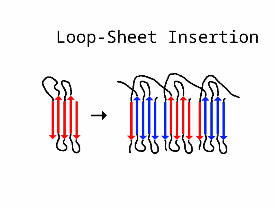
Loop-Sheet Insertion

Loop-sheet Insertion of 1-Antitrypsin
Active (Metastable)
Inactive(Stable)
Loop-sheetInsert
Polymer
Carrell, et al. (1998) Curr. Opin. Struct. Biol. 8:799-809.
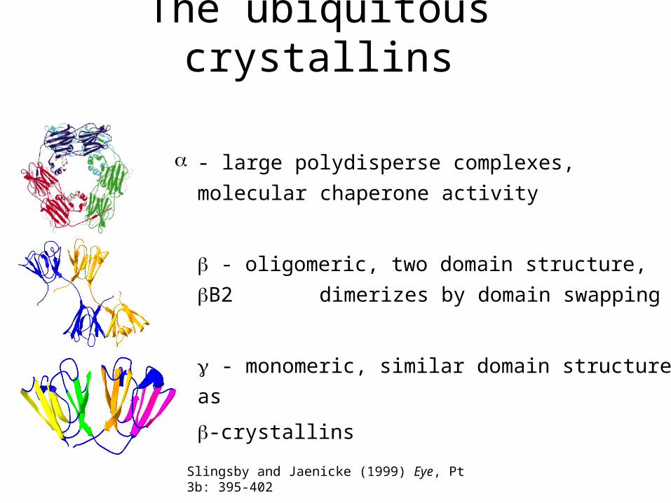
The ubiquitous crystallins
- large polydisperse complexes, molecular
chaperone activity
- oligomeric, two domain structure, B2
dimerizes by domain swapping
- monomeric, similar domain structure as
-crystallins
Slingsby and Jaenicke (1999) Eye, Pt 3b: 395-402
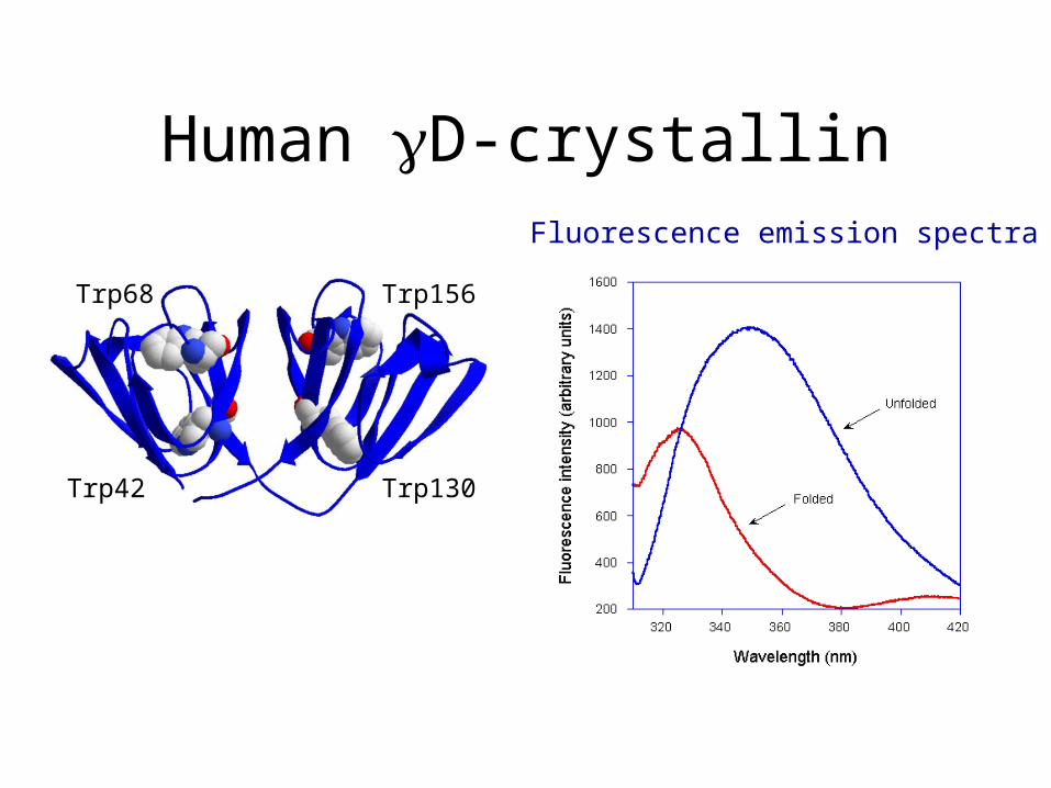
Human D-crystallinFluorescence emission spectra
Trp68
Trp42
Trp156
Trp130
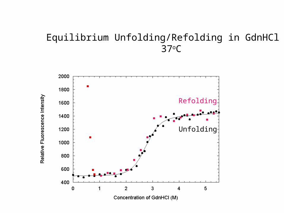
Equilibrium Unfolding/Refolding in GdnHCl at 37oC
Refolding
Unfolding
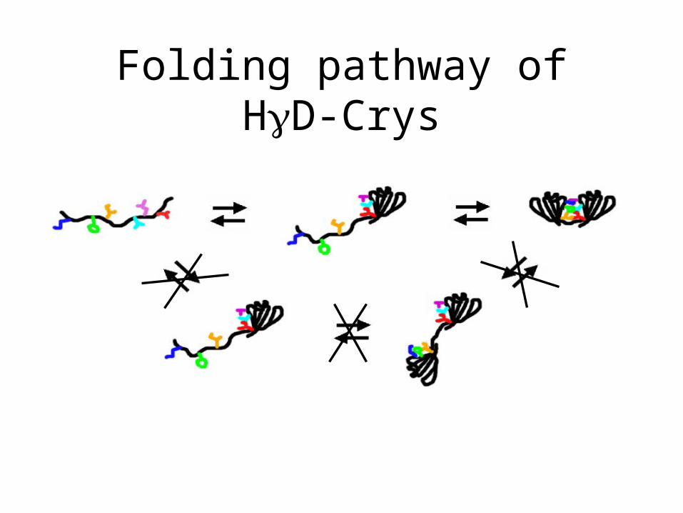
Folding pathway of HD-Crys

Refolding: AFM Time Course
5 minutes30 seconds
24 minutes 54 minutes 54 minutes
Native300 nm
300 nm
1 m
1 m 1 m
1 m1 m
1 m 1 m1 m
1 m 1 m

Hydrophobic domain interface residues
N-terminal domain C-terminal domain
Phe56 - Phe 80%, Val 8.5%, Ile 8.5%, Leu 3%
Val131 - Val 54.3%, Ile 28.5%, Leu 17.2%
Ile81 - Ile 80%, Val 8.5%, Leu 5.5%, Pro 3%, Thr 3%
Leu144 - Leu 68.5%, Tyr 20%, Phe 11.5%
Met43 - Met 77%, Val 11.5%, Ala 8.5%, Ile 3%
Val169 - Val 49%, Ile 42%, Ala 3%, Met 3%, Leu 3%
Conservation among 35 -crystallin sequences:
N-terminal
Met43Phe56Ile81
C-terminal
Leu144Val131Val169
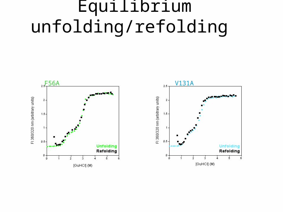
Equilibrium unfolding/refolding
F56A V131A
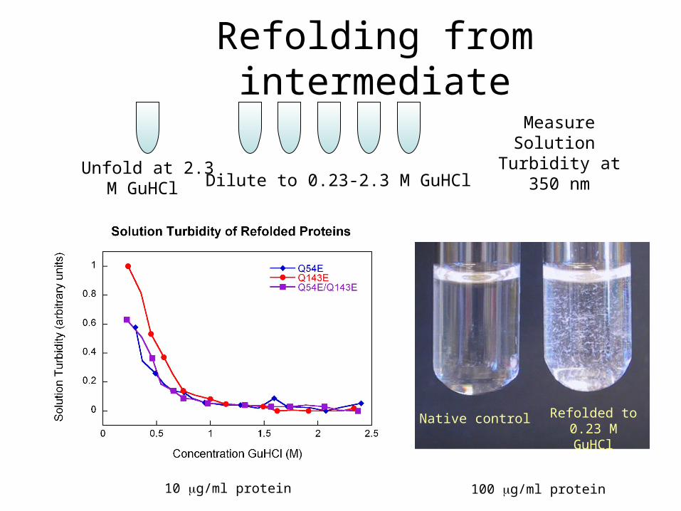
Unfold at 2.3 M GuHCl Dilute to 0.23-2.3 M GuHCl
Measure Solution Turbidity at 350
nm
Native control Refolded to 0.23 M GuHCl
10 g/ml protein 100 g/ml protein
Refolding from intermediate
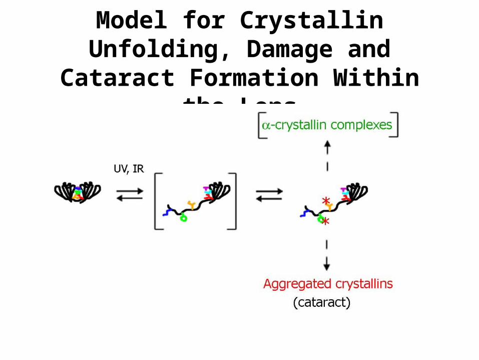
Model for Crystallin Unfolding, Damage and Cataract Formation
Within the Lens