septic shock karasin 1.ppt - Siriraj · PDF file• A subset of severe sepsis (SIRS) and...
61
Septic Shock Col. Danabhand Phiboonbanakit, M.D., Ph.D, Division of Infectious Diseases, Department of Medicine Phramongkutklao Hospital Phramongkutklao Hospital
Transcript of septic shock karasin 1.ppt - Siriraj · PDF file• A subset of severe sepsis (SIRS) and...
Septic Shock
Col. Danabhand Phiboonbanakit, M.D., Ph.D,
Division of Infectious Diseases, Department of Medicine
Phramongkutklao Hospital Phramongkutklao Hospital
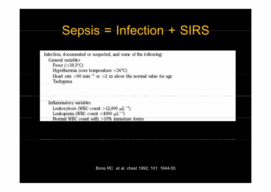
Sepsis = Infection + SIRSSepsis = Infection + SIRS
Levy MM et al. Crit Care Med 2003; 31: 1250-6Bone RC et al. chest 1992; 101: 1644-55
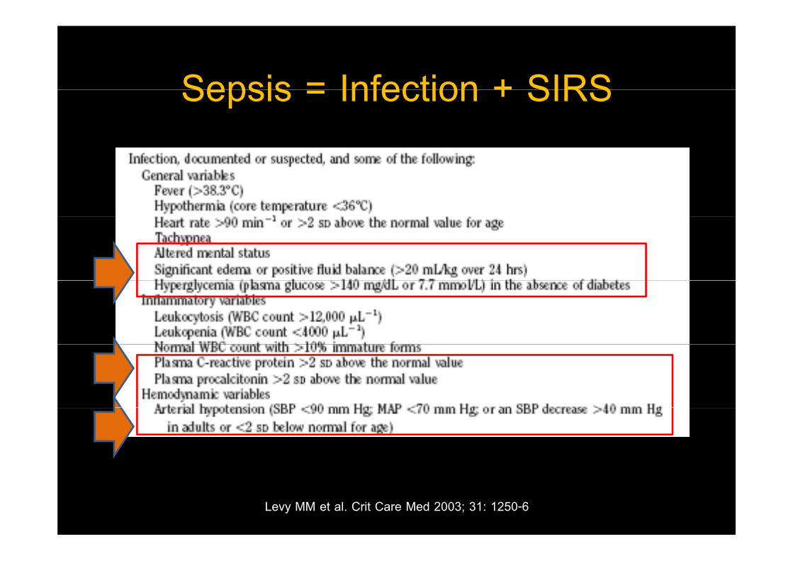
Sepsis = Infection + SIRSSepsis = Infection + SIRS
Levy MM et al. Crit Care Med 2003; 31: 1250-6
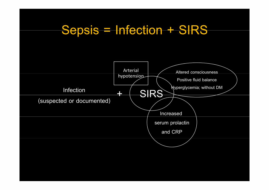
Sepsis = Infection + SIRSSepsis = Infection + SIRS
Altered consciousnessArterial
Infection SIRS
Altered consciousness
Positive fluid balance
Hyperglycemia; without DM
+
hypotension
(suspected or documented)SIRS
Increased
+
serum prolactin
and CRP
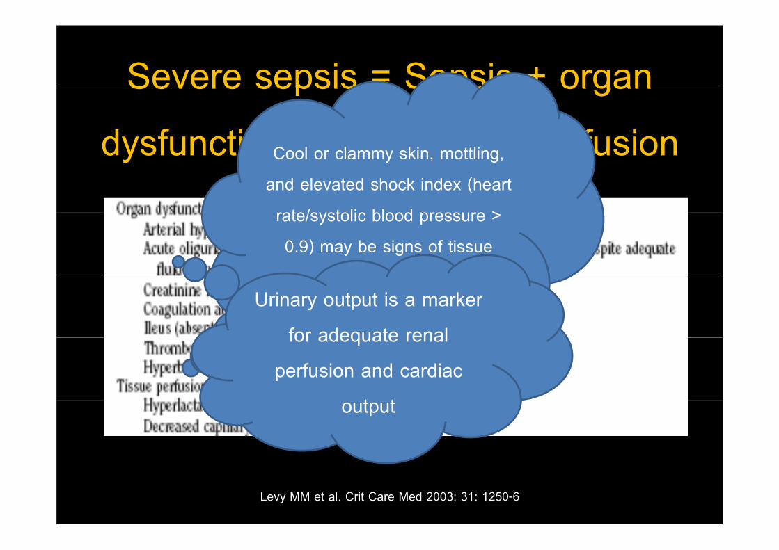
Severe sepsis = Sepsis + organ Severe sepsis Sepsis organ
dysfunction or hypotissue perfusionCool or clamm skin mottling dysfunction or hypotissue perfusionCool or clammy skin, mottling,
and elevated shock index (heart
rate/systolic blood pressure > rate/systolic blood pressure >
0.9) may be signs of tissue
hypo perfusionhypo-perfusionUrinary output is a marker
for adequate renal for adequate renal
perfusion and cardiac
t toutput
Levy MM et al. Crit Care Med 2003; 31: 1250-6
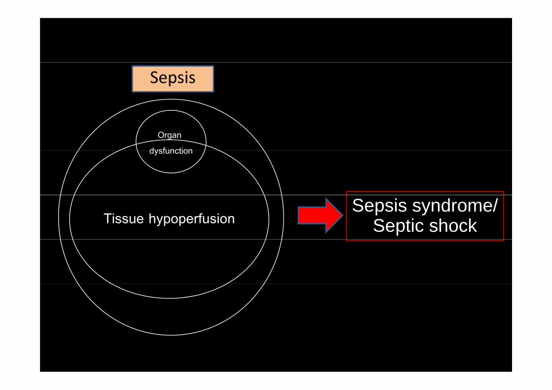
Sepsis
Organ
dysfunctiondysfunction
Tissue hypoperfusionSepsis syndrome/
Septic shock
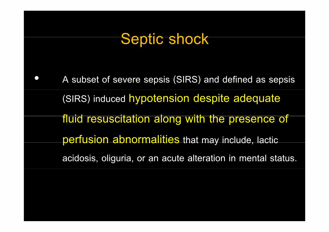
Septic shockSeptic shock
• A subset of severe sepsis (SIRS) and defined as sepsis
(SIRS) induced hypotension despite adequate
fl id it ti l ith th f fluid resuscitation along with the presence of
perfusion abnormalities that may include, lactic perfusion abnormalities that may include, lactic
acidosis, oliguria, or an acute alteration in mental status.
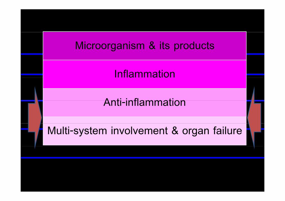
MicroorganismMicroorganism & its productsMicroorganism & its productsIts productsMicroorganism & its products
InflammationInflammation
Its products
Inflammation
Anti inflammationAnti inflammationAnti-inflammationAnti-inflammation
Multi-system involvement & organ failure
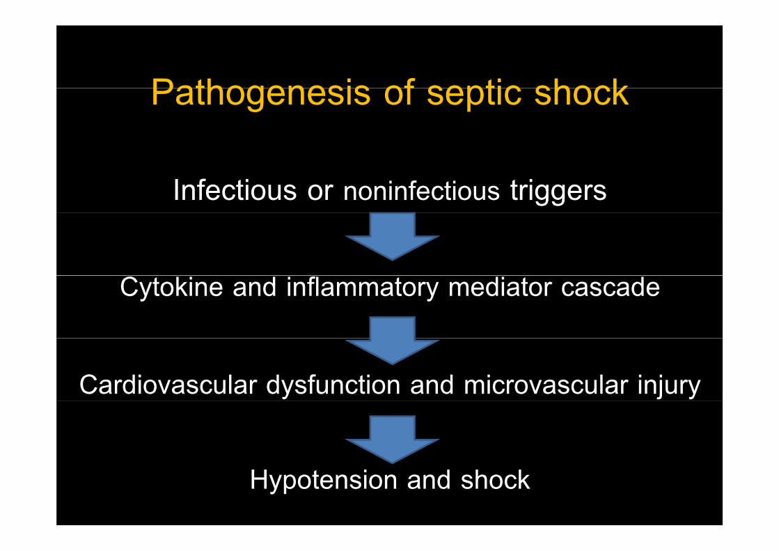
Pathogenesis of septic shockPathogenesis of septic shock
Infectious or noninfectious triggers
Cytokine and inflammatory mediator cascade
Cardiovascular dysfunction and microvascular injury
Hypotension and shock
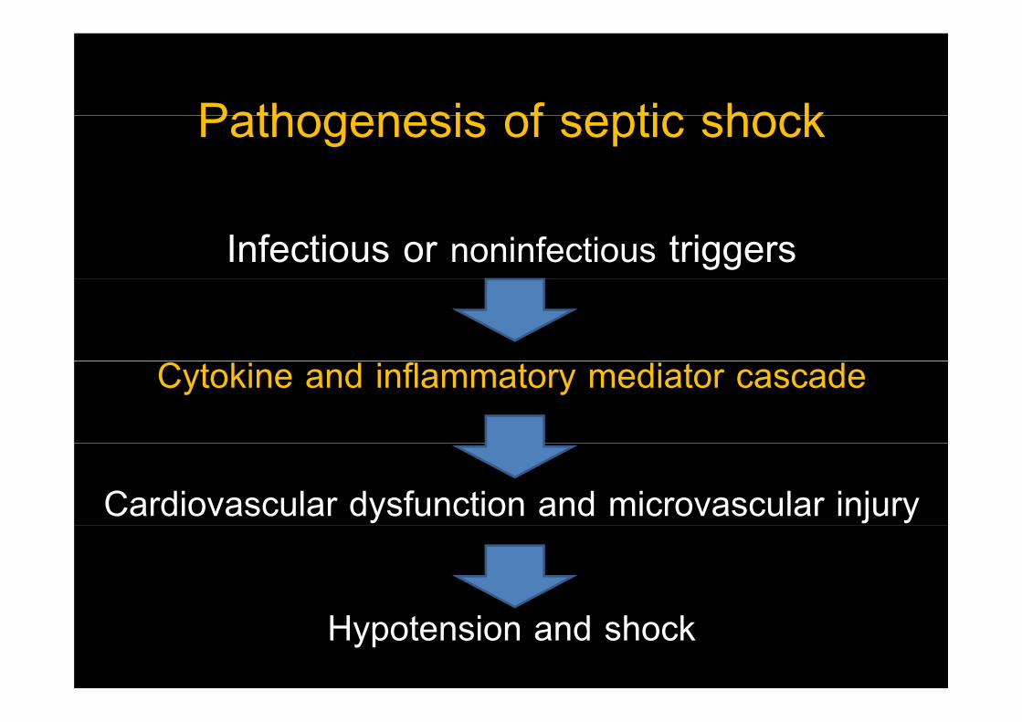
Pathogenesis of septic shockPathogenesis of septic shock
Infectious or noninfectious triggers
Cytokine and inflammatory mediator cascade
Cardiovascular dysfunction and microvascular injury
Hypotension and shock
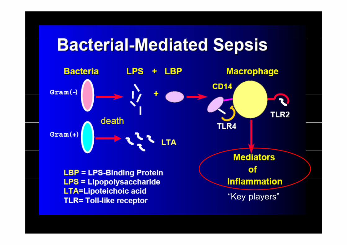
“Key players”
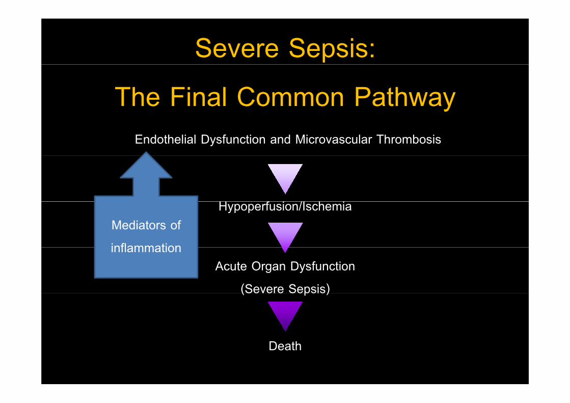
Severe Sepsis:
The Final Common PathwayyEndothelial Dysfunction and Microvascular Thrombosis
H f i /I h iHypoperfusion/IschemiaMediators of
inflammationAcute Organ Dysfunction
(Severe Sepsis)
inflammation
(Severe Sepsis)
Death
The insights in pathogenesisThe insights in pathogenesis
of septic shockof septic shock
• No single biological substance or mechanism is
fully responsible for septic shockfully responsible for septic shock.
• Peripheral vasodilatation and vasopressor-poorly
responded blood vessels are characteristics of
septic shock (Vasodilatory shock)septic shock. (Vasodilatory shock)
Septic shockSeptic shock
• Alternatively, distributive shock is caused by such y, y
conditions as direct arteriovenous shunting and is
characterized by decreased resistance or
increased venous capacity from the increased venous capacity from the
vasomotor dysfunction. y
Circulatory and metabolic Circulatory and metabolic
pathophysiology of septic shock
• Th d i t h d i f t f ti h k
pathophysiology of septic shock
• The predominant hemodynamic feature of septic shock
is arterial vasodilatation is arterial vasodilatation.
• Diminished peripheral arterial vascular tone causesp p
vasodilatation to result in hypotension and shock if
insufficiently compensated by a rise in cardiac
outputoutput.
Circulatory and metabolic Circulatory and metabolic
pathophysiology of septic shockpathophysiology of septic shock
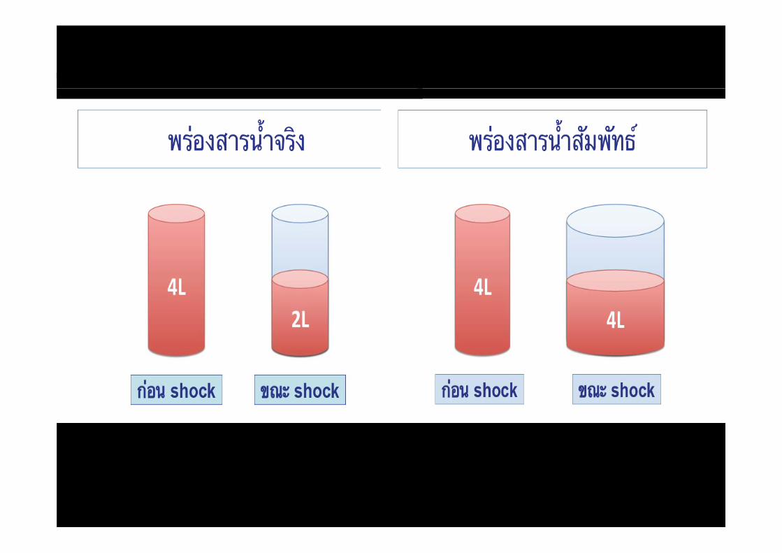
• Early in septic shock, the rise in cardiac output often is
limited by hypovolemia and a fall in preload because of limited by hypovolemia and a fall in preload because of
low cardiac filling pressures.
• When intravascular volume is augmented the cardiac When intravascular volume is augmented, the cardiac
output usually is elevated (the hyperdynamic phase of
sepsis and shock).
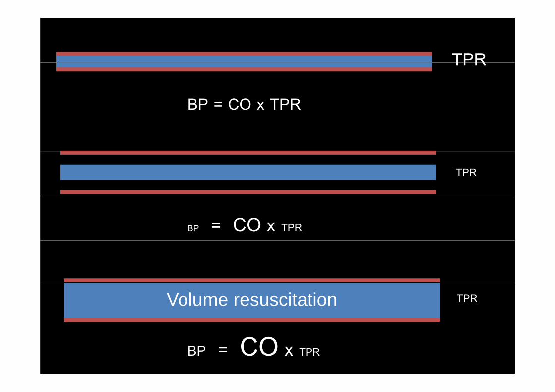
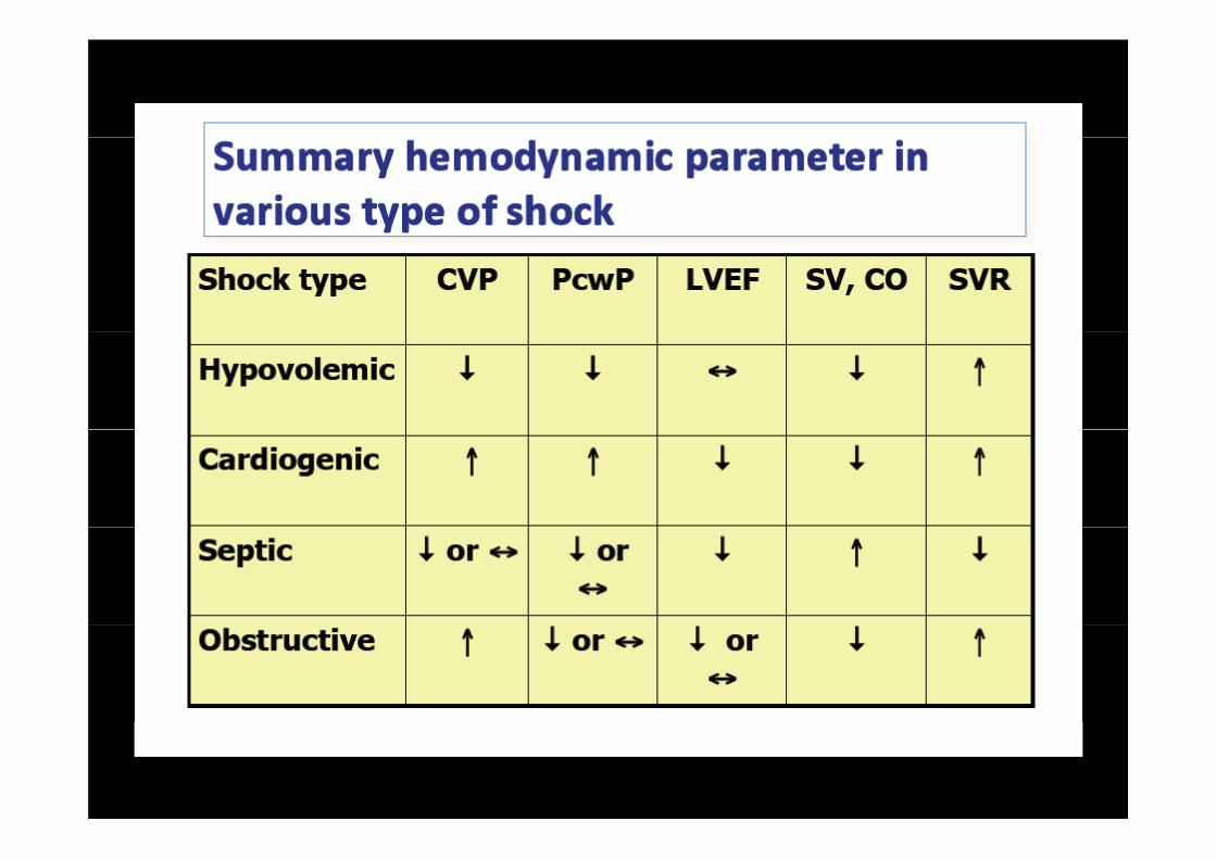
TPRTPR
BP = CO x TPRBP CO x TPR
TPR
BP = CO x TPR
Volume resuscitation TPR
BP = CO x TPR
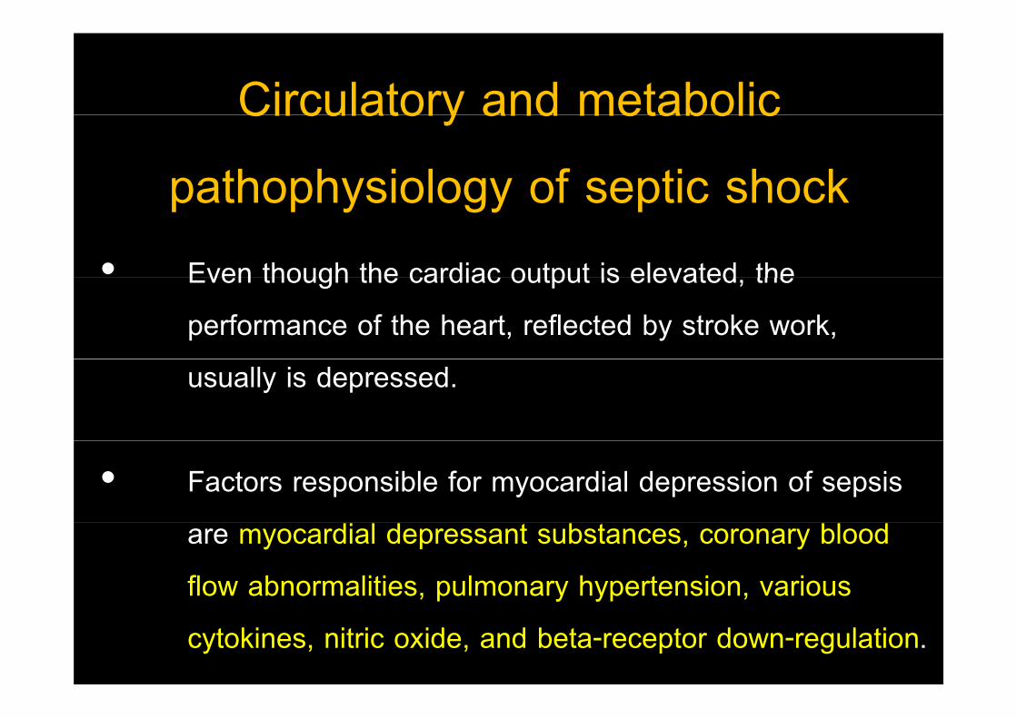
Circulatory and metabolic Circulatory and metabolic
pathophysiology of septic shock• Even though the cardiac output is elevated the
pathophysiology of septic shockEven though the cardiac output is elevated, the
performance of the heart, reflected by stroke work,
usually is depressed.
• Factors responsible for myocardial depression of sepsis
are myocardial depressant substances, coronary blood
flow abnormalities, pulmonary hypertension, various
cytokines, nitric oxide, and beta-receptor down-regulation.
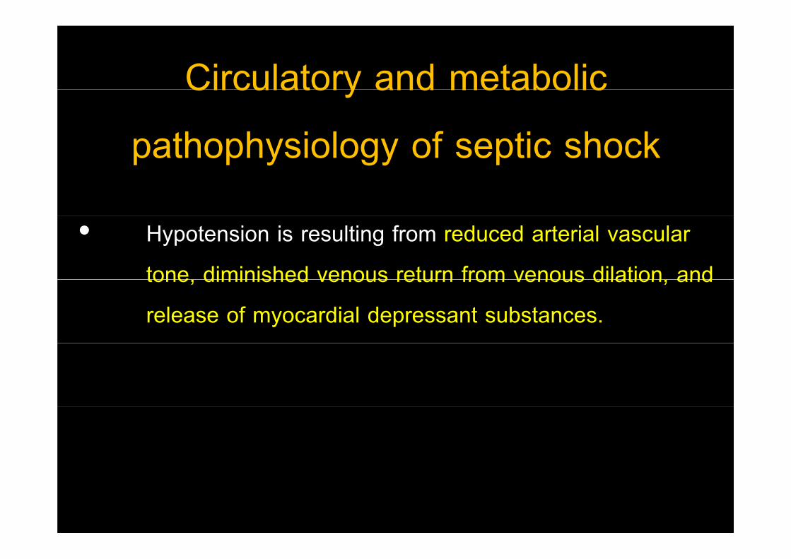
Circulatory and metabolic Circulatory and metabolic
pathophysiology of septic shockpathophysiology of septic shock
• Hypotension is resulting from reduced arterial vascular
tone diminished venous return from venous dilation and tone, diminished venous return from venous dilation, and
release of myocardial depressant substances.
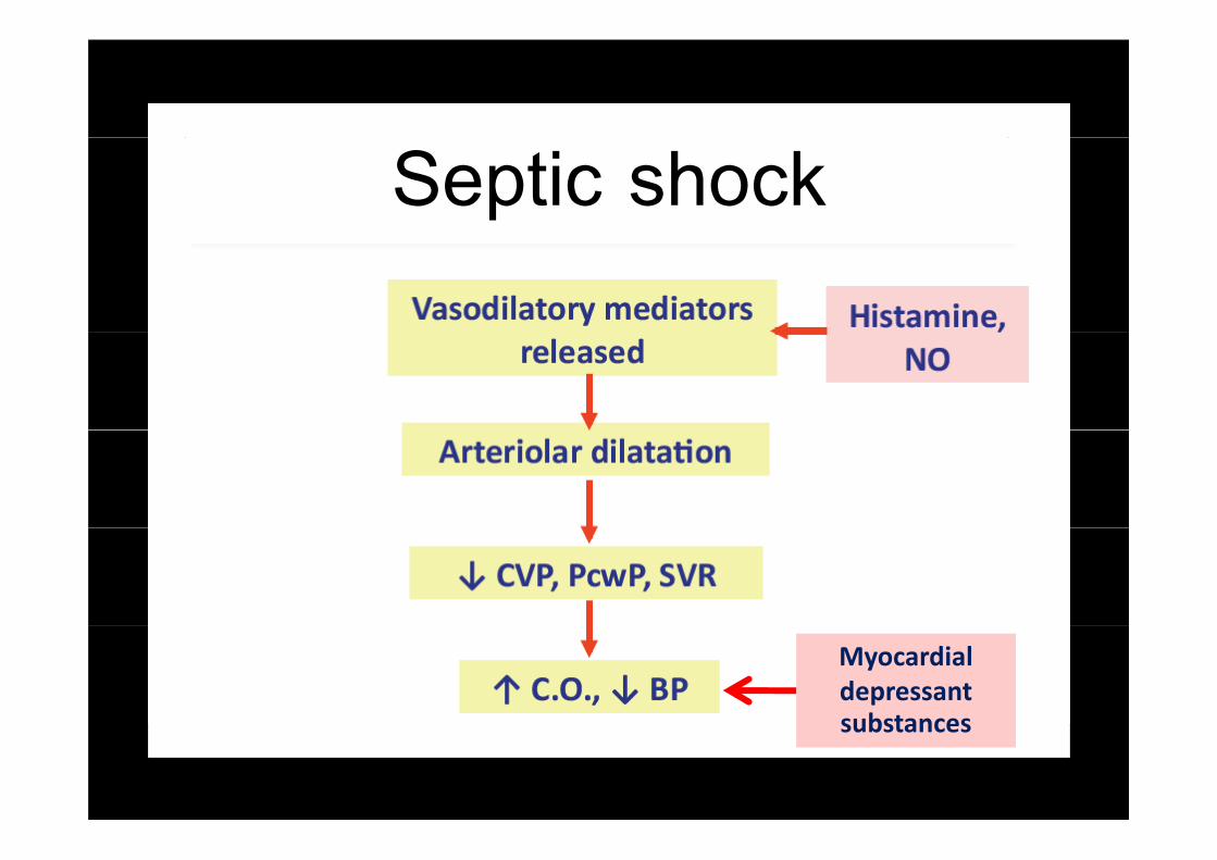
Septic shock
Myocardial depressant substancessubstances
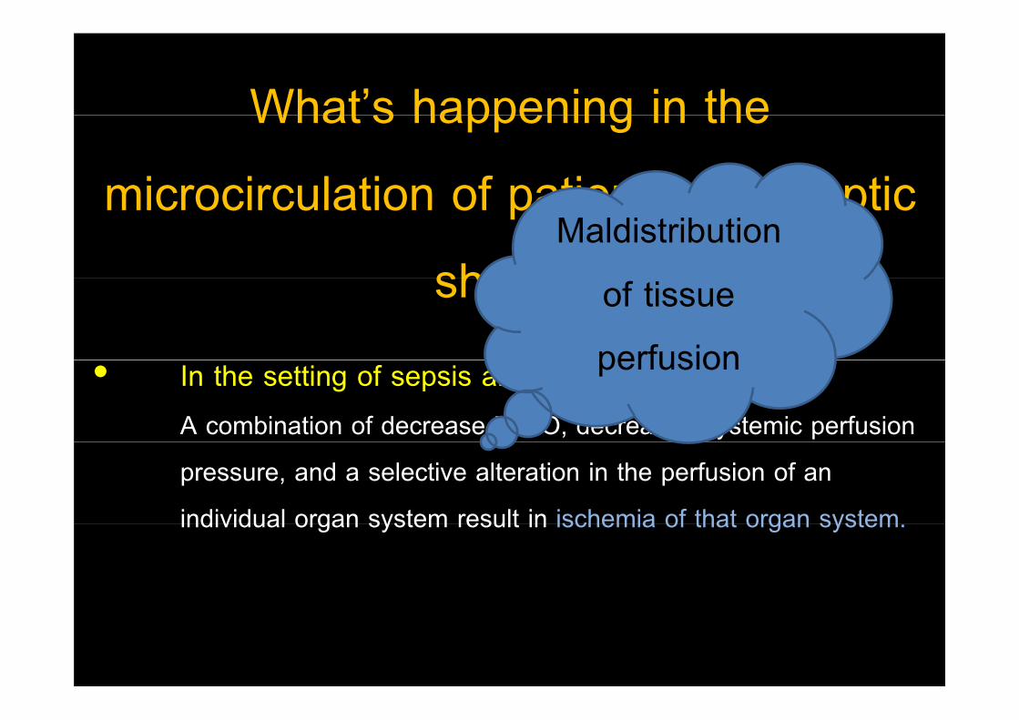
What’s happening in the What s happening in the
microcirculation of patients with septic microcirculation of patients with septic
shock?Maldistribution
shock? of tissue
perfusion• In the setting of sepsis and SIRS: A combination of decrease in CO, decreased systemic perfusion
perfusion
pressure, and a selective alteration in the perfusion of an
individual organ system result in ischemia of that organ system individual organ system result in ischemia of that organ system.
Distribution of blood flowDistribution of blood flow
• The peripheral blood flow : The balance between local
regulation of arterial tone and the activity of central
mechanisms (e.g., autonomic nervous system). g y
• Th i l l ti l f dil ti • The regional regulation, release of vasodilating
substances (e.g., nitric oxide, prostacyclin), and
vasoconstricting substances (e.g. endothelin) affect the
regional blood flow regional blood flow.
What’s happening at the level of
microcirculation?• Significant derangement in the autoregulation of
circulation is typical in patients with sepsis.
• Nitric oxide plays a central role in the vasodilatation of
septic shock Impaired secretion of vasopressin also may septic shock. Impaired secretion of vasopressin also may
occur, which may permit the persistence of vasodilatation.
• Vasoactive mediators also cause an increase in
the microvascular permeability at the site of infection.
What’s happening at the level of What s happening at the level of
i i l ti ?microcirculation?
• Maldistribution of blood flow, disturbances in the
i i l ti d tl i h l h ti f microcirculation, and, consequently, peripheral shunting of
oxygen are responsible for diminished oxygen extraction
and uptake, and lactate acidemia in patients experiencing
septic shockseptic shock.
What’s happening at the level of What s happening at the level of
i i l ti ?microcirculation?
• Development of increased systemic microvascular
permeability also occurs remote from the infectious permeability also occurs, remote from the infectious
focus, contributing to edema of various organs,
particularly the lung microcirculation and development of
ARDSARDS.
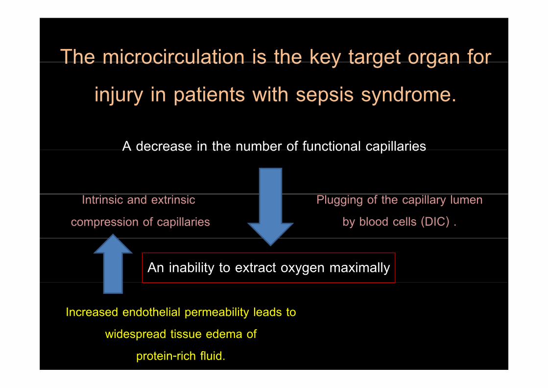
The microcirculation is the key target organ for The microcirculation is the key target organ for
injury in patients with sepsis syndrome.
A decrease in the number of functional capillariesA decrease in the number of functional capillaries
Intrinsic and extrinsic
compression of capillaries
Plugging of the capillary lumen
by blood cells (DIC) .
An inability to extract oxygen maximally
Increased endothelial permeability leads to
widespread tissue edema of
protein-rich fluid.
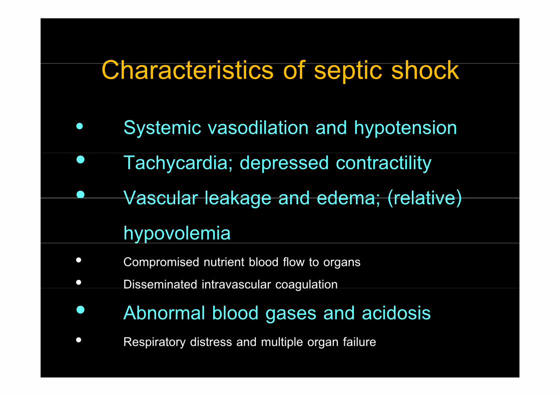
Ch t i ti f ti h kCharacteristics of septic shock
• Systemic vasodilation and hypotension
• Tachycardia; depressed contractility
• Vascular leakage and edema; (relative) Vascular leakage and edema; (relative)
hypovolemiayp• Compromised nutrient blood flow to organs
• Disseminated intravascular coagulationDisseminated intravascular coagulation
• Abnormal blood gases and acidosis• Respiratory distress and multiple organ failure
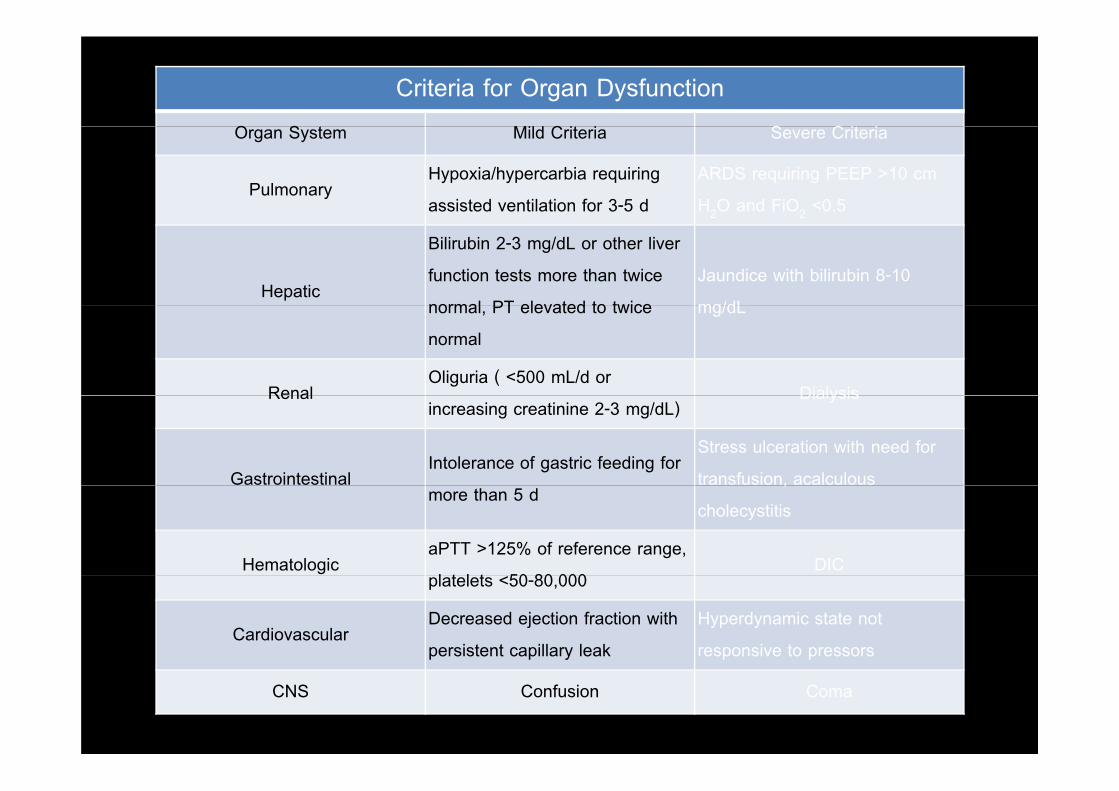
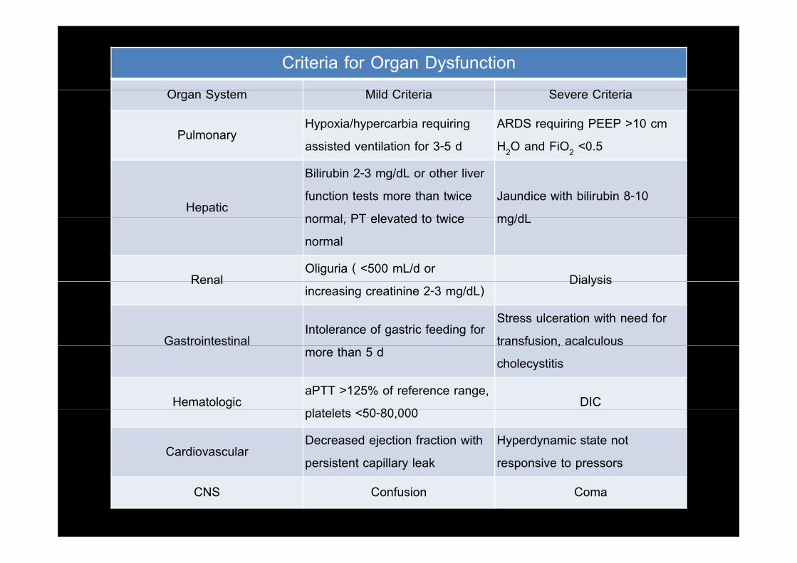
Criteria for Organ DysfunctionO S Mild C i i S C i iOrgan System Mild Criteria Severe Criteria
PulmonaryHypoxia/hypercarbia requiring
assisted ventilation for 3-5 d
ARDS requiring PEEP >10 cm
H2O and FiO2 <0.52 2
Hepatic
Bilirubin 2-3 mg/dL or other liver
function tests more than twice
normal PT elevated to twice
Jaundice with bilirubin 8-10
mg/dLnormal, PT elevated to twice
normal
mg/dL
RenalOliguria ( <500 mL/d or
DialysisRenalincreasing creatinine 2-3 mg/dL)
Dialysis
GastrointestinalIntolerance of gastric feeding for
Stress ulceration with need for
transfusion, acalculousmore than 5 d
,
cholecystitis
HematologicaPTT >125% of reference range,
l t l t 50 80 000DIC
platelets <50-80,000
CardiovascularDecreased ejection fraction with
persistent capillary leak
Hyperdynamic state not
responsive to pressors
CNS Confusion Coma
Multiple Organ Dysfunction Syndrome Multiple Organ Dysfunction Syndrome
(MODS)(MODS)
• Sepsis is described as an autodestructive process
that permits the extension of normal pathophysiologic that permits the extension of normal pathophysiologic
response to infection (involving otherwise normal
tissues), resulting in multiple organ dysfunction
syndrome.y
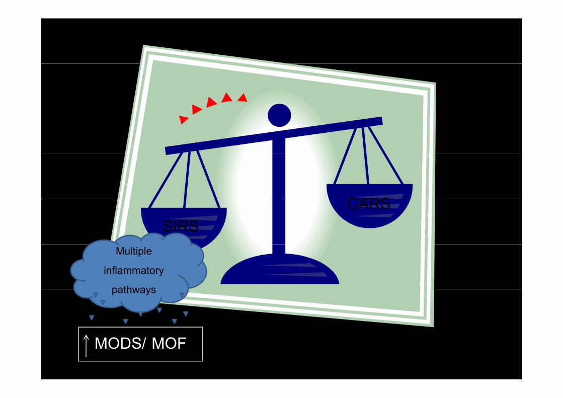
CARSSIRS
CARS
Multiple
inflammatory
pathwayspathways
MODS/ MOF
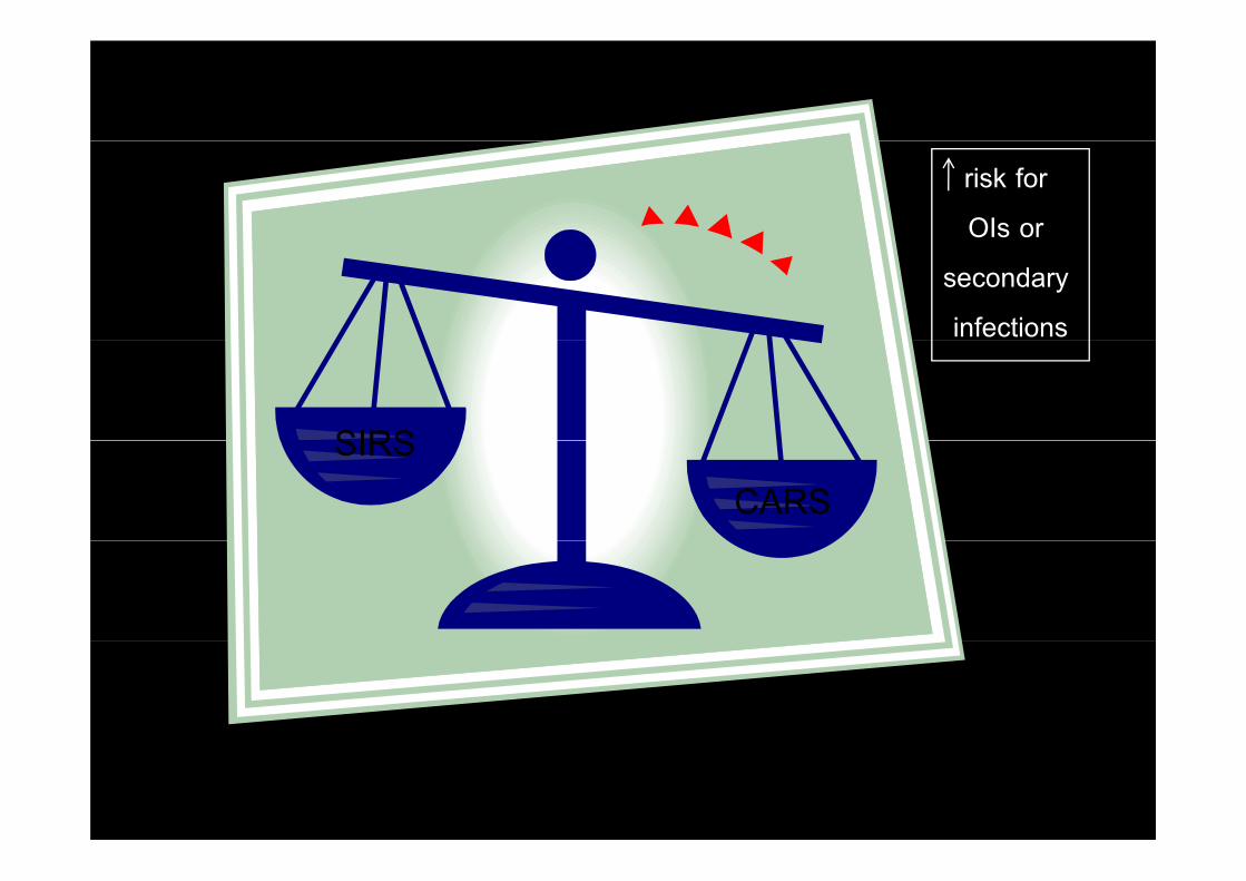
risk for
OIs or
secondary
infections
SIRSSIRSCARS
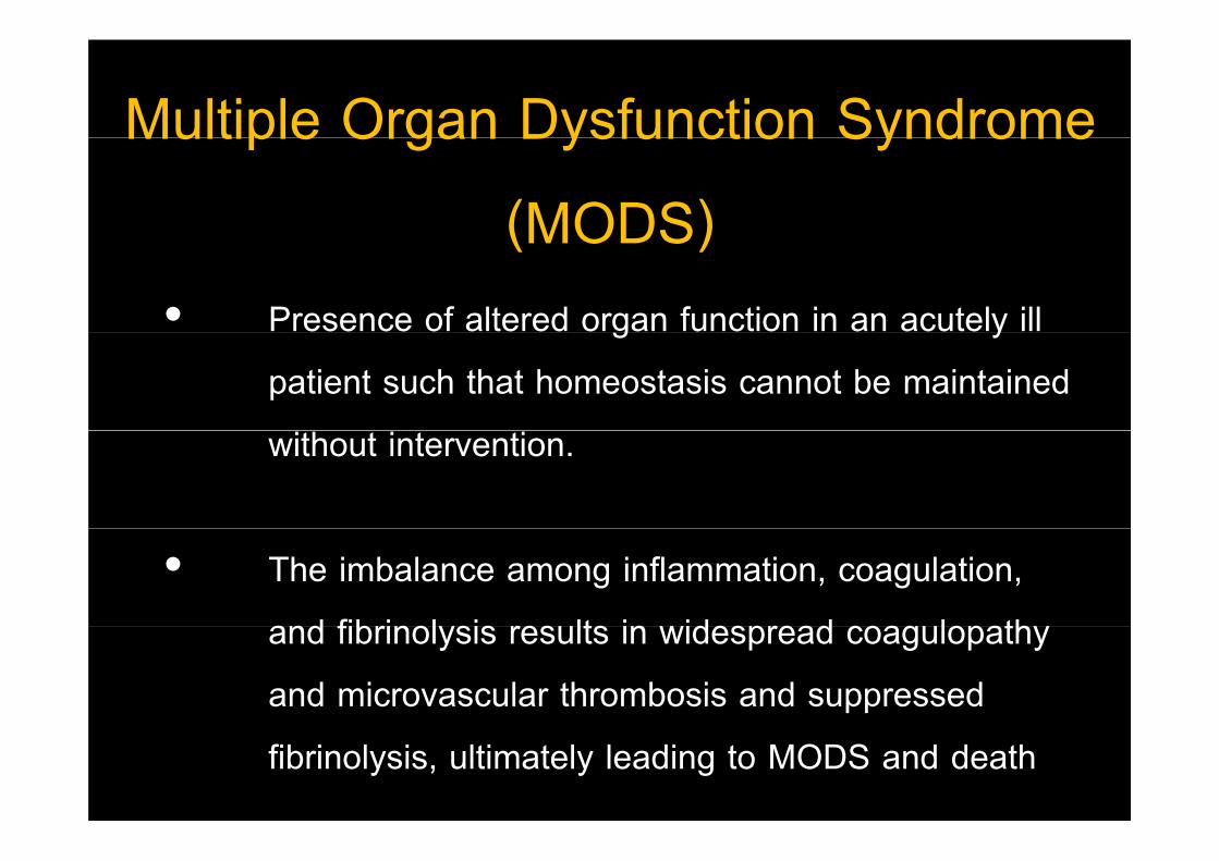
Multiple Organ Dysfunction Syndrome Multiple Organ Dysfunction Syndrome
(MODS)• Presence of altered organ function in an acutely ill
(MODS)g y
patient such that homeostasis cannot be maintained
without intervention.
• The imbalance among inflammation, coagulation,
and fibrinolysis results in widespread coagulopathy and fibrinolysis results in widespread coagulopathy
and microvascular thrombosis and suppressed
fibrinolysis, ultimately leading to MODS and death
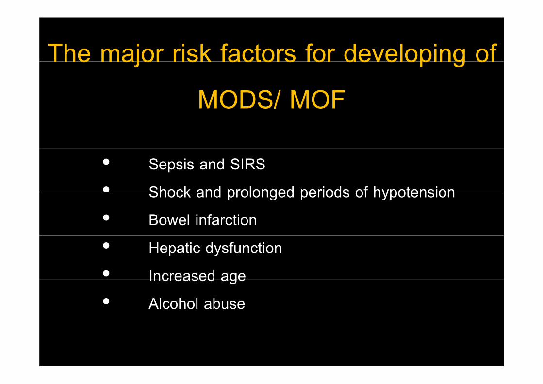
The major risk factors for developing of The major risk factors for developing of
MODS/ MOFMODS/ MOF
• Sepsis and SIRS
• Shock and prolonged periods of hypotension Shock and prolonged periods of hypotension
• Bowel infarction
• Hepatic dysfunction
• Increased ageIncreased age
• Alcohol abuse
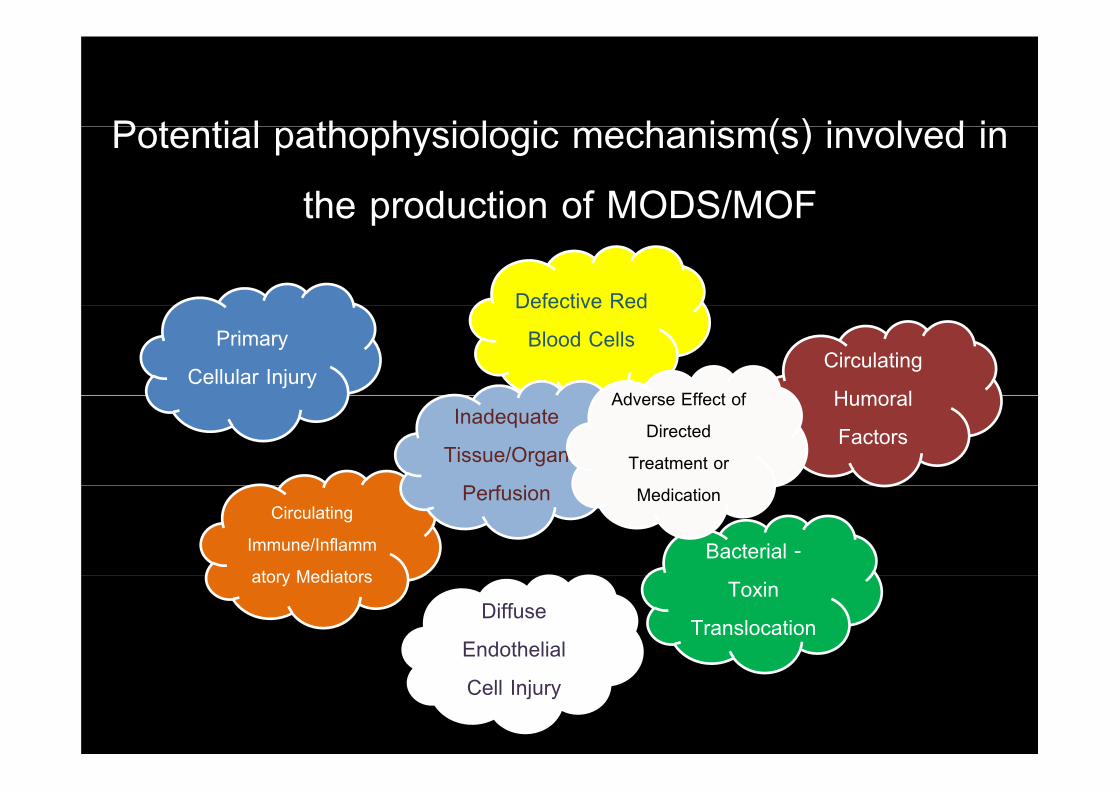
P t ti l th h i l i h i ( ) i l d i Potential pathophysiologic mechanism(s) involved in
the production of MODS/MOFthe production of MODS/MOF
Defective Red
Primary
Cellular Injury
Defective Red
Blood Cells Circulating
HumoralAd Eff t f Humoral
FactorsInadequate
Tissue/Organ
P f i
Adverse Effect of
Directed
Treatment or
Circulating
Immune/Inflamm
atory Mediators
Perfusion
Bacterial -
Medication
atory Mediators Toxin
TranslocationDiffuse
Endothelial
Cell Injury
Criteria for Organ DysfunctionO S Mild C i i S C i iOrgan System Mild Criteria Severe Criteria
PulmonaryHypoxia/hypercarbia requiring
assisted ventilation for 3-5 d
ARDS requiring PEEP >10 cm
H2O and FiO2 <0.52 2
Hepatic
Bilirubin 2-3 mg/dL or other liver
function tests more than twice
normal PT elevated to twice
Jaundice with bilirubin 8-10
mg/dLnormal, PT elevated to twice
normal
mg/dL
RenalOliguria ( <500 mL/d or
DialysisRenalincreasing creatinine 2-3 mg/dL)
Dialysis
GastrointestinalIntolerance of gastric feeding for
Stress ulceration with need for
transfusion, acalculousmore than 5 d
,
cholecystitis
HematologicaPTT >125% of reference range,
l t l t 50 80 000DIC
platelets <50-80,000
CardiovascularDecreased ejection fraction with
persistent capillary leak
Hyperdynamic state not
responsive to pressors
CNS Confusion Coma
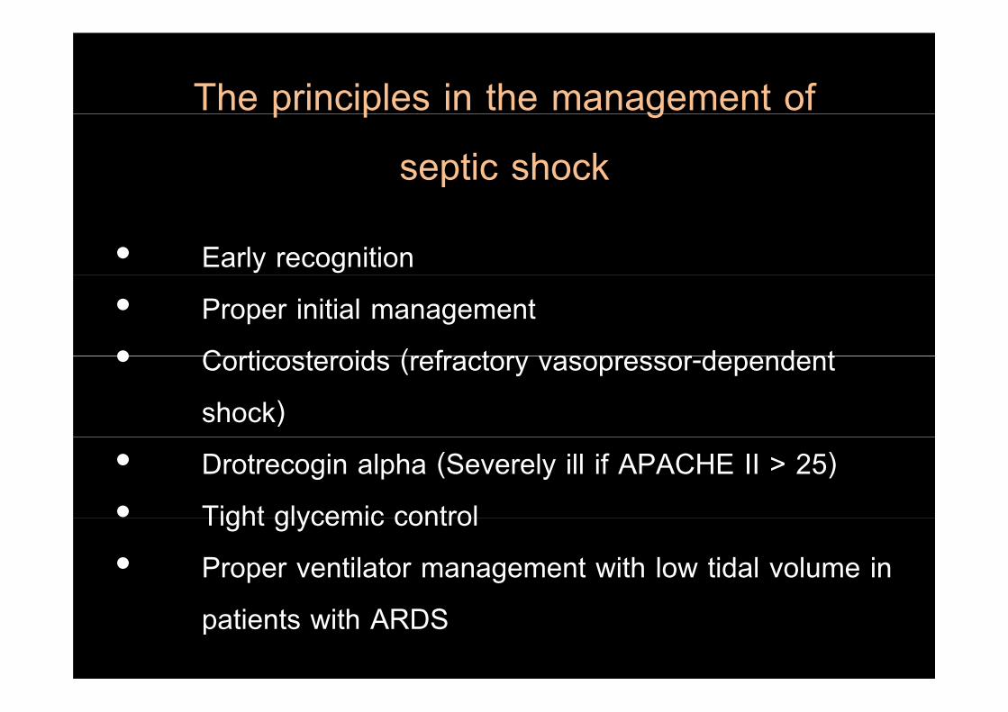
The principles in the management of p p g
septic shock
• Early recognition
• Proper initial management
• Corticosteroids (refractory vasopressor dependent • Corticosteroids (refractory vasopressor-dependent
shock)
• Drotrecogin alpha (Severely ill if APACHE II > 25)
• Tight glycemic control Tight glycemic control
• Proper ventilator management with low tidal volume in
patients with ARDS
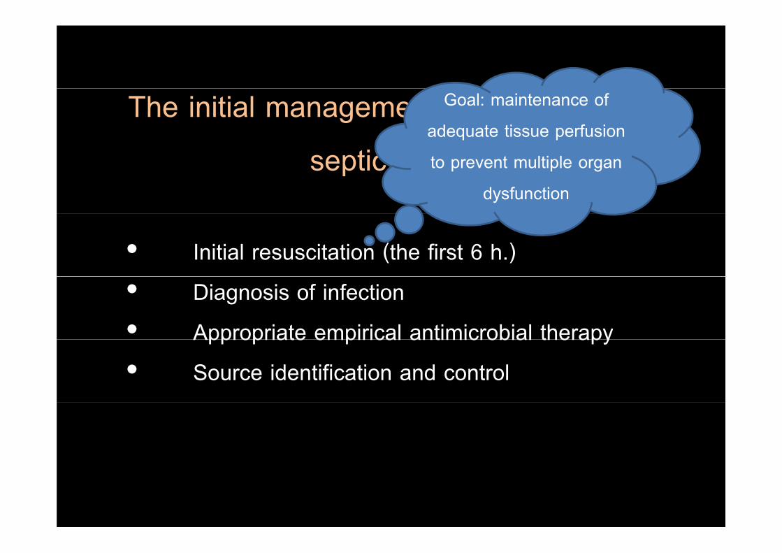
The initial management for patients with
ti h k
Goal: maintenance of
adequate tissue perfusion
septic shockto prevent multiple organ
dysfunction
• Initial resuscitation (the first 6 h.)
• Diagnosis of infection
• Appropriate empirical antimicrobial therapyAppropriate empirical antimicrobial therapy
• Source identification and control
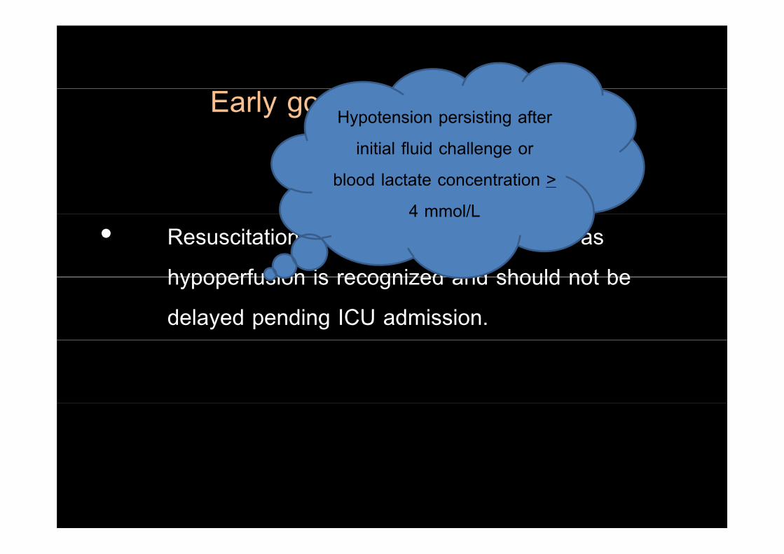
Early goal- directed therapyHypotension persisting after
initial fluid challenge or initial fluid challenge or
blood lactate concentration >
4 mmol/L• Resuscitation should be initiated as soon as
hypoperfusion is recognized and should not be
4 mmol/L
hypoperfusion is recognized and should not be
delayed pending ICU admission.
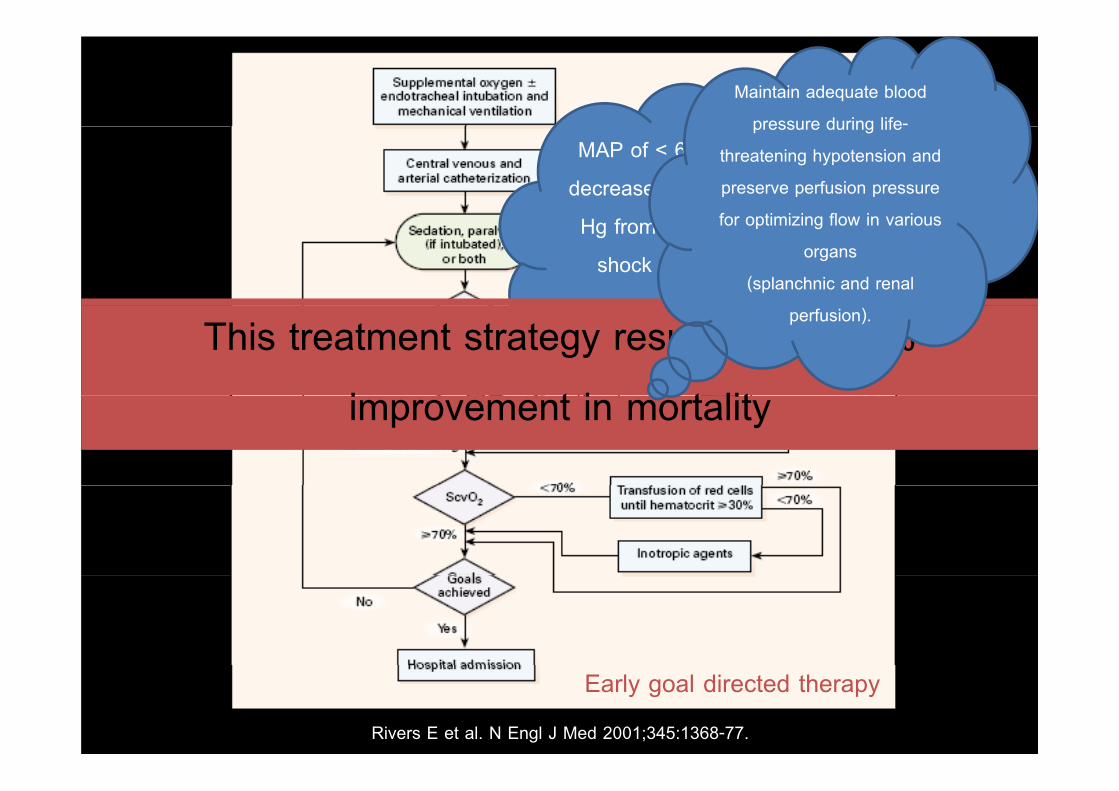
Maintain adequate blood
pressure during life-MAP of < 60 mm Hg or a
decrease in MAP of 40 mm
pressure during life
threatening hypotension and
preserve perfusion pressure
f ti i i fl i i Hg from baseline defines
shock at the bedside.
for optimizing flow in various
organs
(splanchnic and renal
This treatment strategy resulted in a 16%
i t i t lit
perfusion).
improvement in mortality
Early goal directed therapy
Rivers E et al. N Engl J Med 2001;345:1368-77.
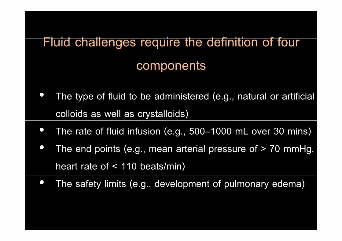
Fl id h ll i th d fi iti f f Fluid challenges require the definition of four
components
• Th t f fl id t b d i i t d ( t l tifi i l
components
• The type of fluid to be administered (e.g., natural or artificial
colloids as well as crystalloids)
• The rate of fluid infusion (e.g., 500–1000 mL over 30 mins)
• The end points (e g mean arterial pressure of > 70 mmHg • The end points (e.g., mean arterial pressure of > 70 mmHg,
heart rate of < 110 beats/min)
• The safety limits (e.g., development of pulmonary edema)
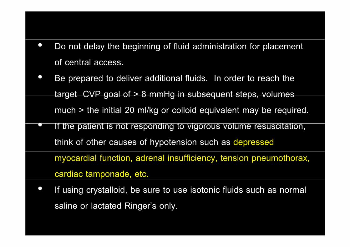
• Do not delay the beginning of fluid administration for placement
of central access.
• Be prepared to deliver additional fluids. In order to reach the
target CVP goal of > 8 mmHg in subsequent steps volumes target CVP goal of > 8 mmHg in subsequent steps, volumes
much > the initial 20 ml/kg or colloid equivalent may be required.
• If th ti t i t di t i l it ti • If the patient is not responding to vigorous volume resuscitation,
think of other causes of hypotension such as depressed
myocardial function, adrenal insufficiency, tension pneumothorax,
cardiac tamponade, etc.
• If using crystalloid, be sure to use isotonic fluids such as normal
saline or lactated Ringer’s only saline or lactated Ringer s only.
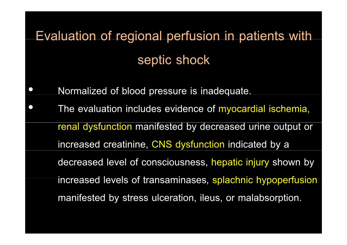
Evaluation of regional perfusion in patients with Evaluation of regional perfusion in patients with
septic shock
• Normalized of blood pressure is inadequate.p q
• The evaluation includes evidence of myocardial ischemia,
renal dysfunction manifested by decreased urine output or
increased creatinine, CNS dysfunction indicated by a
decreased level of consciousness, hepatic injury shown by
increased levels of transaminases splachnic hypoperfusion increased levels of transaminases, splachnic hypoperfusion
manifested by stress ulceration, ileus, or malabsorption.
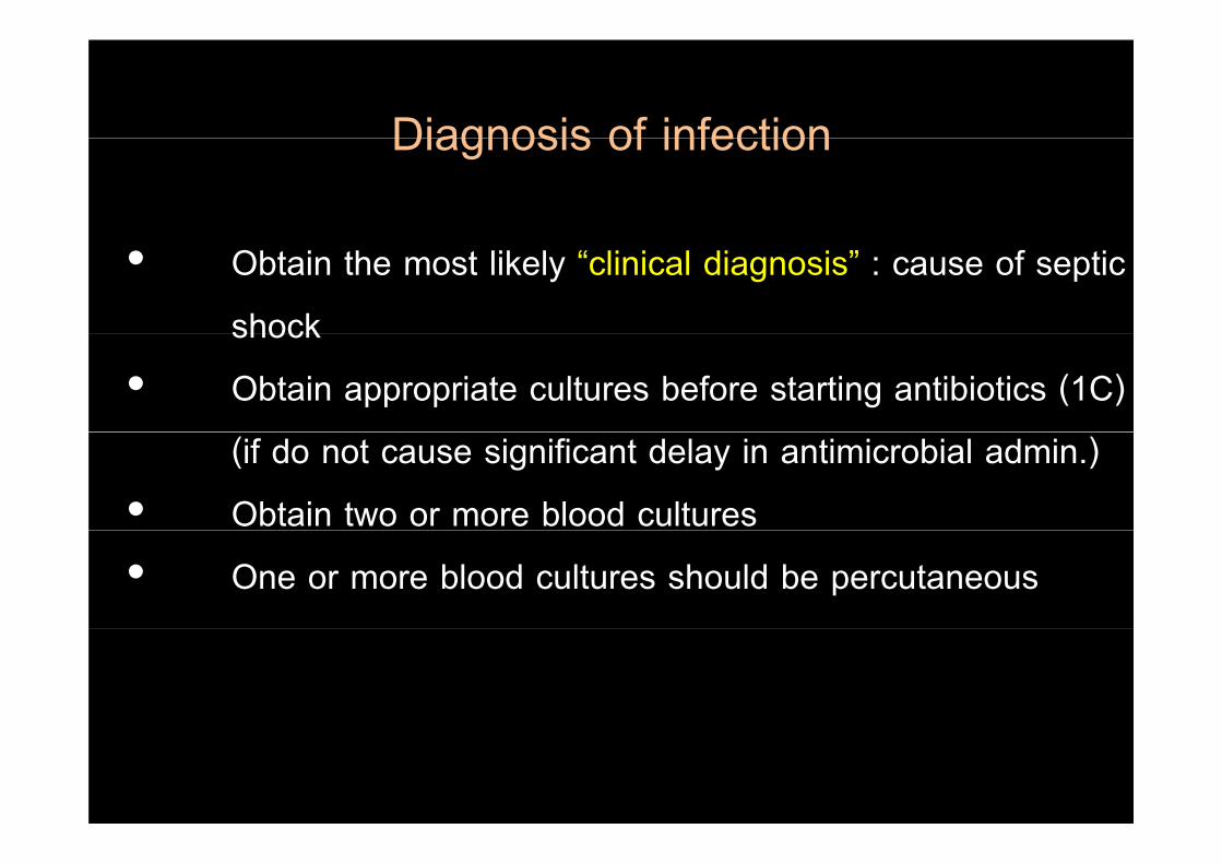
Diagnosis of infectionDiagnosis of infection
• Obtain the most likely “clinical diagnosis” : cause of septic
shockshock
• Obtain appropriate cultures before starting antibiotics (1C)
(if do not cause significant delay in antimicrobial admin.)
• Obtain two or more blood cultures
• One or more blood cultures should be percutaneous
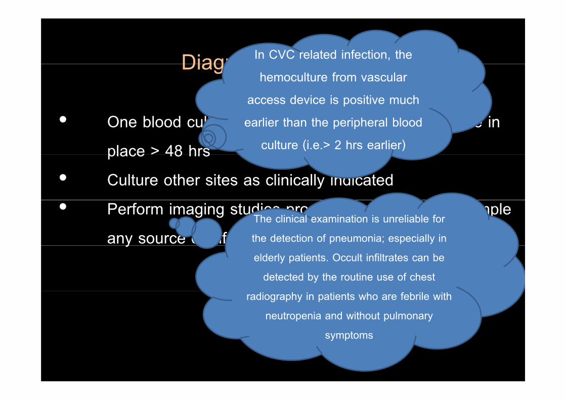
Diagnosis of infectionIn CVC related infection, the Diagnosis of infectionhemoculture from vascular
access device is positive much
• One blood culture from each vascular access device in
place > 48 hrs
earlier than the peripheral blood
culture (i.e.> 2 hrs earlier)place 48 hrs
• Culture other sites as clinically indicated
• Perform imaging studies promptly to confirm and sample
any source of infection, if safe to do so (1C)The clinical examination is unreliable for
the detection of pneumonia; especially in y , ( )elderly patients. Occult infiltrates can be
detected by the routine use of chest
radiography in patients who are febrile with
neutropenia and without pulmonary
symptomssymptoms
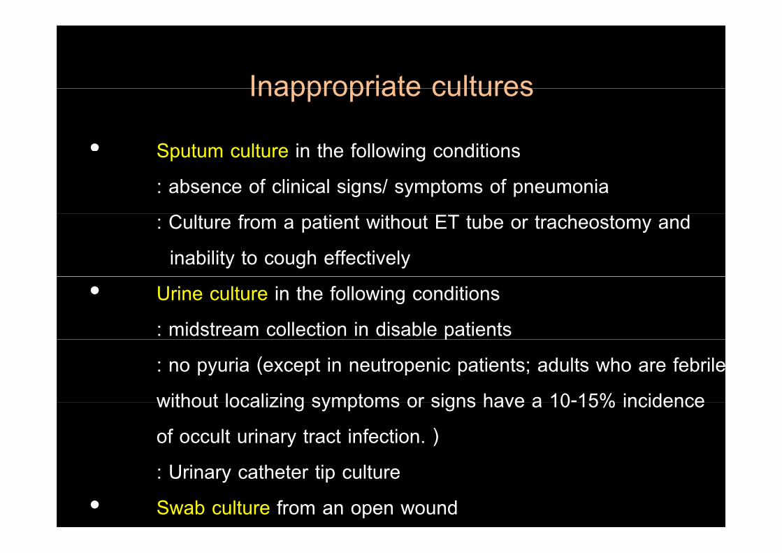
Inappropriate culturesInappropriate cultures
• Sputum culture in the following conditionsSputum culture in the following conditions
: absence of clinical signs/ symptoms of pneumonia
: Culture from a patient without ET tube or tracheostomy and
inability to cough effectively
• Urine culture in the following conditions
: midstream collection in disable patientsp
: no pyuria (except in neutropenic patients; adults who are febrile
without localizing symptoms or signs have a 10-15% incidence without localizing symptoms or signs have a 10-15% incidence
of occult urinary tract infection. )
U i h i l: Urinary catheter tip culture
• Swab culture from an open wound
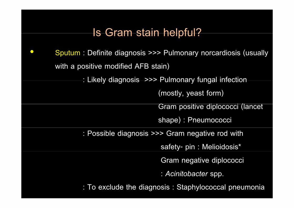
Is Gram stain helpful? Is Gram stain helpful? • Sputum : Definite diagnosis >>> Pulmonary norcardiosis (usually
with a positive modified AFB stain)
: Likely diagnosis >>> Pulmonary fungal infection: Likely diagnosis >>> Pulmonary fungal infection
(mostly, yeast form)
G iti di l i (l tGram positive diplococci (lancet
shape) : Pneumococci
: Possible diagnosis >>> Gram negative rod with
safety- pin : Melioidosis*
Gram negative diplococci
: Acinitobacter spp: Acinitobacter spp.
: To exclude the diagnosis : Staphylococcal pneumonia
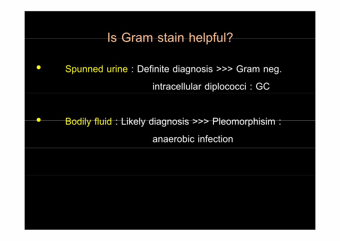
Is Gram stain helpful? Is Gram stain helpful?
• S d i D fi it di i G • Spunned urine : Definite diagnosis >>> Gram neg.
intracellular diplococci : GC
• Bodily fluid : Likely diagnosis >>> Pleomorphisim : • Bodily fluid : Likely diagnosis >>> Pleomorphisim :
anaerobic infection
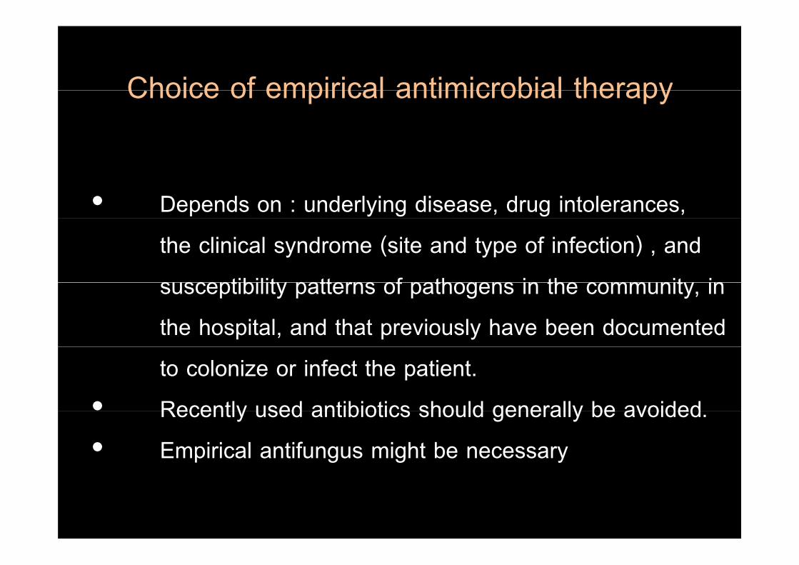
Choice of empirical antimicrobial therapyChoice of empirical antimicrobial therapy
• Depends on : underlying disease, drug intolerances,
the clinical syndrome (site and type of infection) , and
susceptibility patterns of pathogens in the community in susceptibility patterns of pathogens in the community, in
the hospital, and that previously have been documented
to colonize or infect the patient.
• Recently used antibiotics should generally be avoidedRecently used antibiotics should generally be avoided.
• Empirical antifungus might be necessary
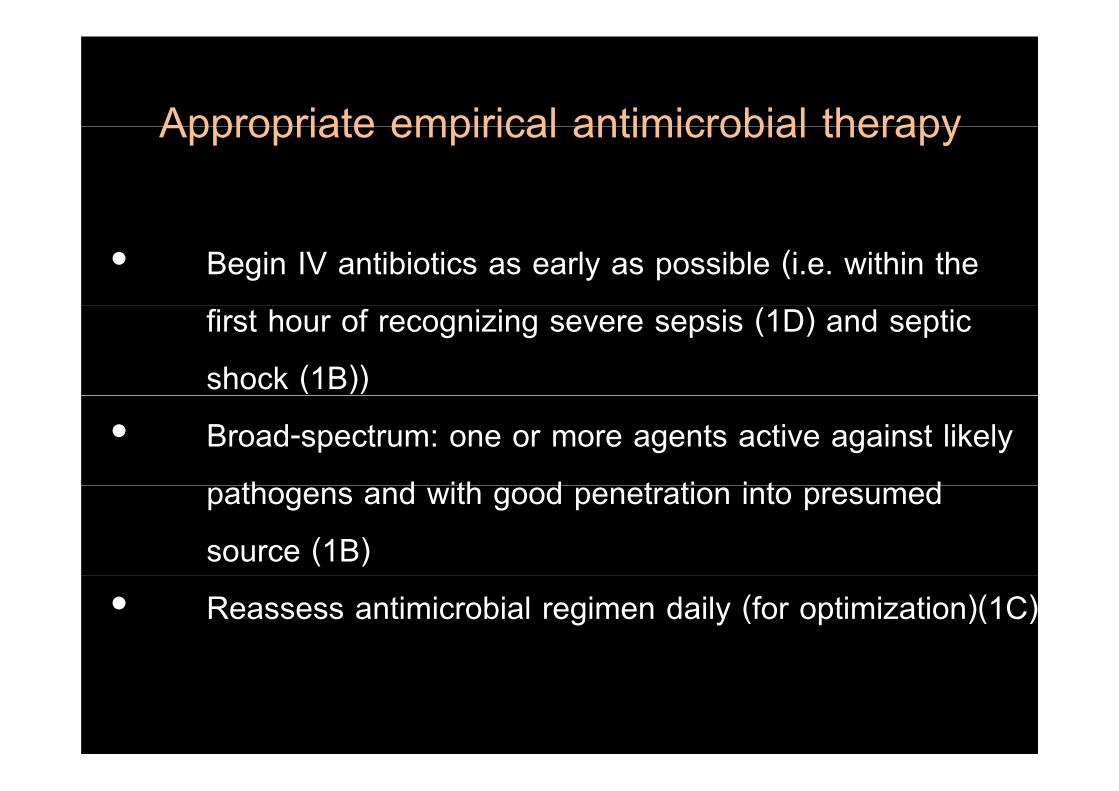
Appropriate empirical antimicrobial therapyAppropriate empirical antimicrobial therapy
• Begin IV antibiotics as early as possible (i.e. within the
first hour of recognizing severe sepsis (1D) and septic
shock (1B))
• Broad-spectrum: one or more agents active against likely
th d ith d t ti i t d pathogens and with good penetration into presumed
source (1B)
• Reassess antimicrobial regimen daily (for optimization)(1C)
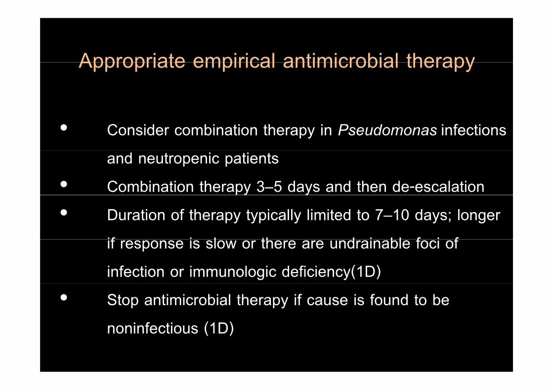
Appropriate empirical antimicrobial therapyAppropriate empirical antimicrobial therapy
• Consider combination therapy in Pseudomonas infections
and neutropenic patients
• Combination therapy 3–5 days and then de-escalationy y
• Duration of therapy typically limited to 7–10 days; longer
if i l th d i bl f i f if response is slow or there are undrainable foci of
infection or immunologic deficiency(1D)
• Stop antimicrobial therapy if cause is found to be
noninfectious (1D)noninfectious (1D)
TipsTips
• Blood cultures will be negative in > 50% of
cases of severe sepsis or septic shock yet many of cases of severe sepsis or septic shock, yet many of
these cases are very likely caused by bacteria or fungi.
• The decisions to continue narrow or stopThe decisions to continue, narrow, or stop
antimicrobial therapy must be made on the basis of
clinician judgment and clinical information.
Source identification and controlSource identification and control
• A specific anatomic site of infection should be established
idl ibl (1C) d ithi fi t 6 h f as rapidly as possible (1C) and within first 6 hrs of
presentation (1D)
• Formally evaluate patient for a focus of infection • Formally evaluate patient for a focus of infection
amenable to source control measures (e.g. abscess
drainage, tissue debridement) (1C)
Source identification and controlSource identification and control
• Implement source control measures as soon as possible
f ll i f l i iti l it ti (1C) ( ti following successful initial resuscitation (1C) (exception:
infected pancreatic necrosis, where surgical intervention
is best delayed)
• Choose source control measure with maximum efficacy Choose source control measure with maximum efficacy
and minimal physiologic upset (1D)
• Remove intravascular access devices if potentially
infected (1C)infected (1C)
Corticosteroid and septic shockCorticosteroid and septic shock
• Consider intravenous hydrocortisone for adult septic
h k h h t i d l t d t shock when hypotension responds poorly to adequate
fluid resuscitation and vasopressors.
• ACTH stimulation test is not recommended to identify the ACTH stimulation test is not recommended to identify the
subset of adults with septic shock who should receive
hydrocortisone.
Corticosteroid and septic shockCorticosteroid and septic shock
• Hydrocortisone is preferred to dexamethasone.
• Fludrocortisone (50 g orally once a day) may be • Fludrocortisone (50 g orally once a day) may be
included if an alternative to hydrocortisone is being used
that lacks significant mineralocorticoid activity.
Corticosteroid and septic shockCorticosteroid and septic shock
• Steroid therapy may be weaned once vasopressors
are no longer required.
• Hydrocortisone dose should be < 300 mg/day (1A)• Hydrocortisone dose should be < 300 mg/day (1A)
• Do not use corticosteroids to treat sepsis in the
absence of shock unless the patient’s endocrine or
corticosteroid history warrants it (1D)corticosteroid history warrants it (1D)
Recombinant human activated protein C and Recombinant human activated protein C and
septic shockp
• Consider rhAPC in adult patients with sepsis-induced
organ dysfunction with clinical assessment of high risk of
death (typically APACHE II > 25 or multiple organ failure) death (typically APACHE II > 25 or multiple organ failure)
if there are no contraindications
• Adult patients with severe sepsis and low risk of death Adult patients with severe sepsis and low risk of death
(typically, APACHE II < 20 or one organ failure) should
not receive rhAPC (1A)
Management of MODS/MOFManagement of MODS/MOF
• Th t f ti t ith MODS/MOF i • The management of patients with MODS/MOF is
predominantly supportive
• The specific treatment is directed at identifying and
treating the underlying disordertreating the underlying disorder
• Prevention of MODS/ MOF is plausible

Th k Y f Y Att tiThank You for Your Attention