Sepsis - Virginia Commonwealth Universitygbearman/Adobe files/SepsisM2.pdf · Inflammation is...
65
Sepsis Gonzalo Bearman MD, MPH Assistant Professor of Medicine Associate Hospital Epidemiologist
Transcript of Sepsis - Virginia Commonwealth Universitygbearman/Adobe files/SepsisM2.pdf · Inflammation is...

SepsisGonzalo Bearman MD, MPH
Assistant Professor of MedicineAssociate Hospital Epidemiologist

Sepsis• Epidemiology• Definitions
– Sepsis– SIRS– Severe sepsis
• Clinical, hematologic and immunologic manifestations
• Management• Clinical example
– Neisseria meningitides

Sands KE et al. Sands KE et al. JAMAJAMA. 1997;278:234. 1997;278:234--4040; ; †Based on data for septicemiaBased on data for septicemia. . §Murphy SL. National Vital Statistics Reports. ‡‡Angus DC et al. Angus DC et al. CritCrit Care MedCare Med. 2001. 2001
Severe Sepsis: A Significant Healthcare
Challenge• Major cause of morbidity and mortality worldwide
– Leading cause of death in noncoronary ICU (US)*– 11th leading cause of death overall (US) †§
• More than 750,000 cases of severe sepsis in US annually‡
• In the US, more than 500 patients die of severe sepsis daily‡

Severe Sepsis: Comparison With Other Major Diseases
Incidence of Severe Sepsis Mortality of Severe Sepsis
0
50
100
150
200
250
300
AIDS* Colon BreastCancer§
CHF† Severe Sepsis‡
Cas
es/1
00,0
00
0
50,000
100,000
150,000
200,000
250,000
Deat
hs/Y
ear
AIDS* SevereSepsis‡
AMI†Breast Cancer§
†National Center for Health Statistics, 2001. §American Cancer Society, 2001. *American Heart Association. 2000. ‡Angus DC et al. Crit Care Med. 2001 (In Press).

Severe Sepsis: An alarming national trendTodayToday
>750,000 cases of severe
sepsis/year in the US*
FutureFuture
200,000
400,000
600,000
800,000
1,000,000
1,200,000
1,400,000
1,600,000
1,800,000
2001 2025 2050
Year
100,000
200,000
300,000
400,000
500,000
600,000Severe Sepsis CasesUS Population
Seps
is C
ases
Tota
l US
Popu
latio
n/1,
000
*Angus DC. *Angus DC. Crit Crit Care Med.Care Med. 2001 (In Press).2001 (In Press).
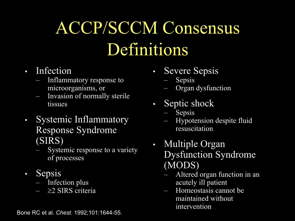
ACCP/SCCM Consensus Definitions
• Infection– Inflammatory response to
microorganisms, or– Invasion of normally sterile
tissues
• Systemic Inflammatory Response Syndrome (SIRS)– Systemic response to a variety
of processes
• Sepsis– Infection plus– ≥2 SIRS criteria
• Severe Sepsis– Sepsis– Organ dysfunction
• Septic shock– Sepsis– Hypotension despite fluid
resuscitation
• Multiple Organ Dysfunction Syndrome (MODS)– Altered organ function in an
acutely ill patient– Homeostasis cannot be
maintained without intervention
Bone RC et al. Chest. 1992;101:1644-55.

Sepsis: A Complex Disease
• Conceptual framework to view the relationships between various components of sepsis.
• The inflammatory changes of sepsis are tightly linked to disturbed hemostasis.
Adapted from: Bone RC et al. Chest. 1992;101:1644-55.Opal SM et al. Crit Care Med. 2000;28:S81-2.

SIRS: More Than Just a Systemic Inflammatory Response
• SIRS: A clinical response arising from a nonspecific insult manifested by ≥2 of the following:
– Temperature ≥38°C or ≤36°C
– HR ≥90 beats/min– Respirations ≥20/min– WBC count ≥12,000/µL or
≤4,000/µL or >10% immature neutrophils
• Recent evidence indicates that hemostatic changes are also involved
Adapted from: Bone RC et al. Chest. 1992;101:1644-55.Opal SM et al. Crit Care Med. 2000;28:S81-2.

Sepsis: More Than Just Inflammation
• Sepsis:– Known or suspected
infection– Two or more
SIRS criteria
• A significant link to disordered hemostasis
Adapted from: Bone RC et al. Chest. 1992;101:1644-55.

Severe Sepsis: Acute Organ Dysfunction and Disordered
Hemostasis• Severe Sepsis:
Sepsis with signs of organ dysfunction in ≥1 of the following systems: – Cardiovascular– Renal– Respiratory– Hepatic– Hemostasis– CNS– Unexplained metabolic
acidosis Adapted from: Bone RC et al. Chest. 1992;101:1644-55.
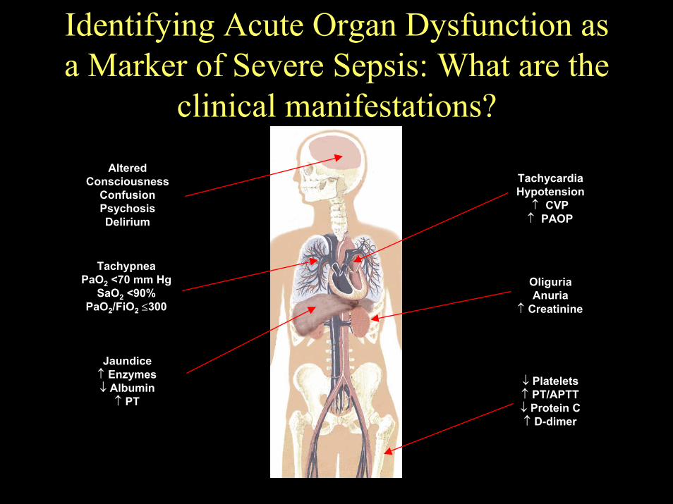
Identifying Acute Organ Dysfunction as a Marker of Severe Sepsis: What are the
clinical manifestations?Altered
ConsciousnessConfusionPsychosisDelirium
TachycardiaHypotension
↑ CVP↑ PAOP
TachypneaPaO2 <70 mm Hg
SaO2 <90%PaO2/FiO2 ≤300
OliguriaAnuria
↑ Creatinine
Jaundice↑ Enzymes↓ Albumin
↑ PT
↓ Platelets↑ PT/APTT↓ Protein C↑ D-dimer

Severe Sepsis: A Complex and Unpredictable Clinical Syndrome• High mortality rate
(28%-50%)
• Heterogeneous patient population
• Unpredictable disease progression
• Unclear etiology and pathogenesis
Angus DC et al. Crit Care Med. 2001; (In Press).Zeni F et al. Crit Care Med. 1997;25:1095-100.Wheeler AP et al. N Engl J Med. 1999;340:207-14.
Systemic Inflammation
Impaired Fibrinolysis
Coagulation
Severe sepsis

Systemic Inflammation and Disordered Homeostasis

Systemic Activation of Inflammation in SepsisInflammation is Activated in Sepsis
14
12
10
8
6
4
2
00 60 120 180 240 300 360
Minutes After LPS Infusion
Endo
toxi
n (n
g/L)
TNF
(ng/
L)
I
L-6
(U/m
L)
Chart adapted from: van Deventer SJ et al. Blood. 1990;76:2520-6.

Activation of Coagulation in Severe Sepsis
Data from: Bernard et al. N Engl J Med. 1997 Mar 27;336(13):912-8
0
20
40
60
80
100
↓ Platel↑ PTT↑ PT
Any OneAny TwoAll Thre↓ Protein C↑ D-Di
Per
cent
of P
atie
nts
mers
ets e

Impairment of Fibrinolysis in Severe Sepsis
0.0
0.2
0.4
0.6
0.8
1.0
1.2
1 4 7
Plas
min
ogen
/ant
ipla
smin
ratio
Time after hospital admission (day)
*P<.05 vs normal valuesP<.05 survivors vs nonsurvivors
** * *
1 4 7PA
I-1 (n
g/m
L)
Time after hospital admission (day)
*P<.05 vs normal values
***
**
*
Survivors (n=23) Nonsurvivors (n=25) Normal Values
Data from: Lorente JA et al. Chest. 1993;103:1536-42.
60
50
40
30
20
10
0
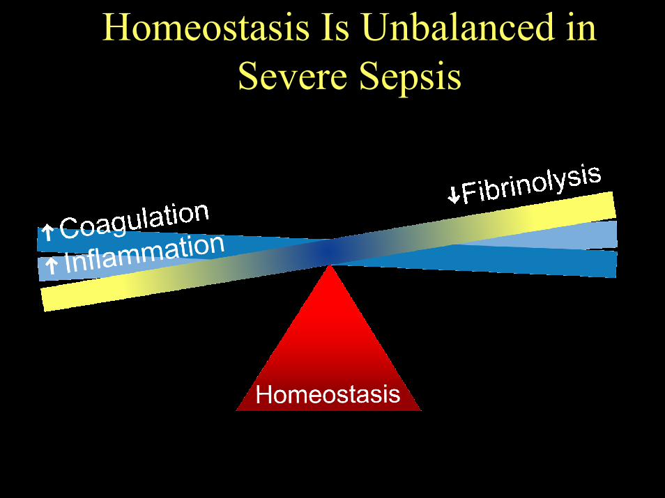
Homeostasis Is Unbalanced in Severe Sepsis
Carvalho AC, Freeman NJ. J Crit Illness. 1994;9:51-75; Kidokoro A et al. Shock.1996;5:223-8; Vervloet MG et al. Semin Thromb Hemost. 1998;24:33-44.
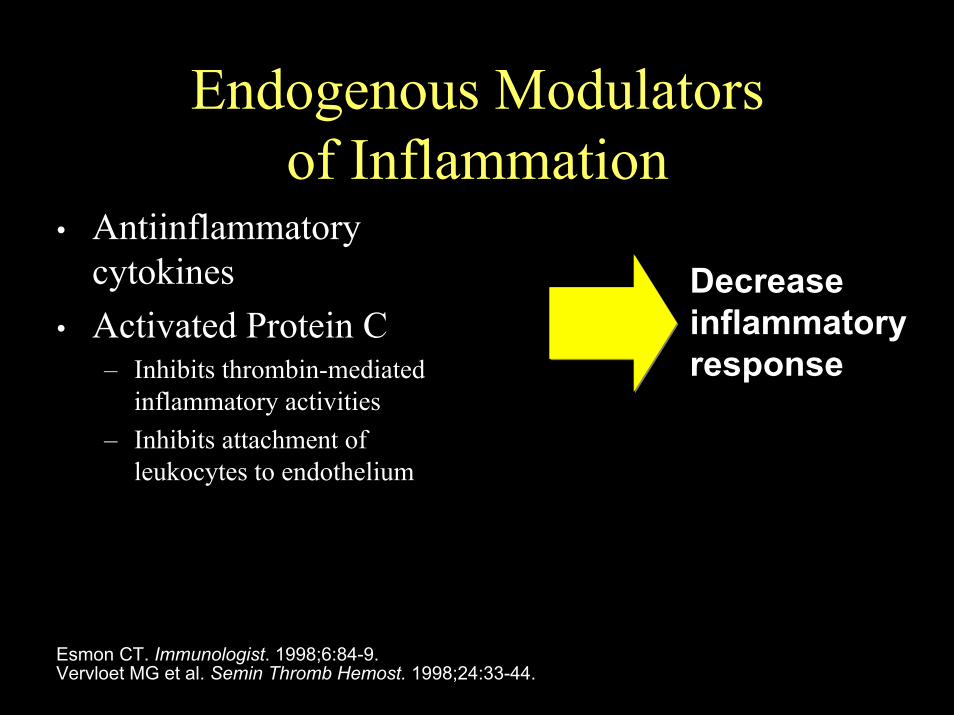
Endogenous Modulators of Inflammation
• Antiinflammatory cytokines
• Activated Protein C– Inhibits thrombin-mediated
inflammatory activities– Inhibits attachment of
leukocytes to endothelium
Esmon CT. Immunologist. 1998;6:84-9.Vervloet MG et al. Semin Thromb Hemost. 1998;24:33-44.
Decrease inflammatory response
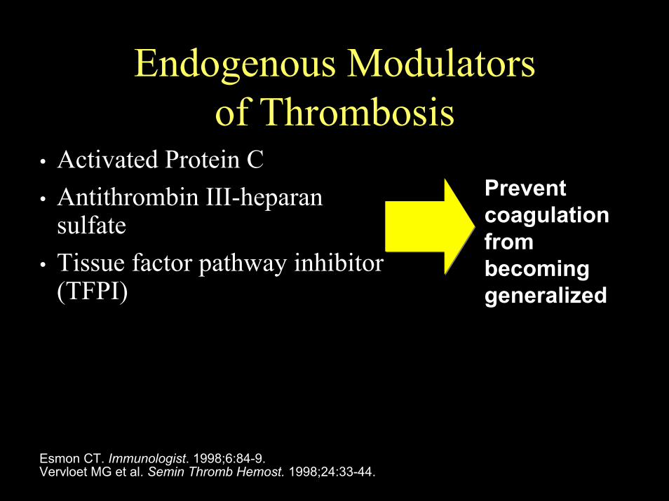
Endogenous Modulators of Thrombosis
• Activated Protein C• Antithrombin III-heparan
sulfate• Tissue factor pathway inhibitor
(TFPI)
Prevent coagulation from becoming generalized
Esmon CT. Immunologist. 1998;6:84-9.Vervloet MG et al. Semin Thromb Hemost. 1998;24:33-44.

Endogenous Modulators of Fibrinolysis
Tissue plasminogen activator (t-PA)Activated Protein C inhibits:
PAI-1TAFI activation
Remove formed microthrombi and maintain blood fluidity
Esmon CT. Immunologist. 1998;6:84-9.Vervloet MG et al. Semin Thromb Hemost. 1998;24:33-44.

Homeostasis
FibrinolysisFibrinolysisCoagulationCoagulationInflammationInflammation
Endogenous Activated Protein C Modulates Coagulation, Fibrinolysis, and Inflammation
in Severe Sepsis
Activated Protein C Activated Protein C
Carvalho AC et al. J Crit Illness. 1994;9:51-75; Kidokoro A et al. Shock. 1996;5:223-8; Vervloet MG et al. Semin Thromb Hemost. 1998;24:33-44.

Severe Sepsis: The Final Common Pathway
Endothelial Dysfunction and Microvascular Thrombosis
Hypoperfusion/Ischemia
Acute Organ Dysfunction (Severe Sepsis)
Death

Sepsis: Management

Severe Sepsis Therapy: Standard Care
• Source control
• Antibiotics
• Hemodynamic support
• Mechanical ventilation
• Renal replacement therapy
• Sedation/analgesia
• Ensure adequate
nutrition
• Provide hematological support
• Other supportive measures
Wheeler AP, Bernard GR. N Engl J Med. 1999;340:207-14.
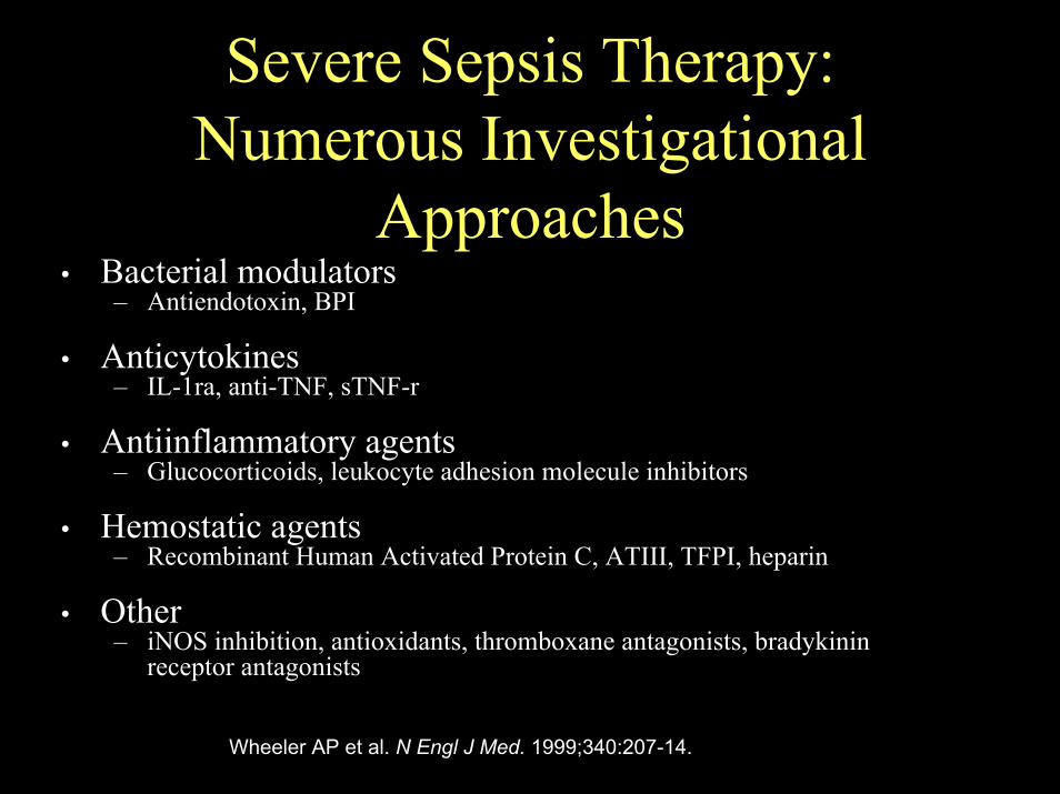
Severe Sepsis Therapy: Numerous Investigational
Approaches• Bacterial modulators
– Antiendotoxin, BPI
• Anticytokines– IL-1ra, anti-TNF, sTNF-r
• Antiinflammatory agents– Glucocorticoids, leukocyte adhesion molecule inhibitors
• Hemostatic agents– Recombinant Human Activated Protein C, ATIII, TFPI, heparin
• Other– iNOS inhibition, antioxidants, thromboxane antagonists, bradykinin
receptor antagonists
Wheeler AP et al. N Engl J Med. 1999;340:207-14.

Role of Activated Protein C in Infection
Bernard GR et al. N Engl J Med 2001;344:699-709.

Bernard GR et al. N Engl J Med 2001;344:699-709.
Figure 2. Kaplan–Meier Estimates of Survival among 850 Patients with Severe Sepsis in theDrotrecogin Alfa Activated Group and 840 Patients with Severe Sepsis in the Placebo Group. Treatment with drotrecogin alfa activated was associated with a significantly higher rate of survival (P=0.006 by the stratified log-rank test).

Bernard GR et al. N Engl J Med 2001;344:699-709.
Figure 3. Changes in Median Plasma D-Dimer Levels in 770 Patients with Severe Sepsis in the Drotrecogin Alfa Activated Group and 729 Patients in the Placebo Group. Only patients with base-line values and at least one subsequent value were included in the analysis. The P values are for the comparison with the placebo group.

Meningococcal Disease

Meningoccal Disease:Recent Cases at MCVH
Discharged on hospital day #23Died on hospital day #3Outcome
PMH
Presentation
Residence
Age/gender
Admit date
PMH: “meningitis” at age 9GSW abdomen 1997→ asplenic
1 day h/o headache, nausea; seizure
1 day h/o headache, fever, myalgias; found unconscious
Virginia Union U. dormitoryRichmond City Jail
18 year old male college student
24 year old male inmate
September 8, 2001August 11, 2001
Case #2Case #1

Meningococcal Disease:Recent Cases at MCVH
Ceftriaxone MICPCN MICSerogroupCultures
0.004 mg/L0.004 mg/L0.25 mg/L0.125 mg/L
YYBlood, CSF: N. meningitidisBlood: N. meningitidis
Case #2Case #1


Microbiology• Gram-negative, biscuit-
shaped diplococci• Usually found
extracellularly & in PMNs
• Usually encapsulated & piliated
• Aerobic• 13 serogroups based
on capsular polysaccharide
• Capacity to exchange genes for capsule production → can switch serogroups
• Humans are the only natural reservoir

CSF Gram stain, patient #2

Virulence Factors• Capsule: prevents desiccation & aids in evasion
of host defenses• Pili: promote adherence to epithelium• Nutrient acquisition factors (e.g., iron)• Endotoxin• Autolysis: releases DNA & cell wall
components which induce the inflammatory cascade
Rosenstein NE et al. N Engl J Med 2001;344:1378-1388.

Meningococcal Cell Wall
Rosenstein NE et al. New Engl J Med 2001;18:1378-1388.

Epidemiology of Meningococcal Disease• 2,400-3,000 cases/year in the US• 500,000 cases/year in the world• 2nd most common cause of meningitis in the US (10-
35% of cases)• >90% of cases occur in pts <45 years old• Numerous outbreaks on college campuses• Meningitis belt: intense
serogroup A epidemics in broad savannah region in Africa from Gambia to Ethiopia

Risk Factors for Meningococcal Disease in College StudentsMatched (3:1) case control study; 96 cases; multivariate analysis
2.3 (1.0-5.3)
4.0 (1.4-11.0)
6.6 (1.2-38.0)
3.6 (1.6-8.5)OR (95% CI)
.04URI in last month
.008Radiator heat
.03White race
.003Freshman in dormitoryPRisk Factor
Bruce MG et al. JAMA 2001;286:688-693.

Meningococcal Disease, US Army, World Wars
4.0%55913,922World War II
31.4%1,8365,839World War IMortality
Number of deaths
Number of cases
US Army, Office of the Surgeon General, 1958.

Host Response to Respiratory Infection with N. meningitidis
• Complete eradication of the organism• Nasopharyngeal carrier state without systemic
invasion• Nasopharyngeal carrier state leads to systemic
disease

Transmission• Person to person by respiratory droplets or direct
contact with secretions• Since respiratory droplet susceptible to drying, close
contact (<3 feet) is necessary for transmission• Most pts have not had contact with a case, thus
asymptomatic carriers are the source of transmission• 300-1000 fold increased risk for invasive disease in
household contacts of an index case (attack rate 0.3-1%)
• 1/1000-1/5000 colonized persons develops invasive disease

Colonization• Site of colonization is
the nasopharynx• 5-10% of adults are
asymptomatic carriers Greenfield S et al. J Infect Dis 1971; 123:67-73.
• Median duration of carriage = 9-10 months DeWals P et al. J Infect 1983;6:147-156; Greenfield S et al. J Infect Dis 1971;123:67-73.
• Carriage is an immunizing process• Carriage rate increases under conditions where
people from different regions are brought together (e.g., military recruits, pilgrims, colleges, jails)

Pathogenesis1. Inhalation of infectious droplet2. Organism passes the mucous barrier;
produces ciliostasis via cytotoxic activity
3. Attachment to nonciliated epithelial mucosal cells via pili
4. Invasion via organism-directed endocytosis
5. Passage to submucosa6. Bloodstream invasion
12
3
4
5
Stephens DS, Farley MM. Rev Infect Dis 1991;13:22-33.

Pathology• Primary lesion: diffuse vascular damage &
intravascular coagulation• Blood vessels blocked by fibrin thrombi with
trapping of WBCs & bacteria → tissue ischemia• Sites: skin, serosal & mucosal surfaces, mediastinum,
epicardium, endocardium, lungs, liver, kidneys, adrenals, intestines, spleen

Clinical Syndromes Associatedwith N. meningitidis• Transient benign bacteremia• Acute meningococcemia without meningitis• Meningitis + meningococcemia• Meningoencephalitis• Respiratory tract infection: pneumonia, epiglottis,
otitis media• Focal infection: conjunctivitis, septic arthritis,
urethritis, pericarditis• Chronic meningococcemia

Clinical Syndromes
Profoundly obtunded, meningeal signs, septic CSF; DTRs, superficial reflexes altered; pathologic reflexes frequently present
Meningoencephalitis
Headache, fever, meningeal signs, cloudy CSF; DTRs, superficial reflexes present; no pathologic reflexes
Meningitis + meningococcemia
Septic picture; headache, fever, rash, malaise, hypotension
Meningococcemia without meningitis
Child presents with upper respiratory illness or viral exanthem; blood cultures surprisingly grow NM but repeat cultures negative; uncomplicated recovery without antibiotics
Bacteremia without sepsis (transient benign bacteremia)
Wolfe RE, Barbara CA. Am J Med 1968;44:243-255.

Meningococcemia
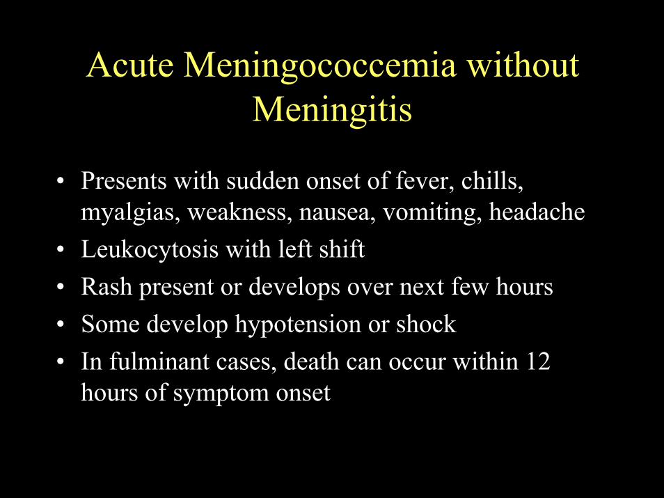
Acute Meningococcemia withoutMeningitis
• Presents with sudden onset of fever, chills, myalgias, weakness, nausea, vomiting, headache
• Leukocytosis with left shift• Rash present or develops over next few hours• Some develop hypotension or shock• In fulminant cases, death can occur within 12
hours of symptom onset

Acute Meningococcemia: Rash• Erythematous maculopapular rash
– Light pink– Indistinct borders– Transient (half hour to 2 days)
• Purpuric rash– Occurs in 40-90%– Always accompanied by DIC– Petechiae, ecchymoses or gross intracutaneous
hemorrhages– Purpura usually appear within 12-36 hours of disease
onset– May lead to purpura fulminans

Meningococcemia Complications
• Purpura fulminans• Autoimmune-like
complications:– Synovitis– Serositis
• Neurologic sequelae (0-15%)– Deafness (4-6%0– CN VI, VII palsies (5-10%)

Meningococcemia Complications• Bilateral adrenal
hemorrhage (Waterhouse-FriderichsenSyndrome)– Found in 30% of
patients with shock secondary to meningococcal disease
– Found in 70% of cases at autopsy
van Deuren M et al. Clin Microb Rev 2000;13:144-166.

Laboratory Studies
• CSF: gram stain positive in 75-80%; culture positive in 90%
• CSF latex agglutination: 70-80% sensitive• Peripheral blood smear: organisms may be seen
indicating high-grade bacteremia; suspect asplenicstate
• Blood culture: positive in 40-75%

Management• Cannulation of large compressible vein (i.e., femoral)• Early fluid resuscitation for patients in shock• Inotropic support• Alkalinization for patients with rhabdomyolysis• Maintain high suspicion for adrenal insufficiency• Empiric corticosteroids for meningococcal meningitis
controversial
van Deuren M et al. Clin Microb Rev 2000;13:144-166.

Management: Antimicrobials• Should not be delayed for diagnostic
procedures• Drug of choice: ceftriaxone 2 g IV q 12 hrs

Protein C• Vitamin-K dependent glycoprotein• Promotes fibrinolysis & inhibits thrombosis &
inflammation• Once activated, protein C requires protein S as
a cofactor for its anticoagulant functions• In severe meningococcal sepsis, protein C
activation is impaired, leading to widespread thrombosis, DIC & purpura fulminans
Faust SN et al. N Engl J Med 2001;345:408-416.

Role of Activated Protein C in Infection• Antiinflammatory:
• inhibits production of inflammatory cytokines (TNF-α, IL-1, IL-6)
• Limits rolling of monocytes & PMNs on injured endothelium
• Antithrombotic:• Inactivates factors
Va & VIIIa, thereby limiting generation of thrombin
• Fibrinolytic:• Inhibits PAI-1 Bernard GR et al. N Engl J Med 2001;344:699-709.

Management: Recombinant Activated Protein C for Severe Sepsis
• Randomized, double-blind, placebo-controlled, multicenter trial
• Severe sepsis, any organism• N=1,690• Reduced risk of death 19.4%
(95% CI 6.6-30.5)• Absolute reduction in risk of
death 6.1% (P=0.005)• Incidence of serious bleeding:
3.5% for APC vs. 2.0% for placebo (P=0.06)
30.8
24.7
0
5
10
15
20
25
30
35
Placebo APC
Mortality (%)
Bernard GR et al. N Engl J Med 2001;344:699-709.

Prognosis• “No other infection so quickly slays…”
Herrick WW. Arch Intern Med 1919;23:409-418.
• Almost all deaths from meningococcal meningitis are due to cerebral edema and brainstem herniation
• Little improvement in outcome over the past few decades despite significant advances in critical care
• Meningitis: 10-15% mortality• Meningococcemia: up to 40% mortality• Sequelae (hearing loss, neurologic disability, limb
loss) in 11-19%

Conclusion•Sepsis: Major cause of morbidity and mortality worldwide-Leading cause of death in noncoronary ICU (US)*
•SIRS: A clinical response arising from a nonspecific insult manifested by ≥2 of the following:
–Temperature ≥38°C or ≤36°C
–HR ≥90 beats/min
–Respirations ≥20/min
–WBC count ≥12,000/mL or ≤4,000/mL or >10% immature neutrophils
• Sepsis:– Known or suspected
infection– Two or more
SIRS criteria• A significant link to
disordered hemostasis
•The management of sepsis is largely supportive with the administration of IV fluids, vasopressors, antibiotics and for some cases, Activated Protein C.
•Meningococcemia is a classic example of gram negative sepsis