Sensory System - Nursing Ed148 CHAPTER 7 Sensory System Home Care (see Box 7-1)A. Maintain...
12
Sensory System 146 B. Evaluate visual health history. 1. Eye pain and headaches. 2. Decreased or blurred vision. 3. Eye infections. 4. Floating or “dots” in field of vision. 5. Chronic illnesses and medications. 6. Surgical procedures. C. Evaluate visual acuity (see Appendix 7-1). OLDER ADULT PRIORITY: It is important for older clients to have regular vision checkups and to report any progressive decrease in vision, “floaters” or specks in their visual field or a partial loss of visual field. DISORDERS OF THE EYE Glaucoma Glaucoma is a group of disorders characterized by an increase in intraocular pressure and progressive loss of peripheral vision. It is a chronic condition and a leading cause of blindness. Types A. Primary open-angle glaucoma (POAG): most common form. Flow of aqueous humor is slowed or stopped by obstruction, thus increasing intraocular pressure; characterized by a slow onset; chronic and progressive. B. Primary angle-closure glaucoma (PACG - acute glau- coma): caused by rapid increase in intraocular pres- sure. Iris is pushed against drainage system, blocking flow of aqueous humor. Immediate treatment is required. C. Secondary glaucoma: caused by trauma or optic neo plasm. D. Treatment can stop progression of condition, but it can not restore lost peripheral vision. Data Collection A. Risk factors. 1. Familial tendency. 2. Aging—occurs most often in clients more than 40 years old. 3. Chronic diseases and eye injury. B. Diagnostics: increased intraocular pressure (greater than 22 mm Hg) (see Appendix 7-1). ✔ ✽ PHYSIOLOGY OF THE EYE A. Structures of the eye 1. Sclera: tough, protective covering of the outside of the eye; the “white” of the eye. 2. Cornea: transparent tissue that covers the front of the eye over the pupils. 3. Ciliary muscle: muscular body that allows the eye to focus through contraction and relaxation. 4. Iris: controls the amount of light; gives the eye its characteristic color. 5. Retina: thin, innermost lining of the eye that con- tains millions of nerve cells to coordinate and transmit signals to the optic nerve. 6. Aqueous humor: fluid that fills anterior and poste- rior chambers; circulates through the pupil and empties into canal of Schlemm. 7. Vitreous humor: fluid that fills the cavity posterior to the lens. 8. Crystalline lens: provides for the convergence and refraction of light rays and images onto the retina; enables vision to be focused. 9. Optic nerve: leaves the eye through the retina at the location of the optic disc. 10. Macula: part of the retina responsible for providing optimal visual focusing. B. Eyelids: protective coverings of the eye. 1. Conjunctiva: thin, transparent mucous membrane that covers the outer surface of the eye and lines the inner surface of the eyelid. 2. Lacrimal gland: excretes lacrimal fluid (tears) to lubricate, clean, and protect the outer surface of the eye. Data Collection A. External data collection. 1. Assess position and alignment of the eyes: Both eyes should fixate on one visual field simultane- ously. 2. Evaluate for presence of ptosis (lid lag). 3. Inspect lids and conjunctivae for discharge or inflammation. 4. Assess color of sclera: normally a thin coating; may yellow with aging. 5. Evaluate size and equality of pupils: should be equal in size and shape. 6. Evaluate pupillary reaction to light. 7. Assess extraocular movement: both eyes move together.
Transcript of Sensory System - Nursing Ed148 CHAPTER 7 Sensory System Home Care (see Box 7-1)A. Maintain...

SensorySystem
146
B. Evaluate visual health history. 1. Eye pain and headaches. 2. Decreased or blurred vision. 3. Eye infections. 4. Floatingor“dots”infieldofvision. 5. Chronic illnesses and medications. 6. Surgical procedures.C. Evaluate visual acuity (see Appendix 7-1).
OLDER ADULT PRIORITY: It is important for older clients to have regular vision checkups and to report any progressive decrease in vision, “floaters” or specks intheirvisualfieldorapartiallossofvisualfield.
DISORDERSOFTHEEYE
GlaucomaGlaucoma is a group of disorders characterized by
anincreaseinintraocularpressureandprogressivelossofperipheralvision.It isachronicconditionanda leadingcauseofblindness.
TypesA. Primary open-angle glaucoma (POAG): most common form. Flow of aqueous humor is slowed or stopped by obstruction, thus increasing intraocular pressure; characterized by a slow onset; chronic and progressive.B. Primary angle-closure glaucoma (PACG - acute glau- coma): caused by rapid increase in intraocular pres- sure. Iris is pushed against drainage system, blocking flowofaqueoushumor.Immediatetreatmentis required.C. Secondary glaucoma: caused by trauma or optic neo plasm.D. Treatment can stop progression of condition, but it can not restore lost peripheral vision.
Data CollectionA. Risk factors. 1. Familial tendency. 2. Aging—occurs most often in clients more than 40 years old. 3. Chronic diseases and eye injury.B. Diagnostics: increased intraocular pressure (greater than 22 mm Hg) (see Appendix 7-1).
✔
✽
PHYSIOLOGYOFTHEEYEA. Structures of the eye 1. Sclera: tough, protective covering of the outside of the eye; the “white” of the eye. 2. Cornea: transparent tissue that covers the front of the eye over the pupils. 3. Ciliary muscle: muscular body that allows the eye to focus through contraction and relaxation. 4. Iris: controls the amount of light; gives the eye its characteristic color. 5. Retina: thin, innermost lining of the eye that con- tains millions of nerve cells to coordinate and transmit signals to the optic nerve. 6. Aqueoushumor:fluidthatfillsanteriorandposte- rior chambers; circulates through the pupil and empties into canal of Schlemm. 7. Vitreoushumor:fluidthatfillsthecavityposterior to the lens. 8. Crystalline lens: provides for the convergence and refraction of light rays and images onto the retina; enables vision to be focused. 9. Optic nerve: leaves the eye through the retina at the location of the optic disc. 10. Macula: part of the retina responsible for providing optimal visual focusing.B. Eyelids: protective coverings of the eye. 1. Conjunctiva: thin, transparent mucous membrane that covers the outer surface of the eye and lines the inner surface of the eyelid. 2. Lacrimalgland:excreteslacrimalfluid(tears)to lubricate, clean, and protect the outer surface of the eye.
Data CollectionA. External data collection. 1. Assess position and alignment of the eyes: Both eyesshouldfixateononevisualfieldsimultane- ously. 2. Evaluate for presence of ptosis (lid lag). 3. Inspect lids and conjunctivae for discharge or inflammation. 4. Assess color of sclera: normally a thin coating; may yellow with aging. 5. Evaluate size and equality of pupils: should be equal in size and shape. 6. Evaluate pupillary reaction to light. 7. Assess extraocular movement: both eyes move together.

CHAPTER7 SensorySystem 147
C. Clinical manifestations of primary open-angle glau- coma—develops slowly and frequently without symp- toms. (Figure 7-1) 1. Gradual loss of peripheral vision (i.e., tunnel vision). 2. Vague headache around lights. 3. Blindness may eventually occur if untreated. 4. Central vision is normal, even with loss of periph- eral vision.
TreatmentA. Medications (see Appendix 7-2). 1. Topical drops. 2. Oral medications to promote diuresis and lower in- traocular pressure.B. Surgical intervention: most often done on outpatient basis with topical anesthetic. 1. Argon laser trabeculoplasty: microscopic laser to openthefluidchannelsfacilitatingoutflowofaque- ous humor. 2. Trabeculectomy:creationofanartificialdrainto bypass the trabecular meshwork, allowing aqueous humortoflow.
TEST ALERT: Assist client to compensate for a sensory impairment (hearing loss or impaired
vision).
Nursing Interventionsv Goal: To prevent progression of visual impairment.A. Client must remain on medication in order to control disease.B. Emphasize the importance of continual follow-up medical care.C. Stress the importance of wearing a medical alert identi- ficationtag.D. Show client correct administration of eye medication and have client return-demonstrate.E. Damage to current vision cannot be corrected. Focus of care is to prevent further damage to vision.v Goal: To decrease intraocular pressure.A. Client should avoid straining while defecating, lifting, and stooping. B. Administer antiemetic, as vomiting causes increased intraocular pressure.C. Administer medications to decrease intraocular pres- sure.v Goal: To assist client to adapt to visual limitations.A. Orientclientstoobjectswithinvisualfield.B. Encourage verbalization regarding fear of blindness and loss of independence.C. Eliminate potential slip, trip, and fall hazards in the home care environment.
v Goal:To provide preoperative nursing care. A. Surgery and treatments are frequently done on an out patient basis and with a local anesthetic. Orient the cli- ent to the surroundings and sounds that will occur during the procedure.B. For outpatient surgery, client should wear comfortable clothes and arrange for someone to provide transporta- tion home.C. Postoperative instructions should be given to the client or family in writing.v Goal:To provide postoperative nursing care (surgery most often done under local anesthesia).A. Administer medications: miotics, antibiotics, and steroids.B. Be sure medication is administered in the eye for which it is ordered; the unaffected eye may be treated with a different medication.C. Client may eat and ambulate as desired after the initial sedative effect is gone.
Home CareA. Emphasize the importance of follow-up care.B. Explain to the client the importance of not rubbing his eyes. C. Demonstrate how to correctly administer eye medica- tion and have the client return the demonstration.D. Advise the client to avoid activities that increase intra- ocular pressure.E. If pain is not easily relieved by analgesics, the client should call the health care provider.
Figure 7-1 Glaucoma (From Zerwekh J, Claborn J, Miller CJ: Memory Notebook of Nursing, ed 3, vol 2, Ingram, Texas 2007, Nursing Education Consultants).

148 CHAPTER7 SensorySystem
Home Care (see Box 7-1)
A. Maintain client’s orientation to surroundings.B. Eye patch may be used for about 24 hours or until the client returns to the physician for a follow-up visit.C. Client should avoid stooping, lifting, or straining in the early postoperative period.D. Client should avoid sleeping on the affected side.E. Teach the client to avoid rubbing the eye; there should be minimal discomfort, report any pain that is not easilyrelievedbyacetaminophenorotheranalgesic.F. Demonstrate to client and/or to family how to admin- ister eye drops (antibiotic and steroid eye drops); have client and/or family demonstrate procedure.G. Client should report any signs of increased redness and discharge.H. Provide written instructions; make sure the type is large enough for client to read.
Data Collection
Retinal Detachment
Aseparationofthetwolayersoftheretinaiscalledretinaldetachment.A. Risk factors. 1. Severe myopia (nearsightedness). 2. Diabetes. 3. Trauma. 4. Degenerative changes in the retina. 5. Previous cataract surgery.B. Diagnostics. 1. Ophthalmoscopic examination. 2. Evaluation of visual acuity.C. Clinical manifestations. 1. Sudden onset. 2. Flashesoflightinvisualfield. 3. Sensation of a veil or “cobwebs” over the eye. 4. Suddenincreaseinthenumberoffloatingspots. 5. May experience area of blank vision. 6. Eventually results in loss of vision.
Treatment There is no medical treatment. The surgical repair may be done on an outpatient basis or may require hospitalization.A. Laser photocoagulation: Laser light beam to create in- flammationandsealingofthetearorbreak.B. Cryotherapy: a “supercooled” probe directed over reti- nalteartoproduceinflammationtosealthetear.C. Scleral buckling: extraocular surgery in which the sclera is depressed from the outside with the application of a silicone “buckle” to weld the retina in contact with the choroid
✽
TEST ALERT: Assist the client to compensate for loss of visual acuity; identify sensory changes in the older client; identify environmental factors that are a safety hazard.
CataractA complete or partial opacity of the lens is known
asacataract.Itmayoccuratbirth(congenitalcataract);however, it occursmost commonly in adultspast middleage(senilecataracts).
Data CollectionA. Clinical manifestations. 1. Painless with gradual decrease in visual acuity. 2. Pupil appears gray to milky white. 3. Decreased perception of colors. 4. Possible diplopia in affected eye.B. Diagnostics (see Appendix 7-1).C. Risk factors. 1. Age: 70% of adults over age 75 years have cataracts. 2. Diabetes. 3. Corticosteroids: long-term systemic use.
TreatmentA. Surgical treatment is only method of correcting prob- lem; surgery is usually performed when client begins to experience problems in activities of daily living.B. Surgical removal of the lens requires lens implant, which usually occurs simultaneously with lens removal.C. Treatment most often done on outpatient bases.
Nursing Interventionsv Goal: To provide preoperative nursing care. A. Orient the client to the surroundings and sounds that will occur during the procedure.B. Client should wear comfortable clothes and arrange for someone to provide transportation home.C. Before surgery, the nurse will instill mydriatic eye drops; frequently, cycloplegic drops are also used; client will be photosensitive.D. Normal for visual acuity to be decreased immediately after surgery.E. Confirmoperativeeyeaccordingtofacilityprocedure.F. Client is awake during procedure; frequently a sedative is given preoperatively.
TEST ALERT: Reinforce client teaching regarding self-administration of medications.
✽

CHAPTER7 SensorySystem 149
Nursing Interventionsv Goal: To prevent further deterioration of vision preop-eratively.A. Restrict activity prior to surgery, especially activities that cause rapid eye movements (e.g., reading, sewing, watching television).B. Assist client to avoid coughing.v Goal: To prevent postoperative complications A. Local anesthesia may be used; client may be discharged
within hours or may remain in hospital—depends on the degree of detachment and the type of repair.B. Prevent nausea and vomiting.C. Avoid activities that increase intraocular pressure: bending, lifting, sneezing, coughing, vomiting.D. Postoperative medications usually include antibiotic eye drops, ophthalmic steroid drops, and dilating agents
Home CareA. Orient client to surroundings.B. Postoperative pain may require narcotic analgesic.C. Client may experience redness and swelling of the lids.D. Warm or cool compresses may be used to promote comfort.E. Havesignificantotherorhomehealthprovidersidentify home environment safety hazards.
Age-Related Macular Degeneration (AMD)
Mostcommoncauseof centralvision loss in clientsover40yearsold;relatedtoretinalaging.
Data CollectionA. Risk factors 1. Familial tendency 2. Long-term exposure to UV lights and eye irritants.B. Clinical manifestations. 1. Blurred, darkened vision. 2. Presenceofscotomas(blindspotsinvisualfield). 3. Distortion of vision. 4. Permanent loss of central vision. 5. Wet exudative—more severe form. a. Development of abnormal vessels in or near macula. b. New vessels begin to leak and form scar tissue. c. Rapid onset. 6. Dry nonexudative—less severe, more common form.C. Diagnostics (see Appendix 7-1).
Treatment Directed toward stopping progression; affected vision cannot be restored.
Nursing Interventionsv Goal:Topromotepreventionand/orearlyidentificationof problem and care of client with decreased visual acuity.A. Encourage regular visual checkups.B. Encourage diet high in leafy green vegetables and vita- mins A and E, beta carotene, and zinc.C. Promote independence in clients with decreased vision (see Box 7-1).
✽
Medications• Use containers that have different shapes (square, round, triangular) when client has to take several medications.
• Obtain medication boxes with raised letters or numbers on them representing the days of the week.
• Obtain a “talking clock” that states the time.
Safety• Remove throw rugs.
• Use unbreakable dishes, cups, and glasses.
• Keep appliance cords short and out of walkways.
• Avoid foot stools, a recliner with a built-in footrest is less of a hazard.
• Cleansers,cleaningfluids,andcausticchemicalsshould be labeled with large, raised lettering.
• Have hand grips installed in bathrooms.
• Putnonskidstrippingonthesurfaceofthetubfloor.
• Encourage use of an electric razor.
Communication• Advise caregivers of client hearing status, encourage everyone who inters the room to always introduce themselves before touching client.
• Teach the client to use telephones that have program- mable automatic dialing features. Be sure to include emergency phone numbers.
• Recognize that much communication is nonverbal; thereforefrequentclarificationofmeaningmaybe required.
Daily Living Considerations• Provide information on Meals on Wheels for delivery of cooked, ready-to-eat meals.
• Encourage use of a microwave for cooking—it is safer than using a standard stove.
• Encourage the use of large-print books, newspapers, and magazines for reading. Also, many publications are available as audiotapes and CDs at local libraries and vision aid services.
• Avoid rearranging furniture and other belongings.
BOX 7-1 OLDER ADULT CARE FOCUS Promoting Independent Activities of Daily Living for the Client with Diminished Vision

150 CHAPTER7 SensorySystem
TEST ALERT: Identify sensory changes that affect the older client, and encourage regular
eye examinations to identify early changes. Identify environmental factors that are a safety hazard to visually impaired client.
Conjunctivitis
Inflammation or infection of the conjunctiva.
A. Clinical manifestations. 1. Usually both eyes are affected. 2. Redness, burning, itching. 3. Purulent discharge (worse in morning). 4. Sensitivity to bright light.
TreatmentA. Ophthalmic antibiotic eye ointment for bacterial infection.B. Drops may be used during day with ointments used at bedtime.
Nursing Interventionsv Goal:To prevent transmission.A. Teach client not to rub eyes, encourage frequent hand washing.B. Cleanse eyelids with warm saline compresses to remove crusts before administering eye medications.C. Always clean eye from inner canthus downward and outward to prevent contamination of other eye.D. Treatment with antibiotic eye drops; instill after eye has been cleaned.E. Teach client to avoid contamination of tip of medication tube.F. Client should discard all eye medications after condi- tion is resolved.G. Highly contagious; spreads easily from one eye to the other and by direct contact from person to person.
Eye TraumaA. Chemical injury: Flush chemicals from the eye. At home use copious amounts of cool tap water; in the hospital use normal saline (see Appendix 7-4).B. Corneal abrasion: May be caused from foreign body or trauma. Fluorescein dye will be used to determine the area of injury. C. Intraocular foreign body: Penetrating foreign bodies must be removed by a physician (ophthalmologist) as soon as possible. Do not attempt to remove foreign objects from eye, but protect from movement while seeking emergency care. If a penetrating foreign body is observed, do not attempt to patch the eye; use a shield if possible, but do not apply pressure to eye.D. Subconjuntival hemorrhage: This eye trauma involves blood between the conjunctiva and the sclera. Most of-
✽
ten clears without intervention. Precipitating cause needs to be evaluated.
Nursing Interventionsv Goal: To prevent further eye damage.A. Have client “rest” the eyes: provide dimly lit room; use eye patches if necessary.B. Irrigate eyes with normal saline solution from inner canthustooutercanthussosolutiondoesnotflowinto unaffected eye.C. Eye may remain irritated after foreign body is removed; eye patch may be necessary.D. If penetrating eye injury is present, decrease activities that cause increased ocular pressure.E. Keep client immobilized until evaluated by an ophthal- mologist.v Goal:To care for a client with an enucleation.A. Immediate: Determine presence of other injuries; moni- tor for bleeding.B. Instillationoftopicalointmentsuntilprosthesisisfitted.C. A clear “conformer” may be placed in the eye socket to allow the area to heal so a permanent prosthesis can befitted.D. Eye prosthesis. 1. Insertion: notched end of prosthesis should be clos- est to the client’s nose; lift upper eyelid (using non- dominant hand) and insert saline solution-rinsed prosthesis into socket area with top edge slipping under upper lid; gently retract lower lid until bottom edge of prosthesis slips behind it. 2. Removal: retract the lower lid and apply slight pressure just below the eye, this should release the suction holding the eye in place; assess the socket for signs of infection. 3. The prosthesis is usually cleansed with normal saline.
PHYSIOLOGYOFTHEEARA. External ear. 1. Auricle or pinna: the cartilage and connective tissue forming the outside of the ear. 2. External auditory canal: collects transmitted sound waves to the tympanic membrane; outer half of canal secretes cerumen (wax) that provides a protective function. 3. Tympanic membrane: a tough membrane separating the external ear and middle ear; transmits vibrations from external ear to the malleus of the middle ear.B. Middle ear. 1. Contains three small articulating bones: malleus, incus, stapes. 2. The eustachian tube in infants and young children is shorter and wider than in adults. 3. Problems in the external and middle ear cause con- ductive hearing loss.

CHAPTER7 SensorySystem 151
C. Inner ear. 1. Assists in maintaining equilibrium. 2. Contains organ of Corti, which is the receptive end organ for hearing. 3. Pathology of the inner ear or nerve pathway can re- sult in sensorineural hearing loss.
Data Collection
A. External assessment of the ear. 1. Assess placement of the ears: Low-set ears may be indicative of congenital anomalies. 2. Movement of the ear lobe should not elicit pain. 3. Note presence of any discharge in the external ear canal. 4. Assess for vertigo: Ask client to close eyes and stand on one foot; have client walk with one foot; client may fall to one side or complain of room spinning.
DISORDERSOFTHEEAR
Otitis MediaOtitismediaisaninfectionofthemiddleearcaused
byaviralorbacterialagent.Infantsandyoungchildrenarepredisposed to thedevelopmentofacuteotitismediabecauseofthephysiologiccharacteristicsoftheear—theeustachiantubeisshorter,wider,andstraighterinchildrenthaninadults.
A. Acuteotitismedia(AOM)maybepurulent(pus-filled) or suppurative (capable of producing pus); repeated or persistent acute infections lead to perforation of the tympanic membrane or more severe complications such as mastoiditis.
B. Otitismediawitheffusion(OME):acollectionoffluid in the middle ear without infection or acute symptoms that results from a blocked eustachian tube; may persist for weeks to months; most common cause of conductive hearing loss.
Data CollectionA. Risk factors. 1. Children: 6 months to 2 years old. 2. History of upper respiratory tract infection. 3. Less common in breast-fed infants. 4. Bottle-feeding in supine position. 5. Respiratory tract allergies. 6. Environment with secondhand smoke.B. Clinical manifestations. 1. Pain from pressure in the middle ear. a. Infants: irritable; pull at their ears; sucking ag- gravates pain. b. Young children verbally complain of severe ear pain.
✽
2. Prevalence of fever varies greatly. 3. Nasal congestion. 4. If tympanic membrane ruptures, purulent drainage may be present in the outer ear; pain will decrease temporarily. 5. Hearing loss with recurrent rupture.
TreatmentA. Medications. 1. Antibiotics: health care provider will determine if infection can be treated with antibiotics. 2. Analgesics, antipyretics, decongestants. 3. Ear drops (antibiotic and steroid combination). 4. OME does not require antibiotics. B. Surgical: myringotomy drainage of the middle ear with insertion of tubes or grommets (or tympanostomy myr- ingotomy) to relieve pressure and promote healing; used for recurrent cases that do not respond to medica- tion; tubes also known as pressure equalizing tubes (PE tubes).
Nursing Interventionsv Goal: To enable parents of clients to describe problem, handle medication schedule, and cope with home care.A. Recurrent infections cause an increased risk of perma- nent hearing loss.B. Antibiotics should be given around the clock, and the prescribed amount should be taken, even after all symptoms are relievedC. Administer acetaminophen or ibuprofen for pain and fever. Aspirin should not be given. D. Pain-relieving ear drops (Auralgan, Pramotic) should be used only when there is no tympanostomy tube or rupture of the tympanic membrane.
NURSING PRIORITY: Teach the parents to administer the full course of antibiotics regardless of the child’simprovedstatus.
E. Decreaseriskofrecurrencebypreventingfluidsfrom pooling in sinuses and upper airways. 1. Hold or elevate child’s head while feeding. 2. Do not prop bottle or allow child to fall asleep while taking a bottle. 3. Encourage juice or water before sleeping.F. Decongestantsmaybeadministeredtodecreasefluid collection, thereby decreasing prevalence of infection and discomfort.v Goal: Care for child after placement of tympanostomy tubes (myringotomy tubes or grommets)A. Do not allow soapy or dirty water to get into the child’s ears; use of earplugs is currently controversial.B. Assure parents that if the ear grommet or tube falls out, itisnotasignificantproblem.
✔

152 CHAPTER7 SensorySystem
Hearing LossHearing loss results from an impairment of the
transmissionofsoundwaves.A. Conductive: results from an impairment in transmission of sound from the outer or middle ear or both. Client willbeabletobenefitfromahearingaid.B. Sensorineural (perceptive): results from a problem in the inner ear, or a nerve pathway. Incoming sound cannotbeanalyzedcorrectly.Clientwillnotbenefit from a hearing aid.C. Otosclerosis: an immobilization of the small bones in the inner ear; it occurs most often in women. D. Impacted cerumen can lead to hearing loss; occurs of- ten in the older adult client.
Data CollectionA. Risk factors. 1. Age-related changes; increased incidence in clients over 65. 2. Prolonged exposure to high-intensity sound waves. 3. Repeated, chronic ear infections. 4. Prenatal problems of rubella and eclampsia. 5. Female with family history of otosclerosis. 6. Ototoxic medications - aminoglycosides, diuretics.
NURSING PRIORITY: Earplugs should be worn whenthepotentialforexposuretoloudnoiseexists.
B. Diagnostics (see Appendix 7-1).C. Clinical manifestations. 1. Speech problems: deterioration of present speech, or delayed speech development. 2. Fails to respond to oral communication or responds inappropriately. 3. Social withdrawal. 4. Decreasedself-confidence. 5. Responds more to facial expressions than to verbal ones. 6. Inappropriate emotional response.
TreatmentA. Hearing aid, if conductive loss.B. Speech therapy.C. Sign language.D. Stapedectomy for otosclerotic lesions.
OLDER ADULT PRIORITY: Identify hearing loss; assist client to compensate and to maintain communication.
✽
✔
✔
Nursing Interventionsv Goal: To promote communication and socialization of the hearing-impaired client (Box 7-2).A. Provide information concerning a TDD (telephone de- vice for the deaf) that transmits typed words over the phone line.B. Suggest light-activated devices rather than sound- activated devices: connect doorbell, smoke, and security alarms to an electrical device that causes lights toflickeronandoff.C. Avoid crowded, noisy environments (such as restau- rants) especially if a hearing aid is used, because it amplifiesbackgroundsound.D. Teach how to care for hearing aid (Box 7-3).
TEST ALERT: Provide care for a client using assistive devices: hearing loss and use of hear-
ing aids are common in older adult clients.
E. Teach client how to remove earwax; if impacted ceru men is a problem, may need ear irrigation (see Appendix 7-3).v Goal: To prevent complications for the client who un-dergoes stapedectomy A. Assess client for dizziness, nausea, and vomiting.B. Teach client to avoid sudden movement to prevent dizziness.C. Maintain safety measures.D. Instruct client that hearing may not improve until edema subsides in the operative area.
Home CareA. Avoid washing hair for about a week; after that keep ear dry.B. Avoidflyingandswimmingforaboutamonthoruntil all edema subsides.
Balance Disorders
The vestibular system of the inner ear maintainsbalanceandcoordination.A. Disorders. 1. Ménière’s disease: an inner ear disorder caused by excess endolymph in the vestibular and semicircular canals. 2. Labyrinthitis:aninflammationoftheinnerear.
AssessmentA. Diagnostics (see Appendix 7-1).B. Clinical manifestations. 1. Vertigo: a sense of moving or spinning that is usu- ally stimulated by sudden movement of the head; may occur when lying down.
✽

CHAPTER7 SensorySystem 153
2. Sudden, severe paroxysmal episodes of vertigo (Ménière’s disease). a. Severe nausea, vomiting. b. Nystagmus. c. Loss of balance. d. No pain or loss of consciousness. 3. Fluctuating hearing loss and tinnitus (Ménière’s disease). 4. Client may have no symptoms between attacks. 5. Manifestations become less severe with time.
TreatmentA. Medications (Ménière’s disease). 1. Atropine may stop an acute attack. 2. Sedatives. 3. Antihistamines, anticholinergics. 4. Diuretics(todecreaseendolymphfluid). 5. Meclizine hydrochloride (Antivert) for prevention and treatment of “motion sickness” symptoms. 6. Antiemetics.B. Diet: low-sodium
Nursing Interventionsv Goal:To provide emotional and physical support dur-ing an acute attack.A. Maintainbedrestinaquiet,dimlylitroom;avoidflick- ering lights and television.B. Position of comfort.C. Avoid unnecessary nursing procedures.D. Minimize stimulation and sudden position changes.E. If client is severely nauseated, administer medications parenterally.F. Maintain fall precautions.
NURSING PRIORITY: As a safety precaution, instruct the client to lie down immediately if an attack feelsimminent.
✔
• Standing in front of client at eye level and speaking with light on your face helps with speech reading (i.e., read- ing lips).
• Get client’s attention by raising your hand or arm.
• Do not walk back and forth in front of client while speaking.
• Speak clearly and in an even tone; do not shout.
• Do not chew gum, cover mouth, or smile excessively while talking.
• Because clients rely on visual cues, watch your facial expressions.
• Encourage professional counseling in speech and sign language.
• If the client is agreeable, assist in obtaining a hearing aid.
• Promote social interaction appropriate for elderly adult.
• Do not avoid conversation with the client; do not depend on family to interpret information.
• Investigate use of TDD (telecommunication device for the deaf).
• Use light-activated devices: door bell, smoke alarms, telephones.
• Client should avoid noisy environments where it is difficulttointerpretsound.
• Provide client instruction in a quiet room with minimal distractions.
BOX 7-2 OLDER ADULT CARE FOCUS Improving Communication with Hearing Imparied Clients
• Keep the hearing aid dry; do not wear it while bathing or swimming or allow it to get wet.
• Clean the ear mold of the hearing aid with soft cloth and recommended cleanser; do not immerse in water.
• Avoid using hair spray, cosmetics, or oils around the ear.
• Always keep extra batteries on hand.
• When not using hearing aid, turn it off and remove the battery before storing it.
• Avoid dropping the hearing aid (has delicate electronics) or exposing it to extremes in temperatures (e.g., leaving it on a window ledge in the sunlight).
• Clean any debris or cerumen from the hole in the middle part that goes in the ear by using a toothpick or pipe cleaner.
• If the hearing aid does not work, change the battery or check the on-off switch, check the connection between the ear mold and receiver, clean it using the steps de- scribed above, or take it to an authorized hearing aid service center.
BOX 7-3 OLDER ADULT CARE FOCUS Hearing Aid Care

154 CHAPTER7 SensorySystem
StudyQuestions:SensorySystem
1. The nurse is preparing a client for a cataract removal. Mydriatic eye drops are ordered. What observation by the nurse will indicate the medication is having the desired effect? 1 The client states his vision is blurred. 2 The pupil on the client’s affected eye is dilated. 3 The client says his eye feels irritated. 4. The pupil on the client’s unaffected eye is pinpoint size.2. What is important in the plan of care for a client who has glaucoma? 1 Prevent secondary infection. 2 Maintain good visual acuity. 3 Prevent injury to unaffected eye. 4 Control intraocular pressure.3. Whatisacommonfindingobtainedduringdatacollec- tion on a client experiencing a problem with Meniere disease? 1 Severe vertigo. 2 Sloshy feeling in ears. 3 Watery drainage from ears. 4 Low-grade temperature.4. What will the nurse identify as the therapeutic response of a miotic eye medication? 1 Constriction of the pupil. 2 Decrease in irritation and pain 3 Increase in visual acuity 4 Dilation of the pupil.5. An infant has a history of frequent otitis media. What would be important teaching information to tell the mother? 1 Avoid the use of decongestants. 2 Encourage formula before sleeping. 3 Stop antibiotics as soon as the fever subsides. 4 Hold the infant and elevate the head while feeding.6. The nurse is instructing the client on administration of eye drops. What nursing observation would indicate the client understands how to administer the drops? 1 Positions himself on his left side to administer drops in the left eye. 2 Instills the drops into the lower conjunctival sac. 3 Blows his nose before administering drops. 4 Cleans the tip of the applicator with a tissue before administering drops.7. What would be important for the nurse to teach the client about chronic primary open angle glaucoma (POAG)? 1 The lens of the eye becomes cloudy. 2 There is an increase in pressure inside the eye. 3 The cornea becomes stretched, causing blurred vision. 4 The pupil becomes constricted, allowing minimal vision.8. Considering the risk factors for hearing loss, what factor would the nurse identify as not being an
increased risk for hearing loss? 1 Prenatal problem of rubella. 2 Repeated, chronic ear infections. 3 Taking penicillin and cephalosporin medications. 4 Exposure to high-intensity sound waves.9. What would be important for the nurse to teach a client regarding the care of the client’s hearing aid? 1 Always keep extra batteries on hand. 2 Clean ear mold part by soaking it in alcohol. 3 Always leave hearing aid turned on when out of the ear. 4 Continue to wear hearing aid even while sleeping.10. What medications would the nurse identify as being contraindicated for a client with a diagnosis of glaucoma? 1 Atropine sulfate (Atropisol). 2 Pilocarpine (Pilocar). 3 Meperidine (Demerol). 4 Fentanyl (Duragesic).11. After a client’s eye has been anesthetized, what would the nurse instruct the client to do? He should: 1 Not watch television for at least 1 day to reduce strain. 2 Not rub the eye for about 30 minutes. 3 Irrigate the eye every hour until sensation is felt. 4 Wear an eye patch for 2 days.12. A client comes to the clinic with decreased hearing. Examination of the ear canal reveals a large amount of earwax. The nurse prepares for removal of the wax by using: 1 Forceps. 2 Normal saline irrigation. 3 D
5W irrigation.
4 A cotton swab applicator.13. The nurse is caring for a client who is hearing- impaired. What nursing action would be least effective in promoting communication? 1 Provide client with a writing pad. 2 Increase your voice until client can hear. 3 Use short sentences or phrases with frequent pauses in the conversation. 4 Stand directly in front of the client to allow them to see your face.14. A nurse is teaching a family about the use of a hearing aid. The nurse bases her teaching on the knowledge that the hearing aid does which of the following? 1 Provides mechanical training for the damaged part of the ear. 2 Amplifiessoundbutdoesnotimprovetheabilityto hear. 3 Amplifiessoundandimprovestheabilitytohear. 4 Assists to heal the damaged part of the ear.
Answers and rationales to these questions are in the section at the end of the book titled Chapter Study Questions: An-swersandRationales.

CHAPTER7 SensorySystem 155
Appendix 7-1 OPHTHALMIC AND HEARING DIAGNOSTICS
OPHTHALMIC DIAGNOSTICS Snellenchart is used in screening for visual acuity problems. Client is placed 20 feet from the chart, and visual acuity is expressed as a ratio (what the client should see at 20 feet compared with what he or she can see at 20 feet). A ratio of 20/50 means that the client can see at 20 feet what he or she should see at 50 feet.
Anoncontacttonometeris used to measure intraocular pressure. Normal pressure is 12 to 22 mm Hg. A puff of air is directed toward the cornea, which causes indentation and allows measurement of intraocular pressure, a screen tool for diagnosis of glaucoma.
Directophthalmoscopy is examination of the back portion of the interior of the eyeball, which provides for visual evaluation of the retina, vascular patterns, and optic disk.
Biomicroscopy (slit-lamp examination) is used to assess the anterior eye for problems of the cornea, iris, and lens and to evaluate the depth of the anterior chamber.
Refractiveerrorevaluation determines what refractive errors have occurred because light is not correctly focused on the retina. Conditions that occur in refractive errors are:
• Myopia (nearsightedness)—sees near objects clearly; has problem seeing distant objects.
• Hyperopia (farsightedness)—sees distant objects clearly; has problem seeing close objects.
• Presbyopia—a decrease in the elasticity of the lens that causes poor accommodation for near vision (common in older adults)
• Astigmatism—an uneven curvature of the cornea; light rays do not focus on the retina at the same time
HEARING DIAGNOSTICS Audiometryis used to measure a client’s hearing by the use of various tones and intensities of sound produced by an audiometer.
156 CHAPTER7 SensorySystem
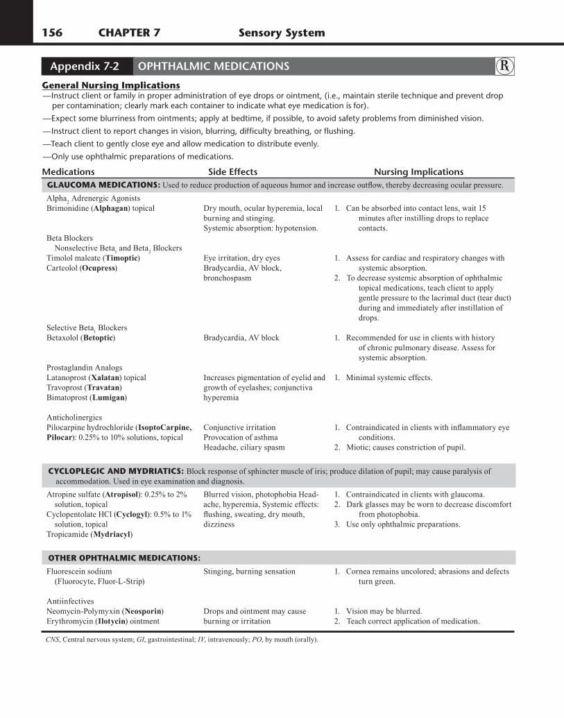
Appendix 7-2 OPHTHALMIC MEDICATIONS
GeneralNursingImplications—Instruct client or family in proper administration of eye drops or ointment, (i.e., maintain sterile technique and prevent drop per contamination; clearly mark each container to indicate what eye medication is for).
—Expect some blurriness from ointments; apply at bedtime, if possible, to avoid safety problems from diminished vision.
—Instruct client to report changes in vision, blurring, difficulty breathing, or flushing.
—Teach client to gently close eye and allow medication to distribute evenly.
—Only use ophthalmic preparations of medications.
Medications Side Effects Nursing ImplicationsGLAuCOmAmEDICATIONS: Usedtoreduceproductionofaqueoushumorandincreaseoutflow,therebydecreasingocularpressure.
Alpha2 Adrenergic Agonists
Brimonidine (Alphagan) topical
Beta Blockers Nonselective Beta
1 and Beta
2 Blockers
Timolol maleate (Timoptic)Carteolol (Ocupress)
Selective Beta1 Blockers
Betaxolol (Betoptic)
Prostaglandin Analogs Latanoprost (Xalatan) topicalTravoprost (Travatan)Bimatoprost (Lumigan)
AnticholinergicsPilocarpine hydrochloride (IsoptoCarpine,Pilocar): 0.25% to 10% solutions, topical
Dry mouth, ocular hyperemia, local burning and stinging.Systemic absorption: hypotension.
Eye irritation, dry eyesBradycardia, AV block,bronchospasm
Bradycardia, AV block
Increases pigmentation of eyelid and growth of eyelashes; conjunctiva hyperemia
Conjunctive irritationProvocation of asthmaHeadache, ciliary spasm
1. Can be absorbed into contact lens, wait 15 minutes after instilling drops to replace contacts.
1. Assess for cardiac and respiratory changes with systemic absorption.2. To decrease systemic absorption of ophthalmic topical medications, teach client to apply gentle pressure to the lacrimal duct (tear duct) during and immediately after instillation of drops.
1. Recommended for use in clients with history of chronic pulmonary disease. Assess for systemic absorption.
1. Minimal systemic effects.
1.Contraindicatedinclientswithinflammatoryeye conditions.2. Miotic; causes constriction of pupil.
CYCLOPLEGICANDmYDRIATICS:Block response of sphincter muscle of iris; produce dilation of pupil; may cause paralysis of accommodation. Used in eye examination and diagnosis.
Atropine sulfate (Atropisol): 0.25% to 2% solution, topicalCyclopentolate HCl (Cyclogyl): 0.5% to 1% solution, topicalTropicamide (Mydriacyl)
Blurred vision, photophobia Head-ache, hyperemia, Systemic effects: flushing,sweating,drymouth,dizziness
1. Contraindicated in clients with glaucoma.2. Dark glasses may be worn to decrease discomfort from photophobia.3. Use only ophthalmic preparations.
OTHEROPHTHALmICmEDICATIONS:
Fluorescein sodium (Fluorocyte, Fluor-L-Strip)
AntiinfectivesNeomycin-Polymyxin (Neosporin)Erythromycin (Ilotycin) ointment
Stinging, burning sensation
Drops and ointment may cause burning or irritation
1. Cornea remains uncolored; abrasions and defects turn green.
1. Vision may be blurred.2. Teach correct application of medication.
CNS, Central nervous system; GI, gastrointestinal; IV, intravenously; PO, by mouth (orally).

CHAPTER7 SensorySystem 157
Appendix 7-3 NURSING PROCEDURE: EAR AND EYE IRRIGATION
EARIRRIGATIONS COMMON SOLUTIONS
• Warm tap water or normal saline solution• Hydrogen peroxide.
✓KEY POINTS: Irrigation• Before irrigation, visually inspect the external ear canal with otoscope to ensure that tympanic membrane is intact and that auditory canal is not obstructed by a foreign body. Do not irrigate an ear with otitis media present.
• Temperature of irrigating solution should be near body temperature (37° C, approximately 98° F). If too cold or hot, dizziness and/or
nausea may occur.
• Cerumen may be softened by adding a few drops of warm mineral oil or OTC preparation.
• A rubber bulb syringe or a water pressure device may be used.
• Straighten the ear canal by either pulling the outer ear up for adults or down for children under 3 years.
• Directwaterflowtowardthetopoftheearcanaltocreateacircularmotion.
• Donotforcefullypushfluidintotheearcanal,becausethismayrupturetheeardrum.Ifseverepain,nausea,vomiting,ordizziness develop, stop the irrigation immediately.
• Position client with irrigated ear dependent to facilitate drainage.
EYEIRRIGATIONSCOMMON SOLUTIONS
• Normal saline or eye irrigation solution
✓ KEY POINTS: Eye Irrigation• Place client in position in which solution does not run into unaffected eye.
• Smallamountoffluid:useacottonballmoistenedinsolution.
• Moderateamountoffluid:useaplasticsqueezebottletodirectfluidalongconjunctivaandovertheeyeballfrominnertooutercanthus.
• Largeamountoffluid:bagsofintravenoussolutionsmaybeusedtoprovideconstantstreamandadequateflushingofchemicalfromthe eye.
• Do not allow tip of irrigating equipment to touch the eye.
• Immersion technique: place entire face in basin of lukewarm water and have client open and close eyes repeatedly.
• Avoid use of contact lenses for a period of time after eye irritation.