Sensory Physiology. Sensory Receptors Perceptions created by the brain from action potentials sent...
46
Sensory Physiology
-
Upload
gilbert-norris -
Category
Documents
-
view
240 -
download
4
Transcript of Sensory Physiology. Sensory Receptors Perceptions created by the brain from action potentials sent...
Sensory Physiology
Sensory Receptors
Perceptions created by the brain from action potentials sent from sensory receptors.
Sensory receptors respond to environmental stimuli.
Receptors transduce (change) different stimuli nerve impulses that are conducted to CNS.
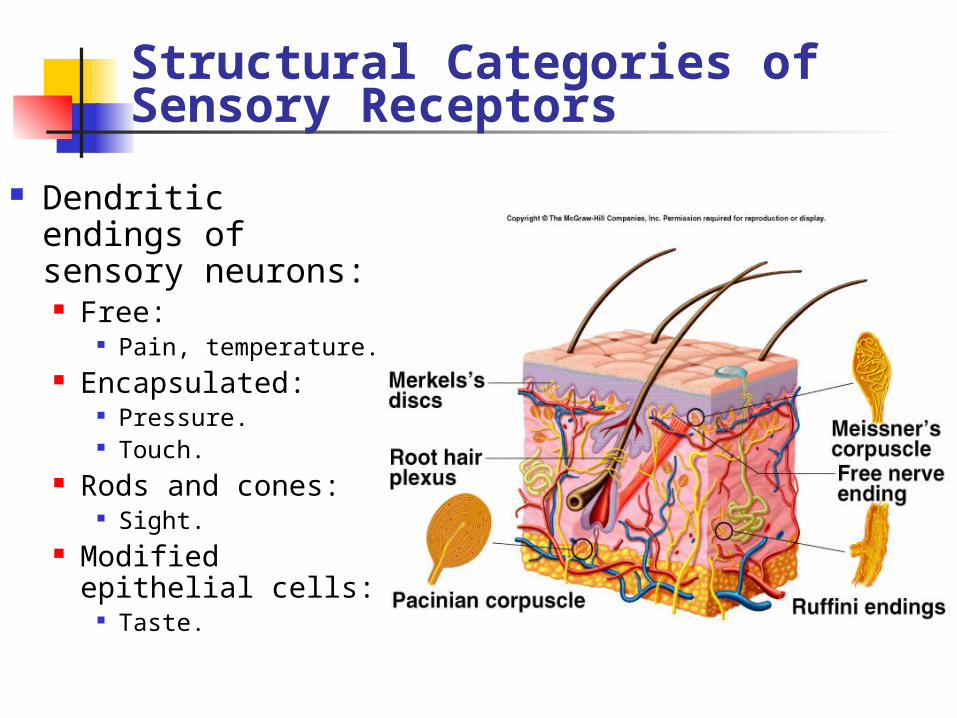
Structural Categories of Sensory Receptors
Dendritic endings of sensory neurons: Free:
Pain, temperature. Encapsulated:
Pressure. Touch.
Rods and cones: Sight.
Modified epithelial cells:
Taste.
Functional Categories of Sensory Receptors
Grouped according to type of stimulus energy they transduce.
Chemoreceptors: Chemical stimuli in environment or
blood (pH, C02). Photoreceptors:
Rods and cones. Thermoreceptors:
Temperature. Mechanoreceptors:
Touch and pressure. Nociceptors:
Pain. Proprioceptors:
Body position.
Categorized according to type of sensory information delivered to brain:
Cutaneous receptors:Touch, pressure, temperature, pain.
Special senses:Sight, hearing, equilibrium.
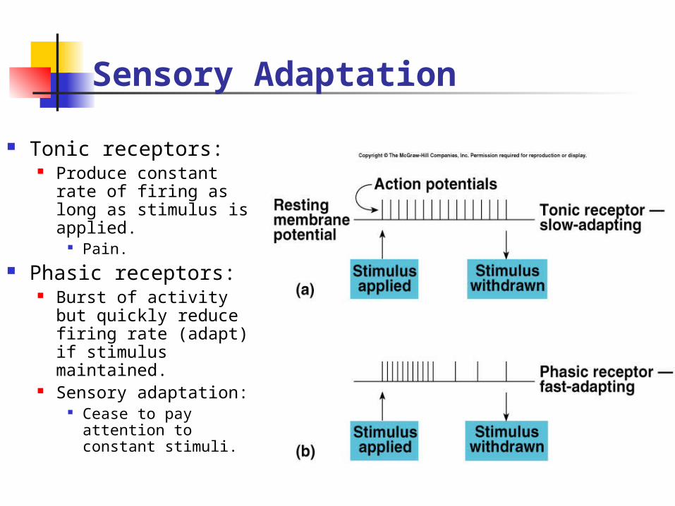
Sensory Adaptation
Tonic receptors: Produce constant
rate of firing as long as stimulus is applied.
Pain. Phasic receptors:
Burst of activity but quickly reduce firing rate (adapt) if stimulus maintained.
Sensory adaptation: Cease to pay
attention to constant stimuli.
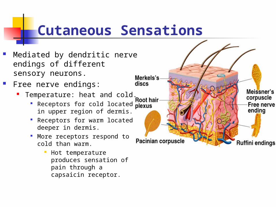
Cutaneous Sensations
Mediated by dendritic nerve endings of different sensory neurons.
Free nerve endings: Temperature: heat and cold.
Receptors for cold located in upper region of dermis.
Receptors for warm located deeper in dermis.
More receptors respond to cold than warm.
Hot temperature produces sensation of pain through a capsaicin receptor.
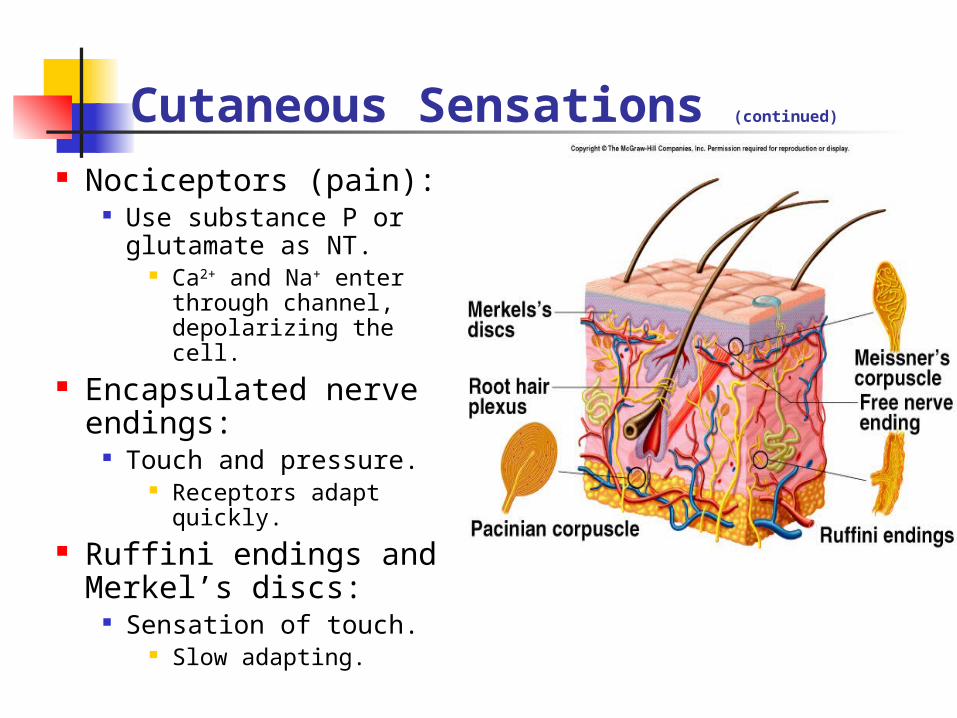
Cutaneous Sensations (continued)
Nociceptors (pain): Use substance P or
glutamate as NT. Ca2+ and Na+ enter
through channel, depolarizing the cell.
Encapsulated nerve endings:
Touch and pressure. Receptors adapt
quickly. Ruffini endings and
Merkel’s discs: Sensation of touch.
Slow adapting.
Receptive Fields
Area of skin whose stimulation results in changes in the firing rate of the neuron. Area of each receptor field varies inversely
with the density of receptors in the region. Back and legs have few sensory
endings. Receptive field is large.
Fingertips have large # of cutaneous receptors. Receptive field is small.
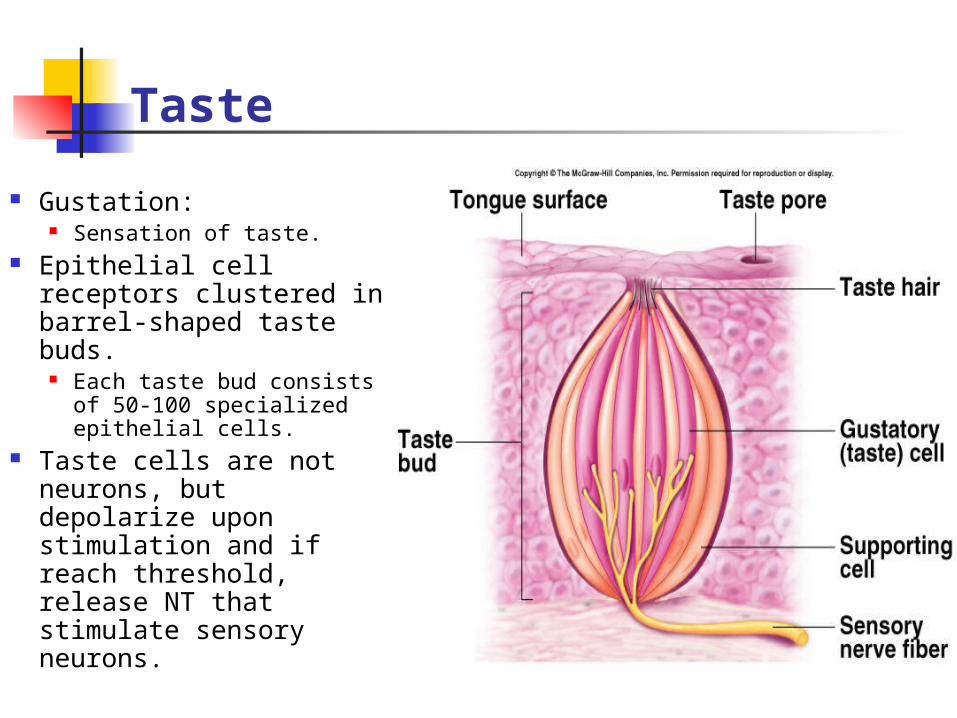
Taste
Gustation: Sensation of taste.
Epithelial cell receptors clustered in barrel-shaped taste buds.
Each taste bud consists of 50-100 specialized epithelial cells.
Taste cells are not neurons, but depolarize upon stimulation and if reach threshold, release NT that stimulate sensory neurons.
Taste (continued)
Each taste bud contains taste cells responsive to each of the different taste categories.
A given sensory neuron may be stimulated by more than 1 taste cell in # of different taste buds.
One sensory fiber may not transmit information specific for only 1 category of taste.
Brain interprets the pattern of stimulation with the sense of smell; so that we perceive the complex tastes.
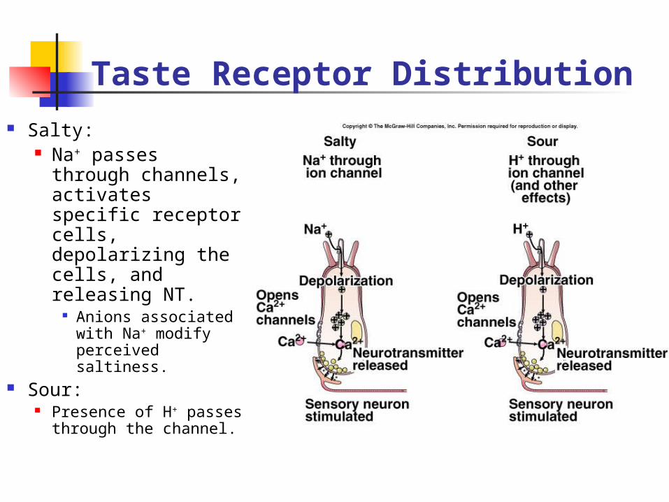
Taste Receptor Distribution Salty:
Na+ passes through channels, activates specific receptor cells, depolarizing the cells, and releasing NT.
Anions associated with Na+ modify perceived saltiness.
Sour: Presence of H+
passes through the channel.
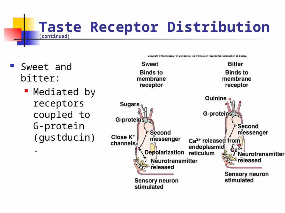
Taste Receptor Distribution (continued)
Sweet and bitter: Mediated by
receptors coupled to G-protein (gustducin).

Taste Receptor Distribution (continued)
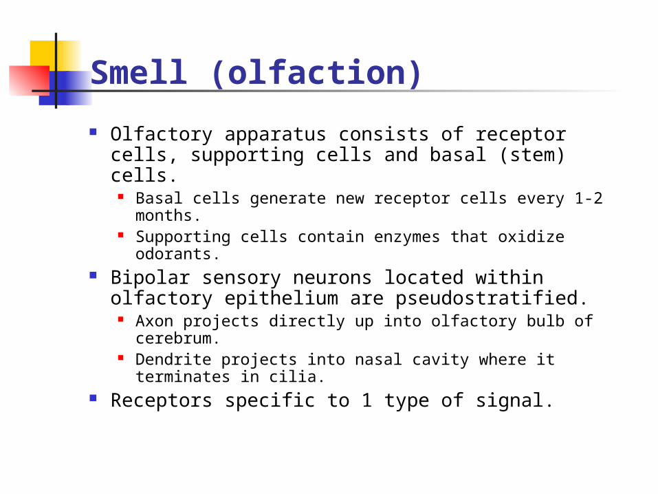
Smell (olfaction)
Olfactory apparatus consists of receptor cells, supporting cells and basal (stem) cells.
Basal cells generate new receptor cells every 1-2 months.
Supporting cells contain enzymes that oxidize odorants.
Bipolar sensory neurons located within olfactory epithelium are pseudostratified.
Axon projects directly up into olfactory bulb of cerebrum.
Dendrite projects into nasal cavity where it terminates in cilia.
Receptors specific to 1 type of signal.
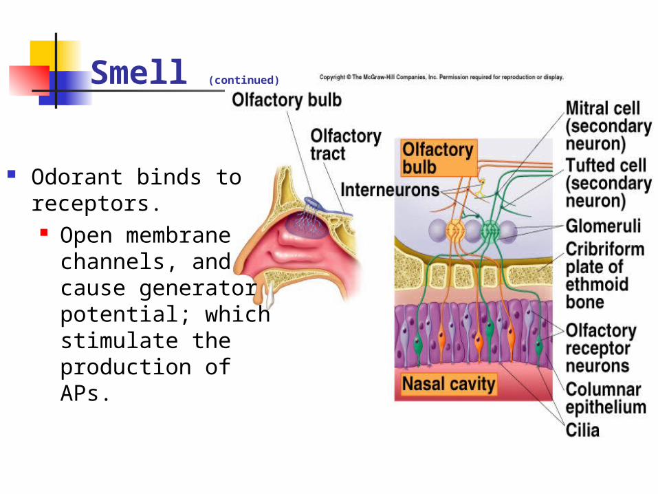
Smell (continued)
Odorant binds to receptors. Open membrane
channels, and cause generator potential; which stimulate the production of APs.
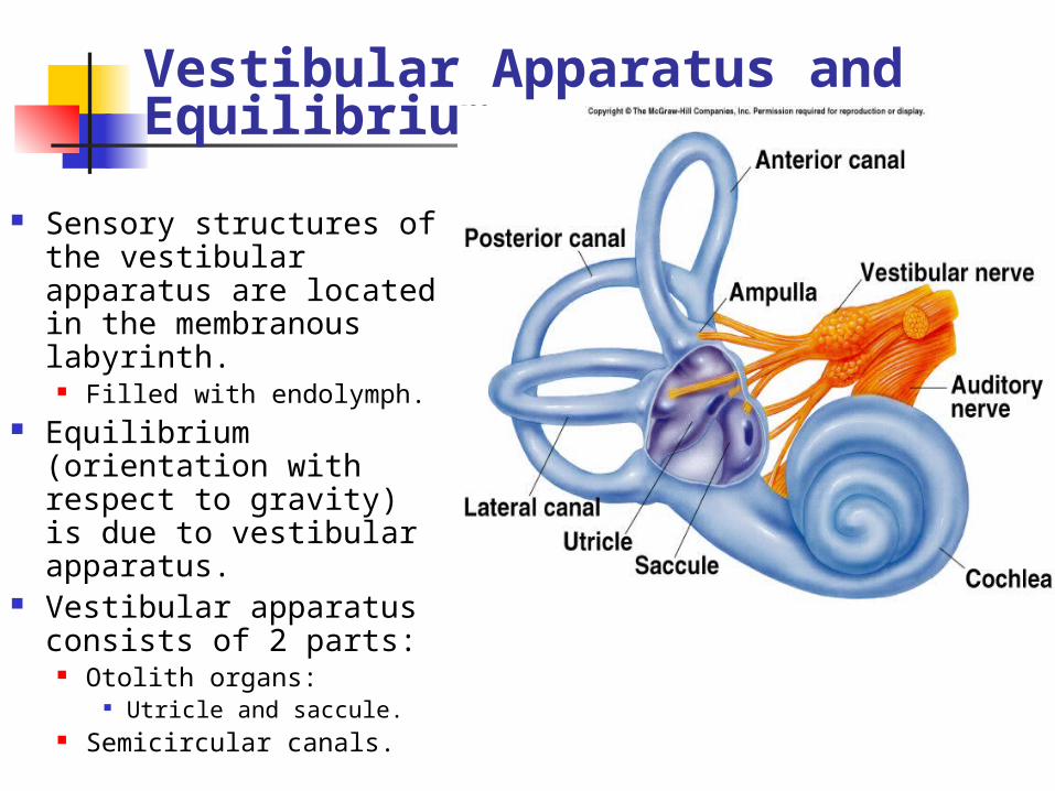
Vestibular Apparatus and Equilibrium
Sensory structures of the vestibular apparatus are located in the membranous labyrinth.
Filled with endolymph. Equilibrium (orientation
with respect to gravity) is due to vestibular apparatus.
Vestibular apparatus consists of 2 parts:
Otolith organs: Utricle and saccule.
Semicircular canals.
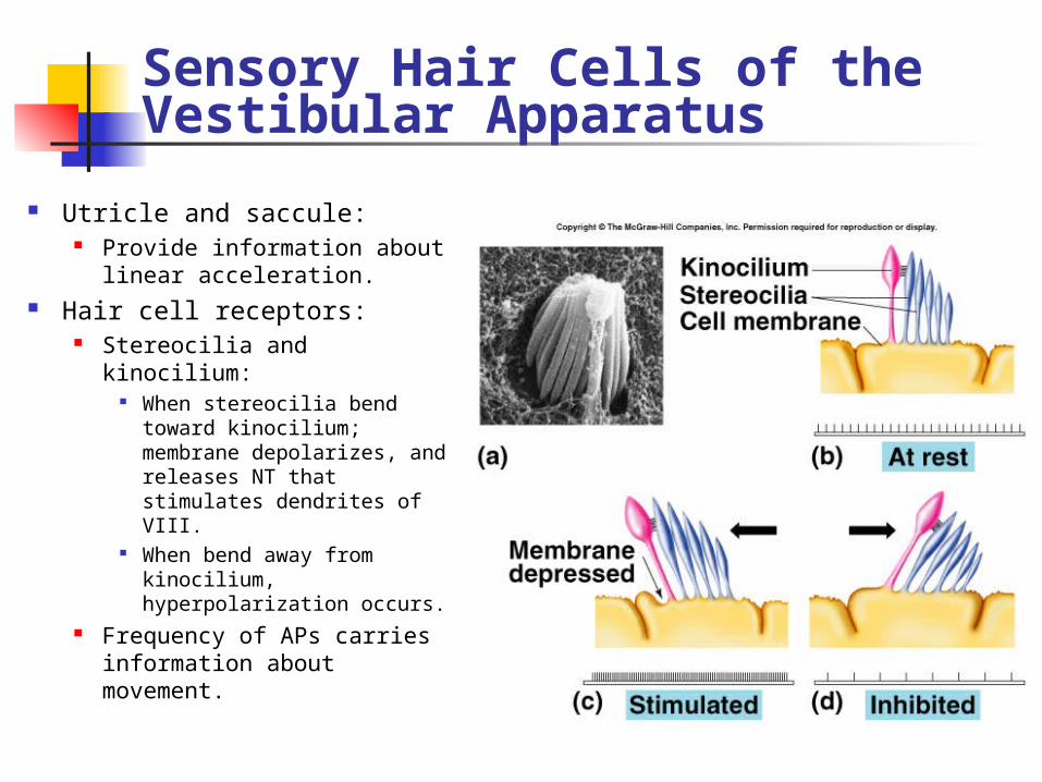
Sensory Hair Cells of the Vestibular Apparatus
Utricle and saccule: Provide information about
linear acceleration. Hair cell receptors:
Stereocilia and kinocilium: When stereocilia bend
toward kinocilium; membrane depolarizes, and releases NT that stimulates dendrites of VIII.
When bend away from kinocilium, hyperpolarization occurs.
Frequency of APs carries information about movement.
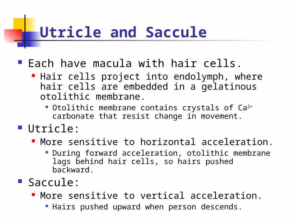
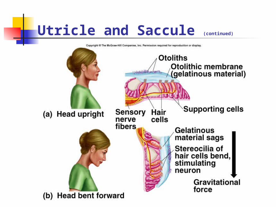
Utricle and Saccule
Each have macula with hair cells. Hair cells project into endolymph, where hair
cells are embedded in a gelatinous otolithic membrane.
Otolithic membrane contains crystals of Ca2+ carbonate that resist change in movement.
Utricle: More sensitive to horizontal acceleration.
During forward acceleration, otolithic membrane lags behind hair cells, so hairs pushed backward.
Saccule: More sensitive to vertical acceleration.
Hairs pushed upward when person descends.
Utricle and Saccule (continued)
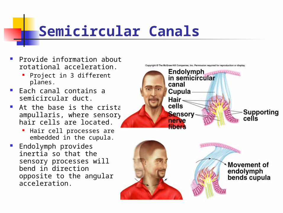
Semicircular Canals
Provide information about rotational acceleration.
Project in 3 different planes.
Each canal contains a semicircular duct.
At the base is the crista ampullaris, where sensory hair cells are located.
Hair cell processes are embedded in the cupula.
Endolymph provides inertia so that the sensory processes will bend in direction opposite to the angular acceleration.
Neural Pathways
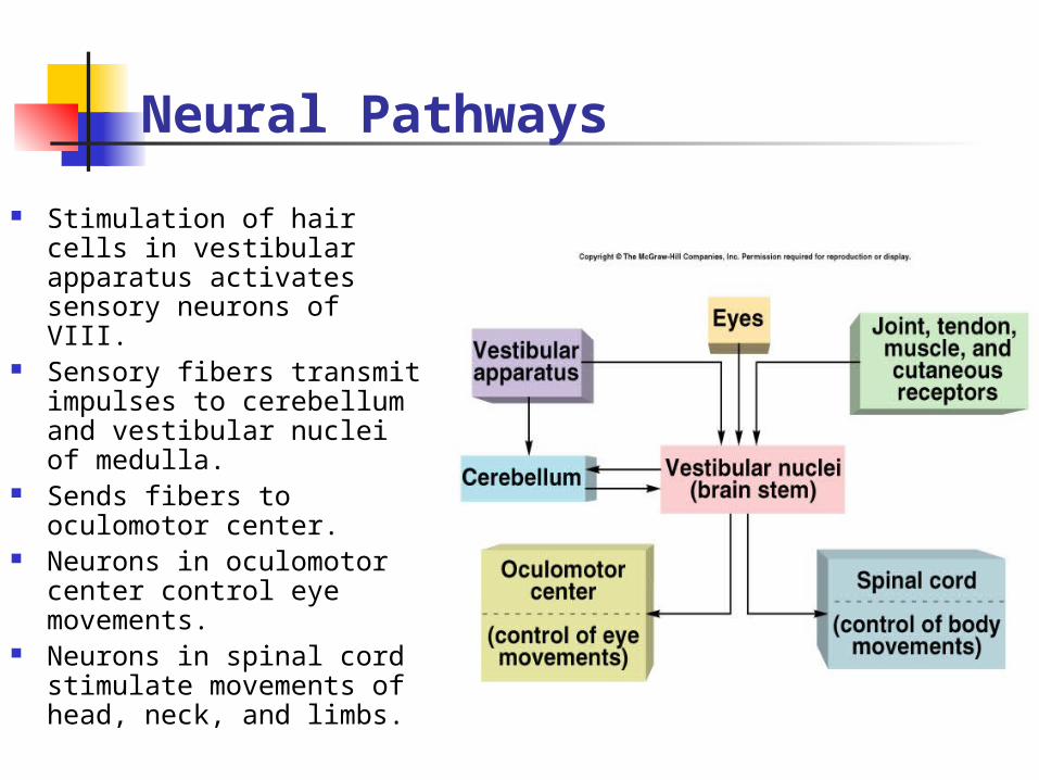
Stimulation of hair cells in vestibular apparatus activates sensory neurons of VIII.
Sensory fibers transmit impulses to cerebellum and vestibular nuclei of medulla.
Sends fibers to oculomotor center.
Neurons in oculomotor center control eye movements.
Neurons in spinal cord stimulate movements of head, neck, and limbs.
Nystagmus and Vertigo
Nystagmus:
Vertigo: Loss of equilibrium when spinning.
May be caused by anything that alters firing rate. Pathologically, viral infections.
Ears and Hearing
Sound waves travel in all directions from their source.
Waves are characterized by frequency and intensity. Frequency:
Measured in hertz (cycles per second). Pitch is directly related to frequency.
Greater the frequency the higher the pitch. Intensity (loudness):
Directly related to amplitude of sound waves. Measured in decibels.
Outer Ear
Sound waves are funneled by the pinna (auricle) into the external auditory meatus.
External auditory meatus channels sound waves to the tympanic membrane. Increases sound wave intensity.
Middle Ear
Cavity between tympanic membrane and cochlea. Malleus:
Attached to tympanic membrane. Vibrations of membrane are transmitted to the malleus
and incus to stapes. Stapes:
Attached to oval window. Vibrates in response to vibrations in tympanic
membrane. Vibrations transferred through 3 bones:
Provides protection and prevents nerve damage. Stapedius muscle contracts and dampens vibrations.
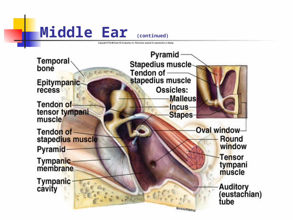
Middle Ear (continued)
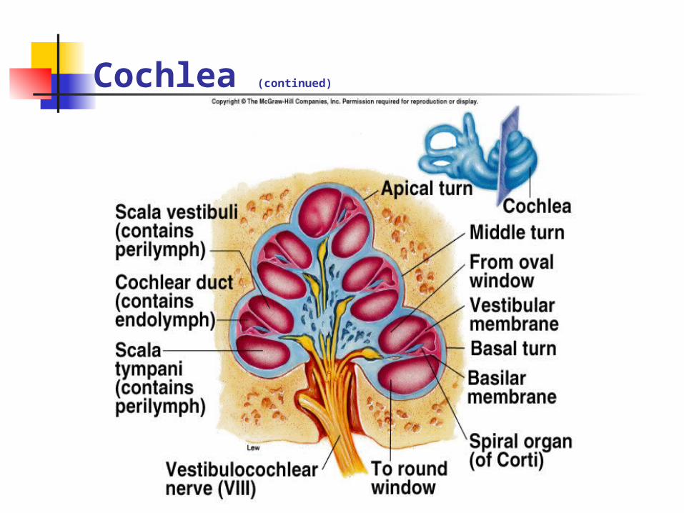
Cochlea
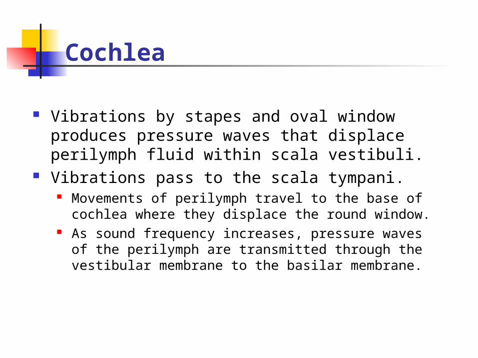
Vibrations by stapes and oval window produces pressure waves that displace perilymph fluid within scala vestibuli.
Vibrations pass to the scala tympani. Movements of perilymph travel to the base of cochlea
where they displace the round window. As sound frequency increases, pressure waves of the
perilymph are transmitted through the vestibular membrane to the basilar membrane.
Cochlea (continued)

Effects of Different Frequencies
Displacement of basilar membrane is central to pitch discrimination.
Waves in basilar membrane reach a peak at different regions depending upon pitch of sound.
Sounds of higher frequency cause maximum vibrations of basilar membrane.
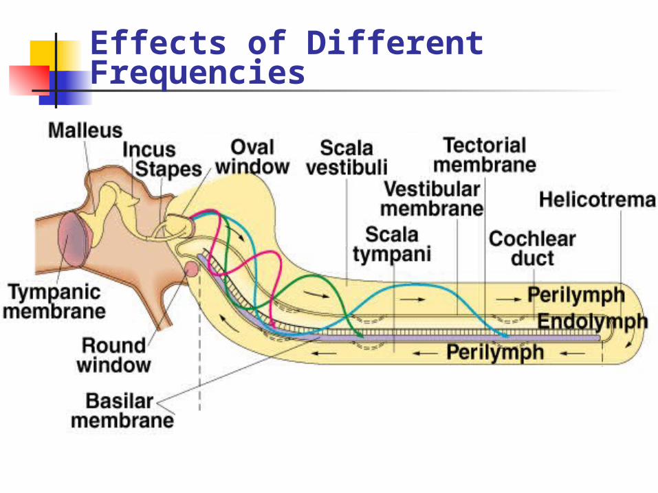
Effects of Different Frequencies

Organ of Corti (continued)
Neural Pathway for Hearing
Sensory neurons in cranial nerve VIII synapse with neurons in brain.
Each area of cortex represents a different part of the basilar membrane and a different pitch.
Hearing Impairments
Conduction deafness: Transmission of sound waves through
middle ear to oval window impaired. Impairs all sound frequencies.
Hearing aids.
Sensorineural (perception) deafness: Transmission of nerve impulses is
impaired. Impairs ability to hear some pitches more than
others. Cochlear implants.
Vision
Eyes transduce energy in the electrmagnetic spectrum into APs. Only wavelengths of 400 – 700 nm
constitute visible light. Neurons in the retina contribute
fibers that are gathered together at the optic disc, where they exit as the optic nerve.
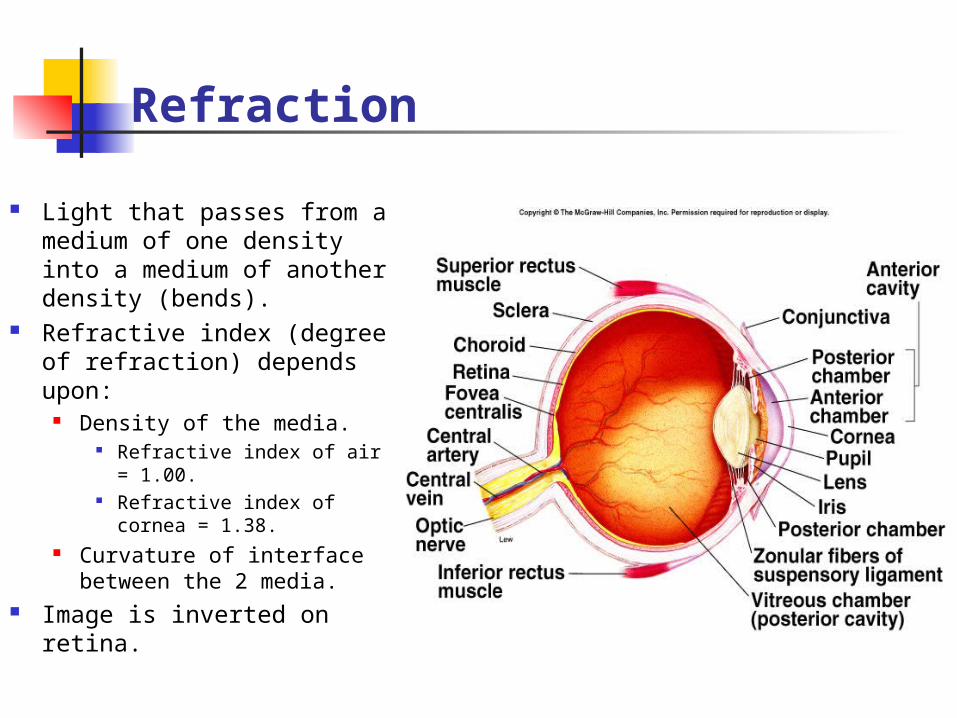
Refraction
Light that passes from a medium of one density into a medium of another density (bends).
Refractive index (degree of refraction) depends upon:
Density of the media. Refractive index of air =
1.00. Refractive index of
cornea = 1.38. Curvature of interface
between the 2 media. Image is inverted on
retina.
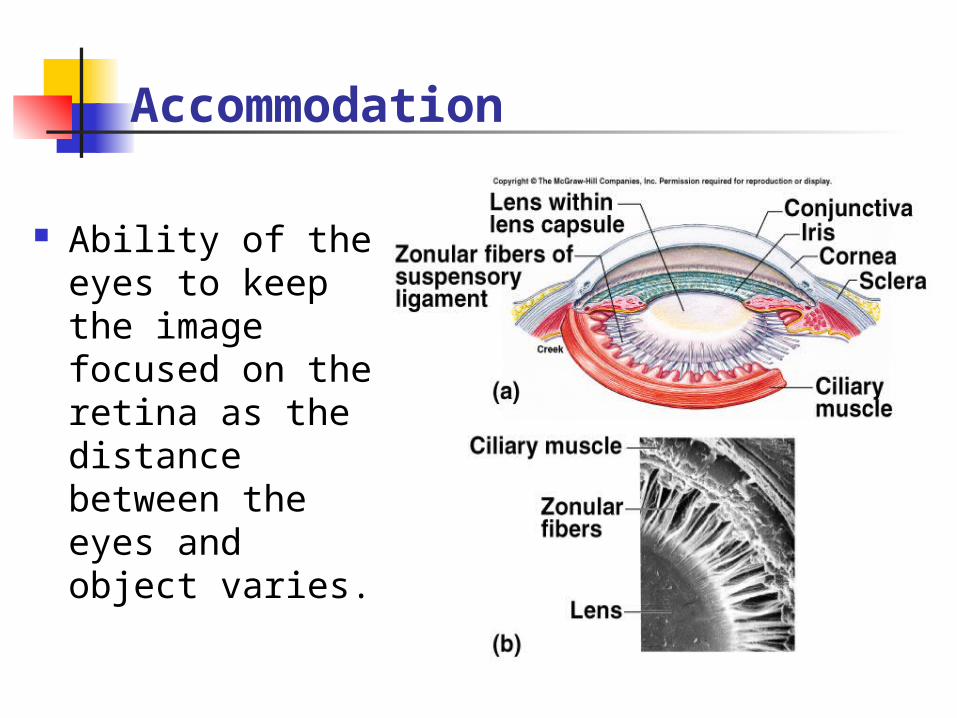
Accommodation
Ability of the eyes to keep the image focused on the retina as the distance between the eyes and object varies.
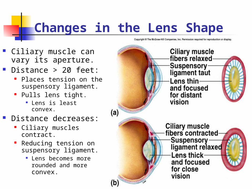
Changes in the Lens Shape
Ciliary muscle can vary its aperture.
Distance > 20 feet: Places tension on the
suspensory ligament. Pulls lens tight.
Lens is least convex. Distance decreases:
Ciliary muscles contract.
Reducing tension on suspensory ligament.
Lens becomes more rounded and more convex.
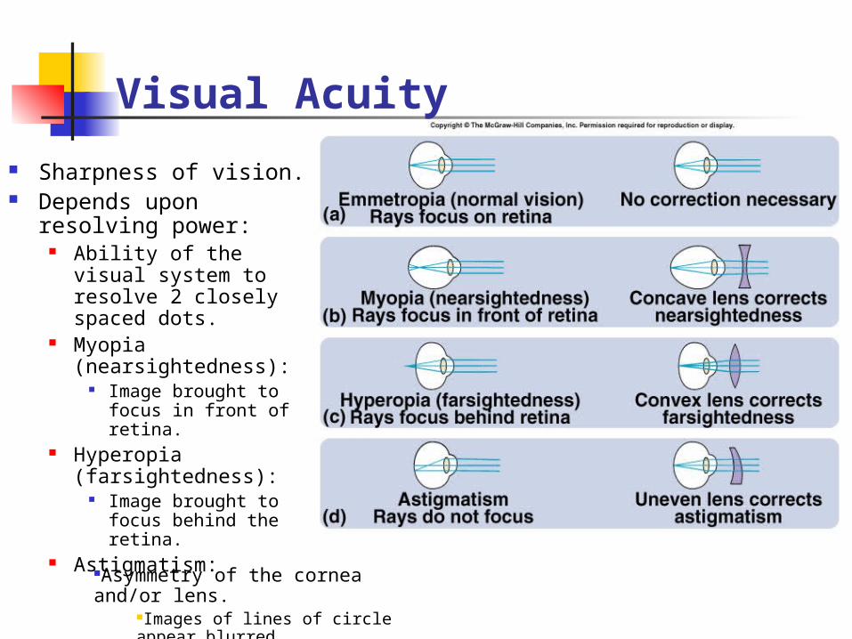
Visual Acuity
Sharpness of vision. Depends upon
resolving power: Ability of the visual
system to resolve 2 closely spaced dots.
Myopia (nearsightedness):
Image brought to focus in front of retina.
Hyperopia (farsightedness):
Image brought to focus behind the retina.
Astigmatism:Asymmetry of the cornea and/or lens.
Images of lines of circle appear blurred.
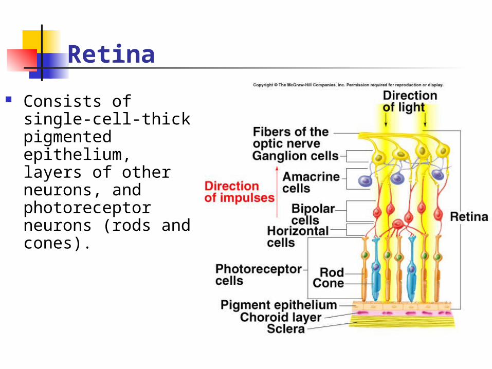
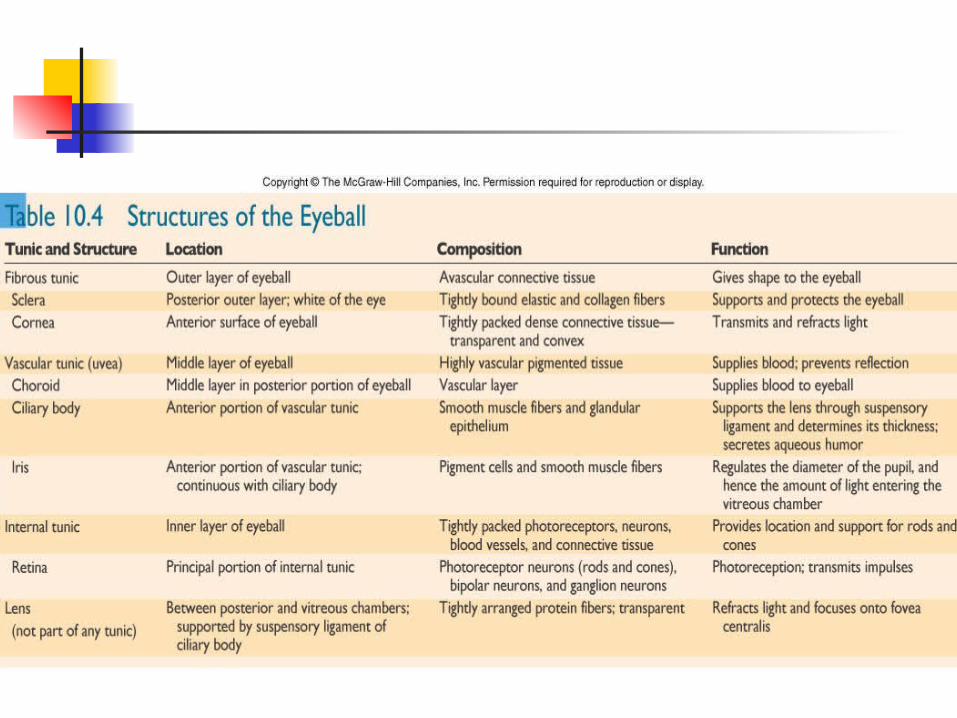
Retina
Consists of single-cell-thick pigmented epithelium, layers of other neurons, and photoreceptor neurons (rods and cones).
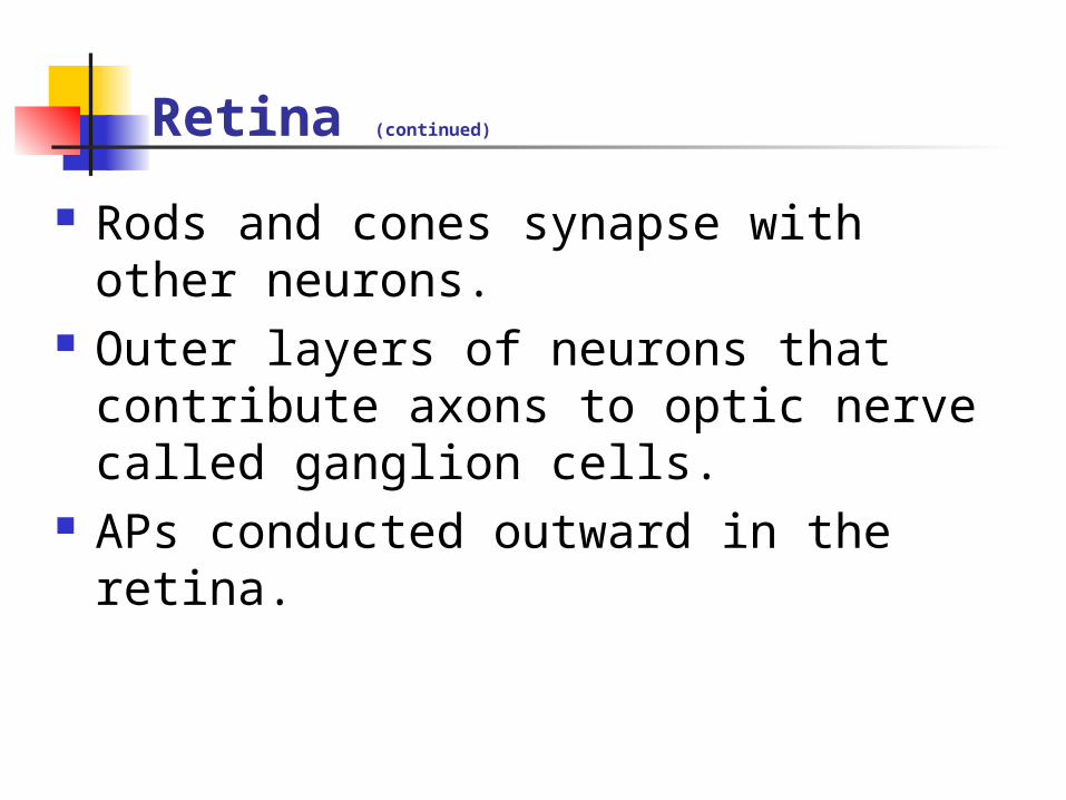
Retina (continued)
Rods and cones synapse with other neurons.
Outer layers of neurons that contribute axons to optic nerve called ganglion cells.
APs conducted outward in the retina.
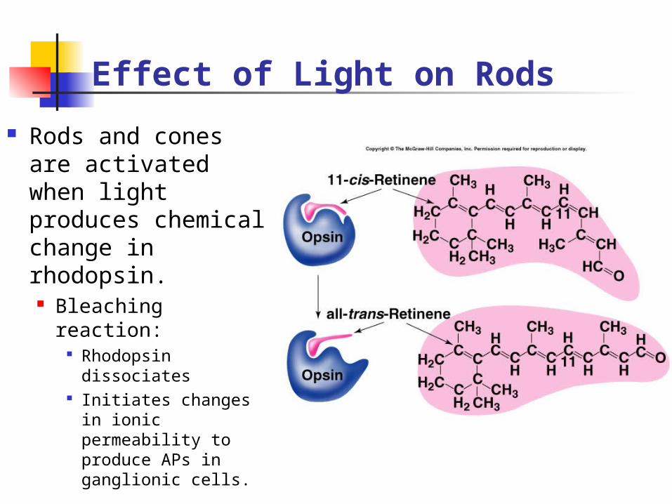
Effect of Light on Rods
Rods and cones are activated when light produces chemical change in rhodopsin. Bleaching reaction:
Rhodopsin dissociates
Initiates changes in ionic permeability to produce APs in ganglionic cells.
Dark Adaptation Gradual increase in photoreceptor
sensitivity when entering a dark room. Maximal sensitivity reached in 20 min.
Increased amounts of visual pigments produced in the dark. Increased pigment in cones produces
slight dark adaptation in 1st 5 min. Increased rhodopsin in rods produces
greater increase in sensitivity. 100,00-fold increase in light sensitivity in
rods.
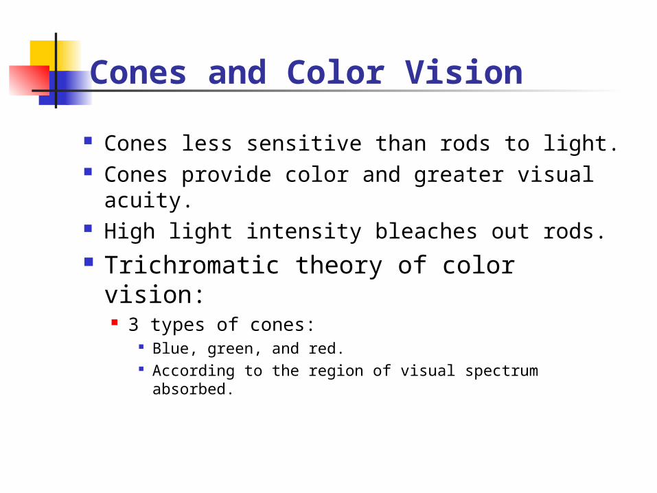
Cones and Color Vision
Cones less sensitive than rods to light. Cones provide color and greater visual
acuity. High light intensity bleaches out rods. Trichromatic theory of color vision:
3 types of cones: Blue, green, and red. According to the region of visual spectrum
absorbed.
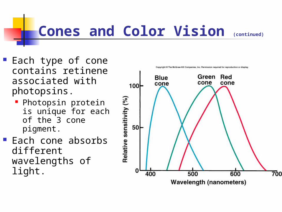
Cones and Color Vision (continued)
Each type of cone contains retinene associated with photopsins. Photopsin protein
is unique for each of the 3 cone pigment.
Each cone absorbs different wavelengths of light.
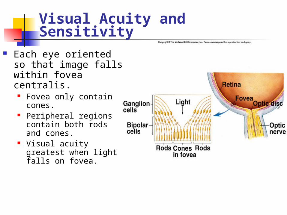
Visual Acuity and Sensitivity Each eye oriented so
that image falls within fovea centralis.
Fovea only contain cones.
Peripheral regions contain both rods and cones.
Visual acuity greatest when light falls on fovea.