Sensory Cranial Nerves
45
Sensory Cranial Nerves
description
Sensory Cranial Nerves. Cranial Nerves. Special Sense Nerves I,II,VIII Somatic Motor Nerves Eye—III,IV,VI Tongue--XII “Rest of body” nerves IX,X,XI Face and jaws VII, V. Human Anatomy, Frolich, Head/Neck IV: Cranial Nerves. SENSORY SpecialGeneral Smellskin Visionteeth - PowerPoint PPT Presentation
Transcript of Sensory Cranial Nerves
Sensory Cranial Nerves
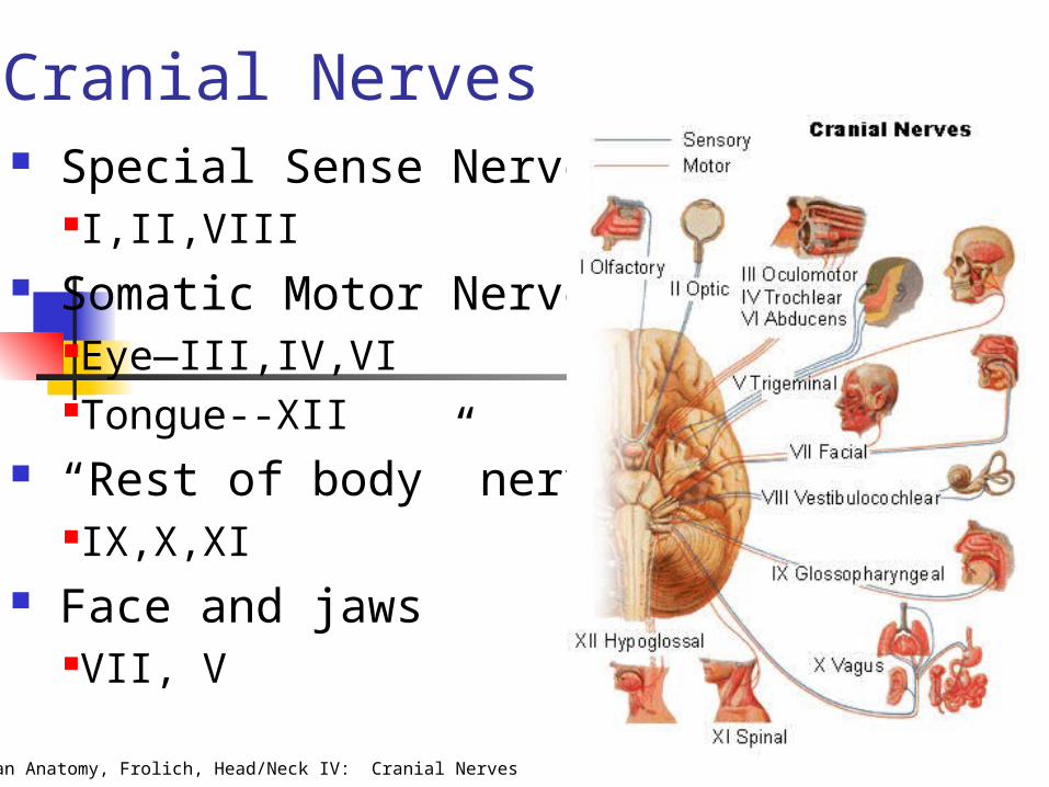
Human Anatomy, Frolich, Head/Neck IV: Cranial Nerves
Cranial Nerves Special Sense Nerves
I,II,VIII Somatic Motor Nerves
Eye—III,IV,VITongue--XII
“Rest of body” nervesIX,X,XI
Face and jawsVII, V
Human Anatomy, Frolich, Head/Neck IV: Cranial Nerves
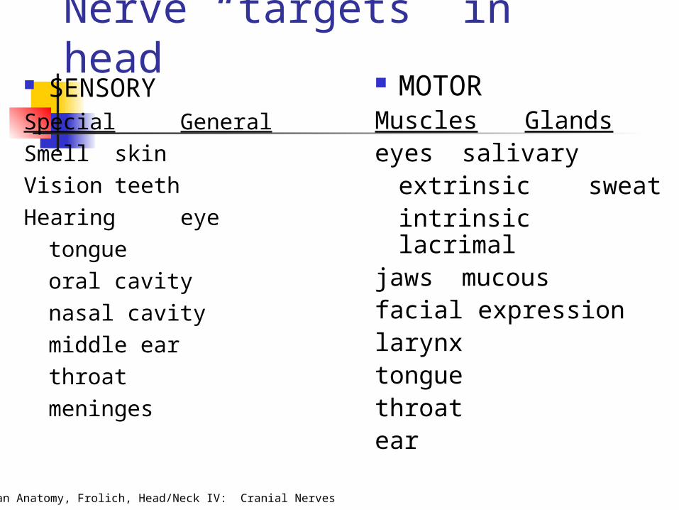
Nerve “targets” in head SENSORYSpecial GeneralSmell skinVision teethHearing eye
tongueoral cavitynasal cavitymiddle earthroatmeninges
MOTORMuscles Glandseyes salivary
extrinsic sweatintrinsic lacrimal
jaws mucousfacial expressionlarynxtonguethroatear

Human Anatomy, Frolich, Head/Neck IV: Cranial Nerves
Base of the skull—cranial nerves out Ethmoid (olfactory)
I. Olfactory Sphenoid (optic)
II. OpticIII. OculomotorIV. TrochlearVI. Abducens
Temporal (otic)VII. Acoustic/Auditory/
Vestibulocochlear Face/Jaws
V. TrigeminalVII. Facial
Throat (rest of body)IX GlossopharyngealX. VagusXI. Spinal AccessoryXII. Hypoglosal
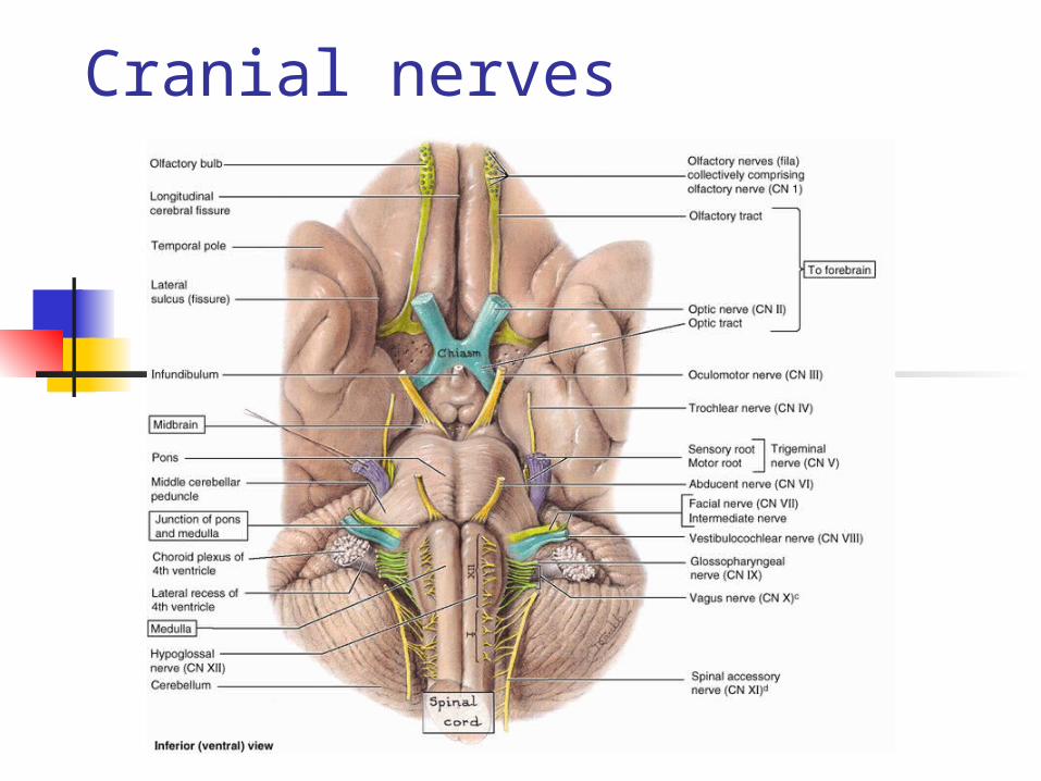
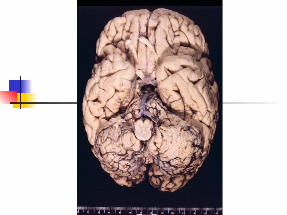
Cranial nerves

Cranial nerves

Human Anatomy, Frolich, Head/Neck IV: Cranial Nerves
Special Sense Nerves
Internal auditory meatus (temporal)
Inner earVIII. Auditory
Optic canal (sphenoid)
RetinaII. Optic
Cribiform plate (ethmoid)
Olfactory epithelium
I. Olfactory
EXIT FROM CRANIAL CAVITY
TARGETNERVE

Human Anatomy, Frolich, Head/Neck IV: Cranial Nerves
Somatic Motor Nerves(eye muscles and tongue)
EXIT CR. CAVITYTARGETNERVE
Hypoglossal canal
(occipital)
Intrinsic, extrinsic mm. of tongue
XII. Hypoglossal
“•Sup.,med.,inf.rectus• Inferior Oblique•Levator palpebrae superioris
III. Oculomotor(Also parasympathetic to ciliary mm, constrictor pupillae)
“Lateral rectusVI. Abducens
Sup. Orbital fissure (sphenoid)
Superior oblique m. (with trochlea)
IV. Trochlear

Human Anatomy, Frolich, Head/Neck IV: Cranial Nerves
“Rest of body” nerves(all exit from jugular foramen)
NERVE TARGET
X: Vagus Somatic motor to larynx/pharynx Parasympathetic to most of gut Taste to back posterior pharynx
XI: (Spinal) Accesory
Motor to traps, sternocleidomastoid
IX: Glosso-pharyngeal
Sensory to carotid body/sinus Taste to posterior tongue Sensory to ear opening/middle
ear Parotid salivary gland

Human Anatomy, Frolich, Head/Neck IV: Cranial Nerves

Cranial Nerves I: Olfactory II: Optic III: Oculomotor IV: Trochlear V: Trigeminal VI: Abducens
VII: Facial VIII:Vestibulocochlear
Acoustic IX:
Glossopharyngeal X: Vagus XI: Accessory XII: Hypoglossal
http://www.gwc.maricopa.edu/class/bio201/cn/cranial.htm
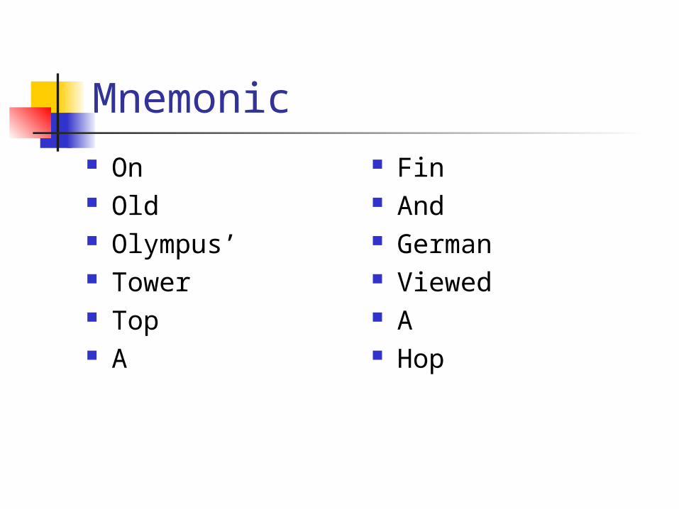
Mnemonic On Old Olympus’ Tower Top A
Fin And German Viewed A Hop
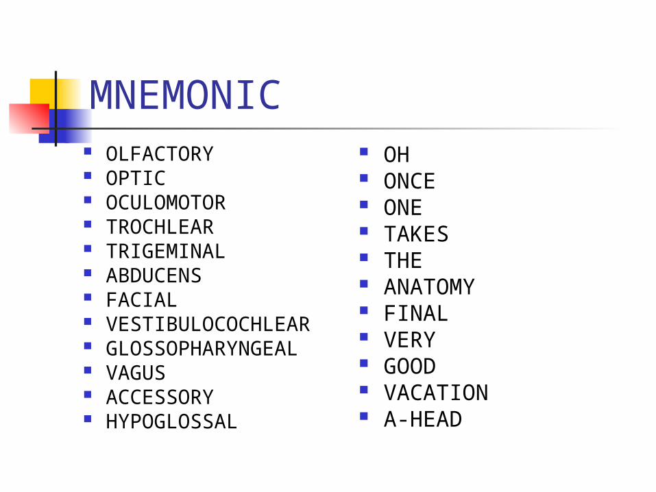
MNEMONIC OLFACTORY OPTIC OCULOMOTOR TROCHLEAR TRIGEMINAL ABDUCENS FACIAL VESTIBULOCOCHLEAR GLOSSOPHARYNGEAL VAGUS ACCESSORY HYPOGLOSSAL
OH ONCE ONE TAKES THE ANATOMY FINAL VERY GOOD VACATION A-HEAD
SENSORY NERVES
Cranial nerve I OlfactoryCranial nerve II Optic
Cranial nerve VIII Acoustovestibular
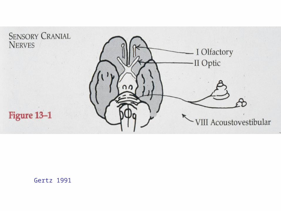
Gertz 1991

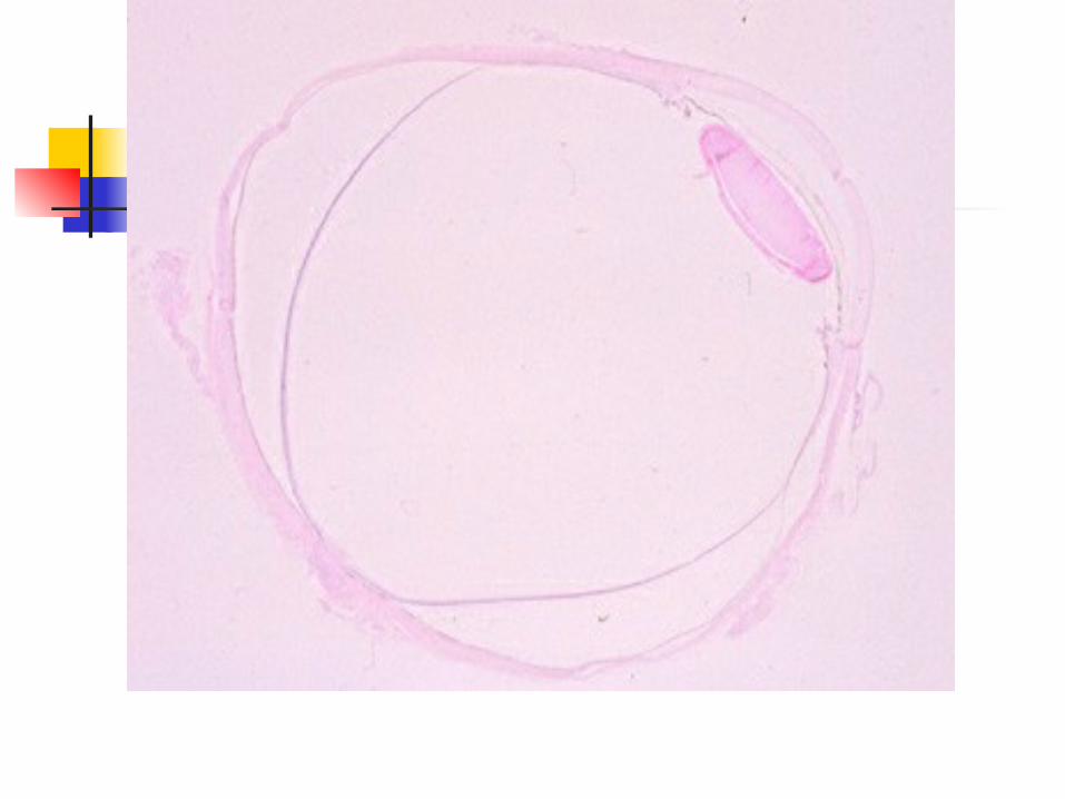
CN I: OLFACTORY Cranial nerve I Function:
smell Clinical test for
damage: determine
whether a person can smell something aromatic
Human Anatomy, Frolich, Head/Neck IV: Cranial Nerves
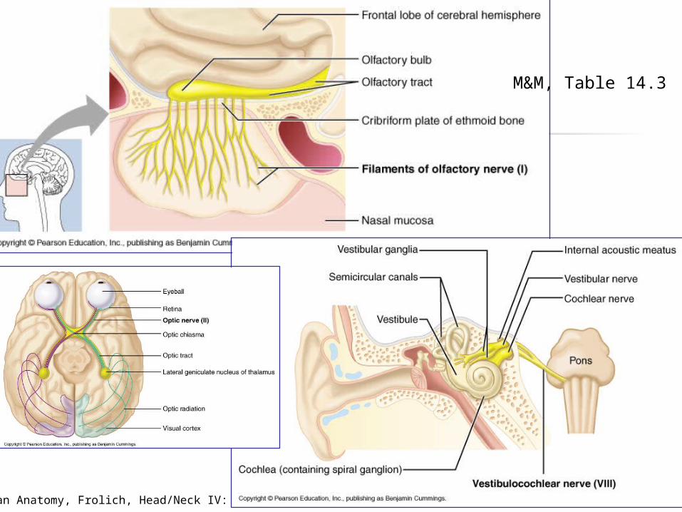
M&M, Table 14.3

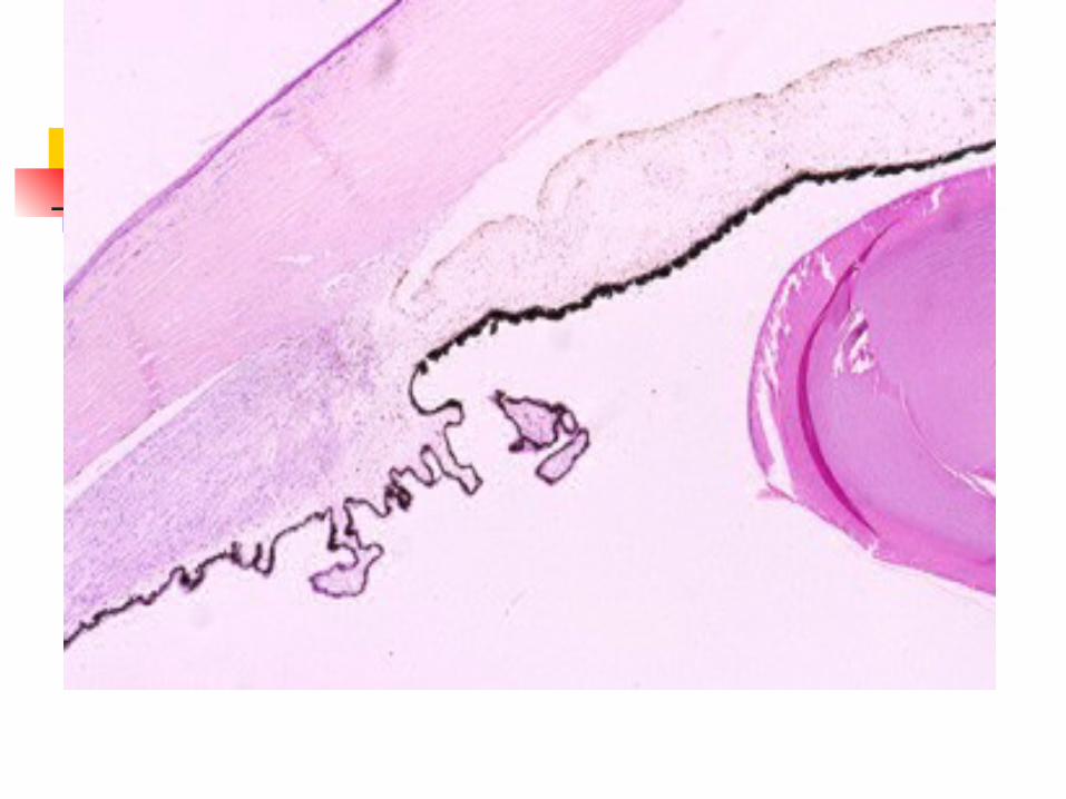
CN II: OPTIC Cranial nerve II Function:
vision Clinical test for
damage: tests peripheral
vision and visual acuity
Effects of damage: blindness in part or
all of the visual field

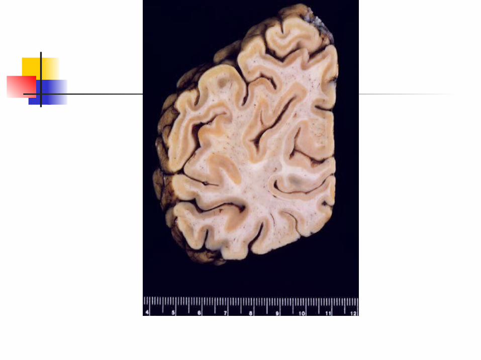
CN VIII: VESTIBULOCOCHLEAR
Cranial Nerve VIII Function: hearing and equilibrium Clinical tests: test hearing, balance,
and ability to walk a straight line Effects of damage: deafness,
dizziness, nausea, loss of balance, and nystagmus
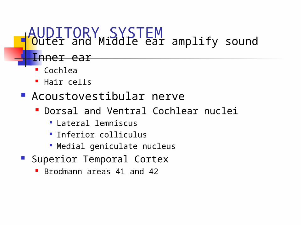
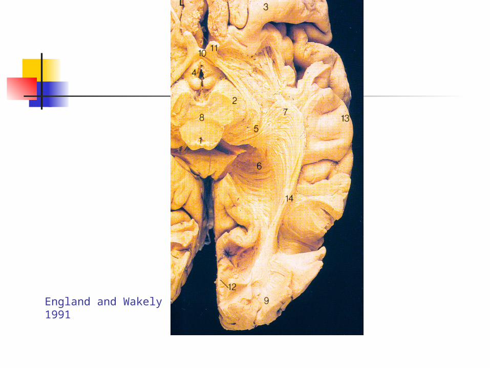
AUDITORY SYSTEM Outer and Middle ear amplify sound Inner ear
Cochlea Hair cells
Acoustovestibular nerve Dorsal and Ventral Cochlear nuclei
Lateral lemniscus Inferior colliculus Medial geniculate nucleus
Superior Temporal Cortex Brodmann areas 41 and 42
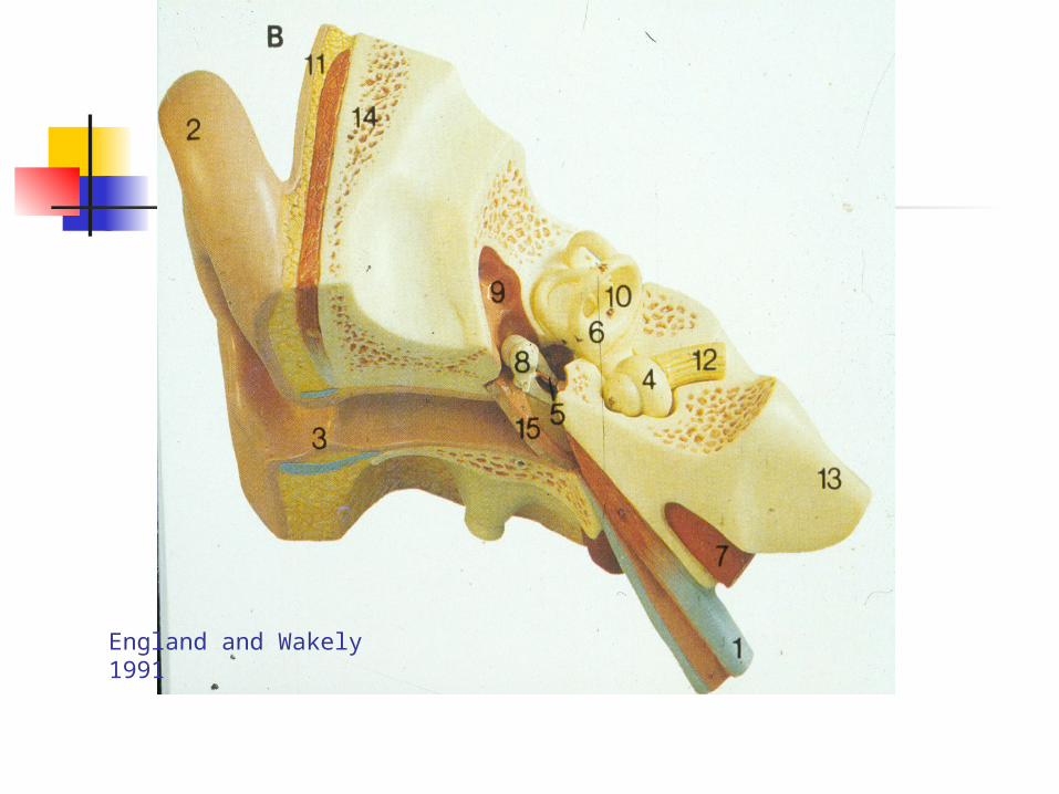
England and Wakely 1991
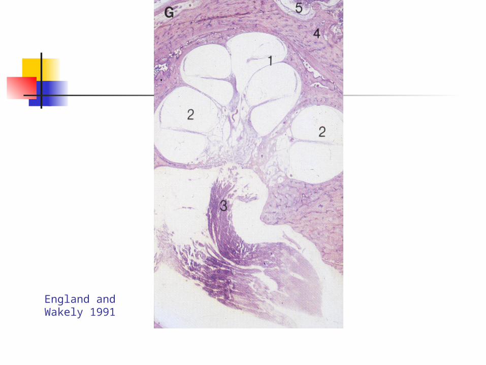
England and Wakely 1991
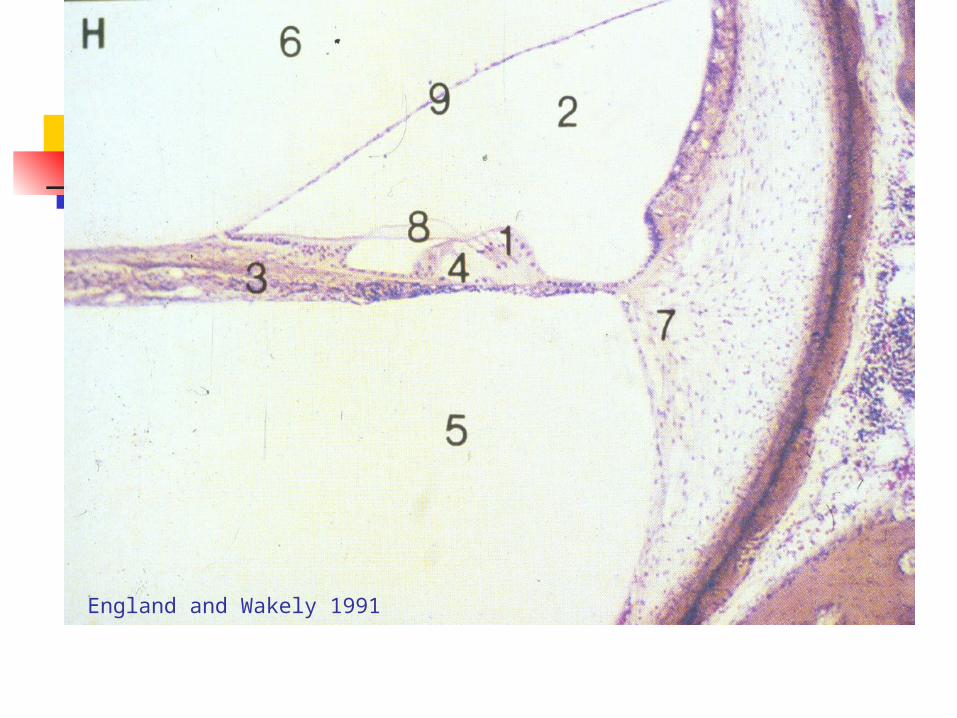
England and Wakely 1991


Gertz 1991
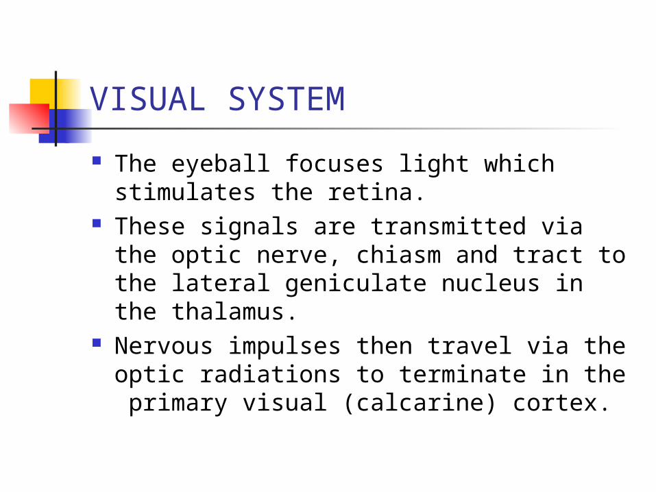
VISUAL SYSTEM
The eyeball focuses light which stimulates the retina.
These signals are transmitted via the optic nerve, chiasm and tract to the lateral geniculate nucleus in the thalamus.
Nervous impulses then travel via the optic radiations to terminate in the primary visual (calcarine) cortex.

www.visionsofjoy.org


England and Wakely 1991

Gertz 1991

Gertz 1991
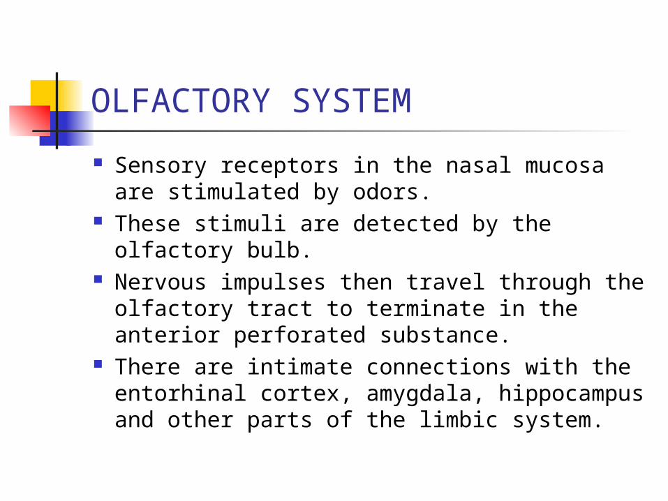
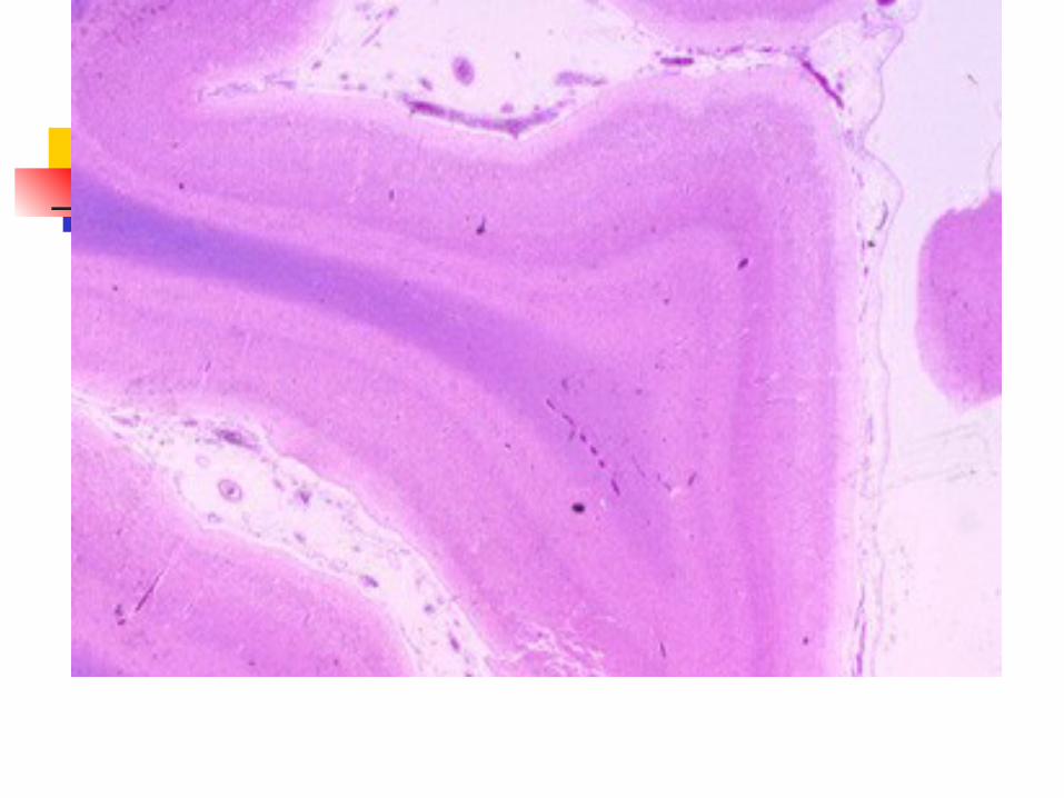
OLFACTORY SYSTEM
Sensory receptors in the nasal mucosa are stimulated by odors.
These stimuli are detected by the olfactory bulb.
Nervous impulses then travel through the olfactory tract to terminate in the anterior perforated substance.
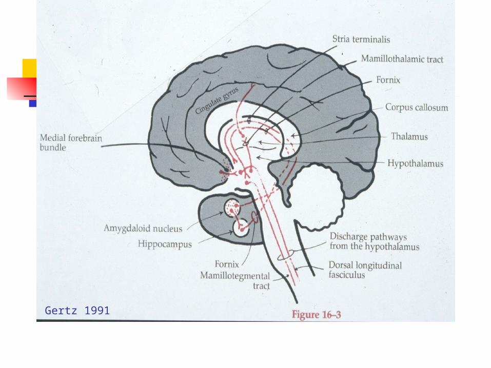
There are intimate connections with the entorhinal cortex, amygdala, hippocampus and other parts of the limbic system.

www.colorado.edu
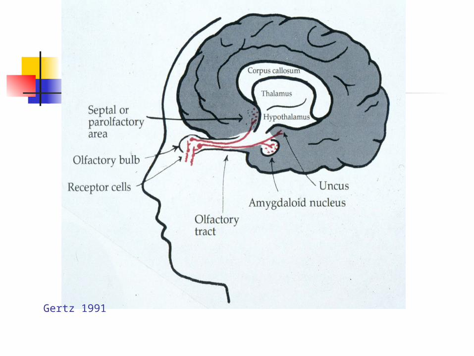
Gertz 1991

Gertz 1991
Gertz 1991