
sensitively monitor nitric oxide release Supporting … monitor nitric oxide release Yan-Ling Liu,...
13
1 Supporting Information for Functionalized graphene-based biomimetic microsensor interfacing with living cells to sensitively monitor nitric oxide release Yan-Ling Liu, Xue-Ying Wang, Jia-Quan Xu, Chong Xiao, Yan-Hong Liu, Xin-Wei Zhang, Jun-Tao Liu and Wei-Hua Huang* Key Laboratory of Analytical Chemistry for Biology and Medicine (Ministry of Education), College of Chemistry and Molecular Sciences, Wuhan University, Wuhan, 430072, China E-mail: [email protected] RECEIVED DATE: Phone: (86)2768752149 Fax: (86)2768754067 Electronic Supplementary Material (ESI) for Chemical Science. This journal is © The Royal Society of Chemistry 2015
Transcript of sensitively monitor nitric oxide release Supporting … monitor nitric oxide release Yan-Ling Liu,...

1
Supporting Information forFunctionalized graphene-based biomimetic microsensor interfacing with living cells to
sensitively monitor nitric oxide release
Yan-Ling Liu, Xue-Ying Wang, Jia-Quan Xu, Chong Xiao, Yan-Hong Liu,
Xin-Wei Zhang, Jun-Tao Liu and Wei-Hua Huang*
Key Laboratory of Analytical Chemistry for Biology and Medicine (Ministry of
Education), College of Chemistry and Molecular Sciences, Wuhan University,
Wuhan, 430072, China
E-mail: [email protected]
RECEIVED DATE:
Phone: (86)2768752149
Fax: (86)2768754067
Electronic Supplementary Material (ESI) for Chemical Science.This journal is © The Royal Society of Chemistry 2015

2
METHODS
Materials. ITO conductive glass (film thickness: 180 nm, conductivity: 10 Ω/sq) was
purchased Crystal Great Technology Co., Ltd (Shenzhen, China). Graphene oxide
(GO) was obtained from Ji Cang Nano Technology Co., Ltd. (Nanjing, China). Fe
(III) meso-tetra (4-carboxyphenyl) porphyrin (FeTCP) was purchased from Frontier
Scientific, Inc. Hydrazine solution (85%) and ammonia solution (25 wt %) were
provided by Beijing Chemicals Inc (Beijing, China). The cell culture medium RPMI
1640, L-glutamine and HEPES for HUVECs culture were purchased from GIBCO
(USA). NOS inhibitor Nω-nitro-L-arginine methyl ester hydrochloride (L-NAME),
N-(3-dimethylaminopropyl)-N'-ethylcarbodiimide (EDC), N-hydrosulfosuccinimide
sodium salt (NHS), 3-aminophenylboronic acid (APBA) and L- arginine (L-Arg)
were purchased from Sigma (USA). The human umbilical vein endothelial cell
(HUVEC) lines were obtained from CHI Scientific, Inc (Shanghai China). 3',6'-Di(o-
acetyl)-4',5'-bis[N,N-bis(carboxymethyl)-aminomethyl] fluorescein,
tetraacetoxymethylester (Calcein-AM) and 3,8-diamino-5-[3-(diethylmethylammonio)
propyl]-6-phenylphenanthridinium diiodide (PI) for cell staining were obtained from
Dojindo laboratory (USA). All other chemicals unless specified were reagent grade
and used without further purification.
Preparation of FGHNs and rGO. FGHNs was prepared similarly to the literature.1
Briefly, 4 mg GO and 6.5 mg FeTCP powder were ultrasonically dissolved in 8 ml
H2O for 1 h. The whole solution was then vigorously stirred at 70 overnight.

3
Subsequently, 4 μL hydrazine solution and 50 μL ammonia solution were added to the
above solution and the mixture was put in a water bath (95 ) for 1 h under agitation.
The resulting product was stable black dispersion, subsequently filtered through a
Nylon membrane (0.22 μm) and thoroughly washed with water until the pH of the
filtrate reached 7.0. The as-prepared nanocomposite was ultrasonically dispersed in
ultrapure water to obtain a FGHNs solution (0.1 mg/mL). Additionally, the
preparation of rGO solution was similar to FGHNs except no addition of FeTCP.
Fabrication of ITO microelectrode array. The ITO film (5 cm × 5 cm) was
patterned into 200-µm wide stripes terminating in 1 by 10 mm rectangles to allow
connection to conducting lines. Photolithography and wet-etching techniques were
adapted to pattern ITO (Figure S3) according to previous literature.2 Briefly, AZ 4620
photoresist (Clariant, USA) was spin-coated on the ITO-glass slide at 3000 rpm and
baked at 65 for 3 min and then 105 for 5 min on a hot plate. It was next exposed
under UV light using a high-resolution (20000 dpi) transparency film as a photomask.
The photoresist was then developed in the recommended developer solution.
Concentrated hydrochloric acid was used to etch the portion of the ITO that was not
protected by photoresist. The photoresist covered on ITO electrodes was exposed
through the designed insulating mask using microscope for mask alignment and the
photoresist was developed, as above. After thoroughly rinsed with ultrapure water and
dried with N2 gas, insulating layer Si3N4 was deposited by magnetron sputtering
techniques. The undeveloped photoresist was then removed by “lift-off” with acetone,
revealing the active area and wiring part. Besides, a PDMS frame (1 cm × 1 cm),

4
designed as cell culture chamber, was made by casting PDMS prepolymer on the ITO
electrodes and curing at 75 for 1 h.
Modifying ITO microelectrode array with APBA/FGHNs. FGHNs was
immobilize onto ITO microelectrodes array by electrophoretic deposition. Briefly,
200 μL FGHNs solution (0.1 mg/mL) was drop into the PDMS chamber. The ITO
microelectrode (100 μm in diameter) and a platinum wire were employed as the
positive electrode and the negative electrode, respectively. Under a constant potential
of +3 V for 300 s, the FGHNs films were deposited onto ITO microelectrodes from
the aqueous solution of FGHNs. After the rest solution was removed, the ITO was
repeatedly washed with deionized water and dried at room temperature. The ITO
microelectrodes were immersed in a solution of 4 mM EDC for 0.5 h, then 10 mM of
NHS was added for activation. 1 h later, 10 mg APBA was then added and the
solution was slightly stirred to dissolve APBA. After left overnight at room
temperature, ITO microelectrodes were rinsed with pH 7.4 phosphate buffer.
Human umbilical vein endothelial cell culture and manipulation. Human
umbilical vein endothelial cells (HUVECs) were routinely cultured using RPMI 1640
culture medium with 12% fetal bovine serum, 0.292 mg/mL L-glutamine, 4.766
mg/mL HEPES, 0.85 mg/mL NaHCO3, penicillin and streptomycin (100 U) in the
culture flask at 37 in a humidified incubator (95% air with 5% CO2). For cells
detection, HUVECs were seeded on APBA/FGHNs/ITO microelectrode at a density
of ~1×10⁶ cell/cm2. It was kept in the incubator for 1 h to allow cells to be adhered

5
onto the microelectrode and loosely bounded HUVECs were washed away. Cells
cultured for 10 h were chosen for NO release study.
Apparatus and characterization. AFM image was taken by a SPM-9500J3
microscope (Shimadzu, Japan) operating in the tapping mode with standard silicon
nitride tips, the sample was obtained by drop-casting diluted FGHNs solution onto
freshly cleaved mica. XPS measurement was performed on an XSAM800
photoelectron spectrometer (Kratos, UK) with Al Kα X-ray radiation as the X-ray
source for excitation. UV-vis absorption spectra were recorded on a UV-3600
spectrophotometer (Shimadzu, Japan). ATR-IR spectra were measured on a FIRT
iS10 spectrometer (Thermo, USA). Electrochemical measurements were carried out
on CHI660A electrochemical workstation (CHI Instruments, Shanghai). A three-
electrode system was used in the experiment including a bare or modified ITO
working electrode (0.5 cm × 0.5 cm), Ag/AgCl reference electrode and Pt counter
electrode. A two-electrode system was adopted in microelectrode arrays test with ITO
microelectrodes (100 μm in diameter) and an Ag/AgCl electrode. An Axiovert 200M
inverted fluorescent microscope (Zeiss, Germany) was utilized for observation and a
TransferMan NK2 micromanipulator (Eppendorf, Germany) was employed for NO
and stimulus solution injection.

6
Fig S1 Amperometric response curves of the FGHNs/ITO electrode (black line) and
after its functionalization with APBA (red line) for a series of NO concentration
increases in a stirred cell medium.

7
Fig S2 Amperometric response curves of FGHNs/ITO (a) and APBA/FGHNs/ITO (c)
electrodes to a series of interferences (2 μM) and NO solutions (100 nM and 1 μM) in
a stirred deaerated PBS solution, and the corresponding selective profiles of
FGHNs/ITO (b) and APBA/FGHNs/ITO (d).
8
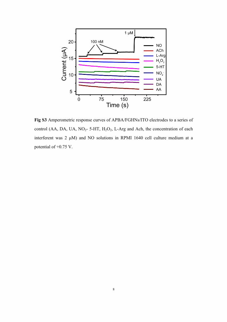
Fig S3 Amperometric response curves of APBA/FGHNs/ITO electrodes to a series of
control (AA, DA, UA, NO2- 5-HT, H2O2, L-Arg and Ach, the concentration of each
interferent was 2 μM) and NO solutions in RPMI 1640 cell culture medium at a
potential of +0.75 V.

9
Fig S4 (a) Microscopic images of HUVECs left on ITO (I), rGO/ITO (II),
FeTCP/ITO (III), FGHNs/ITO (IV) and APBA/FGHNs/ITO (V) after cultured for 1 h
and then washed with PBS solution for 3 times and (b) the corresponding statistical
data of cell numbers on different substrates (n=8).
10
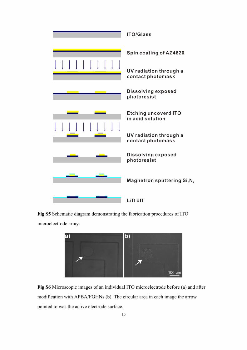
Fig S5 Schematic diagram demonstrating the fabrication procedures of ITO
microelectrode array.
Fig S6 Microscopic images of an individual ITO microelectrode before (a) and after
modification with APBA/FGHNs (b). The circular area in each image the arrow
pointed to was the active electrode surface.
11
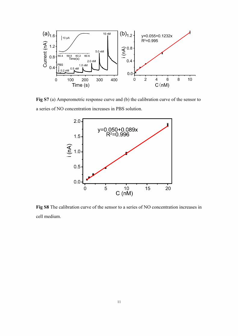
Fig S7 (a) Amperometric response curve and (b) the calibration curve of the sensor to
a series of NO concentration increases in PBS solution.
Fig S8 The calibration curve of the sensor to a series of NO concentration increases in
cell medium.
12
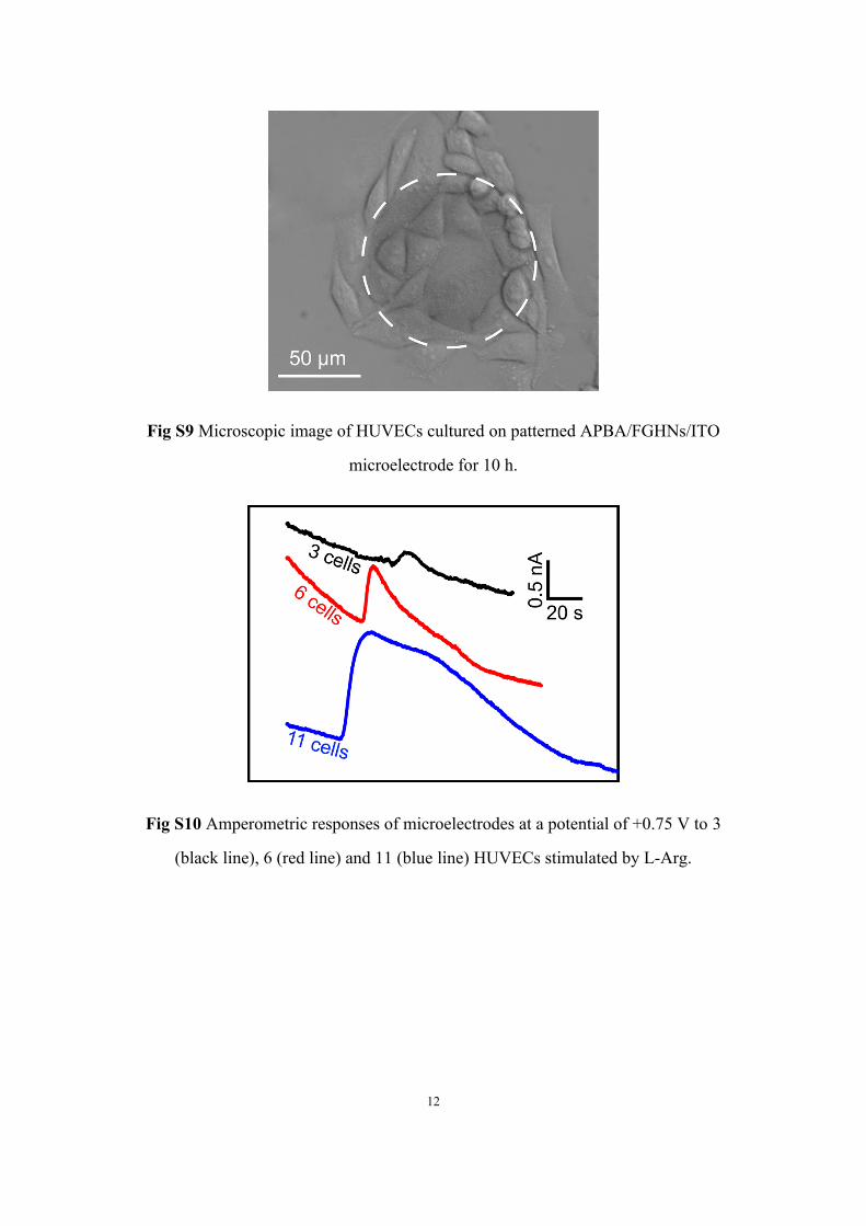
Fig S9 Microscopic image of HUVECs cultured on patterned APBA/FGHNs/ITO
microelectrode for 10 h.
Fig S10 Amperometric responses of microelectrodes at a potential of +0.75 V to 3
(black line), 6 (red line) and 11 (blue line) HUVECs stimulated by L-Arg.
13
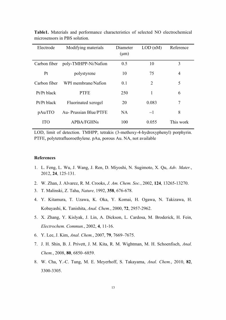
Table1. Materials and performance characteristics of selected NO electrochemical microsensors in PBS solution.
Electrode Modifying materials Diameter (μm)
LOD (nM) Reference
Carbon fiber
Pt
Carbon fiber
Pt/Pt black
Pt/Pt black
pAu/ITO
ITO
poly-TMHPP-Ni/Nafion
polystyrene
WPI membrane/Nafion
PTFE
Fluorinated xerogel
Au- Prussian Blue/PTFE
APBA/FGHNs
0.5
10
0.1
250
20
NA
100
10
75
2
1
0.083
~1
0.055
3
4
5
6
7
8
This work
LOD, limit of detection. TMHPP, tetrakis (3-methoxy-4-hydroxyphenyl) porphyrin. PTFE, polytetrafluoroethylene. pAu, porous Au. NA, not available
References
1. L. Feng, L. Wu, J. Wang, J. Ren, D. Miyoshi, N. Sugimoto, X. Qu, Adv. Mater., 2012, 24, 125-131.
2. W. Zhan, J. Alvarez, R. M. Crooks, J. Am. Chem. Soc., 2002, 124, 13265-13270.3. T. Malinski, Z. Taha, Nature, 1992, 358, 676-678.
4. Y. Kitamura, T. Uzawa, K. Oka, Y. Komai, H. Ogawa, N. Takizawa, H.
Kobayashi, K. Tanishita, Anal. Chem., 2000, 72, 2957-2962.
5. X. Zhang, Y. Kislyak, J. Lin, A. Dickson, L. Cardosa, M. Broderick, H. Fein,
Electrochem. Commun., 2002, 4, 11-16.
6. Y. Lee, J. Kim, Anal. Chem., 2007, 79, 7669–7675.
7. J. H. Shin, B. J. Privett, J. M. Kita, R. M. Wightman, M. H. Schoenfisch, Anal.
Chem., 2008, 80, 6850–6859.
8. W. Cha, Y.-C. Tung, M. E. Meyerhoff, S. Takayama, Anal. Chem., 2010, 82,
3300-3305.