Seminars in Cell & Developmental Biology - Medical...
7
Seminars in Cell & Developmental Biology 21 (2010) 19–25 Contents lists available at ScienceDirect Seminars in Cell & Developmental Biology journal homepage: www.elsevier.com/locate/semcdb Review Carcinoma-associated fibroblasts are a rate-limiting determinant for tumour progression Masayuki Shimoda a,b , Kieran T. Mellody b , Akira Orimo b,∗ a Department of Pathology, School of Medicine, Keio University, 35 Shinanomachi, Shinjuku-ku, Tokyo 160-8582, Japan b CR-UK Stromal-Tumor Interaction Group, Paterson Institute for Cancer Research, The University of Manchester, Wilmslow Road, Manchester M20 4BX, UK article info Article history: Available online 24 October 2009 Keywords: Carcinoma-associated fibroblasts (CAFs) Stromal myofibroblasts Wound healing Fibrosis TGF- SDF-1 abstract Tumours are highly complex tissues composed of carcinoma cells and surrounding stroma, which is constructed by various different types of mesenchymal cells and an extracellular matrix (ECM). Carcinoma-associated fibroblasts (CAFs), which consist of both fibroblasts and myofibroblasts, are fre- quently observed in the stroma of human carcinomas, and their presence in large numbers is often associated with the development of high-grade malignancies and poor prognoses. Moreover, in human tumour xenograft models, CAFs extracted from the tumour are more capable of promoting tumour growth through their interactions with carcinoma cells when compared to those isolated from non-cancerous stroma. Taken together, these observations strongly suggest that CAFs actively contribute to tumour pro- gression. In this review we highlight the emerging roles of these cells in promoting tumourigenesis, and we discuss the molecular mechanisms underlying their tumour-promoting capabilities and their cellular origin. © 2009 Elsevier Ltd. All rights reserved. Contents 1. Introduction .......................................................................................................................................... 19 2. Myofibroblasts, a hallmark of activated fibroblasts, in promoting tumourigenesis ................................................................ 20 3. Somatic genetic and epigenetic alterations in tumour-associated stromal cells ................................................................... 21 4. Heterogeneity of tumour stromal fibroblasts ....................................................................................................... 21 5. The role of tumour stromal fibroblasts in promoting tumour progression ......................................................................... 22 5.1. Tumour stromal fibroblasts boost neoangiogenesis ......................................................................................... 22 5.2. Tumour stromal fibroblasts promote tumour cell invasion ................................................................................. 23 6. Perspective ........................................................................................................................................... 23 Acknowledgements .................................................................................................................................. 24 References ........................................................................................................................................... 24 1. Introduction It has long been established that cancer consists of transformed cells that harbour genetic and/or epigenetic aberrances in onco- genes and/or tumour suppressor genes. Accumulation of these alterations can endow carcinoma cells with greater proliferative, invasive and survival propensities in a cell-autonomous fashion [1]. However, it is widely recognised that in addition to carcinoma cells, tumours contain large numbers of various non-transformed cells and a specialised ECM which are collectively referred to as ∗ Corresponding author. Tel.: +44 161 446 3030. E-mail address: [email protected] (A. Orimo). the stroma. The auxiliary cells found within the stroma include fibroblasts, myofibroblasts, leukocytes, endothelial cells and bone marrow-derived cells, all of which collaborate to create the com- plexity of the tumour microenvironment. Recent evidence also suggests that tumourigenesis is dependant upon contextual signals received from the closely apposed tumour- associated stroma [2–8]. The stroma actively provides continuous support to carcinoma cells throughout the different pathophysi- ological processes that modulate tumour progression. In normal tissue the stroma may actually act as a barrier in promoting tumourigenesis by constraining tumour cell proliferation. During tumourigenesis however, the stroma overtime evolves to actively support tumour growth in response to molecular signals derived from carcinoma cells and other host cell types. 1084-9521/$ – see front matter © 2009 Elsevier Ltd. All rights reserved. doi:10.1016/j.semcdb.2009.10.002
Transcript of Seminars in Cell & Developmental Biology - Medical...
Seminars in Cell & Developmental Biology 21 (2010) 19–25
Contents lists available at ScienceDirect
Seminars in Cell & Developmental Biology
journa l homepage: www.e lsev ier .com/ locate /semcdb
Review
Carcinoma-associated fibroblasts are a rate-limiting determinant fortumour progression
Masayuki Shimodaa,b, Kieran T. Mellodyb, Akira Orimob,∗
a Department of Pathology, School of Medicine, Keio University, 35 Shinanomachi, Shinjuku-ku, Tokyo 160-8582, Japanb CR-UK Stromal-Tumor Interaction Group, Paterson Institute for Cancer Research, The University of Manchester, Wilmslow Road, Manchester M20 4BX, UK
a r t i c l e i n f o
Article history:Available online 24 October 2009
Keywords:Carcinoma-associated fibroblasts (CAFs)Stromal myofibroblastsWound healingFibrosisTGF-�SDF-1
a b s t r a c t
Tumours are highly complex tissues composed of carcinoma cells and surrounding stroma, whichis constructed by various different types of mesenchymal cells and an extracellular matrix (ECM).Carcinoma-associated fibroblasts (CAFs), which consist of both fibroblasts and myofibroblasts, are fre-quently observed in the stroma of human carcinomas, and their presence in large numbers is oftenassociated with the development of high-grade malignancies and poor prognoses. Moreover, in humantumour xenograft models, CAFs extracted from the tumour are more capable of promoting tumour growththrough their interactions with carcinoma cells when compared to those isolated from non-cancerousstroma. Taken together, these observations strongly suggest that CAFs actively contribute to tumour pro-gression. In this review we highlight the emerging roles of these cells in promoting tumourigenesis, andwe discuss the molecular mechanisms underlying their tumour-promoting capabilities and their cellularorigin.
© 2009 Elsevier Ltd. All rights reserved.
Contents
1. Introduction . . . . . . . . . . . . . . . . . . . . . . . . . . . . . . . . . . . . . . . . . . . . . . . . . . . . . . . . . . . . . . . . . . . . . . . . . . . . . . . . . . . . . . . . . . . . . . . . . . . . . . . . . . . . . . . . . . . . . . . . . . . . . . . . . . . . . . . . . . 192. Myofibroblasts, a hallmark of activated fibroblasts, in promoting tumourigenesis . . . . . . . . . . . . . . . . . . . . . . . . . . . . . . . . . . . . . . . . . . . . . . . . . . . . . . . . . . . . . . . . 203. Somatic genetic and epigenetic alterations in tumour-associated stromal cells . . . . . . . . . . . . . . . . . . . . . . . . . . . . . . . . . . . . . . . . . . . . . . . . . . . . . . . . . . . . . . . . . . . 214. Heterogeneity of tumour stromal fibroblasts . . . . . . . . . . . . . . . . . . . . . . . . . . . . . . . . . . . . . . . . . . . . . . . . . . . . . . . . . . . . . . . . . . . . . . . . . . . . . . . . . . . . . . . . . . . . . . . . . . . . . . . 215. The role of tumour stromal fibroblasts in promoting tumour progression . . . . . . . . . . . . . . . . . . . . . . . . . . . . . . . . . . . . . . . . . . . . . . . . . . . . . . . . . . . . . . . . . . . . . . . . . 22
5.1. Tumour stromal fibroblasts boost neoangiogenesis . . . . . . . . . . . . . . . . . . . . . . . . . . . . . . . . . . . . . . . . . . . . . . . . . . . . . . . . . . . . . . . . . . . . . . . . . . . . . . . . . . . . . . . . . 22
5.2. Tumour stromal fibroblasts promote tumour cell invasion . . . . . . . . . . . . . . . . . . . . . . . . . . . . . . . . . . . . . . . . . . . . . . . . . . . . . . . . . . . . . . . . . . . . . . . . . . . . . . . . . 236. Perspective . . . . . . . . . . . . . . . . . . . . . . . . . . . . . . . . . . . . . . . . . . . . . . . . . . . . . . . . . . . . . . . . . . . . . . . . . . . . . . . . . . . . . . . . . . . . . . . . . . . . . . . . . . . . . . . . . . . . . . . . . . . . . . . . . . . . . . . . . . . 23. . . . .. . . . . .
1
cgai[cc
1d
Acknowledgements . . . . . . . . . . . . . . . . . . . . . . . . . . . . . . . . . . . . . . . . . . . . . . . . . . .References . . . . . . . . . . . . . . . . . . . . . . . . . . . . . . . . . . . . . . . . . . . . . . . . . . . . . . . . . . . .
. Introduction
It has long been established that cancer consists of transformedells that harbour genetic and/or epigenetic aberrances in onco-enes and/or tumour suppressor genes. Accumulation of theselterations can endow carcinoma cells with greater proliferative,
nvasive and survival propensities in a cell-autonomous fashion1]. However, it is widely recognised that in addition to carcinomaells, tumours contain large numbers of various non-transformedells and a specialised ECM which are collectively referred to as∗ Corresponding author. Tel.: +44 161 446 3030.E-mail address: [email protected] (A. Orimo).
084-9521/$ – see front matter © 2009 Elsevier Ltd. All rights reserved.oi:10.1016/j.semcdb.2009.10.002
. . . . . . . . . . . . . . . . . . . . . . . . . . . . . . . . . . . . . . . . . . . . . . . . . . . . . . . . . . . . . . . . . . . . . . . . . . 24. . . . . . . . . . . . . . . . . . . . . . . . . . . . . . . . . . . . . . . . . . . . . . . . . . . . . . . . . . . . . . . . . . . . . . . . . 24
the stroma. The auxiliary cells found within the stroma includefibroblasts, myofibroblasts, leukocytes, endothelial cells and bonemarrow-derived cells, all of which collaborate to create the com-plexity of the tumour microenvironment.
Recent evidence also suggests that tumourigenesis is dependantupon contextual signals received from the closely apposed tumour-associated stroma [2–8]. The stroma actively provides continuoussupport to carcinoma cells throughout the different pathophysi-ological processes that modulate tumour progression. In normaltissue the stroma may actually act as a barrier in promoting
tumourigenesis by constraining tumour cell proliferation. Duringtumourigenesis however, the stroma overtime evolves to activelysupport tumour growth in response to molecular signals derivedfrom carcinoma cells and other host cell types.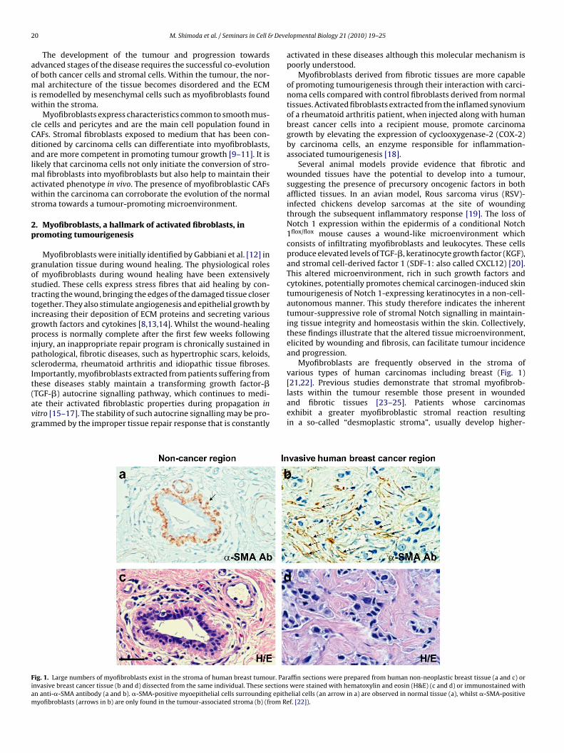
2 Deve
aomiw
cCdalmaws
2p
gosttigpipsIt(avg
Fiam
0 M. Shimoda et al. / Seminars in Cell &
The development of the tumour and progression towardsdvanced stages of the disease requires the successful co-evolutionf both cancer cells and stromal cells. Within the tumour, the nor-al architecture of the tissue becomes disordered and the ECM
s remodelled by mesenchymal cells such as myofibroblasts foundithin the stroma.
Myofibroblasts express characteristics common to smooth mus-le cells and pericytes and are the main cell population found inAFs. Stromal fibroblasts exposed to medium that has been con-itioned by carcinoma cells can differentiate into myofibroblasts,nd are more competent in promoting tumour growth [9–11]. It isikely that carcinoma cells not only initiate the conversion of stro-
al fibroblasts into myofibroblasts but also help to maintain theirctivated phenotype in vivo. The presence of myofibroblastic CAFsithin the carcinoma can corroborate the evolution of the normal
troma towards a tumour-promoting microenvironment.
. Myofibroblasts, a hallmark of activated fibroblasts, inromoting tumourigenesis
Myofibroblasts were initially identified by Gabbiani et al. [12] inranulation tissue during wound healing. The physiological rolesf myofibroblasts during wound healing have been extensivelytudied. These cells express stress fibres that aid healing by con-racting the wound, bringing the edges of the damaged tissue closerogether. They also stimulate angiogenesis and epithelial growth byncreasing their deposition of ECM proteins and secreting variousrowth factors and cytokines [8,13,14]. Whilst the wound-healingrocess is normally complete after the first few weeks following
njury, an inappropriate repair program is chronically sustained inathological, fibrotic diseases, such as hypertrophic scars, keloids,cleroderma, rheumatoid arthritis and idiopathic tissue fibroses.mportantly, myofibroblasts extracted from patients suffering from
hese diseases stably maintain a transforming growth factor-�TGF-�) autocrine signalling pathway, which continues to medi-te their activated fibroblastic properties during propagation initro [15–17]. The stability of such autocrine signalling may be pro-rammed by the improper tissue repair response that is constantlyig. 1. Large numbers of myofibroblasts exist in the stroma of human breast tumour. Parnvasive breast cancer tissue (b and d) dissected from the same individual. These sectionsn anti-�-SMA antibody (a and b). �-SMA-positive myoepithelial cells surrounding epithyofibroblasts (arrows in b) are only found in the tumour-associated stroma (b) (from R
lopmental Biology 21 (2010) 19–25
activated in these diseases although this molecular mechanism ispoorly understood.
Myofibroblasts derived from fibrotic tissues are more capableof promoting tumourigenesis through their interaction with carci-noma cells compared with control fibroblasts derived from normaltissues. Activated fibroblasts extracted from the inflamed synoviumof a rheumatoid arthritis patient, when injected along with humanbreast cancer cells into a recipient mouse, promote carcinomagrowth by elevating the expression of cyclooxygenase-2 (COX-2)by carcinoma cells, an enzyme responsible for inflammation-associated tumourigenesis [18].
Several animal models provide evidence that fibrotic andwounded tissues have the potential to develop into a tumour,suggesting the presence of precursory oncogenic factors in bothafflicted tissues. In an avian model, Rous sarcoma virus (RSV)-infected chickens develop sarcomas at the site of woundingthrough the subsequent inflammatory response [19]. The loss ofNotch 1 expression within the epidermis of a conditional Notch1flox/flox mouse causes a wound-like microenvironment whichconsists of infiltrating myofibroblasts and leukocytes. These cellsproduce elevated levels of TGF-�, keratinocyte growth factor (KGF),and stromal cell-derived factor 1 (SDF-1: also called CXCL12) [20].This altered microenvironment, rich in such growth factors andcytokines, potentially promotes chemical carcinogen-induced skintumourigenesis of Notch 1-expressing keratinocytes in a non-cell-autonomous manner. This study therefore indicates the inherenttumour-suppressive role of stromal Notch signalling in maintain-ing tissue integrity and homeostasis within the skin. Collectively,these findings illustrate that the altered tissue microenvironment,elicited by wounding and fibrosis, can facilitate tumour incidenceand progression.
Myofibroblasts are frequently observed in the stroma ofvarious types of human carcinomas including breast (Fig. 1)
[21,22]. Previous studies demonstrate that stromal myofibrob-lasts within the tumour resemble those present in woundedand fibrotic tissues [23–25]. Patients whose carcinomasexhibit a greater myofibroblastic stromal reaction resultingin a so-called “desmoplastic stroma”, usually develop higher-affin sections were prepared from human non-neoplastic breast tissue (a and c) orwere stained with hematoxylin and eosin (H&E) (c and d) or immunostained withelial cells (an arrow in a) are observed in normal tissue (a), whilst �-SMA-positive
ef. [22]).

Deve
g[
osumctdimcri
gwacetfocatg
uCOTnopwmm
stht
mfintcfib
3t
fatmpacp
M. Shimoda et al. / Seminars in Cell &
rade malignancies associated with poor prognostic outcome11,26,27].
CAFs have been extracted from a number of different typesf human carcinomas including breast, prostate, ovary, pancreas,kin, colon and esophagus [22,28–33]. DNA microarray analyses,sing either cultured CAFs or tumour-associated stromal tissuesicrodissected from the regions adjacent to human carcinoma
ells, show gene expression profiles distinct to those in their con-rol groups [34–37]. CAFs isolated from different sample sets showifferences in the expression of large varieties of genes encod-
ng for a number of cytokines, growth factors, enzymes, ECM anduscle-related proteins. These differences may result from dis-
rete alterations in different signalling pathways within CAFs thategulate their phenotype and have been modified through theirnteractions with carcinoma cells.
The ability of tumour stromal fibroblasts to promote carcino-enesis was investigated using a tumour xenograft model inhich tumour cells were implanted into immunodeficient mice,
long with CAFs, counterpart fibroblasts extracted from the non-ancerous tissue in the same individual, or normal fibroblastsxtracted from healthy donors [18,22,28,29,33]. Those tumourshat developed from carcinoma cells injected with CAFs show aaster growth rate, compared to those tumours admixed with eitherf the control sets of fibroblasts. Considerable numbers of CAFs orontrol fibroblasts can be observed alongside carcinoma cells indvanced tumour xenografts [22,38]. CAF-secreted paracrine fac-ors likely act upon these cancer cells and encourage carcinomarowth during tumour development.
CAFs secrete elevated levels of the cytokine SDF-1 that stim-lates carcinoma cell proliferation in vivo, acting through theXCR4 receptor expressed on the surface of carcinoma cells [22,39].ther studies have also shown that CAFs secrete high levels ofGF-�1 that increases CXCR4 expression on human prostate pre-eoplastic cells [39–41]. These studies highlight the importancef stroma-derived SDF-1/CXCR4 and TGF-� paracrine signalling inromoting tumourigenesis. Understanding the molecular crosstalkhich occurs between CAFs and carcinoma cells is essential anday in the future provide novel therapeutic targets for the treat-ent of cancer.Myofibroblasts derived from both tumour and fibrotic tissues
hare similar molecular and cellular traits. The cellular processeshat occur within both the tumour and the diseased tissues are per-aps best epitomised by the statement that “tumours are woundshat do not heal” [23–25].
TGF-� autocrine signalling plays a central role in forming andaintaining the myofibroblastic phenotype of cells involved in
brosis. However, it is unclear if such a TGF-� autocrine sig-alling pathway is also responsible for mediating this phenotype inumour-associated myofibroblasts, and if these cells possess spe-ific genetic alteration(s) that are not shared with those present inbrotic tissue. This issue warrants further consideration and wille addressed in the following section.
. Somatic genetic and epigenetic alterations inumour-associated stromal cells
In human epithelial carcinomas, cellular transformation resultsrom genetic aberrances that lead to tumour developmentnd progression. Although it is now widely accepted thatumour-associated stromal cells can influence tumourigenesis, the
echanism(s) by which they acquire these tumour-promotingroperties are not known. The presence of somatic genetic alter-tions observed in tumour-associated stromal cells has beenontroversial [5,42,43], and the functional relevance of these pro-osed alterations to their phenotypes remains unclear.
lopmental Biology 21 (2010) 19–25 21
Genetic alterations, such as chromosomal loss of heterozygos-ity (LOH) and somatic mutations, have been reported in stromalregions microdissected from various human carcinomas includ-ing those of breast [44–47], ovarian [48], colon [45], bladder [49],and head and neck [50]. Indeed, analyses performed on microdis-sected regions of stromal tissues from within the tumour identifysomatic TP53 mutations within 25.6% of the hereditary group, and19.4% of the sporadic group of breast cancer patients [47]. Thereis also a higher risk of regional lymph node metastases occurringin these patients. Moreover, tumour-associated stromal regionsmicrodissected from prostate tumours, when raised in epithe-lial cell-specific oncogene-driven transgenic mice with a TP53+/−background, display frequent loss of the wild type TP53 allele [51].A complementary study finds that, when human breast carcinomacells are implanted into p53+/− recipient mice, the host stromalcells isolated from the developing tumour lose their wild type TP53allele resulting in p53−/− tumour-associated stromal cells [52].
Although loss of p53 activity within stromal cells appearsinstrumental in the development of a tumour-promoting stroma,it is unclear if this can trigger the differentiation of fibroblastsand/or progenitor cells into myofibroblasts. Furthermore, althoughaberrations in p53 signalling are often associated with a higherincidence of mesenchymal tumours in patients with Li–Fraumenisyndrome and in p53-mutant mice, it is unclear why carcinosar-comas, which are malignant biphasic epithelial and mesenchymaltumours, very rarely (∼0.2%) develop in human breast carcinomapatients [53,54].
The above studies detected genetic alterations within thestroma of paraffin-fixed tumour tissues. Conversely, genome-widegenetic analyses, including single nucleotide polymorphism (SNP)and comparative genomic hybridisation (CGH) arrays using freshlyfrozen tissues, fail to detect any such significant somatic geneticalterations within the stroma of human breast tumours. This ishighlighted in one important study that detected a LOH event onchromosome 22 in only 1 out of 35 human breast and ovariancarcinoma-associated stroma samples [55].
Specifically in CAFs, such analyses have also failed to detectsubstantial genetic alterations [34,55,56]. Our own observationsindicate that CAFs themselves are non-neoplastic as they typicallysenesce in vitro, and show no detectable chromosomal abnormal-ities and no cellular phenotypes characteristic of malignant celltypes [22]. Nonetheless, there is a possibility that only a small num-ber of CAFs may actually possess genetic alterations, and thesecells may be difficult to detect in a large heterogenous fibroblastpopulation. Other studies, however, have reported the presenceof epigenetic modifications, such as DNA methylation within thegenome of CAFs that may give rise to their tumour-promoting phe-notype [57,58].
The biological relevance of genetic and/or epigenetic alterationswithin the tumour stroma and the cellular and molecular mecha-nisms that underlie the tumour-promoting phenotype of CAFs haveyet to be elucidated. The detection of somatic alterations in somestudies, whilst not in others, may be in part due to the differentmethods used to acquire and prepare tissues for analysis, the useof paraffin-fixed tissues instead of frozen tissues, or the differenttypes of genomic analyses performed. Future independent analysesare required in order to clarify these issues.
4. Heterogeneity of tumour stromal fibroblasts
Within a murine renal fibrosis model, significant numbers ofmyofibroblasts found in affected tissues are derived from epithe-lial cells, endothelial cells and circulating bone marrow cells[59]. In tumour tissue, myofibroblasts are also proposed to orig-inate from different cell types including pre-existing fibroblasts,
22 M. Shimoda et al. / Seminars in Cell & Developmental Biology 21 (2010) 19–25
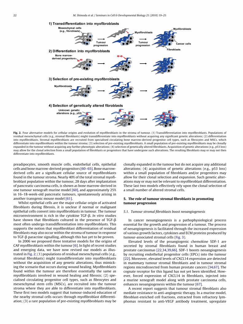
Fig. 2. Four alternative models for cellular origins and evolution of myofibroblasts in the stroma of tumour. (1) Transdifferentiation into myofibroblasts. Populations ofresidual mesenchymal cells (e.g., stromal fibroblasts) might transdifferentiate into myofibroblasts without acquiring any significant genetic alterations; (2) differentiationinto myofibroblasts. Stromal myofibroblasts are recruited from specialised circulating bone marrow-derived progenitor cell types, such as fibrocytes and MSCs, whichd istinge electiom tors thd
pcdfbooia
fiemhmsfit
CatswifmcmsTta
ifferentiate into myofibroblasts within the tumour stroma; (3) selection of pre-exxpanded in the tumour without acquiring any further phenotypic alterations; (4) say allow for the clonal selection from a small population of fibroblasts or progeni
ifferentiate into myofibroblasts.
readipocytes, smooth muscle cells, endothelial cells, epithelialells and bone marrow-derived progenitors [60–65]. Bone marrow-erived cells are a significant cellular source of myofibroblastsound in the tumour stroma. Nearly 40% of the total stromal myofi-roblast population within the tumour, 28 days after implantationf pancreatic carcinoma cells, is shown as bone marrow-derived inne tumour xenograft murine model [60], and approximately 25%n 16–18-week-old pancreatic tumours, spontaneously arising innother transgenic mouse model [61].
Whilst epithelial cells are the major cellular origin of activatedbroblasts during fibrosis, it is unclear if normal or malignantpithelial cells convert into myofibroblasts in tumour. The tumouricroenvironment is rich in the cytokine TGF-�. In vitro studies
ave shown that fibroblasts cultured in the presence of TGF-�ost often undergo transdifferentiation into myofibroblasts. This
upports the notion that myofibroblast differentiation of residualbroblasts may also occur within the stroma of tumour in responseo TGF-� paracrine signalling, although this has yet to be proven.
In 2006 we proposed three tentative models for the origins ofAF myofibroblasts within the tumour [6]. In light of recent studiesnd emerging data, we have now revised our models as illus-rated in Fig. 2; (1) populations of residual mesenchymal cells (e.g.,tromal fibroblasts) might transdifferentiate into myofibroblastsithout the acquisition of any genetic alterations, thus mimick-
ng the scenario that occurs during wound healing. Myofibroblastsound within the tumour are therefore essentially the same as
yofibroblasts involved in wound healing and fibrosis; (2) spe-ialised circulating progenitor cell types, such as fibrocytes and
esenchymal stem cells (MSCs), are recruited into the tumourtroma where they are able to differentiate into myofibroblasts.hese first two models suggest that tumour-induced education ofhe nearby stromal cells occurs through myofibroblast differenti-tion; (3) a rare population of pre-existing myofibroblasts may be
myofibroblasts. A small population of pre-existing myofibroblasts may be clonallyn of genetically altered fibroblasts. Acquisition of genetic alterations (e.g., p53 loss)at have undergone such alterations. The resulting fibroblasts may or may not then
clonally expanded in the tumour but do not acquire any additionalalterations; (4) acquisition of genetic alterations (e.g., p53 loss)within a small population of fibroblasts and/or progenitors mayallow for their clonal selection and expansion. Such genetic alter-ations may or may not be relevant to myofibroblast differentiation.These last two models effectively rely upon the clonal selection ofa small number of altered stromal cells.
5. The role of tumour stromal fibroblasts in promotingtumour progression
5.1. Tumour stromal fibroblasts boost neoangiogenesis
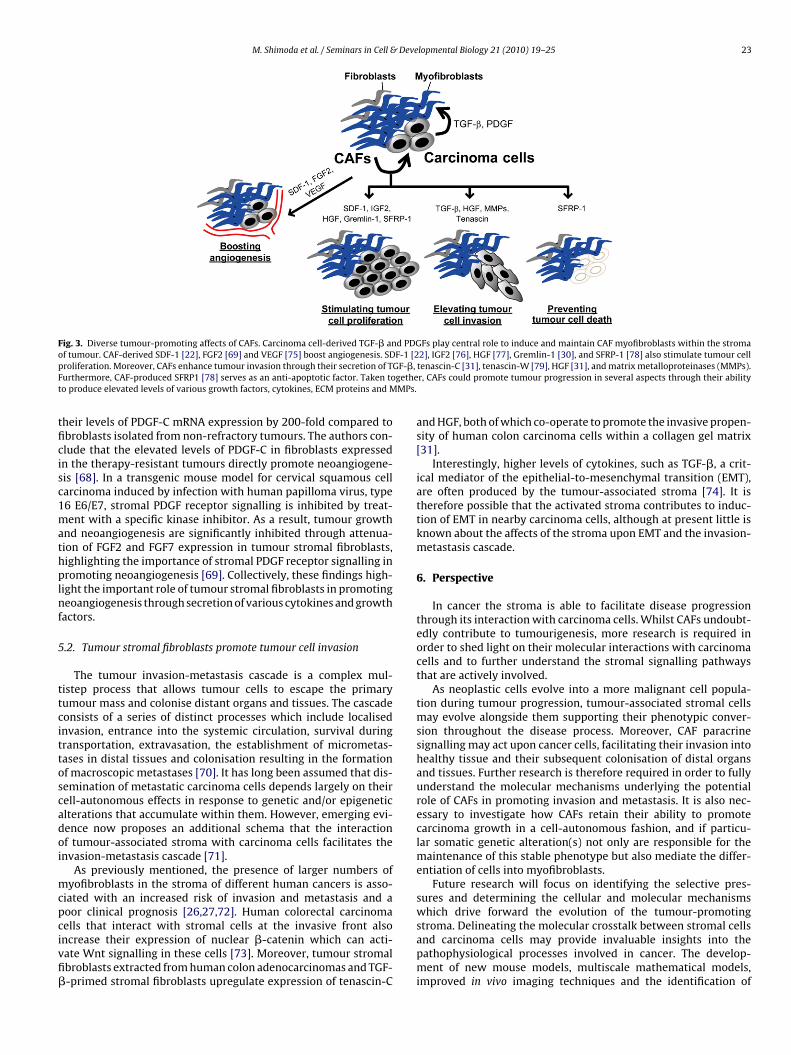
In cancer neoangiogenesis is a pathophysiological processessential for the growth and progression of a tumour. The processof neoangiogenesis is facilitated through the increased expressionof various growth factors, cytokines and ECM proteins produced bytumour-associated stromal cells (Fig. 3).
Elevated levels of the proangiogenic chemokine SDF-1 aresecreted by stromal fibroblasts found in human breast andprostate carcinomas [22,34,39,66]. SDF-1 boosts neoangiogenesisby recruiting endothelial progenitor cells (EPCs) into the tumour[22]. Moreover, elevated levels of CXCL14 expression are detectedin mammary tumour stromal fibroblasts and in tumour stromalregions microdissected from human prostate cancers [34,67]. Thecognate receptor for this ligand has not yet been identified. How-ever, forced expression of CXCL14 in fibroblasts, injected intoa murine xenograft model along with prostate carcinoma cells,
enhances neoangiogenesis within the tumour [67].A recent report suggests that tumour stromal fibroblasts alsomediate resistance to anti-angiogenic therapy. In a murine model,fibroblast-enriched cell fractions, extracted from refractory lym-phomas resistant to anti-VEGF antibody treatment, upregulate
M. Shimoda et al. / Seminars in Cell & Developmental Biology 21 (2010) 19–25 23
Fig. 3. Diverse tumour-promoting affects of CAFs. Carcinoma cell-derived TGF-� and PDGFs play central role to induce and maintain CAF myofibroblasts within the stromao F-1 [2p GF-�,F gethet MPs.
tficisc1mathplnf
5
ttcittoscadoi
mcpcivfi�
f tumour. CAF-derived SDF-1 [22], FGF2 [69] and VEGF [75] boost angiogenesis. SDroliferation. Moreover, CAFs enhance tumour invasion through their secretion of Turthermore, CAF-produced SFRP1 [78] serves as an anti-apoptotic factor. Taken too produce elevated levels of various growth factors, cytokines, ECM proteins and M
heir levels of PDGF-C mRNA expression by 200-fold compared tobroblasts isolated from non-refractory tumours. The authors con-lude that the elevated levels of PDGF-C in fibroblasts expressedn the therapy-resistant tumours directly promote neoangiogene-is [68]. In a transgenic mouse model for cervical squamous cellarcinoma induced by infection with human papilloma virus, type6 E6/E7, stromal PDGF receptor signalling is inhibited by treat-ent with a specific kinase inhibitor. As a result, tumour growth
nd neoangiogenesis are significantly inhibited through attenua-ion of FGF2 and FGF7 expression in tumour stromal fibroblasts,ighlighting the importance of stromal PDGF receptor signalling inromoting neoangiogenesis [69]. Collectively, these findings high-
ight the important role of tumour stromal fibroblasts in promotingeoangiogenesis through secretion of various cytokines and growth
actors.
.2. Tumour stromal fibroblasts promote tumour cell invasion
The tumour invasion-metastasis cascade is a complex mul-istep process that allows tumour cells to escape the primaryumour mass and colonise distant organs and tissues. The cascadeonsists of a series of distinct processes which include localisednvasion, entrance into the systemic circulation, survival duringransportation, extravasation, the establishment of micrometas-ases in distal tissues and colonisation resulting in the formationf macroscopic metastases [70]. It has long been assumed that dis-emination of metastatic carcinoma cells depends largely on theirell-autonomous effects in response to genetic and/or epigeneticlterations that accumulate within them. However, emerging evi-ence now proposes an additional schema that the interactionf tumour-associated stroma with carcinoma cells facilitates thenvasion-metastasis cascade [71].
As previously mentioned, the presence of larger numbers ofyofibroblasts in the stroma of different human cancers is asso-
iated with an increased risk of invasion and metastasis and aoor clinical prognosis [26,27,72]. Human colorectal carcinoma
ells that interact with stromal cells at the invasive front alsoncrease their expression of nuclear �-catenin which can acti-ate Wnt signalling in these cells [73]. Moreover, tumour stromalbroblasts extracted from human colon adenocarcinomas and TGF--primed stromal fibroblasts upregulate expression of tenascin-C2], IGF2 [76], HGF [77], Gremlin-1 [30], and SFRP-1 [78] also stimulate tumour celltenascin-C [31], tenascin-W [79], HGF [31], and matrix metalloproteinases (MMPs).r, CAFs could promote tumour progression in several aspects through their ability
and HGF, both of which co-operate to promote the invasive propen-sity of human colon carcinoma cells within a collagen gel matrix[31].
Interestingly, higher levels of cytokines, such as TGF-�, a crit-ical mediator of the epithelial-to-mesenchymal transition (EMT),are often produced by the tumour-associated stroma [74]. It istherefore possible that the activated stroma contributes to induc-tion of EMT in nearby carcinoma cells, although at present little isknown about the affects of the stroma upon EMT and the invasion-metastasis cascade.
6. Perspective
In cancer the stroma is able to facilitate disease progressionthrough its interaction with carcinoma cells. Whilst CAFs undoubt-edly contribute to tumourigenesis, more research is required inorder to shed light on their molecular interactions with carcinomacells and to further understand the stromal signalling pathwaysthat are actively involved.
As neoplastic cells evolve into a more malignant cell popula-tion during tumour progression, tumour-associated stromal cellsmay evolve alongside them supporting their phenotypic conver-sion throughout the disease process. Moreover, CAF paracrinesignalling may act upon cancer cells, facilitating their invasion intohealthy tissue and their subsequent colonisation of distal organsand tissues. Further research is therefore required in order to fullyunderstand the molecular mechanisms underlying the potentialrole of CAFs in promoting invasion and metastasis. It is also nec-essary to investigate how CAFs retain their ability to promotecarcinoma growth in a cell-autonomous fashion, and if particu-lar somatic genetic alteration(s) not only are responsible for themaintenance of this stable phenotype but also mediate the differ-entiation of cells into myofibroblasts.
Future research will focus on identifying the selective pres-sures and determining the cellular and molecular mechanismswhich drive forward the evolution of the tumour-promoting
stroma. Delineating the molecular crosstalk between stromal cellsand carcinoma cells may provide invaluable insights into thepathophysiological processes involved in cancer. The develop-ment of new mouse models, multiscale mathematical models,improved in vivo imaging techniques and the identification of
2 Deve
ntatat
A
FC
R
[
[
[
[
[
[
[
[
[
[
[
[
[
[
[
[
[
[
[
[
[
[
[
[
[
[
[
[
[
[
[
[
[
[
[
[
[
[
[
[
[
[
[
[
4 M. Shimoda et al. / Seminars in Cell &
ew molecules important in regulating stroma-tumour interac-ions will further our understanding of cancer in the future. Suchdvancements in research may lead to the development of novelherapeutic approaches to treating cancer by targeting the inter-ctions between the stroma and carcinoma cells that mediateumourigenesis.
cknowledgements
This work was supported by Keio Gijuku Fukuzawa Memorialund for the Advancement of Education and Research (M.S.) andancer Research UK (CR-UK) grant number C147/A6058 (A.O.).
eferences
[1] Hanahan D, Weinberg RA. The hallmarks of cancer. Cell 2000;100:57–70.[2] Bissell MJ, Radisky D. Putting tumours in context. Nat Rev Cancer 2001;1:46–54.[3] Mueller MM, Fusenig NE. Friends or foes—bipolar effects of the tumour stroma
in cancer. Nat Rev Cancer 2004;4:839–49.[4] Bhowmick NA, Neilson EG, Moses HL. Stromal fibroblasts in cancer initiation
and progression. Nature 2004;432:332–7.[5] Polyak K, Haviv I, Campbell IG. Co-evolution of tumor cells and their microen-
vironment. Trends Genet 2009;25:30–8.[6] Orimo A, Weinberg RA. Stromal fibroblasts in cancer: a novel tumor-promoting
cell type. Cell Cycle 2006;5:1597–601.[7] Coussens LM, Werb Z. Inflammation and cancer. Nature 2002;420:860–7.[8] Kalluri R, Zeisberg M. Fibroblasts in cancer. Nat Rev Cancer 2006;6:392–401.[9] Guo X, Oshima H, Kitmura T, Taketo MM, Oshima M. Stromal fibroblasts acti-
vated by tumor cells promote angiogenesis in mouse gastric cancer. J Biol Chem2008;283:19864–71.
10] Noma K, Smalley KSM, Lioni M, Naomoto Y, Tanaka N, El-Deiry W, et al. Theessential role of fibroblasts in esophageal squamous cell carcinoma-inducedangiogenesis. Gastroenterology 2008;134:1981–93.
11] Kellermann MG, Sobral LM, da Silva SD, Zecchin KG, Graner E, Lopes MA, etal. Mutual paracrine effects of oral squamous cell carcinoma cells and normaloral fibroblasts: induction of fibroblast to myofibroblast transdifferentiationand modulation of tumor cell proliferation. Oral Oncol 2008;44:509–17.
12] Gabbiani G, Ryan GB, Majne G. Presence of modified fibroblasts in gran-ulation tissue and their possible role in wound contraction. Experientia1971;27:549–50.
13] Serini G, Gabbiani G. Mechanisms of myofibroblast activity and phenotypicmodulation. Exp Cell Res 1999;250:273–83.
14] Hinz B, Phan SH, Thannickal VJ, Galli A, Bochaton-Piallat ML, Gabbiani G. Themyofibroblast: one function, multiple origins. Am J Pathol 2007;170:1807–16.
15] Varga J. Scleroderma and Smads: dysfunctional Smad family dynamics culmi-nating in fibrosis. Arthritis Rheum 2002;46:1703–13.
16] Kim KK, Wei Y, Szekeres C, Kugler MC, Wolters PJ, Hill ML, et al. Epithelial cellalpha3beta1 integrin links beta-catenin and Smad signaling to promote myofi-broblast formation and pulmonary fibrosis. J Clin Invest 2009;119:213–24.
17] Mori Y, Chen SJ, Varga J. Expression and regulation of intracellular SMAD sig-naling in scleroderma skin fibroblasts. Arthritis Rheum 2003;48:1964–78.
18] Hu M, Peluffo G, Chen H, Gelman R, Schnitt S, Polyak K. Role of COX-2 inepithelial-stromal cell interactions and progression of ductal carcinoma in situof the breast. Proc Natl Acad Sci USA 2009;106:3372–7.
19] Sieweke MH, Thompson NL, Sporn MB, Bissell MJ. Mediation of wound-relatedRous sarcoma virus tumorigenesis by TGF-beta. Science 1990;248:1656–60.
20] Demehri S, Turkoz A, Kopan R. Epidermal Notch1 loss promotes skin tumorige-nesis by impacting the stromal microenvironment. Cancer Cell 2009;16:55–66.
21] Sappino AP, Skalli O, Jackson B, Schurch W, Gabbiani G. Smooth-muscle differ-entiation in stromal cells of malignant and non-malignant breast tissues. Int JCancer 1988;41:707–12.
22] Orimo A, Gupta PB, Sgroi DC, Arenzana-Seisdedos F, Delaunay T, Naeem R, etal. Stromal fibroblasts present in invasive human breast carcinomas promotetumor growth and angiogenesis through elevated SDF-1/CXCL12 secretion. Cell2005;121:335–48.
23] Dvorak HF. Tumors: wounds that do not heal. Similarities between tumorstroma generation and wound healing. N Engl J Med 1986;315:1650–9.
24] Ronnov-Jessen L, Petersen OW, Bissell MJ. Cellular changes involved in conver-sion of normal to malignant breast: importance of the stromal reaction. PhysiolRev 1996;76:69–125.
25] Schafer M, Werner S. Cancer as an overhealing wound: an old hypothesis revis-ited. Nat Rev Mol Cell Biol 2008;9:628–38.
26] Maeshima AM, Niki T, Maeshima A, Yamada T, Kondo H, Matsuno Y. Modifiedscar grade: a prognostic indicator in small peripheral lung adenocarcinoma.Cancer 2002;95:2546–54.
27] Cardone A, Tolino A, Zarcone R, Borruto Caracciolo G, Tartaglia E. Prognosticvalue of desmoplastic reaction and lymphocytic infiltration in the managementof breast cancer. Panminerva Med 1997;39:174–7.
28] Olumi AF, Grossfeld GD, Hayward SW, Carroll PR, Tlsty TD, Cunha GR.Carcinoma-associated fibroblasts direct tumor progression of initiated humanprostatic epithelium. Cancer Res 1999;59:5002–11.
[
[
lopmental Biology 21 (2010) 19–25
29] Hwang RF, Moore T, Arumugam T, Ramachandran V, Amos KD, Rivera A, et al.Cancer-associated stromal fibroblasts promote pancreatic tumor progression.Cancer Res 2008;68:918–26.
30] Sneddon JB, Zhen HH, Montgomery K, van de Rijn M, Tward AD, West R, etal. Bone morphogenetic protein antagonist gremlin 1 is widely expressed bycancer-associated stromal cells and can promote tumor cell proliferation. ProcNatl Acad Sci USA 2006;103:14842–7.
31] De Wever O, Nguyen QD, Van Hoorde L, Bracke M, Bruyneel E, Gespach C, et al.Tenascin-C and SF/HGF produced by myofibroblasts in vitro provide convergentpro-invasive signals to human colon cancer cells through RhoA and Rac. FASEBJ 2004;18:1016–8.
32] Zhang C, Fu L, Fu J, Hu L, Yang H, Rong TH, et al. Fibroblast growth fac-tor receptor 2-positive fibroblasts provide a suitable microenvironment fortumor development and progression in esophageal carcinoma. Clin Cancer Res2009;15:4017–27.
33] Yang G, Rosen DG, Zhang Z, Bast Jr RC, Mills GB, Colacino JA, et al. The chemokinegrowth-regulated oncogene 1 (Gro-1) links RAS signaling to the senescenceof stromal fibroblasts and ovarian tumorigenesis. Proc Natl Acad Sci USA2006;103:16472–7.
34] Allinen M, Beroukhim R, Cai L, Brennan C, Lahti-Domenici J, Huang H, et al.Molecular characterization of the tumor microenvironment in breast cancer.Cancer Cell 2004;6:17–32.
35] Mercier I, Casimiro MC, Wang C, Rosenberg AL, Quong J, Minkeu A, et al. Humanbreast cancer-associated fibroblasts (CAFs) show caveolin-1 downregulationand RB tumor suppressor functional inactivation: implications for the responseto hormonal therapy. Cancer Biol Ther 2008;7:1212–25.
36] Micke P, Kappert K, Ohshima M, Sundquist C, Scheidl S, Lindahl P, et al. In situidentification of genes regulated specifically in fibroblasts of human basal cellcarcinoma. J Invest Dermatol 2007;127:1516–23.
37] Dakhova O, Ozen M, Creighton CJ, Li R, Ayala G, Rowley D, et al. Global geneexpression analysis of reactive stroma in prostate cancer. Clin Cancer Res2009;15:3979–89.
38] Yang F, Tuxhorn JA, Ressler SJ, McAlhany SJ, Dang TD, Rowley DR. Stromalexpression of connective tissue growth factor promotes angiogenesis andprostate cancer tumorigenesis. Cancer Res 2005;65:8887–95.
39] Ao M, Franco OE, Park D, Raman D, Williams K, Hayward SW. Cross-talk between paracrine-acting cytokine and chemokine pathways promotesmalignancy in benign human prostatic epithelium. Cancer Res 2007;67:4244–53.
40] San Francisco IF, DeWolf WC, Peehl DM, Olumi AF. Expression of trans-forming growth factor-beta 1 and growth in soft agar differentiate prostatecarcinoma-associated fibroblasts from normal prostate fibroblasts. Int J Cancer2004;112:213–8.
41] Rosenthal E, McCrory A, Talbert M, Young G, Murphy-Ullrich J, Gladson C. Ele-vated expression of TGF-beta1 in head and neck cancer-associated fibroblasts.Mol Carcinogen 2004;40:116–21.
42] Haviv I, Polyak K, Qiu W, Hu M, Campbell I. Origin of carcinoma associatedfibroblasts. Cell Cycle 2009;8:589–95.
43] Weinberg RA. Coevolution in the tumor microenvironment. Nat Genet2008;40:494–5.
44] Moinfar F, Man YG, Arnould L, Bratthauer GL, Ratschek M, Tavassoli FA.Concurrent and independent genetic alterations in the stromal and epithe-lial cells of mammary carcinoma: implications for tumorigenesis. Cancer Res2000;60:2562–6.
45] Wernert N, Locherbach C, Wellmann A, Behrens P, Hugel A. Presence of geneticalterations in microdissected stroma of human colon and breast cancers. Anti-cancer Res 2001;21:2259–64.
46] Kurose K, Gilley K, Matsumoto S, Watson PH, Zhou XP, Eng C. Frequent somaticmutations in PTEN and TP53 are mutually exclusive in the stroma of breastcarcinomas. Nat Genet 2002;32:355–7.
47] Patocs A, Zhang L, Xu Y, Weber F, Caldes T, Mutter GL, et al. Breast-cancerstromal cells with TP53 mutations and nodal metastases. N Engl J Med2007;357:2543–51.
48] Tuhkanen H, Anttila M, Kosma VM, Yla-Herttuala S, Heinonen S, Kuronen A, etal. Genetic alterations in the peritumoral stromal cells of malignant and border-line epithelial ovarian tumors as indicated by allelic imbalance on chromosome3p. Int J Cancer 2004;109:247–52.
49] Paterson RF, Ulbright TM, MacLennan GT, Zhang S, Pan CX, Sweeney CJ, etal. Molecular genetic alterations in the laser-capture-microdissected stromaadjacent to bladder carcinoma. Cancer 2003;98:1830–6.
50] Weber F, Xu Y, Zhang L, Patocs A, Shen L, Platzer P, et al. Microenvironmentalgenomic alterations and clinicopathological behavior in head and neck squa-mous cell carcinoma. JAMA 2007;297:187–95.
51] Hill R, Song Y, Cardiff RD, Van Dyke T. Selective evolution of stromalmesenchyme with p53 loss in response to epithelial tumorigenesis. Cell2005;123:1001–11.
52] Kiaris H, Chatzistamou I, Trimis G, Frangou-Plemmenou M, Pafiti-Kondi A,Kalofoutis A. Evidence for nonautonomous effect of p53 tumor suppressor incarcinogenesis. Cancer Res 2005;65:1627–30.
53] Tokudome N, Sakamoto G, Sakai T, Sarumaru S, Okuyama N, Hori F, et al. A case
of carcinosarcoma of the breast. Breast Cancer 2005;12:149–53.54] Bolton B, Sieunarine K. Carcinosarcoma: a rare tumour of the breast. Aust N Z JSurg 1990;60:917–9.
55] Qiu W, Hu M, Sridhar A, Opeskin K, Fox S, Shipitsin M, et al. No evidence of clonalsomatic genetic alterations in cancer-associated fibroblasts from human breastand ovarian carcinomas. Nat Genet 2008;40:650–5.
Deve
[
[
[
[
[
[
[
[
[
[
[
[
[
[
[
[
[
[
[[
[
[
M. Shimoda et al. / Seminars in Cell &
56] Walter K, Omura N, Hong SM, Griffith M, Goggins M. Pancreatic cancer associ-ated fibroblasts display normal allelotypes. Cancer Biol Ther 2008;7:882–8.
57] Jiang L, Gonda TA, Gamble MV, Salas M, Seshan V, Tu S, et al. Globalhypomethylation of genomic DNA in cancer-associated myofibroblasts. CancerRes 2008;68:9900–8.
58] Hu M, Yao J, Cai L, Bachman KE, van den Brule F, Velculescu V, et al. Dis-tinct epigenetic changes in the stromal cells of breast cancers. Nat Genet2005;37:899–905.
59] Kalluri R, Weinberg RA. The basics of epithelial–mesenchymal transition. J ClinInvest 2009;119:1420–8.
60] Ishii G, Sangai T, Oda T, Aoyagi Y, Hasebe T, Kanomata N, et al. Bone-marrow-derived myofibroblasts contribute to the cancer-induced stromal reaction.Biochem Biophys Res Commun 2003;309:232–40.
61] Direkze NC, Hodivala-Dilke K, Jeffery R, Hunt T, Poulsom R, Oukrif D, et al.Bone marrow contribution to tumor-associated myofibroblasts and fibroblasts.Cancer Res 2004;64:8492–5.
62] Mishra PJ, Mishra PJ, Humeniuk R, Medina DJ, Alexe G, Mesirov JP, et al.Carcinoma-associated fibroblast-like differentiation of human mesenchymalstem cells. Cancer Res 2008;68:4331–9.
63] Zeisberg EM, Potenta S, Xie L, Zeisberg M, Kalluri R. Discovery of endothelialto mesenchymal transition as a source for carcinoma-associated fibroblasts.Cancer Res 2007;67:10123–8.
64] Radisky DC, Kenny PA, Bissell MJ. Fibrosis and cancer: do myofibroblasts comealso from epithelial cells via EMT? J Cell Biochem 2007;101:830–9.
65] Ronnov-Jessen L, Petersen OW, Koteliansky VE, Bissell MJ. The origin of themyofibroblasts in breast cancer. Recapitulation of tumor environment in cul-ture unravels diversity and implicates converted fibroblasts and recruitedsmooth muscle cells. J Clin Invest 1995;95:859–73.
66] Tait LR, Pauley RJ, Santner SJ, Heppner GH, Heng HH, Rak JW, et al. Dynamicstromal–epithelial interactions during progression of MCF10DCIS.comxenografts. Int J Cancer 2007;120:2127–34.
67] Augsten M, Hagglof C, Olsson E, Stolz C, Tsagozis P, Levchenko T, et al. CXCL14 isan autocrine growth factor for fibroblasts and acts as a multi-modal stimulatorof prostate tumor growth. Proc Natl Acad Sci USA 2009;106:3414–9.
[
[
lopmental Biology 21 (2010) 19–25 25
68] Crawford Y, Kasman I, Yu L, Zhong C, Wu X, Modrusan Z, et al. PDGF-C medi-ates the angiogenic and tumorigenic properties of fibroblasts associated withtumors refractory to anti-VEGF treatment. Cancer Cell 2009;15:21–34.
69] Pietras K, Pahler J, Bergers G, Hanahan D. Functions of paracrine PDGF signal-ing in the proangiogenic tumor stroma revealed by pharmacological targeting.PLoS Med 2008;5:e19.
70] Fidler IJ. The pathogenesis of cancer metastasis: the ‘seed and soil’ hypothesisrevisited. Nat Rev Cancer 2003;3:453–8.
71] Karnoub AE, Dash AB, Vo AP, Sullivan A, Brooks MW, Bell GW, et al. Mesenchy-mal stem cells within tumour stroma promote breast cancer metastasis. Nature2007;449:557–63.
72] Surowiak P, Suchocki S, Gyorffy B, Gansukh T, Wojnar A, Maciejczyk A, et al.Stromal myofibroblasts in breast cancer: relations between their occurrence,tumor grade and expression of some tumour markers. Folia Histochem Cytobiol2006;44:111–6.
73] Le NH, Franken P, Fodde R. Tumour-stroma interactions in colorectal can-cer: converging on beta-catenin activation and cancer stemness. Br J Cancer2008;98:1886–93.
74] Weinberg RA. The biology of cancer. New York: Garland Science; 2007.75] Fukumura D, Xavier R, Sugiura T, Chen Y, Park EC, Lu N, et al. Tumor induction
of VEGF promoter activity in stromal cells. Cell 1998;94:715–25.76] Zhu CQ, Popova SN, Brown ER, Barsyte-Lovejoy D, Navab R, Shih W, et al.
Integrin alpha 11 regulates IGF2 expression in fibroblasts to enhance tumori-genicity of human non-small-cell lung cancer cells. Proc Natl Acad Sci USA2007;104:11754–9.
77] Tokunou M, Niki T, Eguchi K, Iba S, Tsuda H, Yamada T, et al. c-MET expressionin myofibroblasts: role in autocrine activation and prognostic significance inlung adenocarcinoma. Am J Pathol 2001;158:1451–63.
78] Joesting MS, Perrin S, Elenbaas B, Fawell SE, Rubin JS, Franco OE, et al. Identi-fication of SFRP1 as a candidate mediator of stromal-to-epithelial signaling inprostate cancer. Cancer Res 2005;65:10423–30.
79] Degen M, Brellier F, Kain R, Ruiz C, Terracciano L, Orend G, et al. Tenascin-W isa novel marker for activated tumor stroma in low-grade human breast cancerand influences cell behavior. Cancer Res 2007;67:9169–79.