Seminar on chronic osteomyelitis sch
173
CHRONIC OSTEOMYELITIS CHRONIC OSTEOMYELITIS Chair Person- Dr.C.V.Mudga Dr.C.V.Mudga l Speaker – Dr.S.C.Hiremath. PG In orthopedics KIMS Hubli
-
Upload
sharanayya-hiremath -
Category
Documents
-
view
294 -
download
5
Transcript of Seminar on chronic osteomyelitis sch
CHRONIC OSTEOMYELITISCHRONIC OSTEOMYELITIS
Chair Person-Dr.C.V.MudgaDr.C.V.Mudgal Speaker – Dr.S.C.Hiremath. PG In orthopedics KIMS Hubli

IntroductionIntroduction
Definition: “ A severe,persistent and incapacitating
infection of bone and bone marrow ” Chronic osteomyelitis is often defined as
the presence of ongoing bone infection for longer than 1 month in the presence of devitalized bone

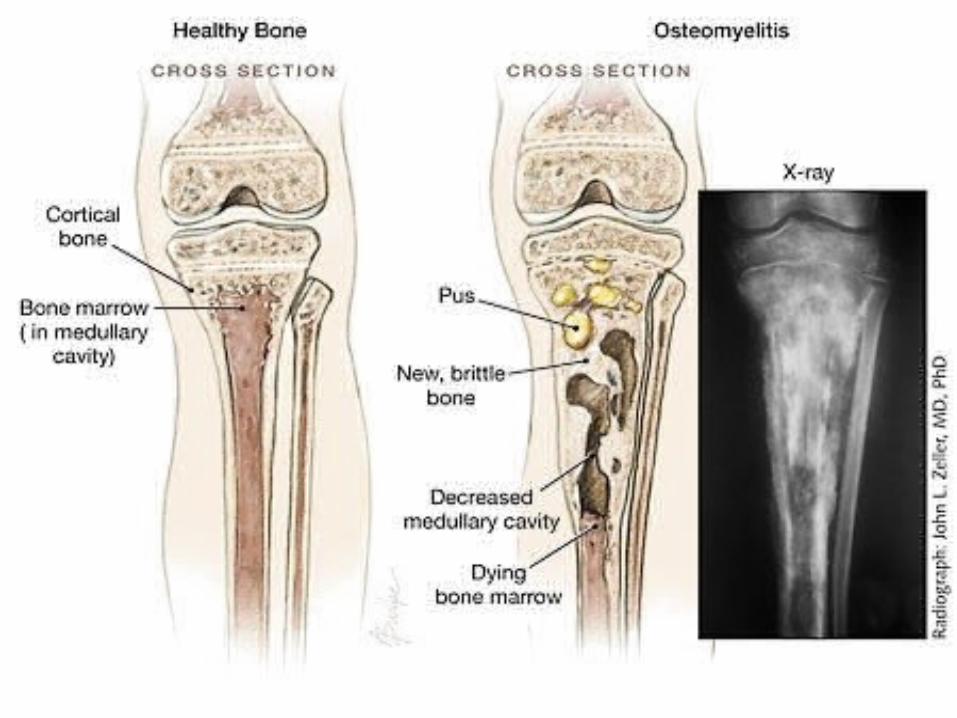
OsteomyelitisOsteomyelitis
Nelaton (1834) : coined osteomyelitis
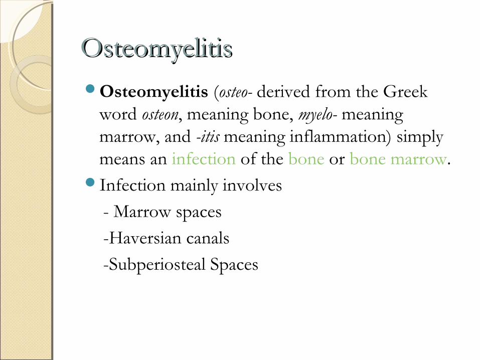
OsteomyelitisOsteomyelitisOsteomyelitis (osteo- derived from the Greek
word osteon, meaning bone, myelo- meaning marrow, and -itis meaning inflammation) simply means an infection of the bone or bone marrow.
Infection mainly involves
- Marrow spaces
-Haversian canals
-Subperiosteal Spaces
Bone and joint infections pose a formidable challenge to the Orthopaedic surgeon
The high success rate obtained with antibiotic therapy in most bacterial diseases has not been obtained in bone and joint infections because of the physiological and anatomical characteristics of bone

The mere presence of bacteria in bone, whether from bacteremia or from direct inoculation, is insufficient to produce osteomyelitis
Morrissy and Haynes have shown the relationship of trauma to osteomyelitis. Illness, malnutrition, and inadequacy of the immune system also can cause bone and joint infections.
As in other parts of the body, bones and joints produce inflammatory and immune responses to infection.
Osteomyelitis occurs when an adequate number of a sufficiently virulent organism overcomes the host's natural defenses (inflammatory and immune responses) and establishes a focus of infection, for example; local skeletal factors also play a role in the development of infection
The relative absence of phagocytic cells in the metaphyses of bones in children may explain why acute hematogenous osteomyelitis is more common in this location
The peculiarity of an abscess in bone is that it is contained within a firm structure with little chance of tissue expansion
As infection progresses, purulent material works its way through the haversian system and Volkmann canals and lifts the periosteum off the surface of bone
The combination of pus in the medullary cavity and in the subperiosteal space causes necrosis of cortical bone.
This necrotic cortical bone, known as a sequestrum, can continue to harbor bacteria despite antibiotic treatment. Antibiotics and inflammatory cells cannot adequately access this avascular area, resulting in failure of medical treatment of osteomyelitis
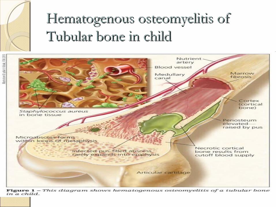
Hematogenous osteomyelitis of Hematogenous osteomyelitis of Tubular bone in childTubular bone in child
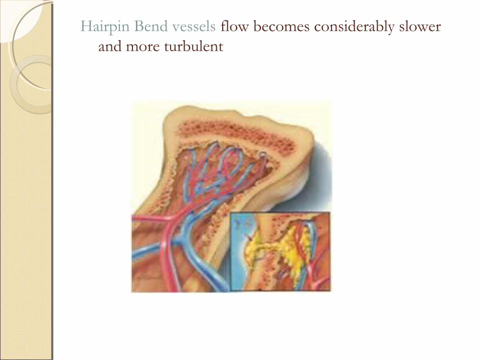
Hairpin Bend vessels flow becomes considerably slower and more turbulent
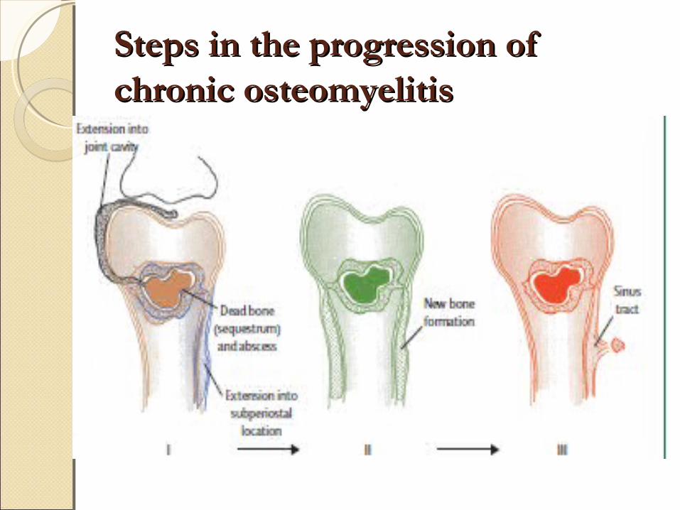
Steps in the progression of Steps in the progression of chronic osteomyelitischronic osteomyelitis
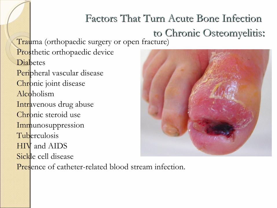
Factors That Turn Acute Bone Infection Factors That Turn Acute Bone Infection to Chronic Osteomyelitisto Chronic Osteomyelitis::
Trauma (orthopaedic surgery or open fracture)Prosthetic orthopaedic deviceDiabetesPeripheral vascular diseaseChronic joint diseaseAlcoholismIntravenous drug abuseChronic steroid useImmunosuppressionTuberculosisHIV and AIDSSickle cell diseasePresence of catheter-related blood stream infection.
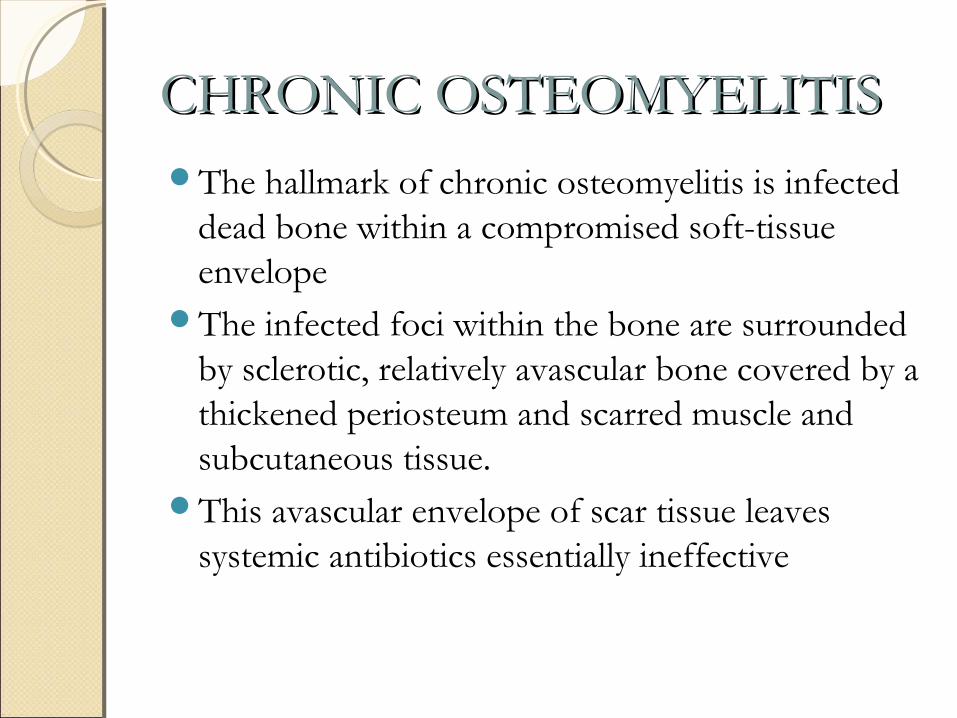
CHRONIC OSTEOMYELITISCHRONIC OSTEOMYELITISThe hallmark of chronic osteomyelitis is infected
dead bone within a compromised soft-tissue envelope
The infected foci within the bone are surrounded by sclerotic, relatively avascular bone covered by a thickened periosteum and scarred muscle and subcutaneous tissue.
This avascular envelope of scar tissue leaves systemic antibiotics essentially ineffective
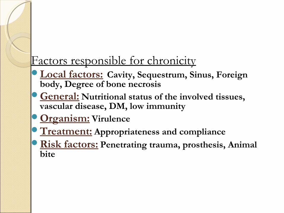
Factors responsible for chronicityLocal factors: Cavity, Sequestrum, Sinus, Foreign
body, Degree of bone necrosisGeneral: Nutritional status of the involved tissues,
vascular disease, DM, low immunity Organism: Virulence Treatment: Appropriateness and complianceRisk factors: Penetrating trauma, prosthesis, Animal
bite
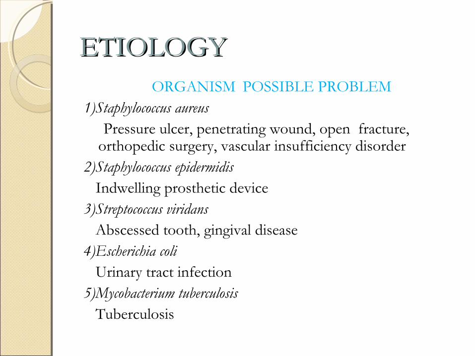
ETIOLOGYETIOLOGYORGANISM POSSIBLE PROBLEM
1)Staphylococcus aureus Pressure ulcer, penetrating wound, open fracture,
orthopedic surgery, vascular insufficiency disorder2)Staphylococcus epidermidis Indwelling prosthetic device3)Streptococcus viridans Abscessed tooth, gingival disease4)Escherichia coli Urinary tract infection5)Mycobacterium tuberculosis Tuberculosis

6)Neisseria gonorrhoeae
Gonorrhea
7)Pseudomonas sp.
Puncture wounds, intravenous drugs
8)Salmonella sp.
Sickle cell disease
9)Fungi, mycobacteria
Immunocompromised host
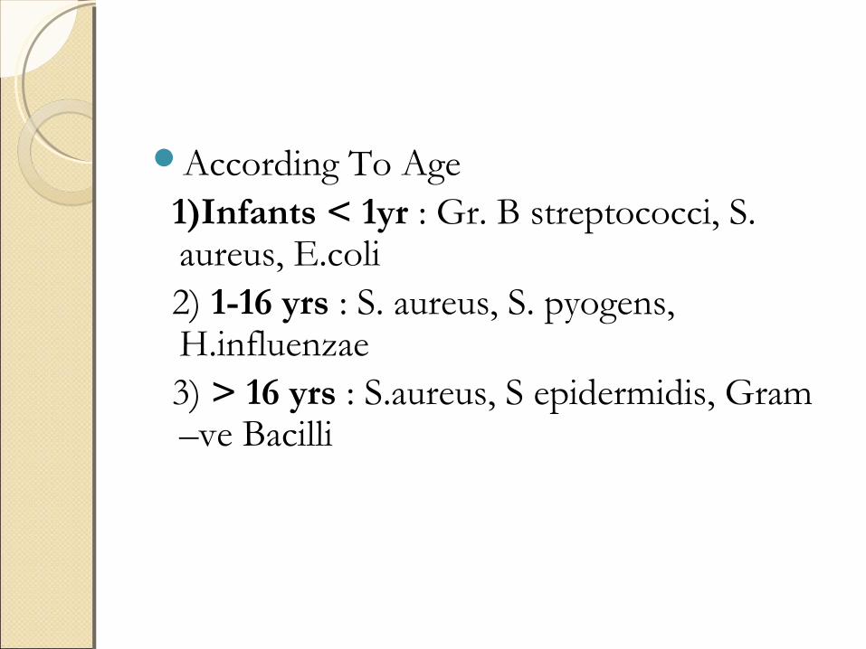
According To Age 1)Infants < 1yr : Gr. B streptococci, S.
aureus, E.coli 2) 1-16 yrs : S. aureus, S. pyogens,
H.influenzae 3) > 16 yrs : S.aureus, S epidermidis, Gram
–ve Bacilli
PATHOGENESISPATHOGENESIS
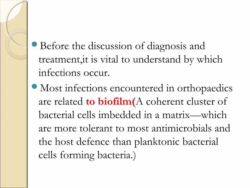
Before the discussion of diagnosis and treatment,it is vital to understand by which infections occur.
Most infections encountered in orthopaedics are related to biofilm(A coherent cluster of bacterial cells imbedded in a matrix—which are more tolerant to most antimicrobials and the host defence than planktonic bacterial cells forming bacteria.)
Biofilm bacteria exist in one of the two states
1. Planktonic state2. Stationary state
Planktonic state bacteria are free floating in the extra cellular matrix and are isolated and relatively small in quantity.
In this state,body host defences can easily eradicate the organism through the usual immunological mechanisms.
It is rare for planktonic bacteria to survive long in the extra cellular matrix despite numerous and repeat occurences of entry.
However,if the bacterial load is large and sustained,they can overwhelm the host defences and escape the effects of antibiotics.
Then they can invade tissue and blood,leading to septicemia and death.
Planktonic bacteria are metabolically active and reproductive.
This is important consideration for antibiotics treatments that work by either interfering with cellwall or protien synthesis or with reproduction.
If planktonic bacteria encounter a suitable inert surface such as dead or necrotic tissue,foreign bodies or avascular body part by either dircet contamination,contiguous spreading or hematogenous seeding,they can attach and began the process of colonisation.

Juxta position of the bacteria with a surface or biomaterial is accomplished by vander waal’s forces,which allow bacteria to develop irreversible crosslinks with the surface (adhesion-receptor interaction).
Following adhesion,bacteria begin to create a mucopolysachharide layer called biofilm or slime.
Then they develop into colonies.
These colonies exhibit remarkably resilient behaviour.
In the early stages of colonisation,sessile bacteria can be killed or neutralised by the host defences.
However,some of these bacteria may ecape destruction and potentially act as a nidus for future infection.
Transition from colonisation to infection usually requires other condition to exist.
This might occur if there was an inoculum that was larger than threshold levels,impaired host immune defence mechanisms,traumatised or necrotic tissues,foreign bodies or an a acellular or inanimate surface such as dead bone,cartilage or biomaterials.
Damaged bone,being relatively acellular,acts as a suitable surface for bacterial adhesion and colonisation.
Devitalised bone divide of normal periosteum presents a collagen matrix to which bacteria can bind.
Moreover,it has been suggested that bone sialo protein can act as a ligand for bacterial binding to bone.

Biomaterials and other foreign bodies are usually inert and susceptible to bacterial colonisation because they are inanimate.
Following bacterial adherence and colonisation,the resistance to antibiotics appears to increase.
Bacterial colonies can undergo phenotypic changes and appear to hybernate.
They can survive in a dormant state without causing infection,and this can explain the recovery of bacteria from asymptomatic hardware removal.
So while colonisation is a necessary antecedent for infection,colonisation alone does not necessarily lead to infection.

Once colonisation occurs,body defences continue to identify bacteria as foreign.
The subsequent collection of inflammatory cells brought into wall off the bacteria via chemotaxis manifest as purulence,which is a symptom of host’s attempt to isolate and destroy the infection.
The accute inflammatory cells will also release a spectrum oxidative and enzymatic products in an attempt to penetrate the glycocalyx.

These mediators and enzymes are nonspecific and may be toxic to host tissues.
Increased host tissue damage can lead to more surface substrate for local bacteria,creating cycle of tissue damage,host response,and exacerbation of infection.
The host tissues will eventually react to limit the spread of infection macroscopically as well as microscopically.
The clinical manifestation of a sequestered infection is an abcess or involucrum.
Alternatively,if the infection grows and reaches the skin or an internal epithealial surafce,a sinus tract forms as a route to dispel detritus.
While the appearance of sinus tract is a manifestation of a locally devastating disease process and indicates severe underlying infection,it should be remembered that it can also prevent the accumulation of internal collection which can lead to bacterimia and septicimia.
Eventually the equilibrium may exist in the form of chronic infection,which is what many surgeons see in practice.
There is usually history of intermittent symptoms and drainage that has responded to some type of antibiotic regimen.
Longstanding infections that were tolerated by healthy individuals may suddenly become limb or life threatening as the individual’s age.

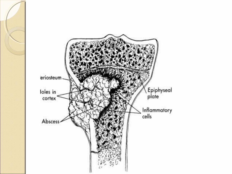
Bacteria settle down in metaphysisPrimary focus in metaphysis (form abscess in
metaphysis)Subperiosteal abscessSequestrum formation (dead bone )Involucrum formation (new bone formed by
cambium layer of periosteum)Pus eventually perforates periosteum and forms
abscess in soft tissues.Ultimately abscess burst on surface and forms
discharging sinus
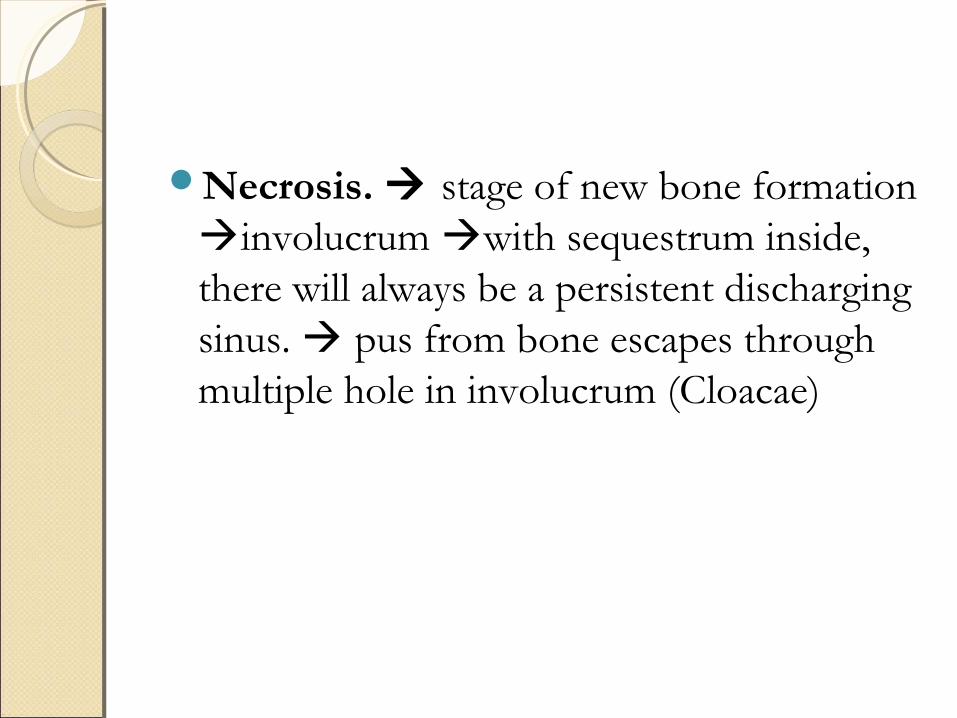
Necrosis. stage of new bone formation involucrum with sequestrum inside, there will always be a persistent discharging sinus. pus from bone escapes through multiple hole in involucrum (Cloacae)
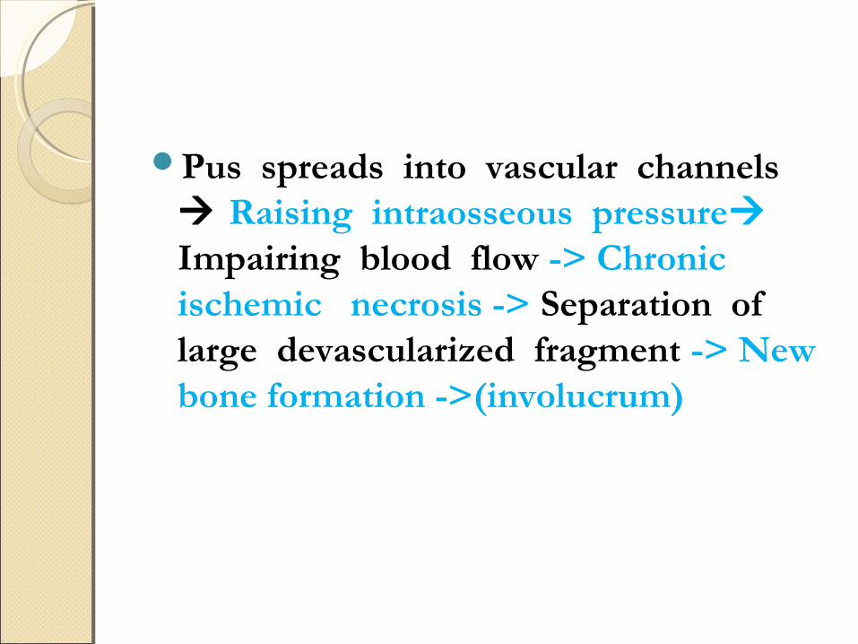
Pus spreads into vascular channels Raising intraosseous pressure Impairing blood flow -> Chronic ischemic necrosis -> Separation of large devascularized fragment -> New bone formation ->(involucrum)

Chronic osteomylitis involving metaphysis of Chronic osteomylitis involving metaphysis of tibiatibia
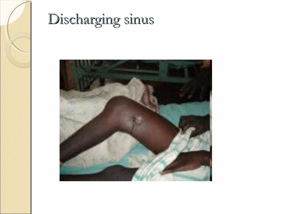
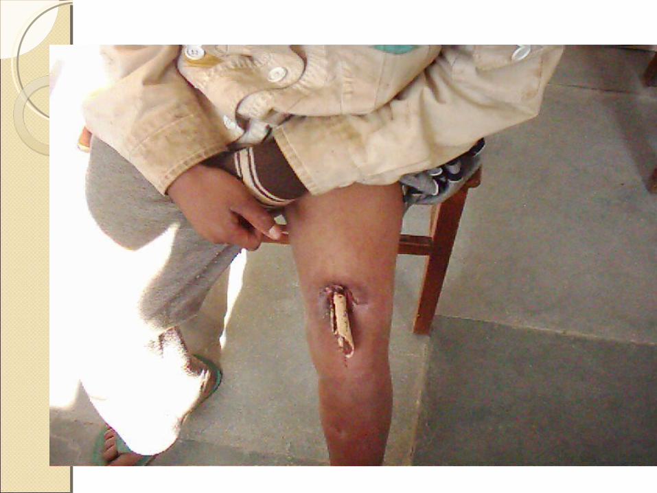
Discharging sinusDischarging sinus
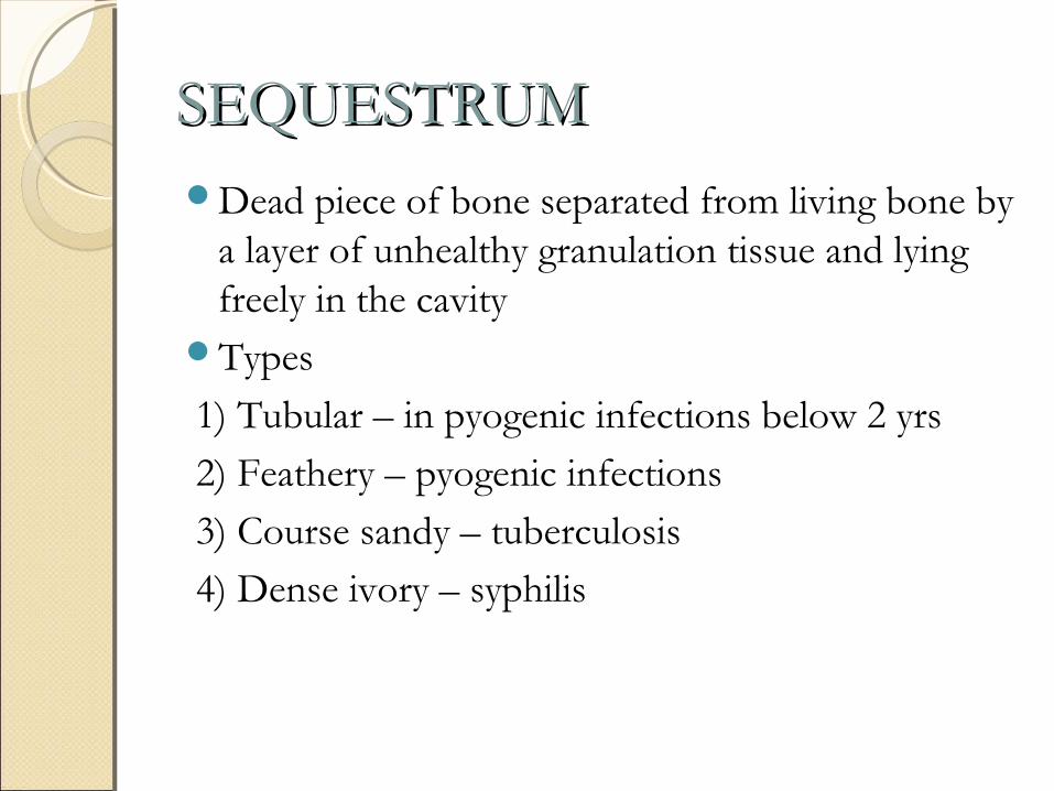
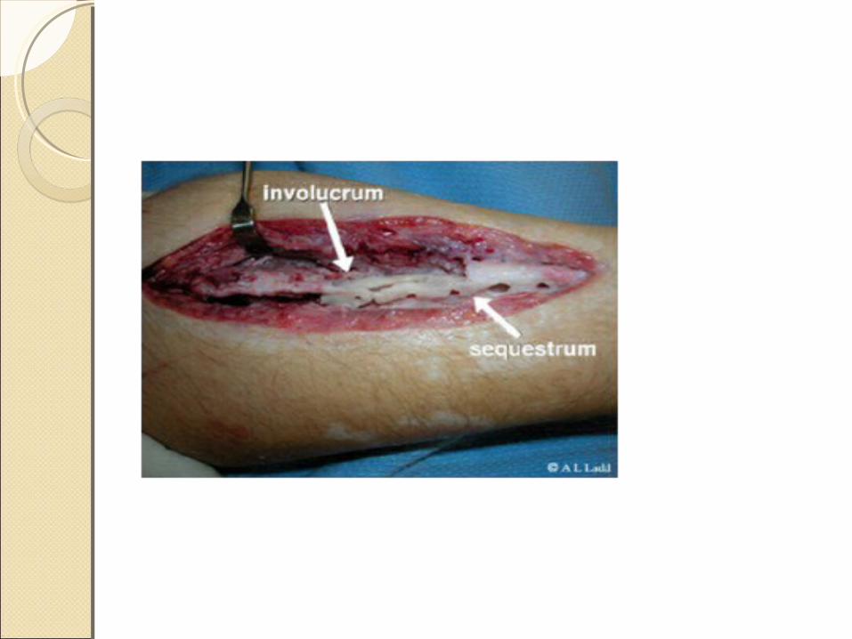
SEQUESTRUMSEQUESTRUMDead piece of bone separated from living bone by
a layer of unhealthy granulation tissue and lying freely in the cavity
Types
1) Tubular – in pyogenic infections below 2 yrs
2) Feathery – pyogenic infections
3) Course sandy – tuberculosis
4) Dense ivory – syphilis
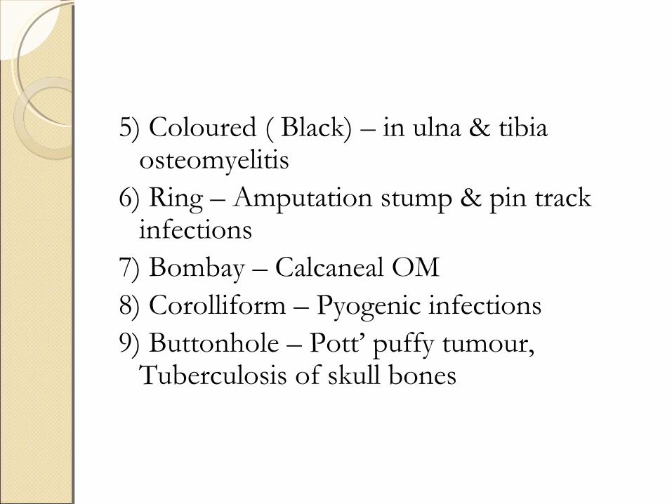
5) Coloured ( Black) – in ulna & tibia osteomyelitis
6) Ring – Amputation stump & pin track infections
7) Bombay – Calcaneal OM8) Corolliform – Pyogenic infections9) Buttonhole – Pott’ puffy tumour,
Tuberculosis of skull bones
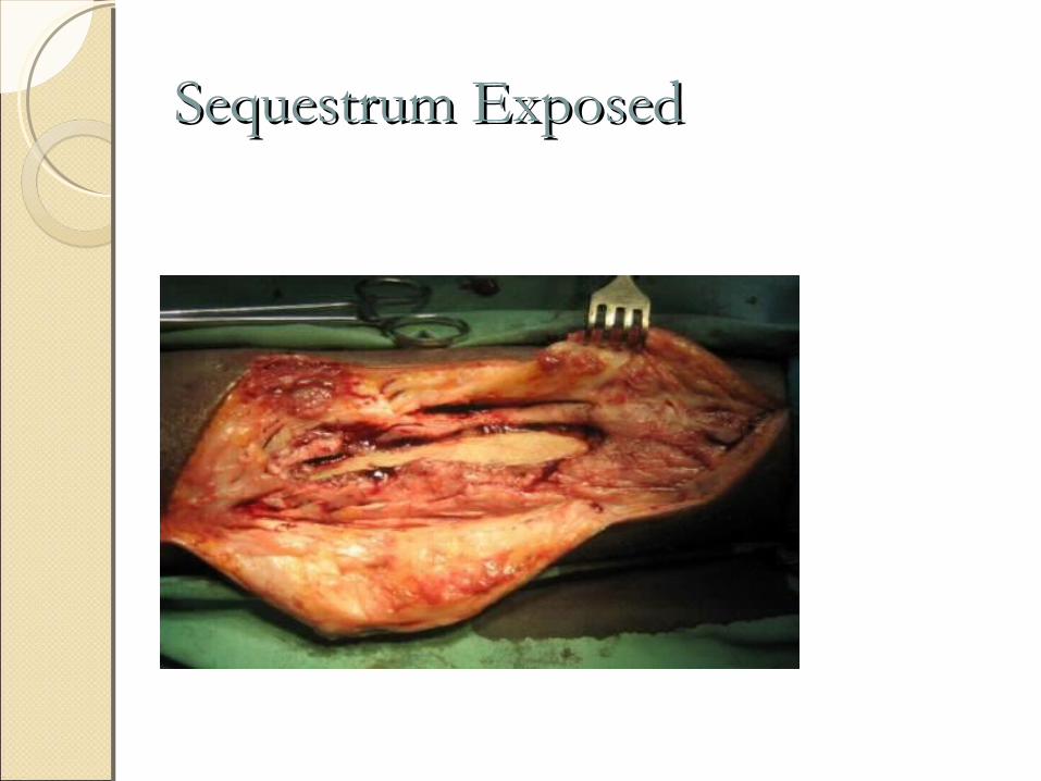
Sequestrum ExposedSequestrum Exposed
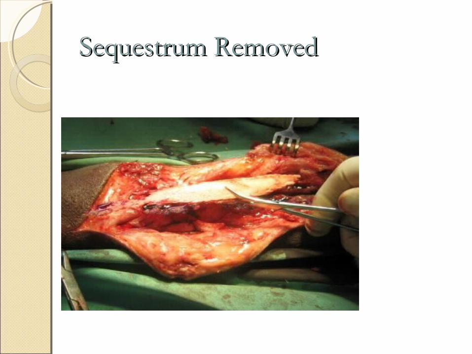
Sequestrum RemovedSequestrum Removed
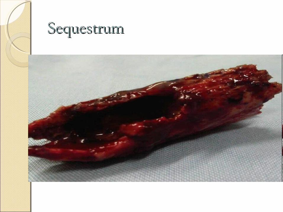
SequestrumSequestrum
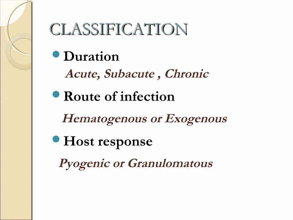
CLASSIFICATIONCLASSIFICATIONDuration Acute, Subacute , Chronic
Route of infection
Hematogenous or Exogenous
Host response
Pyogenic or Granulomatous
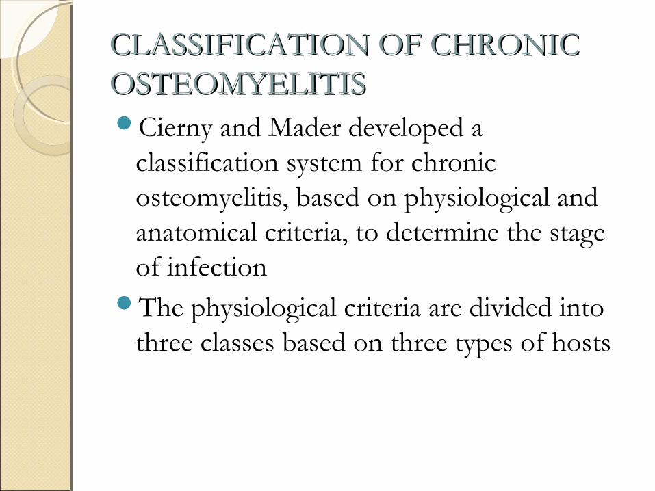
CLASSIFICATION OF CHRONIC CLASSIFICATION OF CHRONIC OSTEOMYELITISOSTEOMYELITISCierny and Mader developed a
classification system for chronic osteomyelitis, based on physiological and anatomical criteria, to determine the stage of infection
The physiological criteria are divided into three classes based on three types of hosts
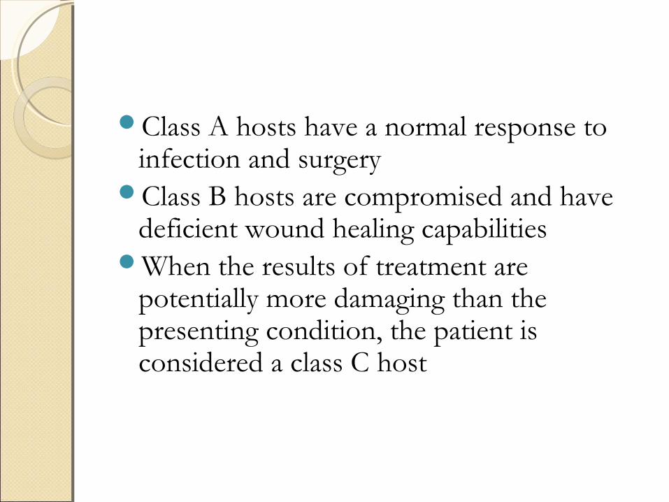
Class A hosts have a normal response to infection and surgery
Class B hosts are compromised and have deficient wound healing capabilities
When the results of treatment are potentially more damaging than the presenting condition, the patient is considered a class C host
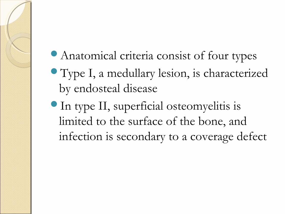
Anatomical criteria consist of four typesType I, a medullary lesion, is characterized
by endosteal diseaseIn type II, superficial osteomyelitis is
limited to the surface of the bone, and infection is secondary to a coverage defect
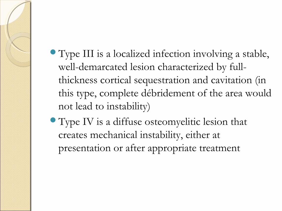
Type III is a localized infection involving a stable, well-demarcated lesion characterized by full-thickness cortical sequestration and cavitation (in this type, complete débridement of the area would not lead to instability)
Type IV is a diffuse osteomyelitic lesion that creates mechanical instability, either at presentation or after appropriate treatment

Cierney & Mader Class.Cierney & Mader Class.
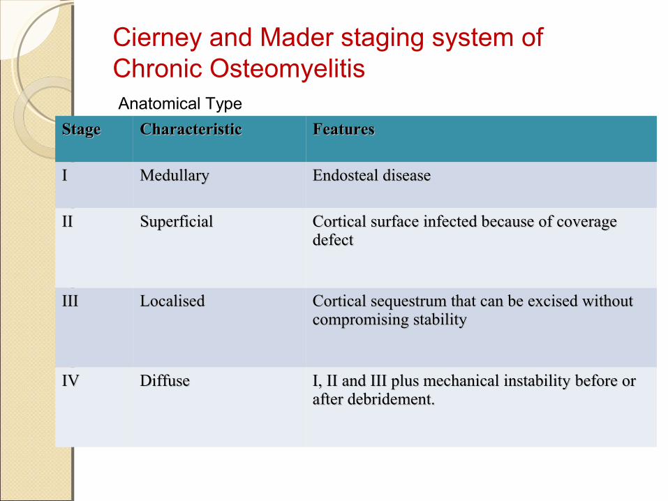
StageStage CharacteristicCharacteristic FeaturesFeatures
II MedullaryMedullary Endosteal diseaseEndosteal disease
IIII Superficial Superficial Cortical surface infected because of coverage Cortical surface infected because of coverage defectdefect
IIIIII LocalisedLocalised Cortical sequestrum that can be excised without Cortical sequestrum that can be excised without compromising stabilitycompromising stability
IVIV DiffuseDiffuse I, II and III plus mechanical instability before or I, II and III plus mechanical instability before or after debridement.after debridement.
Anatomical Type
Cierney and Mader staging system of Chronic Osteomyelitis
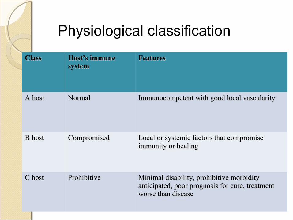
ClassClass Host’s immune Host’s immune systemsystem
FeaturesFeatures
A hostA host NormalNormal Immunocompetent with good local vascularityImmunocompetent with good local vascularity
B host B host CompromisedCompromised Local or systemic factors that compromise Local or systemic factors that compromise immunity or healingimmunity or healing
C hostC host ProhibitiveProhibitive Minimal disability, prohibitive morbidity Minimal disability, prohibitive morbidity anticipated, poor prognosis for cure, treatment anticipated, poor prognosis for cure, treatment worse than diseaseworse than disease
Physiological classification
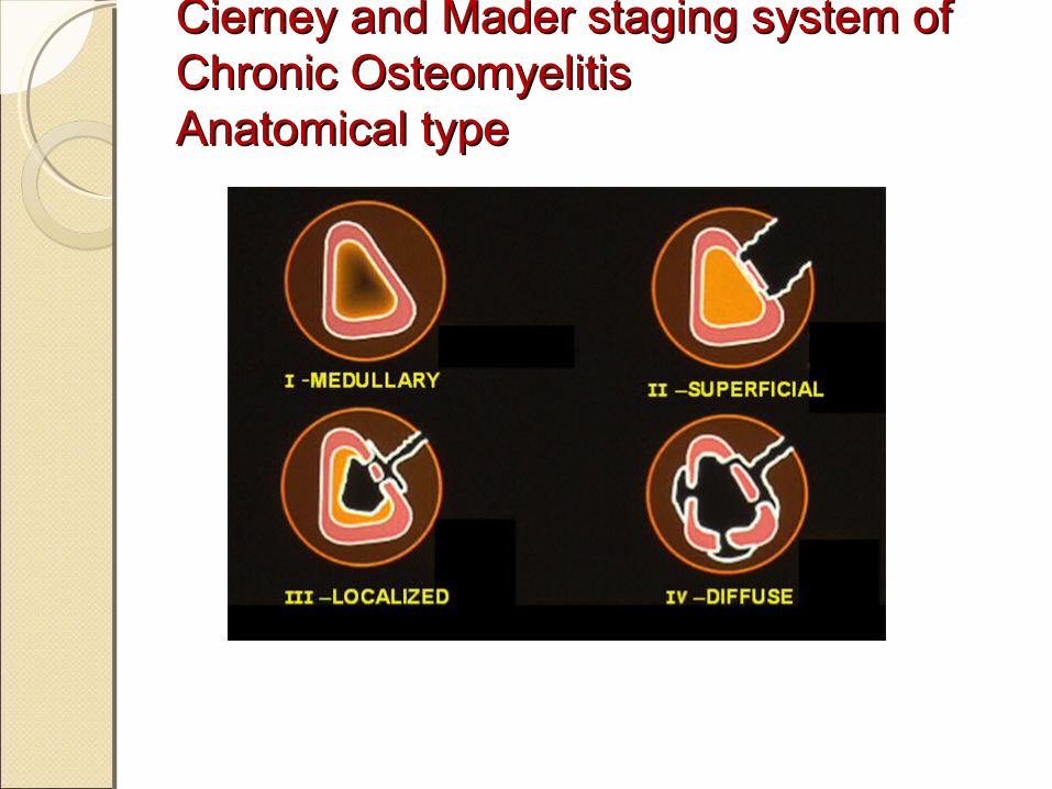
Cierney and Mader staging system of Cierney and Mader staging system of Chronic OsteomyelitisChronic OsteomyelitisAnatomical typeAnatomical type
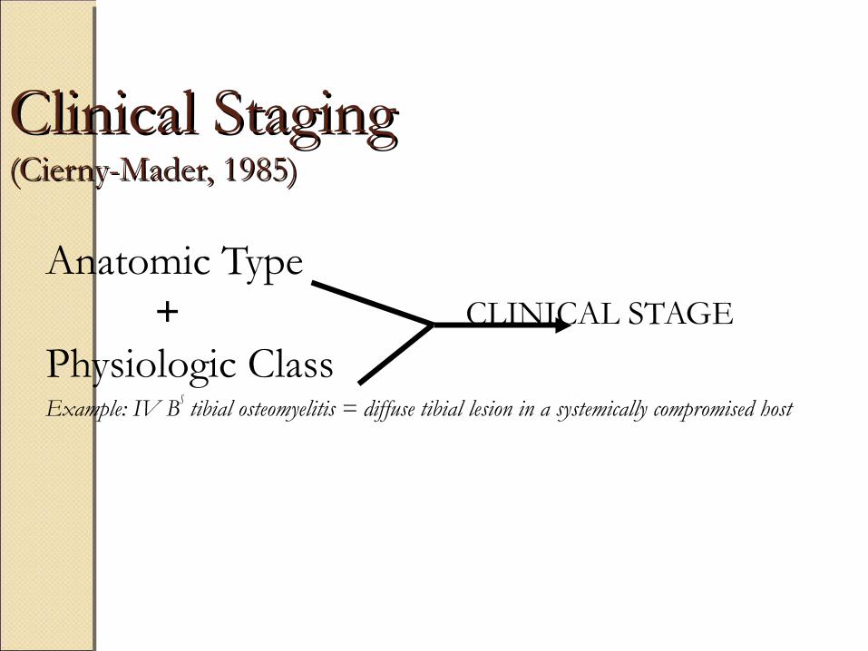
Clinical StagingClinical Staging(Cierny-Mader, 1985)(Cierny-Mader, 1985)
Anatomic Type + CLINICAL STAGE
Physiologic ClassExample: IV BS tibial osteomyelitis = diffuse tibial lesion in a systemically compromised host
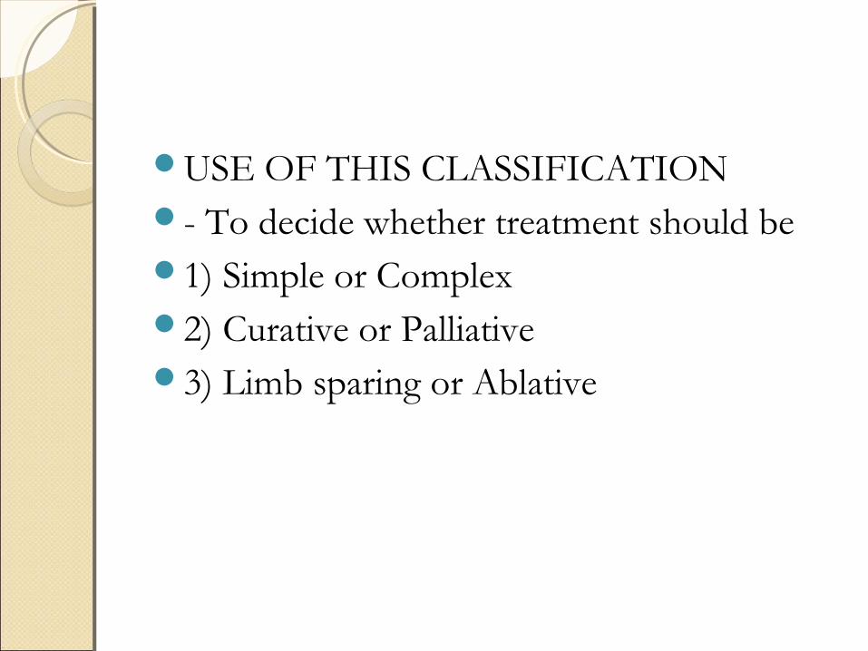
USE OF THIS CLASSIFICATION - To decide whether treatment should be1) Simple or Complex2) Curative or Palliative3) Limb sparing or Ablative
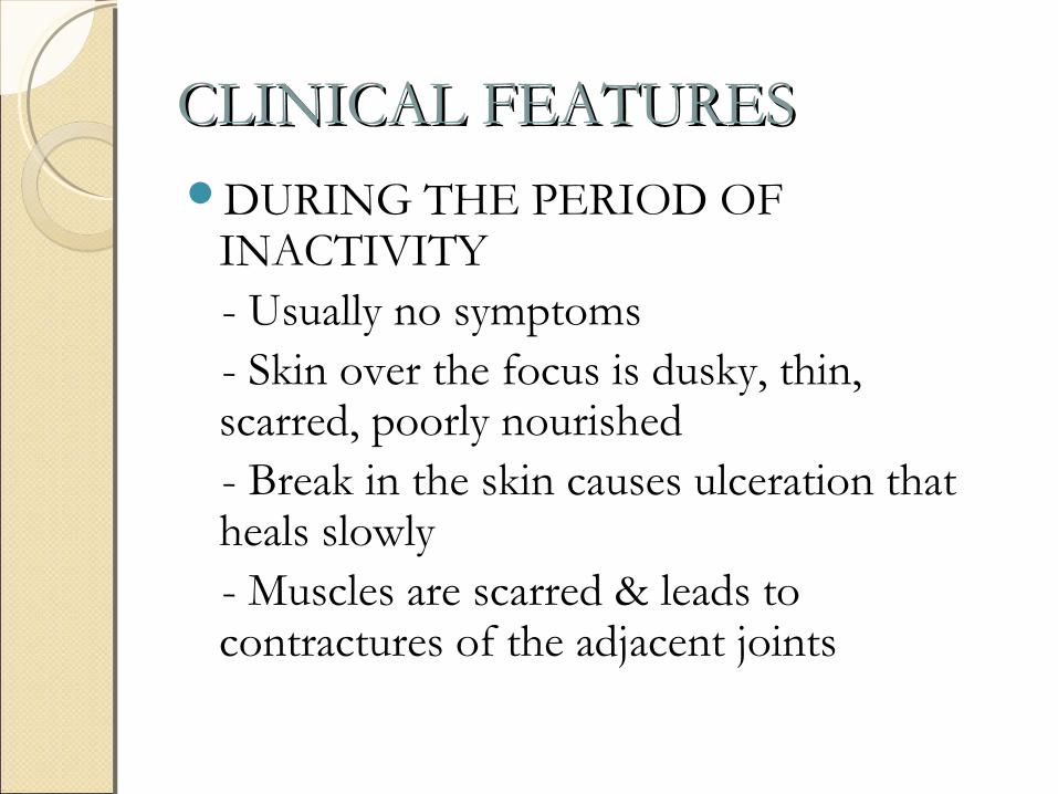
CLINICAL FEATURESCLINICAL FEATURESDURING THE PERIOD OF
INACTIVITY - Usually no symptoms - Skin over the focus is dusky, thin,
scarred, poorly nourished - Break in the skin causes ulceration that
heals slowly - Muscles are scarred & leads to
contractures of the adjacent joints
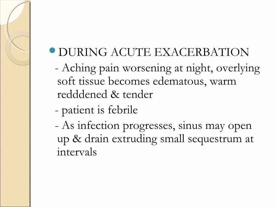
DURING ACUTE EXACERBATION - Aching pain worsening at night, overlying
soft tissue becomes edematous, warm redddened & tender
- patient is febrile - As infection progresses, sinus may open
up & drain extruding small sequestrum at intervals
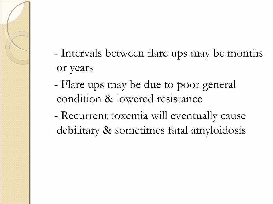
- Intervals between flare ups may be months or years
- Flare ups may be due to poor general condition & lowered resistance
- Recurrent toxemia will eventually cause debilitary & sometimes fatal amyloidosis

DIAGNOSISDIAGNOSIS
The diagnosis of chronic osteomyelitis is based on CLINICAL, LABORATORY & IMAGING studies
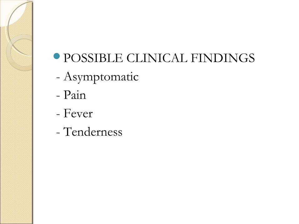
POSSIBLE CLINICAL FINDINGS - Asymptomatic - Pain - Fever - Tenderness
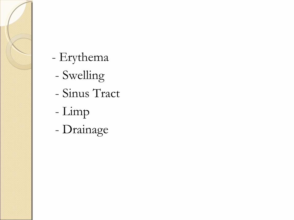
- Erythema - Swelling - Sinus Tract - Limp - Drainage
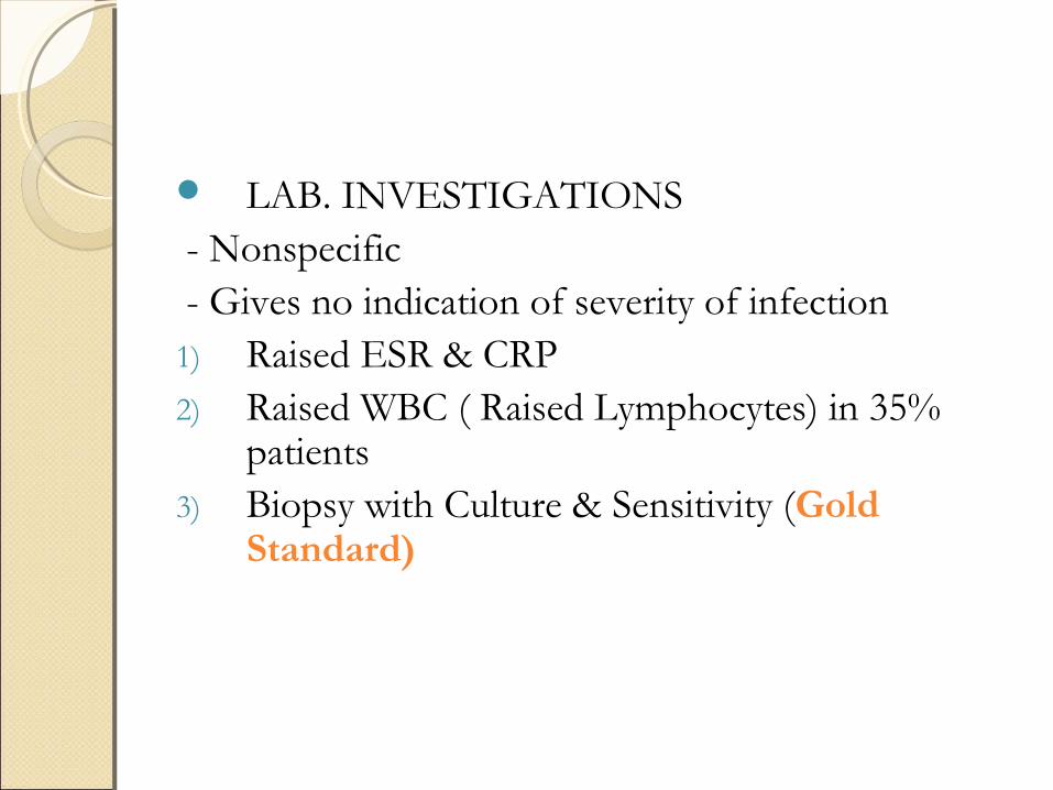
LAB. INVESTIGATIONS - Nonspecific - Gives no indication of severity of infection1) Raised ESR & CRP2) Raised WBC ( Raised Lymphocytes) in 35%
patients3) Biopsy with Culture & Sensitivity (Gold
Standard)

IMAGINGIMAGING
For confirmation of the diagnosisTo prepare for surgical treatmentHowever no technique can absolutely
confirm or exclude the presence of osteomyelitis
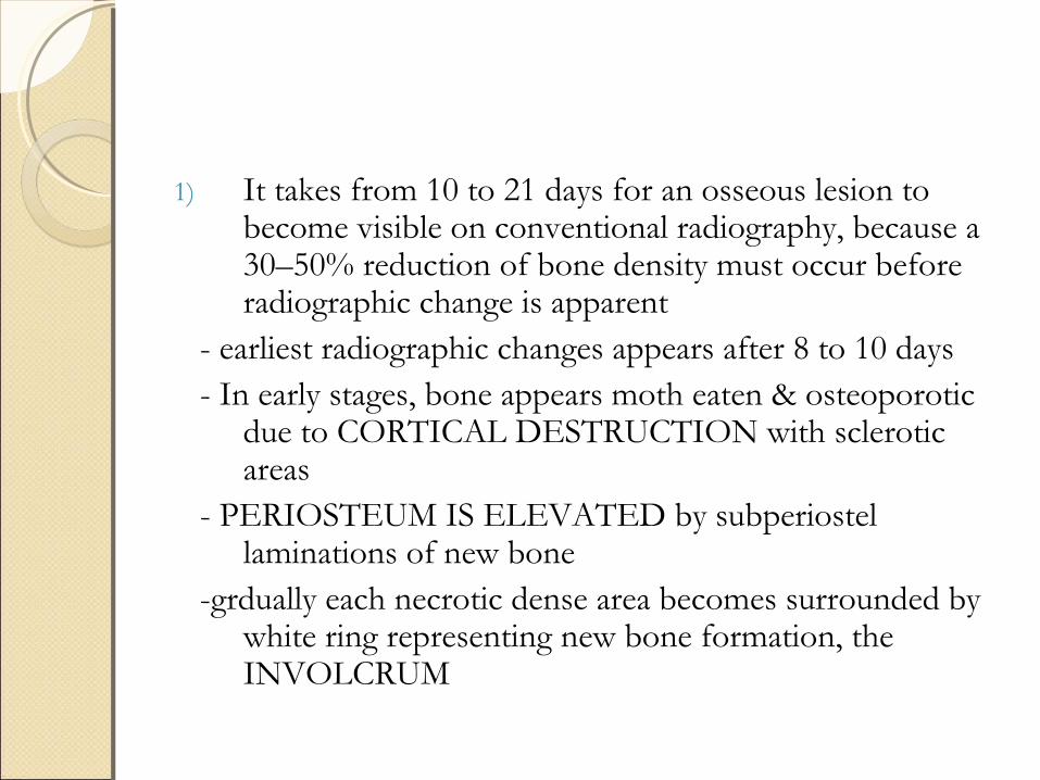
1) It takes from 10 to 21 days for an osseous lesion to become visible on conventional radiography, because a 30–50% reduction of bone density must occur before radiographic change is apparent
- earliest radiographic changes appears after 8 to 10 days - In early stages, bone appears moth eaten & osteoporotic
due to CORTICAL DESTRUCTION with sclerotic areas
- PERIOSTEUM IS ELEVATED by subperiostel laminations of new bone
-grdually each necrotic dense area becomes surrounded by white ring representing new bone formation, the INVOLCRUM
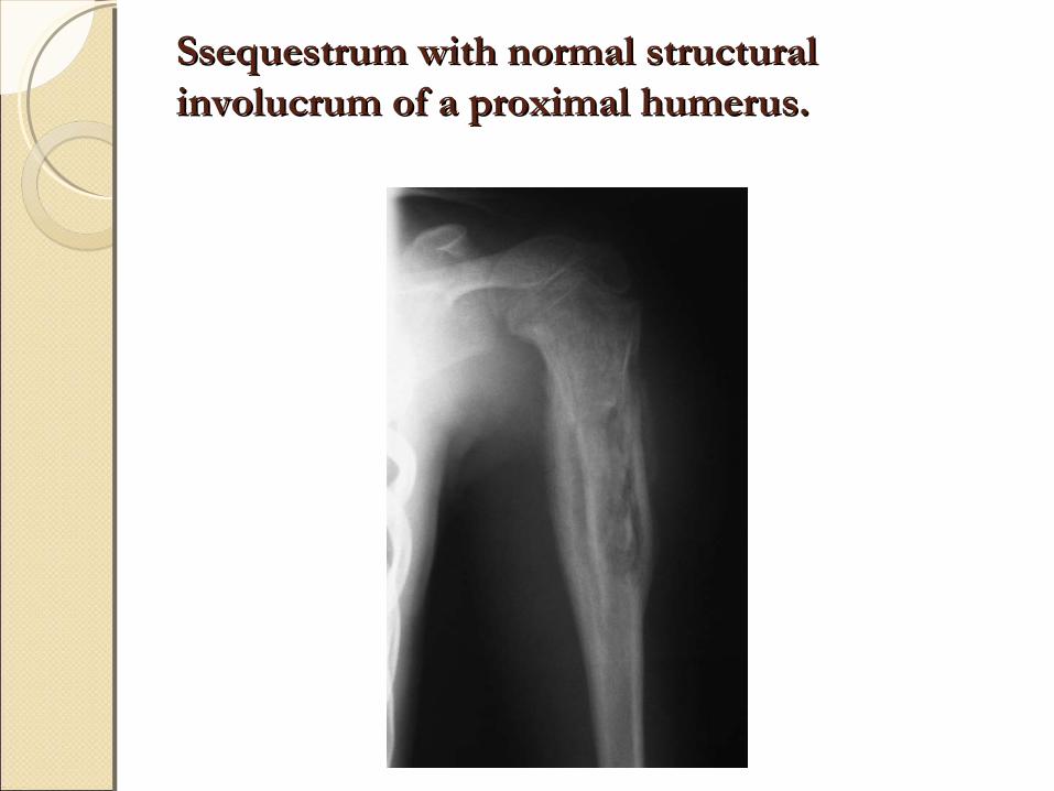
Ssequestrum with normal structural Ssequestrum with normal structural involucrum of a proximal humerus.involucrum of a proximal humerus.
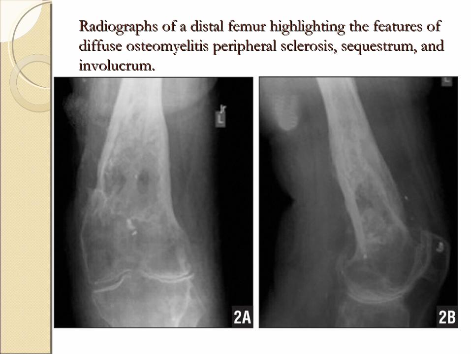
Radiographs of a distal femur highlighting the features of Radiographs of a distal femur highlighting the features of diffuse osteomyelitis peripheral sclerosis, sequestrum, and diffuse osteomyelitis peripheral sclerosis, sequestrum, and involucrum.involucrum.
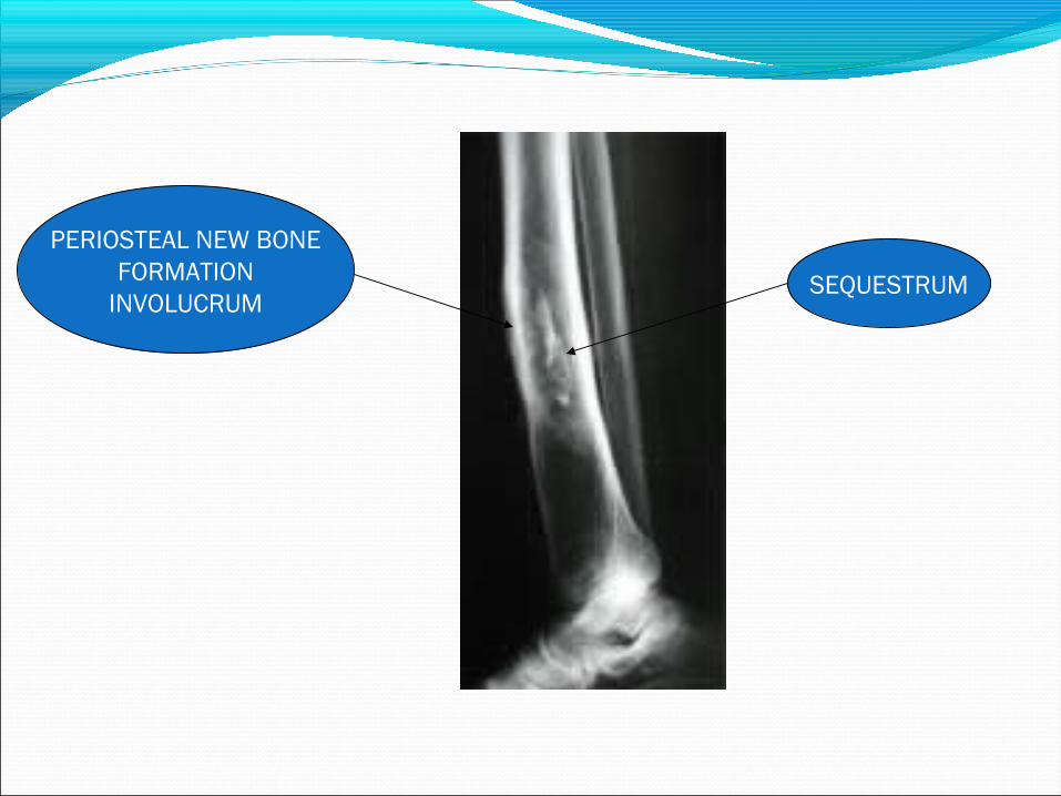
SEQUESTRUM
PERIOSTEAL NEW BONEFORMATION
INVOLUCRUM
SINOGRAPHYSINOGRAPHY
Helpful if the sinus is present in locating focus of the infection in chronic osteomyelitis
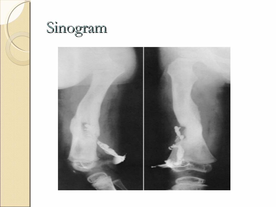
SinogramSinogram

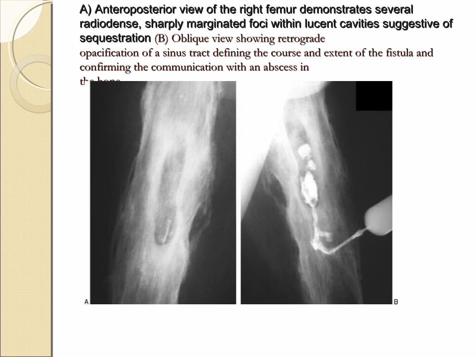
A) Anteroposterior view of the right femur demonstrates severalA) Anteroposterior view of the right femur demonstrates severalradiodense, sharply marginated foci within lucent cavities suggestive of radiodense, sharply marginated foci within lucent cavities suggestive of sequestration sequestration (B) Oblique view showing retrograde(B) Oblique view showing retrogradeopacification of a sinus tract defining the course and extent of the fistula and opacification of a sinus tract defining the course and extent of the fistula and confirming the communication with an abscess inconfirming the communication with an abscess inthe bone.the bone.
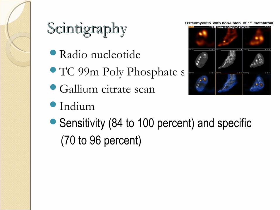
ScintigraphyScintigraphy
Radio nucleotide TC 99m Poly Phosphate scan Gallium citrate scan Indium Sensitivity (84 to 100 percent) and specific (70 to 96 percent)
1)99M Tc Scan - Action
binds to hydroxyapetite crystals - Shows increased uptake in the areas of
increased blood flow or oseoblastic activity
- lacks specificity
2) 67Ga Scan - shows increased uptake in areas where
leucocytes or bacteria accumulate - normal Ga scan excludes presence of
osteomyelitis - useful as follow up examination after
surgery
3) 111Indium leucocyte scan - more sensitive than Tc or Ga scans - especially useful in differentiating
chronic osteomyelitis from neuropathic arthropathy like diabetic foot
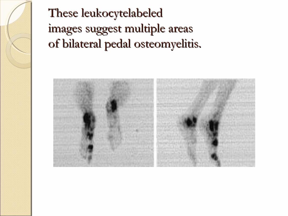
These leukocytelabeledThese leukocytelabeledimages suggest multiple areasimages suggest multiple areasof bilateral pedal osteomyelitis.of bilateral pedal osteomyelitis.
Various combinations of aforementioned scintigraphic method remain the gold standard diagnostic method in post traumatic infections.
This is especially true because the presence of metallic implants limits the usefullness of MRI scanning.
Flourodeoxy glucose (FDG)-PET scan enables noninvasive detection and demonstration of the chronic osteomyelitis with 97% accuracy.
18FDG PET Scan
Meta-analysis showed –Fluorodeoxyglucose positron emission tomography has the highest accuracy for confirming or excluding the diagnosis of Chr OM
CT SCAN - Provides excellent difinition of cortical
bone - especially useful in identification of
Sequestra
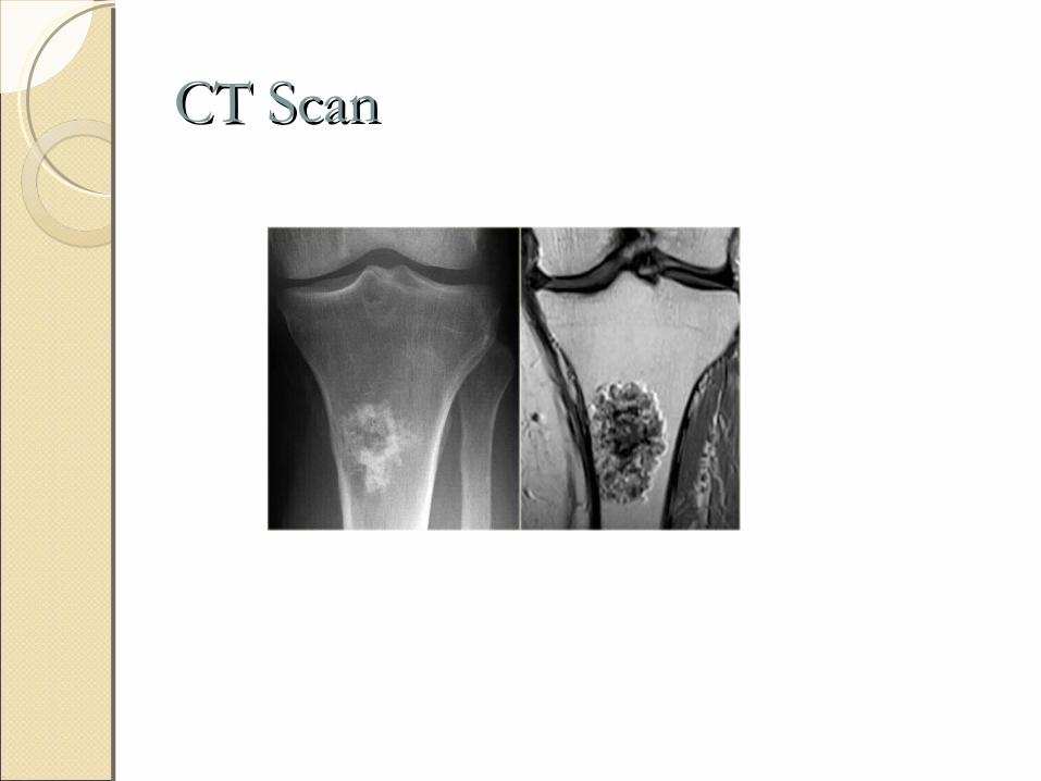
CT ScanCT Scan
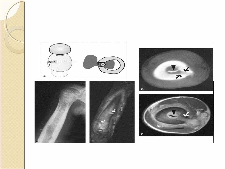
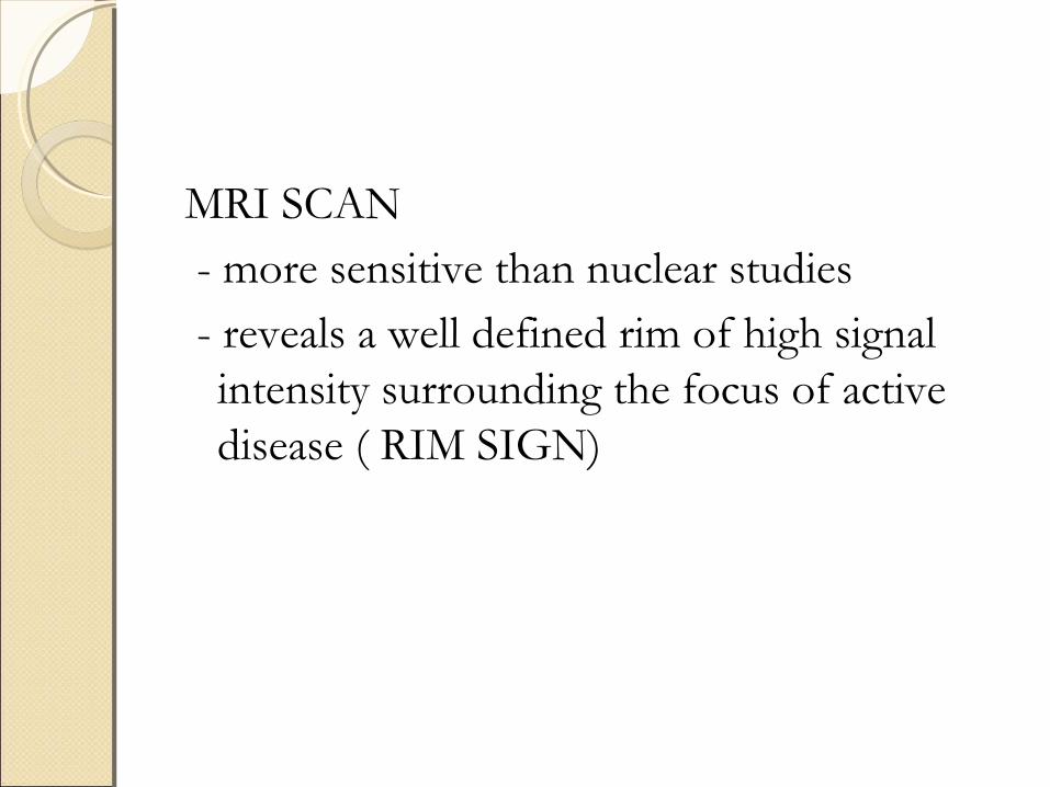
MRI SCAN - more sensitive than nuclear studies - reveals a well defined rim of high signal
intensity surrounding the focus of active disease ( RIM SIGN)
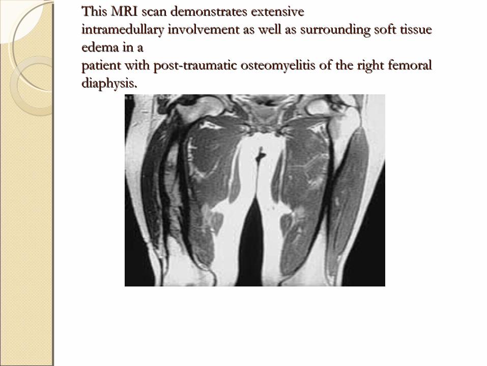
This MRI scan demonstrates extensiveThis MRI scan demonstrates extensiveintramedullary involvement as well as surrounding soft tissue intramedullary involvement as well as surrounding soft tissue edema in aedema in apatient with post-traumatic osteomyelitis of the right femoral patient with post-traumatic osteomyelitis of the right femoral diaphysis.diaphysis.
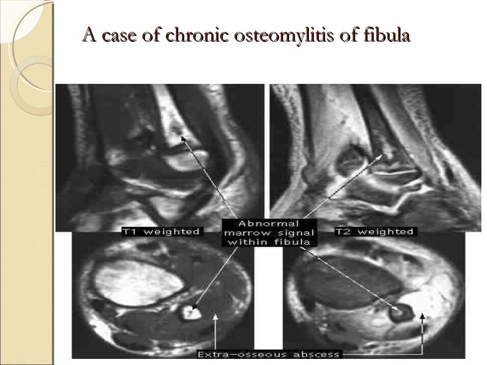
A case of chronic osteomylitis of fibulaA case of chronic osteomylitis of fibula
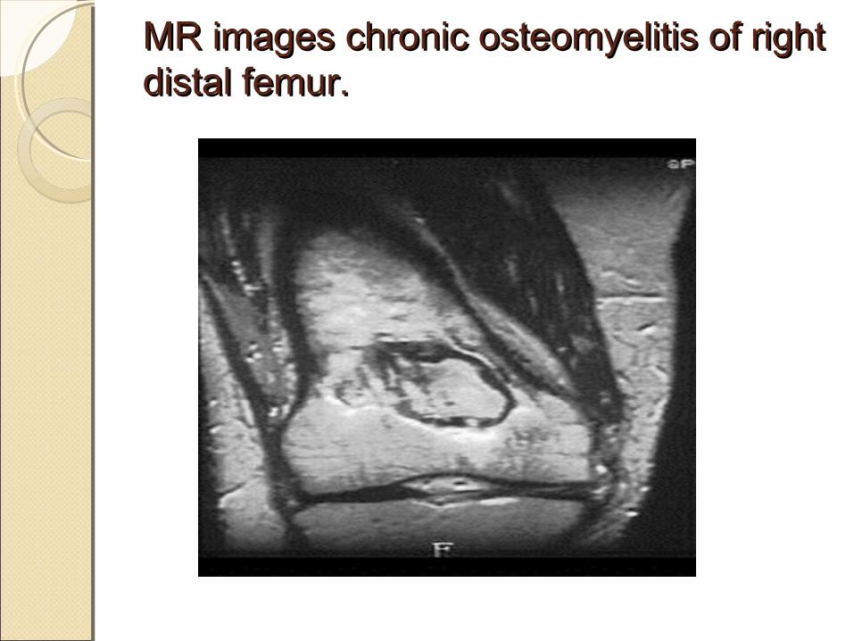
MR images chronic osteomyelitis of right MR images chronic osteomyelitis of right distal femur. distal femur.
MOLECULAR DIAGNOSTICSMOLECULAR DIAGNOSTICS
Identification procedures based on molecular analysis and RNA or DNA typing are currently in development to facilitate diagnosis in osteomyelitis.
The most commonly used method is PCR technique.
Unfortunately,PCR cannot easily delineate between nuclear materials from living or dead bacteria.
This increases likelihood of false positive results.
TREATMENTTREATMENT
To wait is to invite disaster .
Chronic OM generally cannot be eradicated without surgical treatment
Surgery for Chronic OM consists of sequestrectomy & resection of scarred & infected bone & soft tissue
THE GOALS OF SURGERY - Eradication of infection by achieving a
viable & vascular environment- Radical debridement- Prevent recurrences
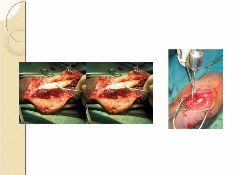
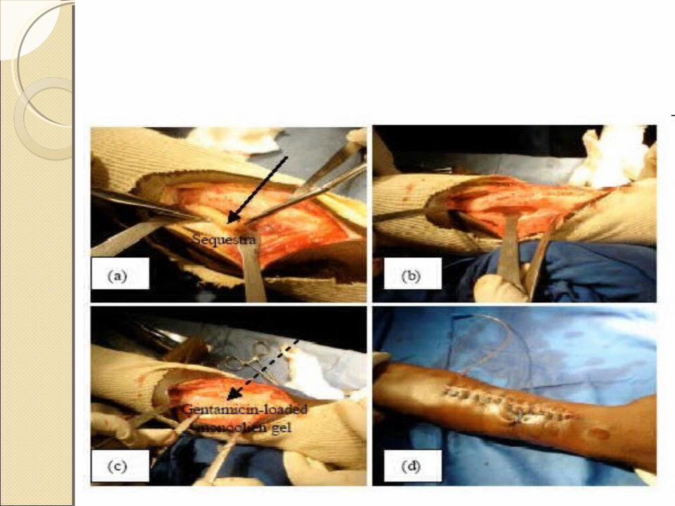
SEQUESTRECTOMY & CURETTAGESEQUESTRECTOMY & CURETTAGE
1) All sinus tracts are excised completely along with sequestra, purulent material & scarred and necrotic tissue
if sclerosis bone seals off a cavity within the medullary canal, it is opened into the canal in both directions to allow blood vessels to grow into the cavity
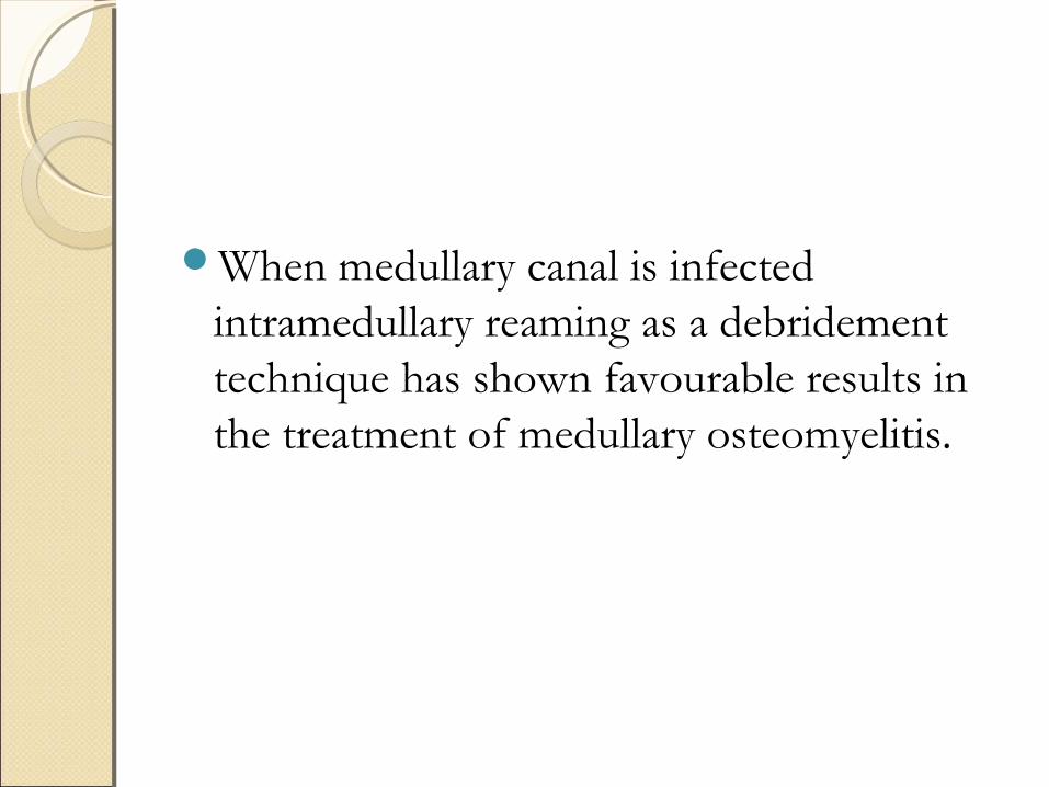
When medullary canal is infected intramedullary reaming as a debridement technique has shown favourable results in the treatment of medullary osteomyelitis.
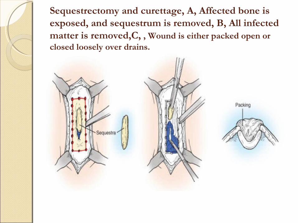
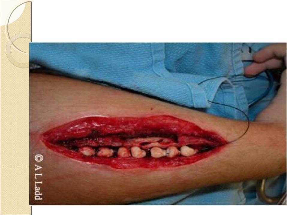
Sequestrectomy and curettage, A, Affected bone is exposed, and sequestrum is removed, B, All infected matter is removed,C, , Wound is either packed open or closed loosely over drains.
AFTER TREATMENT
- Limb is splinted until the wound is healed & then it is protected to prevent pahological fracture
- prolonged antibiotic therapy is given usually for 6 weeks
- bony & soft tissue defects must be filled to reduce chances of continued infection & loss of function
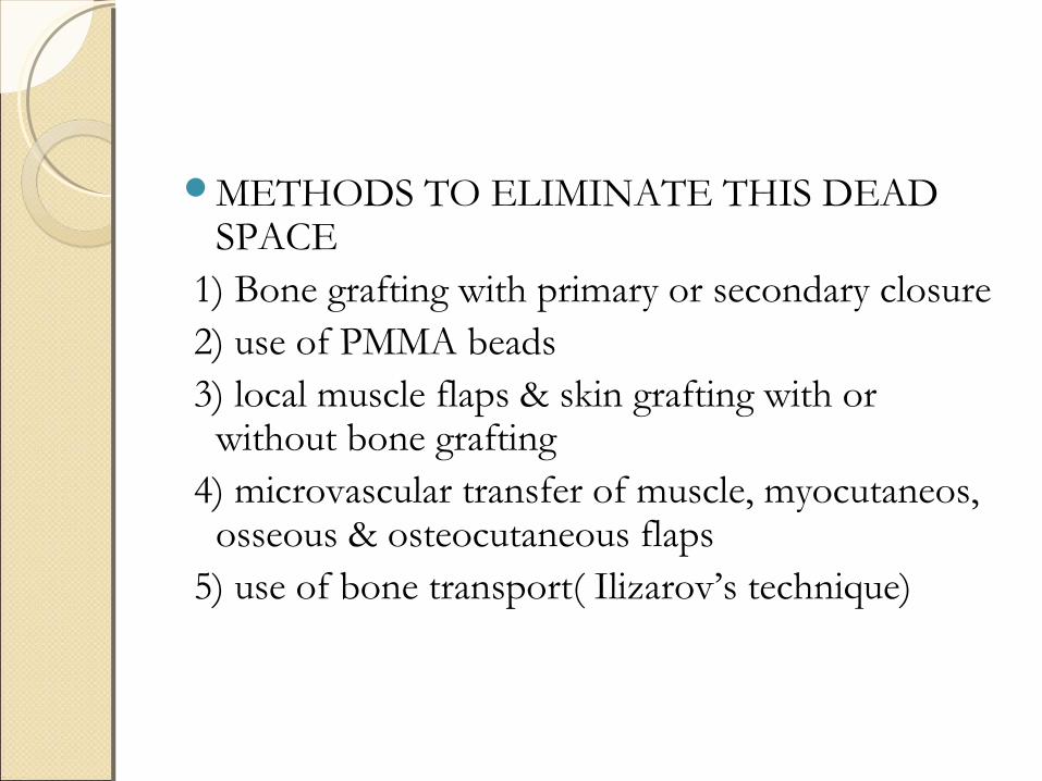
METHODS TO ELIMINATE THIS DEAD SPACE
1) Bone grafting with primary or secondary closure 2) use of PMMA beads 3) local muscle flaps & skin grafting with or
without bone grafting 4) microvascular transfer of muscle, myocutaneos,
osseous & osteocutaneous flaps 5) use of bone transport( Ilizarov’s technique)
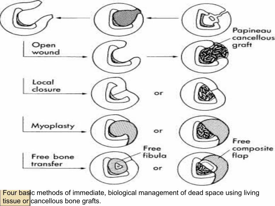
Four basic methods of immediate, biological management of dead space using living tissue or cancellous bone grafts.

OPEN BONE GRAFTING( PAPINEAU TECHNIQUE)
This procedure is based on the following principles: - granulation tissue markedly resists infection - autogenous cancellous bone grafts are rapidly
revascularized and are resistant to infection -the infected area is completely excised -adequate drainage is provided -adequate immobilization is provided -antibiotics are used for prolonged periods
DONE IN 3 STAGES 1)Excision of infected tissue without or with
stabilization using an external fixator or an intramedullary rod
2) Cancellous autografting 3) skin closure
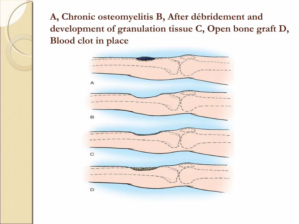
A, Chronic osteomyelitis B, After débridement and development of granulation tissue C, Open bone graft D, Blood clot in place
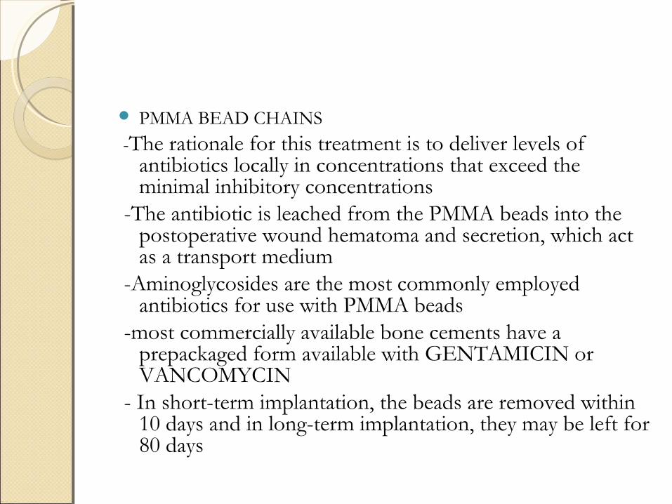
PMMA BEAD CHAINS
-The rationale for this treatment is to deliver levels of antibiotics locally in concentrations that exceed the minimal inhibitory concentrations
-The antibiotic is leached from the PMMA beads into the postoperative wound hematoma and secretion, which act as a transport medium
-Aminoglycosides are the most commonly employed antibiotics for use with PMMA beads
-most commercially available bone cements have a prepackaged form available with GENTAMICIN or VANCOMYCIN
- In short-term implantation, the beads are removed within 10 days and in long-term implantation, they may be left for 80 days
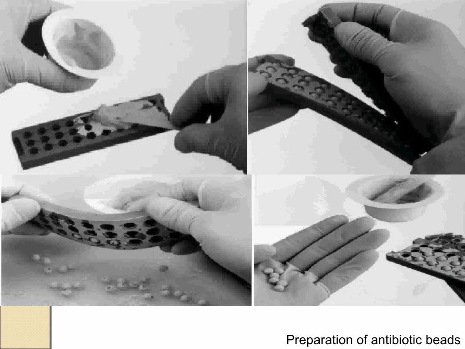
Preparation of antibiotic beads
BIODEGRADABLE ANTIBIOTIC DELIVERY SYSTEMS
-Biodegradable substrates contain osteoconductive and osteoinductive materials, which can be used to promote new bone formation
- Bioabsorbable substrates (calcium sulfate or calcium phosphate) that can be mixed with antibiotics ( Vancomycin or Tobramycin)
-These beads typically resorb by about 8 weeks after surgery

Polymethylmethacrylate (PMMA) beads connected Polymethylmethacrylate (PMMA) beads connected together in a chain are the most widely used drug together in a chain are the most widely used drug delivery system.delivery system.
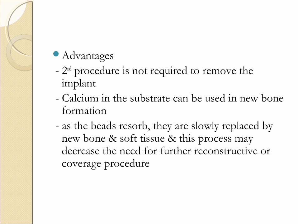
Advantages - 2nd procedure is not required to remove the
implant - Calcium in the substrate can be used in new bone
formation - as the beads resorb, they are slowly replaced by
new bone & soft tissue & this process may decrease the need for further reconstructive or coverage procedure
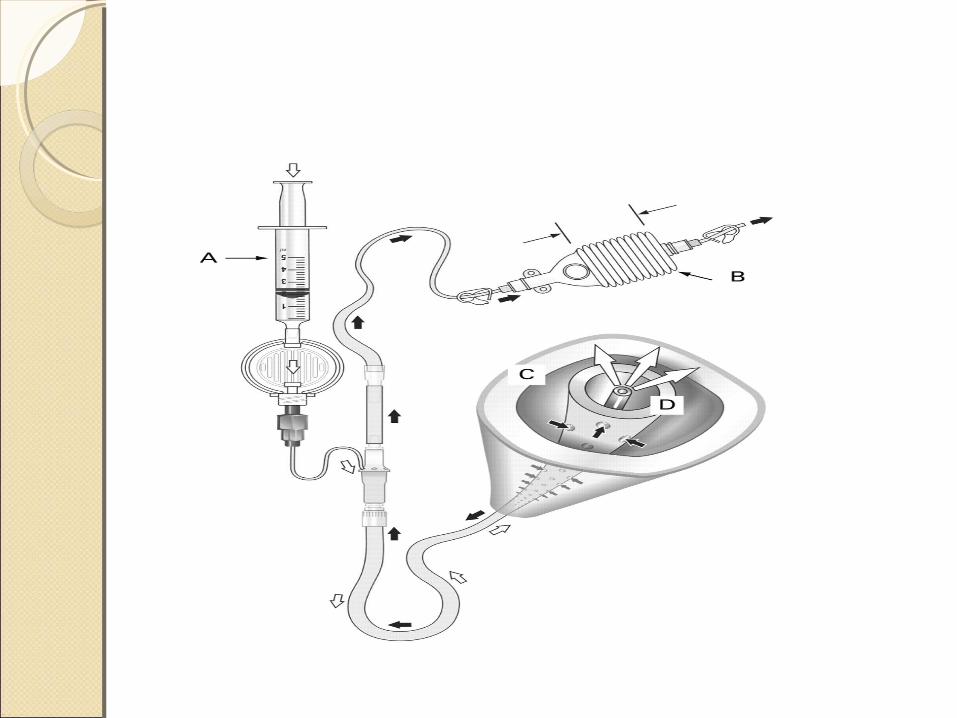
CLOSED IRRIGATION & SUCTION
-For resistant focal infections, topical instillation of solution containing mild detergent ( eg. Alevaire) & one or more antibiotic seems to be effective
- detergent inhibits the formation of penicillinase
- so penicillins used with detergents becomes effective against resistant, bacterial strains

-Closed suction antibiotic ingress and egress high-volume irrigation systems(Lautenbach continuous irrigation method) can be used over 3 to 21 days
- the material collected through suction tube is cultured every day when 3 successive negative cultures are obtained, the antibiotic detergent solution is discontinued
Detergents or soaps have been showed to be the only irrigation solutions that remove additional bacteria beyond the affect of mechanical irrigation alone.
Castille soap has recently been reported to be useful in this situation.
SOFT TISSUE TRANSFER - Soft-tissue transfers to fill dead space left behind
after extensive debridement may range from a localized muscle flap on a vascular pedicle to microvascular free tissue transfer
- transfer of vascularized muscle tissue improves the local biological environment by bringing in a blood supply that is important in the host's defense mechanisms and for antibiotic delivery and osseous and soft-tissue healing.
- Most commonly, a local muscle flap is used in the treatment of chronic osteomyelitis of the tibia
- The gastrocnemius muscle is used for defects around the proximal third of the tibia
- the soleus muscle is used for defects around the middle third
- A microvascular free muscle transfer is required for defects around the distal third of the tibia

- When a microvascular free muscle flap is used, and segmental bone loss has occurred, autogenous cancellous bone grafting can be done about 6 weeks after the initial free flap transfer
- free fibular graft can be used for segmental bone loss of the tibia
- If chronic osteomyelitis involves segmental bone loss of the tibia and the fibula, the results of a free fibular graft are not good, and amputation or reconstruction by the Ilizarov technique is advised
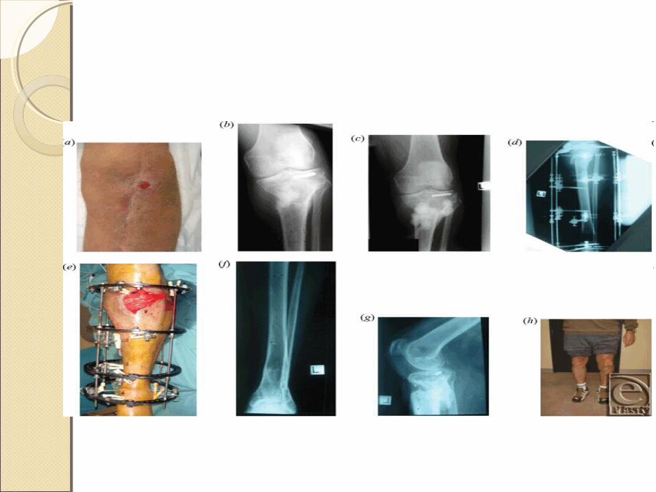
ILIZAROV’S TECHNIQUE- The Ilizarov technique has been helpful in the treatment
of chronic osteomyelitis and infected nonunions- This technique allows radical resection of the infected
bone- A corticotomy is performed through normal bone
proximal and distal to the area of disease. The bone is transported until union is achieved
- Disadvantages of this technique include the time required to achieve a solid union and the high incidence of associated complications
-
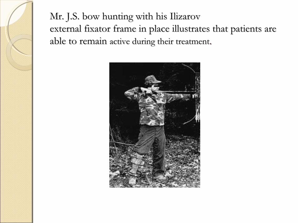
Mr. J.S. bow hunting with his IlizarovMr. J.S. bow hunting with his Ilizarovexternal fixator frame in place illustrates that patients are external fixator frame in place illustrates that patients are able to remain able to remain active during their treatmentactive during their treatment..
BELFAST TECHNIQUE 2 Stage Technique - in 1st stage, all necrotic & infected tissues
are debrided & is covered with soft tissue transfer
- in 2nd stage , at later time autogenous cancellous grfating is done after infection has been completely subsided
SILVER IONTOPHORESIS - Electrically generated silver ions have been
shown to have antibacterial properties - can be used as adjuvent treatment in
management of chronic osteomyelitis
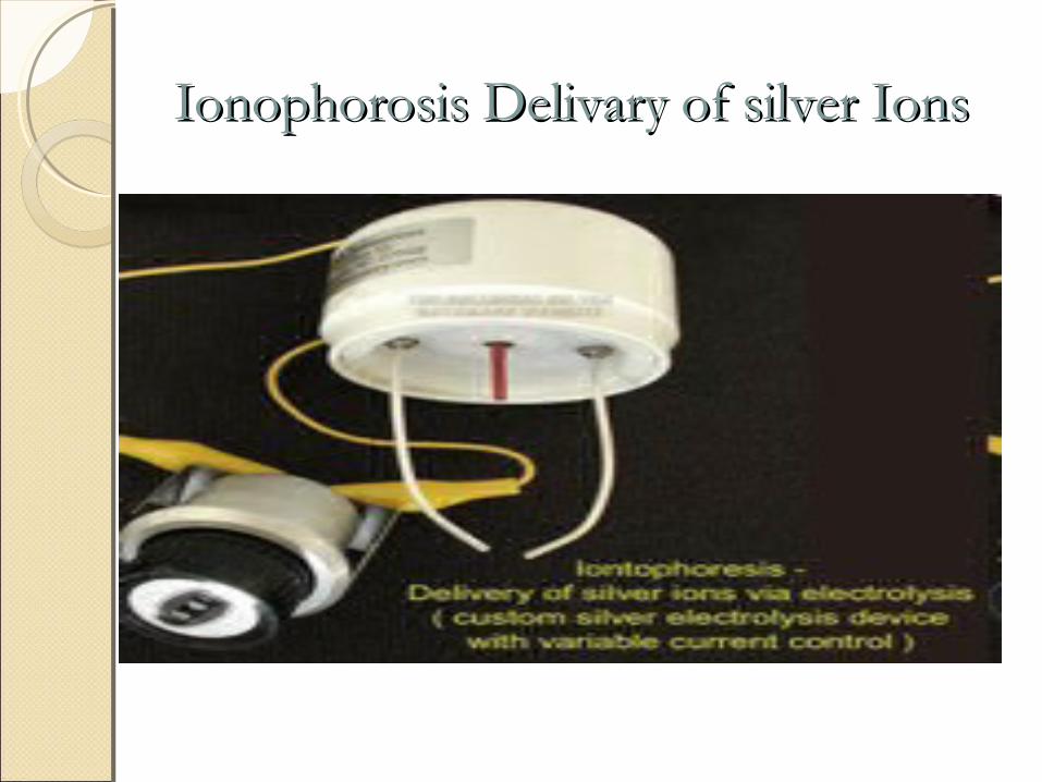
Ionophorosis Delivary of silver IonsIonophorosis Delivary of silver Ions
HYPERBARIC OXYGEN THERAPYHYPERBARIC OXYGEN THERAPY
Used as adjuvent to other methods of treatment
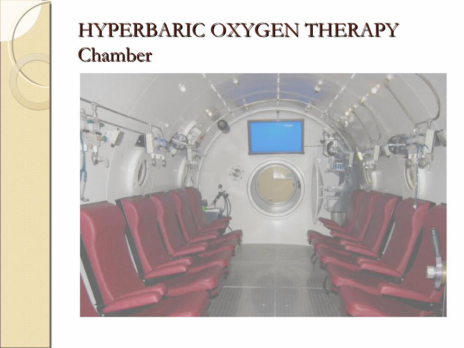
HYPERBARIC OXYGEN THERAPY HYPERBARIC OXYGEN THERAPY ChamberChamber
HBO enhances oxygen-dependent leukocyte killing through the production of hydrogen peroxide and superoxide by providing increased oxygen tension in the hypoxic tissue.
Secondly, optimal tissue oxygen tension enhances osteogenesis and neovascularizationnto fill the dead space with new bone and soft tissues.
HBO has also been shown to enhance osteoclastic activity to remove bony debris.
Finally, HBO also potentiates the antimicrobial effects of aminoglycosides, and possibly sulpha drugs and vancomycin, in the killing of susceptible bacteria
Patients with osteomyelitis are usually treated at 2.0-2.5 ATA for 90-120 min per day and typically receive 20-40 treatments
AMPUTATION FOR CHRONIC OSTEOMYELITIS
- Malignant change of sinus tract - Madura Mycosis - Joint contractures & stiffness
COMPLICATIONSCOMPLICATIONSGrowth disturbancesPathological fracturesMuscle contracturesSecondary septicemiaEpitheliomaMalignant changes( Squamous cell ca,Reticulum
cell ca, Fibrosarcoma)Joint stiffnessAmyloidosis
Novel techniques underdevelopment include the use of compounds that promote detachment of stationary colonised bacteria into the planktonic state,where they are easier to eradicate.
The techniques include surface treatments that inhibit colonisation and the use of light activated dye that destroys ceratain bacteria in wounds.
In this technique indocyanine green,which is a harmless dye,is placed into the wound and activated with near infrared light.
The light resulsts in the dye releasing molecules that are toxic to the MRSA.
The mechanism of action is varied and unlike standard antibiotics that development of resistence is unlikely.
These and other methods under development may provide novel adjunctive treatments to help treat osteomyelitis.
PROPHYLAXISPROPHYLAXIS
Prevention is always better than cure.Prophylactic antibiotics have an important
role in the treatment of closed fractures,the use of which will reduce post operative osteomyelitis in elective cases.
Antibiotic administration is not a substitute for proper aseptic technique,but it is a validated additional measure to reduce post operative infection.
The current recommendation is that antibiotic should be administered 30-60 mins befor the incision is made.
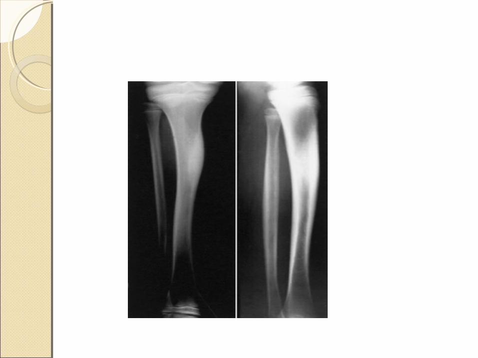
MISCELLANEOUS CONDITIONSMISCELLANEOUS CONDITIONSSCLEROSING OM OF GARRE - Idiopathic cortical sclerosis - mc site Tibia -chronic form of disease in which the bone is
thickened and distended, but abscesses and sequestra are absent
-affects children and young adults -cause is unknown, but it is thought to be an
infection caused by a low-grade, possibly anaerobic bacterium
CLINICAL FEATURES - Intermittent pain - Swelling & Tenderness over affected bone
INVESTIGATIONS - ESR : slightly raised - C/S : usually negative - X Ray : expanded bone with generalised
sclerosis - Biopsy : chronic low grade non specific
OM
DIFFERTIAL DIAGNOSIS - Paget’s disease - Osteoid osteoma
TREATMENT - Fenestration of sclerotic bone - Antibiotics
RESIDUAL STAGE OF RESIDUAL STAGE OF OSTEOMYELITIS OSTEOMYELITIS - characterized by a complete absence of the signs and
symptoms of infection, including drainage -The bone is sclerotic, and its blood supply and strength are
normal -During this stage of osteomyelitis, the bone bears the same
relation to normal bone that scar tissue bears to normal connective tissue
-Adhesions of skin to bone are more common if the bone is subcutaneous
-Injury to such tissues frequently causes skin breakdown and even recurrence of infection
TREATMENT -correcting leg-length inequality or angular
and joint deformities -contracted scars must be released -adherent scars must be replaced by
myocutaneous flaps
PATHOLOGICAL FRACTURE IN PATHOLOGICAL FRACTURE IN OSTEOMYELITIS OSTEOMYELITIS -Because the involucrum is sometimes insufficient, the shaft
of a long bone may fracture during the acute or subacute stage of osteomyelitis before immobilization has been started
-all operations necessary to combat the infection should be carried out thoroughly, and bone fragments are then realigned and immobilized as with any other fracture
-External fixation or cast immobilization usually is preferred -If bone loss is significant, the defect can be filled with
autogenous bone graft, a vascularized osseous graft, or bone transport using the Ilizarov technique
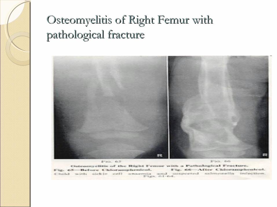
Osteomyelitis of Right Femur with Osteomyelitis of Right Femur with pathological fracturepathological fracture

CHRONIC RECURRENT CHRONIC RECURRENT MULTIFOCAL OSTEOMYELITIS MULTIFOCAL OSTEOMYELITIS CRMO was first described by Giedion and
colleagues in 1972Inflammatory bone disease characterized by
an insidious onset of mild-to-moderate pain with signs of inflammation over the affected parts, which tend to recur
often affects the metaphysis of long bones, especially the tibia, femur, and clavicle
ETIOLOGICAL FACTORS - Infectious disease : Propiobacterium acne - Autoimmune Diseases : Psoriasis & IBD - Genetic Predisposition : LPIN2 gene
CLINICAL FEATURES - Nonspecific : pain, swelling, restricted
mobility - Duration : from days to several years
INVETIGATIONS - raised ESR & CRP with normal WBC
count - Cultures : negative - Biopsy : may be useful
PATHOLOGICAL CONDITIONS PATHOLOGICAL CONDITIONS ASSO. WITH CRMOASSO. WITH CRMO• - Palmoplantar pustulosis• - Psoriasis• -IBD ( Crohn’s disease & Ulcerative colitis) • - SAPHO Syndrome( Synovitis, Acne,
Pustulosis, Hyperostosis, osteitis)• - Majeed syndrome-( CRMO,Cong.
Dyserythropoetic anaemia, inflammatory dermatosis)
- Wegner’s granulomatosis - Takayasu’s arteritis - Pyoderma gangrenosum - Sweet syndrome(Febrile neutrophilic
dermatoses) - Acne fulminans -Spondyloarthropathy
TREATMENTTREATMENTNo effective treatment for CRMO availableCases of disease remission after treatment with
IFN gamma have been reportedResolution of symptoms followed by recurrence
months later is characteristic of this diseaseGenerally, the symptoms continue to recur over 2
years, and the disease generally is self-limitingThe long-term prognosis seems to be good
OSTEOMYELITIS AFTER OSTEOMYELITIS AFTER PUNCTURE WOUND OF THE PUNCTURE WOUND OF THE FOOT FOOT Association of Pseudomonas osteomyelitis
with puncture wounds to the footTreatment consists of surgical drainage,
curettage when indicated, and appropriate antibiotic treatment
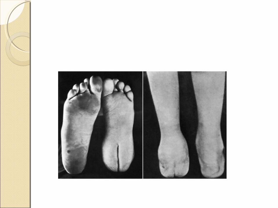
OSTEOMYELITIS OF SPECIFIC OSTEOMYELITIS OF SPECIFIC REGIONS REGIONS 1) CALCANEUM - Cancellous bone with thin periosteum
firmly adherent to the bone - Periosteum is usually perforated - destruction of the cortex is not extensive -sequestrum formation is minimal
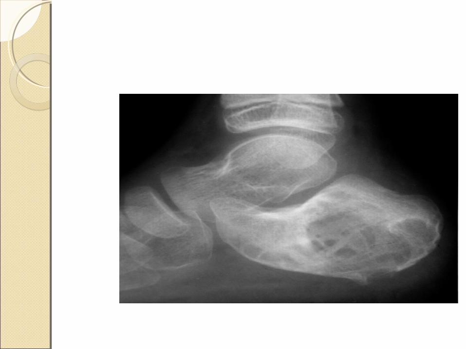
TREATMENT - Gaenslen’s approach: through plantar
surface of heel
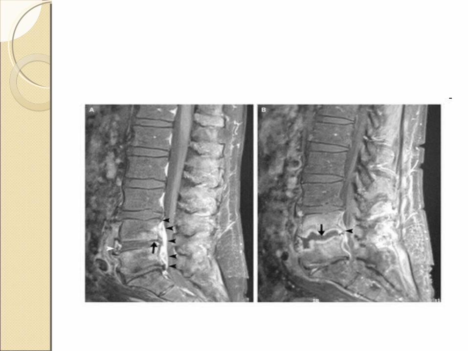
2) SPINE• Commonly affects lumbar spine > thoracic
spine > cervical spine• Usual site : vertebral bodies• Simultaneous involvement of the 2 adjacent
vertebral end plates with intervening discs
ETIOLOGY - richly vascular metaphyseal bone near
anterior longitudinal ligament - avascular disc - end arterial supply of interosseous artery in
adults
X Ray Findings - disc space narrowing with end plate erosion
with vertebral body destruction - vertebral end plate sclerosis with increased
density in subchondral bone - spinal fusion
TREATMENT - IV Antibiotics - Rest - Spinal immobilisation - Surgery
3) DISTAL THIRD FEMUR -Chronic osteomyelitis of the distal third of the
femur is difficult to treat. Because the periosteum may become completely separated posteriorly by a subperiosteal abscess, this part of the bone may lose most of its blood supply, and sinuses often persist
- A mass of scar tissue forms that interferes with revascularization of the bone; the scar tissue is relatively inaccessible surgically because of the proximity of large vessels and nerves

4) ILIUM 1) Treatment during Acute Phase -large subperiosteal abscesses develop on the medial and
lateral cortices -Early symptoms may suggest acute appendicitis or pyogenic
arthritis of the hip joint -Ghahremani reported a relatively high incidence of
osteomyelitis of the ilium in patients with Crohn disease (granulomatous enteritis and colitis)
-He found it was necessary to resect the involved bowel before the osteomyelitis could be cured
2) Treatment during chronic phase
-the entire bone often is involved
-infection is so diffuse that it is impossible to remove the individual sequestra and drain all the abscesses
-Because most of the iliac wing can be removed without causing significant disability, resecting it usually is preferable to less radical operations
4) ISCHIUM & PUBIS -an abscess develops either beneath the external or internal
obturator muscles or in the ischiorectal fossa -Osteomyelitis of the ischial tuberosity is common in
bedridden and paraplegic patients who develop pressure sores with secondary infection, necrosis, and osteomyelitis
-Effective treatment requires debridement of all necrotic tissue and infected bone, and a soft-tissue transfer usually is required to close the defect
SPECIFIC TYPESSPECIFIC TYPES
TUBERCULOUS OM - chronic granulomatous infection caused by
AFB - usually secondary to primary elsewhere - commonly involving metaphysis in children
& epiphysis in adults

- Tubercle follicle is formed of epitheloid cells, lymphocytes, langhans giant cells with central caseation necrosis with peripheral fibrosis by fibroblasts in central portion of shaft
- Caseated pus extends peripherally forms subperiosteal absces, enters soft tissue planes with least resistance to infection as cold abscess
CLINICAL FEATURES -swelling - local tenderness - muscle spasm - cold abscess with chronic discharging sinus
INVESTIGATIONS - Tubeculin test - Biopsy :typical tubercles - Blood : lymphocytosis - X Ray: osteoporosis, loss of outline of
articular cortex, granular foci
TREATMENT - Sinuses with sequestra : curretage &
excision - AKT
To conclude the treatment of chronic osteomyelitis should be in collaboration with other specialities like infectious disease,psychiatry,medicine,micro vascular surgery,social work and physical and occupational therapy.
However,even with access to these experts and appropriate surgery,the success rate for treatment of osteomyelitis is still significantly less than 100%.
There is no doubt that prevention remains the best overall strategy.