Seminar Decompression illness - Deep Diving Academy · Seminar Vol 377 January 8, 2011 155...
12
Seminar www.thelancet.com Vol 377 January 8, 2011 153 Lancet 2010; 377: 153–64 Department of Anesthesiology and Center for Hyperbaric Medicine and Environmental Physiology, Duke University Medical Center, Durham, NC, USA (R D Vann PhD, Prof R E Moon MD); United States Army Institute of Surgical Research, San Antonio, TX, USA (F K Butler MD); and Department of Anaesthesiology, University of Auckland, Auckland, New Zealand (S J Mitchell FANZCA) Correspondence to: Dr Richard D Vann, Box 3823, Center for Hyperbaric Medicine and Environmental Physiology, Duke University Medical Center, Durham, NC 27710, USA [email protected] Decompression illness Richard D Vann, Frank K Butler, Simon J Mitchell, Richard E Moon Decompression illness is caused by intravascular or extravascular bubbles that are formed as a result of reduction in environmental pressure (decompression). The term covers both arterial gas embolism, in which alveolar gas or venous gas emboli (via cardiac shunts or via pulmonary vessels) are introduced into the arterial circulation, and decompression sickness, which is caused by in-situ bubble formation from dissolved inert gas. Both syndromes can occur in divers, compressed air workers, aviators, and astronauts, but arterial gas embolism also arises from iatrogenic causes unrelated to decompression. Risk of decompression illness is affected by immersion, exercise, and heat or cold. Manifestations range from itching and minor pain to neurological symptoms, cardiac collapse, and death. First- aid treatment is 100% oxygen and definitive treatment is recompression to increased pressure, breathing 100% oxygen. Adjunctive treatment, including fluid administration and prophylaxis against venous thromboembolism in paralysed patients, is also recommended. Treatment is, in most cases, effective although residual deficits can remain in serious cases, even after several recompressions. Introduction Decompression illness is caused by bubbles in blood or tissue during or after a reduction in environmental pressure (decompression). It includes two patho- physiological syndromes: arterial gas embolism and the more common decompression sickness. Arterial gas embolism occurs mainly during hyperbaric exposure (eg, diving) and rarely during hypobaric exposure (eg, altitude). Arterial gas embolism occurs when expanding gas stretches and ruptures alveolar capillaries—pulmonary barotrauma—allowing alveolar gas to enter the arterial circulation (figure 1). This syndrome can occur after ascent from a depth as shallow as 1·0–1·5 m if the starting lung volume is close to total lung capacity. 1 It can be caused by gas becoming trapped as a result of airways obstruction in disorders such as asthma 2 or by the presence of pulmonary blebs, cysts, or bullae. 3 Arterial gas embolism can also arise in the absence of decompression through iatrogenic accidents involving vascular catheters and mechanical ventilation. Decompression sickness starts with the formation and increase in size of extravascular and intravascular bubbles when the sum of the dissolved gas tensions (oxygen, carbon dioxide, nitrogen, helium) and water vapour exceeds the local absolute pressure. In diving and during compressed-air tunnel and caisson work, this state of supersaturation is made possible by the increase in tissue inert gas partial pressure that occurs when the gas (usually nitrogen, but occasionally helium) is respired at high pressure. Supersaturation arises during decompression if the rate of ambient pressure reduction exceeds the rate of inert gas washout from tissue. Ascent to altitude in aviation and extravehicular activity during spaceflight involves exposure to decreased barometric pressure. In these settings, supersaturation arises as a result of pre-existing dissolved nitrogen at sea level (partial pressure of nitrogen of about 570 mm Hg), which can also cause bubble formation. Venous gas emboli formed from dissolved gas are easily detected by ultrasonography. In divers, 3·6 m is the minimum dive depth after which venous gas emboli can be seen 4 whereas the decompression sickness threshold, after saturation dives lasting 1–3 days, is about 6 m. 5 During direct decompression from sea level to altitude, the threshold for formation of venous gas emboli is around 3600 m whereas the decompression sickness threshold is about 5500 m. 6,7 Bubbles can have mechanical, embolic, and biochemical effects with manifestations ranging from trivial to fatal. Clinical manifestations can be caused by direct effects from extravascular (autochthonous) bubbles such as mechanical distortion of tissues causing pain, or vascular obstruction causing stroke-like signs and symptoms. Secondary effects can cause delayed symptom onset up to 24 h after surfacing. Endothelial damage by intravascular bubbles can cause capillary leak, extravasation of plasma, and haemoconcentration. 8 Impaired endothelial function, as measured by decreased effects of vasoactive compounds, has been reported in animals 9 and might occur in man. Hypotension can occur in severe cases. 10 Other effects include platelet activation and deposition, 11 leucocyte-endothelial adhesion, 12 and possibly consequences of vascular occlusion believed to occur in thromboembolic stroke such as ischaemia-reperfusion injury, and apoptosis. 13 Arterial gas embolism most often affects the brain but can occasionally affect the heart and other organs. Search strategy and selection criteria We searched PubMed in English with the search terms “decompression illness”, “decompression sickness”, and “arterial gas embolism” for reports mostly published in the past 20 years until January, 2010. Bibliographies of selected articles were reviewed for other relevant references. We also relied on our familiarity with key literature. Pertinent review articles, book chapters, proceedings, and papers older than 20 years were used when judged important, but some conclusions are based on anecdotal reports because randomised trials are rare.
Transcript of Seminar Decompression illness - Deep Diving Academy · Seminar Vol 377 January 8, 2011 155...

Seminar
www.thelancet.com Vol 377 January 8, 2011 153
Lancet 2010; 377: 153–64
Department of Anesthesiology and Center for Hyperbaric Medicine and Environmental Physiology, Duke University Medical Center, Durham, NC, USA (R D Vann PhD, Prof R E Moon MD); United States Army Institute of Surgical Research, San Antonio, TX, USA (F K Butler MD); and Department of Anaesthesiology, University of Auckland, Auckland, New Zealand (S J Mitchell FANZCA)
Correspondence to:Dr Richard D Vann, Box 3823, Center for Hyperbaric Medicine and Environmental Physiology, Duke University Medical Center, Durham, NC 27710, [email protected]
Decompression illnessRichard D Vann, Frank K Butler, Simon J Mitchell, Richard E Moon
Decompression illness is caused by intravascular or extravascular bubbles that are formed as a result of reduction in environmental pressure (decompression). The term covers both arterial gas embolism, in which alveolar gas or venous gas emboli (via cardiac shunts or via pulmonary vessels) are introduced into the arterial circulation, and decompression sickness, which is caused by in-situ bubble formation from dissolved inert gas. Both syndromes can occur in divers, compressed air workers, aviators, and astronauts, but arterial gas embolism also arises from iatrogenic causes unrelated to decompression. Risk of decompression illness is aff ected by immersion, exercise, and heat or cold. Manifestations range from itching and minor pain to neurological symptoms, cardiac collapse, and death. First-aid treatment is 100% oxygen and defi nitive treatment is recompression to increased pressure, breathing 100% oxygen. Adjunctive treatment, including fl uid administration and prophylaxis against venous thromboembolism in paralysed patients, is also recommended. Treatment is, in most cases, eff ective although residual defi cits can remain in serious cases, even after several recompressions.
IntroductionDecompression illness is caused by bubbles in blood or tissue during or after a reduction in environmental pressure (decompression). It includes two patho-physiological syndromes: arterial gas embolism and the more common decompression sickness. Arterial gas embolism occurs mainly during hyperbaric exposure (eg, diving) and rarely during hypobaric exposure (eg, altitude).
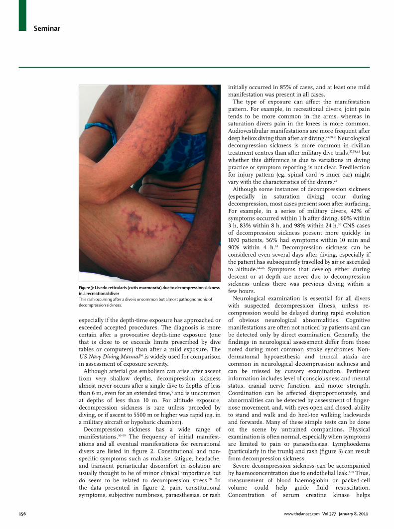
Arterial gas embolism occurs when expanding gas stretches and ruptures alveolar capillaries—pulmonary barotrauma—allowing alveolar gas to enter the arterial circulation (fi gure 1). This syndrome can occur after ascent from a depth as shallow as 1·0–1·5 m if the starting lung volume is close to total lung capacity.1 It can be caused by gas becoming trapped as a result of airways obstruction in disorders such as asthma2 or by the presence of pulmonary blebs, cysts, or bullae.3 Arterial gas embolism can also arise in the absence of decompression through iatrogenic accidents involving vascular catheters and mechanical ventilation.
Decompression sickness starts with the formation and increase in size of extravascular and intravascular bubbles when the sum of the dissolved gas tensions (oxygen, carbon dioxide, nitrogen, helium) and water vapour exceeds the local absolute pressure. In diving and during compressed-air tunnel and caisson work, this state of supersaturation is made possible by the increase in tissue inert gas partial pressure that occurs when the gas (usually nitrogen, but occasionally helium) is respired at high pressure. Supersaturation arises during decompression if the rate of ambient pressure reduction exceeds the rate of inert gas washout from tissue. Ascent to altitude in aviation and extravehicular activity during spacefl ight involves exposure to decreased barometric pressure. In these settings, supersaturation arises as a result of pre-existing dissolved nitrogen at sea level (partial pressure of nitrogen of about 570 mm Hg), which can also cause bubble formation.
Venous gas emboli formed from dissolved gas are easily detected by ultrasonography. In divers, 3·6 m is the
minimum dive depth after which venous gas emboli can be seen4 whereas the decompression sickness threshold, after saturation dives lasting 1–3 days, is about 6 m.5 During direct decompression from sea level to altitude, the threshold for formation of venous gas emboli is around 3600 m whereas the decompression sickness threshold is about 5500 m.6,7
Bubbles can have mechanical, embolic, and biochemical eff ects with manifestations ranging from trivial to fatal. Clinical manifestations can be caused by direct eff ects from extravascular (autochthonous) bubbles such as mechanical distortion of tissues causing pain, or vascular obstruction causing stroke-like signs and symptoms. Secondary eff ects can cause delayed symptom onset up to 24 h after surfacing. Endothelial damage by intravascular bubbles can cause capillary leak, extravasation of plasma, and haemoconcentration.8 Impaired endothelial function, as measured by decreased eff ects of vasoactive compounds, has been reported in animals9 and might occur in man. Hypotension can occur in severe cases.10 Other eff ects include platelet activation and deposition,11 leucocyte-endothelial adhesion,12 and possibly consequences of vascular occlusion believed to occur in thromboembolic stroke such as ischaemia-reperfusion injury, and apoptosis.13
Arterial gas embolism most often aff ects the brain but can occasionally aff ect the heart and other organs.
Search strategy and selection criteria
We searched PubMed in English with the search terms “decompression illness”, “decompression sickness”, and “arterial gas embolism” for reports mostly published in the past 20 years until January, 2010. Bibliographies of selected articles were reviewed for other relevant references. We also relied on our familiarity with key literature. Pertinent review articles, book chapters, proceedings, and papers older than 20 years were used when judged important, but some conclusions are based on anecdotal reports because randomised trials are rare.
Seminar
154 www.thelancet.com Vol 377 January 8, 2011
Decompression sickness produces symptoms related to the eff ects of bubbles on periarticular tissues, spinal cord, brain, lungs, skin, and the audiovestibular system. Concurrent arterial bubbles from arterial gas embolism exacerbate decompression sickness,14 possibly by reducing tissue perfusion and impairing inert gas washout or by increasing bubble size. Decompression sickness symptoms after recreational diving (fi gure 2) typically consist of pain or mild neurological
manifestations such as numbness or paraesthesias. Most patients with altitude-related decompression sickness have similar manifestations,15–18 although cerebral symptoms have been reported in U-2 pilots.19,20
Small quantities of venous gas emboli are common in diving21 although they are usually asymptomatic because most of the time they are eff ectively fi ltered by the pulmonary circulation. However, large numbers of venous gas emboli can cause cough, dyspnoea, and pulmonary oedema (cardiorespiratory decompression sickness, or chokes)22 and can overcome the pulmonary capillary fi lter.23 Moreover, a patent foramen ovale or other right-to-left cardiac shunt is present in about 27% of the normal population,24 and theoretically some venous gas emboli could enter the arterial circulation and reach the CNS, where they could grow from the inward diff usion of supersaturated inert gas.25 Patent foramen ovale has been statistically associated with cerebral, spinal, and vestibulocochlear manifestations,26–36 and with cutaneous manifestations.30
Epidemiology Arterial gas embolism is usually precipitated by rapid ascent, breath-holding, or the presence of lung disease, and thus is rare with an apparently decreasing incidence. The proportion of cases of decompression illness attributable to arterial gas embolism in recreational divers declined from 18% in 1987 to 8% in 1997.37 Of 441 confi rmed or possible incidents of decompression illness in recreational divers reported to the Divers Alert Network, only 3·9% were classifi ed as possible arterial gas embolism.38
If appropriate decompression procedures are followed, decompression sickness is also uncommon. Rate of occurrence (per dive) in operational open water dives from minutes to several hours in duration varies according to the diving population: typically 0·015% for scientifi c divers, 0·01–0·019% for recreational divers, 0·030% for US Navy divers, and 0·095% for commercial divers.39,40 The number of active worldwide recreational divers is not known but is likely to be in the millions. The Divers Alert Network took a sample of 135 000 dives by 9000 recreational divers in which the rate of occurrence of decompression sickness was 0·03%. This rate was much higher during dives to wrecks in cold water than during dives in warm water from diving cruise vessels.38 These numbers are all based on many dives made well within the maximum exposure limits of accepted procedures (decompression tables or computers) and therefore are underestimates of the true rates at the maximum limits. For example, the rate of occurrence of decompression sickness for US Navy dives from 1971 to 1978 at the maximum limits was 1·3%.41 Moreover, for long exposures under stressful thermal and exercise conditions, US Navy dive trials designed to develop new decompression procedures had an occurrence rate of 4·4 cases of decompression sickness per 100 dives.42
Figure 1: Pulmonary barotrauma in a diver during breath-hold ascent
Cerebral air embolism
Subcutaneous emphysema
Pneumomediastinum
Air enters pulmonary capillary
Pneumothorax
Rupture of alveoli
Normal alveoli
Expansion of alveoli

Seminar
www.thelancet.com Vol 377 January 8, 2011 155
Technical diving, a form of recreational diving with deep, long exposures, could be associated with a higher incidence of decompression sickness and more serious manifestations than are the other types of diving, although insuffi cient data are available for accurate estimates. Commercial diving for construction and off shore oil production is another important diving activity. Decompression sickness is reportedly rare in modern commercial saturation dives (working dives lasting several days, with one, extended decompression) although documentation for commercial diving and for compressed air and caisson work is insuffi cient. For hyperbaric medicine attendants, the occurrence rate of decompression sickness is reported as 0·02% per exposure.43,44 In altitude training or fl ight operations, the rate of occurrence of decompression sickness is typically less than 0·1% per exposure, with most individuals reporting only mild symptoms.16,17 However, anonymous surveys of high-altitude Air Force pilots indicated higher frequencies,18 with some cases being quite serious.19,20
Risks of decompression sickness that are thought to be acceptable are a matter of subjective judgment. Acceptable risks specifi ed for commercial diving include 0·1% for mild cases and 0·025% for serious cases,39 whereas for US Navy diving, acceptable risk is 2% for mild cases and 0·1% for serious cases.45
Other than depth and time, risk of decompression sickness is aff ected by other factors that aff ect inert gas exchange and bubble formation, such as immersion (vs dry hyperbaric chamber exposure), exercise, and temperature. Immersion decreases venous pooling and increases venous return and cardiac output.46,47 Warm environments improve peripheral perfusion by promoting vasodilation, whereas cool temperatures decrease perfusion through vasoconstriction. Exercise increases both peripheral perfusion and temperature. The eff ect of environmental conditions on risk of decompression sickness is dependent on the phase of the pressure exposure.39 While under pressure, exercise, immersion, or a hot environment increase inert gas uptake and risk of decompression sickness. During decompression these factors increase inert gas elimination and therefore decrease the risk of decompression sickness.48 Conversely, uptake is reduced during rest or in a cold environment, hence a diver resting in a cold environment on the bottom has decreased risk of decompression sickness. Rest or low temperatures during decompression increase the risk. If exercise occurs after decompression when super-saturation is present, bubble formation increases and risk of decompression sickness rises.39,49
Exercise at specifi c times before a dive can decrease the risk of serious decompression sickness in animals and incidence of venous gas emboli in both animals and man.50–54 The mechanisms of these eff ects are unknown but might involve modulation of nitric oxide production and eff ects on endothelium. Whether appropriately
timed exercise can decrease the probability of decompression sickness in man is unknown. Venous gas emboli and risk of decompression sickness increase slightly with age and body-mass index,21,39 but the eff ect of sex is uncertain.7,55
DiagnosisThe protean nature of decompression illness makes diagnosis diffi cult. Diagnosis is made on a clinical basis, thus accurate history and physical examination of individuals with symptoms after diving or altitude exposure are crucial.
Arterial gas embolism should be suspected if a diver has a new onset of altered consciousness, confusion, focal cortical signs, or seizure during ascent or within a few minutes after surfacing from a compressed gas dive. If the diver spends much time at depth and might have absorbed substantial inert gas before surfacing, arterial gas embolism and serious decompression sickness can coexist, and in such cases, spinal cord manifestations can predominate.14 Other organ systems, such as the heart, can also be aff ected, but the clinical diagnosis of gas embolism is not reliable without CNS manifestations. Arterial gas embolism is rare in altitude exposure; if cerebral symptoms occur after altitude exposure, the cause is usually decompression sickness.
The diagnosis of decompression sickness is based entirely on clinical manifestations. Any new symptom arising shortly after decompression should be considered as possible decompression sickness,
Figure 2: Classifi cation of initial and of all eventual manifestations of decompression illness in 2346 recreational diving accidents reported to the Divers Alert Network from 1998 to 2004 *For all instances of pain, 58% consisted of joint pain, 35% muscle pain, and 7% girdle pain. Girdle pain often portends spinal cord involvement. †Constitutional symptoms included headache, lightheadedness, inappropriate fatigue, malaise, nausea or vomiting, and anorexia. ‡Muscular discomfort included stiff ness, pressure, cramps, and spasm but excluded pain. §Pulmonary manifestations included dyspnoea and cough.
40·668·0
27·463·4
13·640·8
6·119·4
3·818·7
3·49·5
1·36·5
1·27·9
0·95·6
0·87·9
0·41·8
0·32·1
0·31·8
0·042·8
0·040·4
0 20 40 60 80 100Occurrence (% of patients)
Cardiovascular
Bladder, bowel
Lymphatic
Auditory
Consciousness
Coordination
Pulmonary§
Mental status
Muscular discomfort‡
Cutaneous
Motor weakness
Dizziness/vertigo
Constitutional†
Numbness, paraesthesias
Pain*
All symptomsFirst symptom
Seminar
156 www.thelancet.com Vol 377 January 8, 2011
especially if the depth-time exposure has approached or exceeded accepted procedures. The diagnosis is more certain after a provocative depth-time exposure (one that is close to or exceeds limits prescribed by dive tables or computers) than after a mild exposure. The US Navy Diving Manual56 is widely used for comparison in assessment of exposure severity.
Although arterial gas embolism can arise after ascent from very shallow depths, decompression sickness almost never occurs after a single dive to depths of less than 6 m, even for an extended time,5 and is uncommon at depths of less than 10 m. For altitude exposure, decompression sickness is rare unless preceded by diving, or if ascent to 5500 m or higher was rapid (eg, in a military aircraft or hypobaric chamber).
Decompression sickness has a wide range of manifestations.56–59 The frequency of initial manifest-ations and all eventual manifestations for recreational divers are listed in fi gure 2. Constitutional and non-specifi c symptoms such as malaise, fatigue, headache, and transient periarticular discomfort in isolation are usually thought to be of minor clinical importance but do seem to be related to decompression stress.60 In the data presented in fi gure 2, pain, constitutional symptoms, subjective numbness, paraesthesias, or rash
initially occurred in 85% of cases, and at least one mild manifestation was present in all cases.
The type of exposure can aff ect the manifestation pattern. For example, in recreational divers, joint pain tends to be more common in the arms, whereas in saturation divers pain in the knees is more common. Audiovestibular manifestations are more frequent after deep heliox diving than after air diving.25,58,61 Neurological decompression sickness is more common in civilian treatment centres than after military dive trials,57,58,62 but whether this diff erence is due to variations in diving practice or symptom reporting is not clear. Predilection for injury pattern (eg, spinal cord vs inner ear) might vary with the characteristics of the divers.25
Although some instances of decompression sickness (especially in saturation diving) occur during decompression, most cases present soon after surfacing. For example, in a series of military divers, 42% of symptoms occurred within 1 h after diving, 60% within 3 h, 83% within 8 h, and 98% within 24 h.56 CNS cases of decompression sickness present more quickly: in 1070 patients, 56% had symptoms within 10 min and 90% within 4 h.63 Decompression sickness can be considered even several days after diving, especially if the patient has subsequently travelled by air or ascended to altitude.64–66 Symptoms that develop either during descent or at depth are never due to decompression sickness unless there was previous diving within a few hours.
Neurological examination is essential for all divers with suspected decompression illness, unless re-compression would be delayed during rapid evolution of obvious neurological abnormalities. Cognitive manifestations are often not noticed by patients and can be detected only by direct examination. Generally, the fi ndings in neurological assessment diff er from those noted during most common stroke syndromes. Non-dermatomal hypoaesthesia and truncal ataxia are common in neurological decompression sickness and can be missed by cursory examination. Pertinent information includes level of consciousness and mental status, cranial nerve function, and motor strength. Coordination can be aff ected disproportionately, and abnormalities can be detected by assessment of fi nger-nose movement, and, with eyes open and closed, ability to stand and walk and do heel-toe walking backwards and forwards. Many of these simple tests can be done on the scene by untrained companions. Physical examination is often normal, especially when symptoms are limited to pain or paraesthesias. Lymphoedema (particularly in the trunk) and rash (fi gure 3) can result from decompression sickness.
Severe decompression sickness can be accompanied by haemoconcentration due to endothelial leak.8,10 Thus, measurement of blood haemoglobin or packed-cell volume could help guide fl uid resuscitation. Concentration of serum creatine kinase helps
Figure 3: Livedo reticularis (cutis marmorata) due to decompression sickness in a recreational diverThis rash occurring after a dive is uncommon but almost pathognomonic of decompression sickness.

Seminar
www.thelancet.com Vol 377 January 8, 2011 157
distinguish arterial gas embolism from decompression sickness (enzyme concentration in severe cases can be high in pulmonary barotrauma with arterial gas embolism67), although diff erentiation between these disorders before recompression is unnecessary since recompression is indicated for both. Chest radiography is useful for detection of pneumothorax after a suspected arterial gas embolism. For detection of extrapulmonary air, chest CT is more sensitive but is unnecessary because of its high radiation exposure.
Bubbles are rarely detectable with radiography in joints aff ected by pain, and are rarely noted in the brain with either MRI or CT.68 MRI is similarly not useful for detection of abnormalities of the spinal cord related to decompression sickness.69 Thus, although laboratory and radiological analyses are useful for detection of abnormalities in some cases, imaging studies are not useful for assessment of whether a patient needs recompression treatment and should not delay recompression unless there is a strong suspicion of a non-diving related cause (eg, cerebral haemorrhage). Specifi c neurophysiological tests (eg, audiometry and electronystagmography for inner-ear decompression sickness) or imaging can usually be delayed until after recompression. Doppler ultrasonography and echo cardio graphy are valuable for research into venous gas emboli4,21,51–54,70 but not for diagnosis of decompression illness. The diff erential diagnosis of decompression illness includes acute coincidental illness, especially neurological disorders. Specifi c disorders that could be confused with decompression illness are listed in the panel.
Decompression injuries have traditionally been classifi ed as arterial gas embolism, and type 1 and type 2 decompression sickness. Type 1 decompression sickness included pain, cutaneous manifestations, and constitu-tional symptoms, whereas type 2 manifestations includ-ed numbness, tingling, paraesthesias, muscle weakness, paralysis, and mental or motor abnormalities.81 Arterial gas embolism and type 2 decompression sickness were thought to require more aggressive treatment than type 1 decompression sickness. Because the traditional classifi cation system was applied inconsistently and had little predictive value, it has been largely replaced by the inclusive term decompression illness.82 Although identifi cation of the pathophysio logical changes in individuals is useful for epidemi ology and recommen-dations for future diving, the correct pathophysiological changes often cannot be identifi ed and are not usually important for selection of treatment.
Other classifi cation systems that have been proposed were for the creation of severity indices that might be useful to guide treatment and prediction of outcome.83–89 These approaches showed reasonable associations between severity at presentation and outcome,69,85,90–94 but need to be more consistent and simpler for their potential to be reached.95–97
PreventionArterial gas embolism is rare at altitude and is not related to depth-time exposure in diving. The risk of this syndrome can be decreased by avoidance of breath holding, rapid ascent, and diving with pulmonary infections or disease. Risk of decompression sickness is decreased by reduction of exposure or by elimination of inert gas before (eg, with high oxygen concentrations) or during decompression, but adherence to these procedures does not always prevent the syndrome. 100 years ago, serious manifestations and deaths were frequent in diving and caisson work but they decreased greatly when decompression stops were introduced. These stops delay ascent to the surface and allow inert gases to be eliminated in dissolved form rather than as bubbles.6,99 A
Panel: Diff erential diagnosis of decompression illness
Inner-ear barotraumaInner-ear barotrauma usually occurs during descent and results in tinnitus, hearing loss, and persistent vertigo.71,72 Conductive hearing loss is seen in middle-ear barotrauma. Both inner-ear and middle-ear barotrauma are usually preceded by diffi culty in equalising middle-ear pressure. Transient vertigo during compression or decompression can arise because of asymmetric middle-ear pressure equilibration (alternobaric vertigo).61
Middle-ear or maxillary sinus overinfl ationThis disorder is caused by gas expansion during ascent and an obstructed eustachian tube or sinus ostium, resulting in compression of the facial nerve and unilateral upper and lower facial weakness,73 or compression of branches of the trigeminal nerve causing hypoaesthesia of the face.74
Contaminated diving gas and oxygen toxic eff ectsCarbon monoxide poisoning due to contaminated breathing gas can cause encephalopathy and convulsions. Toxic eff ects of oxygen are most common in divers using enriched oxygen breathing mixtures and can cause convulsions at depth.
Musculoskeletal strains or trauma sustained before, during, or after diving56,58,74–76
Time of onset and history of trauma or strain are helpful. Pain due to decompression illness is rarely accompanied by tenderness or position-related or motion-related exacerbation physical examination.
Seafood toxin ingestion (ciguatera, puff er fi sh, paralytic shellfi sh poisoning)Ingestion of toxins is often associated with gastrointestinal symptoms and can cause neurological manifestations within hours after ingestion.77
Immersion pulmonary oedemaThis disorder usually occurs shortly after the start of a dive, while the diver is still at depth, and might be confused with cardiorespiratory decompression sickness, since both cause dyspnoea and cough.78–80 Symptoms of immersion pulmonary oedema typically begin during descent or at depth, whereas the onset of cardiorespiratory decompression sickness occurs after the dive.
Water aspirationWater aspiration could be mistaken for cardiorespiratory decompression sickness. Both cardiorespiratory decompression sickness and water aspiration can cause pulmonary oedema, although the diver is usually aware of aspiration.
Coincidental, unrelated acute neurological disorder (eg, stroke, spinal hematoma)Diagnosis is made with conventional techniques.

Seminar
158 www.thelancet.com Vol 377 January 8, 2011
decompression schedule specifi es the stop times typically at 3 m intervals during ascent according to the maximum dive depth and bottom time. In the 1980s, equally eff ective diver-worn digital computers (dive computers) were developed that continuously track depth-time exposure and specify how slowly a diver should ascend. Gas mixtures with high oxygen partial pressures are sometimes used to decrease inert gas absorption at depth and for faster elimination during decompression. For dives deeper than 45 m, gas mixtures containing helium are used to avoid nitrogen narcosis and to decrease the density of respired gas.
Detection of patent foramen ovale as a way of reducing the risk of serious decompression sickness has been of much interest. Estimates of relative risk for serious neurological decompression sickness associated with a patent foramen ovale range from 2·5 to 6·6.32 Nevertheless, routine screening of candidate divers for patent foramen ovale does not seem warranted since the absolute risk of neurological decompression sickness is small (<0·02%),34 and the cost of screening is high.
Decompression sickness is rare after rapid ascent to altitudes lower than 5500 m with increasing risk at higher altitudes,7 unless the altitude exposure is preceded by diving within several days, in which case decompression sickness can occur at less than 2500 m.64,99,100 Oxygen breathing at sea level before altitude exposure eliminates dissolved tissue nitrogen and permits higher altitudes and longer fl ights. For extravehicular activity during spacefl ight, protocols have been developed that use oxygen breathing and exercise before the activity to accelerate nitrogen elimination before decompression to space-suit pressures of 0·3–0·4 bar.39
TreatmentDecompression illness is rare and only one prospective randomised trial of treatment has been reported so far.101 The following guidelines are therefore based largely on case reports, case series, animal studies, and clinical judgment, and have empirically changed in the past 60 years, especially with respect to fi rst aid and adjunctive treatment.
The principles of basic and advanced life support apply to any obtunded diver, but manifestations of decompression illness are typically mild and non-specifi c, although potentially progressive. Appropriate fi rst aid should be applied as soon as possible. Assistance with diagnosis or management can be obtained from various diving medical services, such as the Divers Alert Network or Duke Dive Medicine.
The best and primary fi rst aid for decompression illness is 100% oxygen delivered for several hours even if manifestations resolve. Pure oxygen washes inert gas from the lungs and establishes the largest possible inert gas gradient from tissue to alveolar gas. This gradient results in rapid removal of inert gas from tissue to lungs by perfusion and from bubble to tissue by diff usion, and
thus removal of bubbles.102 Another advantage of pure oxygen is amelioration of tissue hypoxia caused by bubble-induced ischaemia, mechanical injury, or biochemical damage. In an observational study, patients with decompression sickness who received oxygen during fi rst aid had symptom resolution after fewer recompressions than did those who did not receive oxygen.103 Oxygen given under slight pressure while a diver is returned to the water might be appropriate under some circumstances but is controversial because of the increased risk of seizures caused by toxic eff ects of oxygen on the CNS.104
Although the head-down position has been advocated to prevent distribution of arterial bubbles towards the head, it is not eff ective and can promote cerebral oedema.105 Consensus is for horizontal orientation in a position that helps care of the patient. Fluid administration is important and intravenous fl uids can be benefi cial, especially in serious cases, but few clinical data show benefi t from a specifi c type or amount of intravenous fl uid. Glucose-containing fl uids are best avoided because of the potential for adverse eff ects of hyperglycaemia in neurological injury, and hypotonic fl uids should not be used because they promote intracellular oedema.106 Substantial fl uid resuscitation is not recommended in patients with isolated arterial gas embolism—eg, a patient who suff ers gas embolism after a short, shallow dive.106 Oral rehydration can be used in stable, conscious patients, but is unlikely to help those in greatest need. Nitrous oxide is inappropriate for pain relief or as a component of a general anaesthetic for a coincidental procedure because it can cause an increase in the size of bubbles by inward diff usion.107
Typically, recompression is done in a multiplace chamber in which the diver is accompanied by one or more attendants (fi gure 4). Decompression illness has also been successfully treated in one-occupant monoplace chambers.108 Recompression while breathing 100% oxygen decreases bubble volume as predicted by Boyle’s law and increases the inert gas partial pressure gradients between tissue and alveolar gas. These eff ects lead to quick resolution of bubbles, relieve mechanical pressure on surrounding tissue, and encourage redistribution of bubbles lodged in the microcirculation.109 Hyperbaric oxygen also oxygenates compromised tissues and ameliorates infl ammatory responses that contribute to tissue injury.12
Recompression is usually advised even if manifestations resolve with fi rst aid since untreated decompression sickness can recur days after the initial onset.110 Evacuation for recompression might be needed, especially from remote locations.111 For short distances, this evacuation could be by helicopter at low altitude, but for long distances, an air ambulance pressurised to 1 atm is usually recommended in severe cases.111,112 When pain is the only symptom, short commercial fl ights seem to have no eff ect on outcome; when mild neurological symptoms
For the Divers Alert Network see www.diversalertnetwork.org
For Duke Dive Medicine see www.dukedivemedicine.org

Seminar
www.thelancet.com Vol 377 January 8, 2011 159
are present, short fl ights more than 24 h after diving also seem to be safe.113 Mild initial manifestations can develop into a more serious form, usually within a few hours after surfacing. In serious cases, delayed recompression is probably less eff ective, but the time beyond which recompression is pointless is unknown.94 Decisions about the advisability of recompression should ideally include a physician trained in diving medicine.
Recent decompression and manifestations compatible with decompression illness usually justify emergent recompression with oxygen in a pressure chamber (fi gure 4) unless another cause is obvious.56 Recompres-sion should occur as soon as feasible to avoid late recurrence and increased severity.105,110 Treatment is recommended even in patients with substantially delayed presentation since clinical response often occurs hours or even days after onset.114–116 Some patients need ventilator support and intravenous drug infusion during recompression (fi gure 4).
The most common recompression schedule is US Navy Treatment Table 6 (fi gure 5) or an equivalent procedure such as promulgated by the Royal Navy, in which patients are compressed to 2·8 bar (equivalent to 18 m sea water depth) while breathing 100% oxygen, a pressure with an acceptably low risk of cerebral oxygen-associated toxic eff ects.56,117 The time at 2·8 bar and 1·9 bar (equivalent to 9 m sea water) can be extended with additional cycles of oxygen and air if resolution is not complete within the prescription shown in fi gure 5. If treatment pressures are greater than 2·8 bar, air is used or nitrogen or helium is added to the breathing mix to reduce the risk of oxygen-associated toxic eff ects. In animal studies, faster bubble resolution has been shown with helium than with oxygen,102 but this comparison has been inadequately studied in clinical practice. Anecdotally, increased pressures can improve manifestations that are refractory at 2·8 bar,118 and they have been advocated for both arterial gas emboli (when severe symptoms remain unchanged or worsen within the fi rst 20 min at 2·8 bar)56 and decompression sickness.119 However, supporting evidence is weak for depths greater than 18 m for initial recompression without a demonstrated need to go deeper, and no benefi t has been shown in animal studies.120,121 Many recompression strategies ranging from pressures of 1·9–10·0 bar exist, but there are no human outcome studies for comparison of effi cacy. Short treatment schedules have been tested, and seem eff ective.83,108
If resolution is complete after one treatment, no additional treatments are needed. With residual manifestations after the fi rst treatment, recompression is typically repeated every day (often with short treatment tables such as the US Navy Treatment Table 5)56 until complete symptom resolution or no further improve-ment.105 Most patients with residual neurological manifestations need only two or three treatments to reach a clinical plateau.101 Nonetheless, some severe cases
(eg, important motor weakness) do not reach a plateau until after 15–20 repetitive treatments. In decompression sickness in which the predominant manifestations are sensory or pain, symptoms often wax and wane every day. Unless improvement after each treatment is documented, prolonged hyperbaric treatment is unnecessary.
A few facilities are equipped and staff ed to provide saturation therapy, in which the chamber and its occupants are maintained at a constant elevated ambient pressure (eg, 2·8 bar for US Navy Treatment Table 7).56,122 Decompression starts after clinical resolution or stability is achieved, often in 2–3 days. Saturation treatment is usually reserved for cases of severe neurological impairment, in which resolution was incomplete during initial treatment or deterioration occurred during decompression. Data do not suggest that the outcome for saturation treatment is better than that with repetitive short treatments.
Administration of 100% oxygen at 1 atm is often suffi cient treatment for mild decompression sickness after altitude exposure without a preceding dive.123 Recompression should ideally be off ered to all divers with suspected decompression sickness. If recom-pression facilities are remote and the patient has mild
Figure 4: Recompression chamber(A) Multiplace recompression chamber. The chamber can typically be compressed to 6 bar with air. Treatment gas (oxygen, oxygen and nitrogen, or oxygen and helium mixes) is given to the patient via a head tent, tightly fi tting mask or, for critically ill patients, through endotracheal tube. Photo from SJM. (B) Critical care in a multiplace chamber. A fl uidic or pneumatic ventilator is shown at the left. The infusion pump is contained within a plastic cover, in which 100% nitrogen is used to decrease the fi re risk in the event of an electrical problem. The monitor screen is outside the chamber and can be seen through the viewing port. Photo from Duke University Medical Center, with permission.
Figure 5: US Navy Treatment Table 6From US Navy Diving Manual.56 fsw=feet sea water.
A B
5 15 1520 520 520 30 3060 6060
45
30
15
0
Dept
h (fs
w)
3Time at depth (min)
Descent rate20 ft/min
Ascent rate1 ft/min
Ascent rate1 ft/min
100% oxygenAirOxygen or air
Total elapsed time:285 min4 h 45 min(not including descent time)

Seminar
160 www.thelancet.com Vol 377 January 8, 2011
decompression illness (defi ned as static or remitting limb pain, constitutional symptoms, rash, or non-dermatomal sensory changes that remain stable for 24 h with a normal neurological assessment), the outcome is unlikely to be worse without recompression.111 In a remote location, the risk-benefi t ratio of a hazardous evacuation might not be favourable compared with delayed recompression or even no recompression.
Decompression sickness during decompression from a saturation dive (commercial or military) is usually treated with a small recompression to the pressure at which symptoms are relieved and by administration of oxygen-enriched breathing gas (oxygen partial pressure 1·5–2·8 bar).56
Although recompression is the primary treatment, especially for serious decompression illness, other aspects of care for seriously ill patients should not be neglected—eg, management of airway compromise, coma, haemo-dynamic instability, temperature control, metabolic instability, bladder dysfunction, pain, the risks of immobility, and long-term disability. In patients with leg immobility, prophylaxis is recommended because of the substantial risk of venous thromboembolism.106
Asymptomatic diving routinely causes slight haemoconcentration suggesting dehydration.124 In animals, dehydration can increase the seriousness of decompression sickness,125 and in man, adequate hydration decreases bubble formation after decom-pression.70 Severe decompression sickness can cause substantial haemoconcentration and haemodynamic instability, presumably by widespread endothelial damage or infl ammation.10,126 Such fi ndings encourage the routine use of intravenous rehydration with non-glucose isotonic crystalloid fl uids. Fluid overload is best avoided because it can contribute to cerebral, spinal cord, or pulmonary oedema.
Specifi c drugs have been used as adjuncts to recompression. In a randomised trial, the non-steroidal anti-infl ammatory drug tenoxicam decreased the number
of recompressions needed to achieve symptom resolution or recovery plateau but did not change fi nal outcome.101 The prevalence of use of non-steroidal anti-infl ammatory drugs in this context is unknown. Aspirin has previously been advocated for its antiplatelet eff ects, but it has not been formally studied in this context and is not recommended.106
Interest in intravenous lidocaine for treatment of neurological decompression illness arose out of this drug’s apparent effi cacy in vivo and anecdotal eff ectiveness in divers.127 Neuroprotection has been reported in patients undergoing cardiac surgery,128 but there are no studies in divers, and results of trials in cardiac surgery did not show repeat of initial success.129,130 Lidocaine is usually reserved for serious neurological cases with features typical of arterial gas embolism. High-dose steroids worsen outcome in animals131 and are not recommended in people.106
A promising prospect is intravenous perfl uorocarbon emulsions132 because both oxygen and inert gases are very soluble in these compounds. Perfl uorocarbon emulsions probably work by enhancement of tissue oxygenation and inert gas transport from tissue to lungs.133 Tissue bubbles shrank more rapidly with infusion of perfl uorocarbon emulsions and oxygen breathing than with oxygen breathing alone in a mouse model of decompression sickness.134 Moreover, in a pig model of this disease, morbidity and mortality were greatly reduced with infusion of perfl uorocarbon emulsions at sea level.135 Perfl uorocarbon emulsions and hyperbaric oxygen have not been studied but hypothetically the combination could increase risk of oxygen-related toxic eff ects. The availability of a perfl uorocarbon emulsion that is suitable for use in man is awaited before trials can be done.
OutcomeWhen oxygen treatment tables are used with an initial treatment pressure of 2·8 bar and the delay to treatment is not excessive, symptoms are resolved with a high degree of success.105,108,136,137 67% of 63 divers with spinal cord decompression sickness had complete resolution at 1 month after treatment. Of 30 patients in the same series with motor weakness, cerebral involvement, or cochleovestibular manifestations, only eight (27%) had severe disability 1 month after treatment.94 In a study of 268 patients, including both amateur and professional divers, 230 (86%) had complete resolution or minor symptoms at discharge.93 In a review of 1763 cases of decompression sickness, including several cases with long periods to treatment, 80% showed complete recovery.136 Of 166 patients with decompression sickness resulting from experimental dives, with recompression facilities immediately at hand, 97% had complete relief after initial hyperbaric treatment, and all patients eventually showed complete resolution, despite some serious cases of neurological and cardiorespiratory disorders.136 Long-term outcome data of 69 recreational divers with severe spinal cord decompression sickness are listed in the table.
n %
No residual symptoms 34 49·3%
Any residual symptom 35 50·7%
Mild paraesthesias, weakness, or pain 14 20·3%
Some impairment of daily activities 21 30·4%
Diffi culty walking 11 15·9%
Impaired micturition 13 18·8%
Impaired defecation 15 21·7%
Impaired sexual function 15 21·7%
Data are taken from 51 men, 18 women; mean age 41·2 years (range 19–70 years). Divers were assessed by phone interview after a median of 6·1 years (range 2·3–9·7 years). All but one had received recompression treatment. (Data from the Divers Alert Network.)
Table: Long-term outcomes of 69 divers with spinal cord decompression sickness, by manifestation

Seminar
www.thelancet.com Vol 377 January 8, 2011 161
Guidelines suggest that patients treated for decompression illness should be observed within timely reach of a chamber for 2 h when symptoms are mild or 6 h for severe symptoms.56 US Navy guidelines suggest that patients should be within 1 h travelling time for 24 h after recompression.56 Hospital admission might be necessary for patients with severe or residual symptoms. Worsening or recurrence suggests a need for immediate re-evaluation. Altitude exposure (eg, commercial airplane fl ight) after decompression illness can precipitate recurrence of symptoms. Generally, patients who were recompressed with complete relief should not fl y for at least 72 h. Consultation with a trained hyperbaric physician is suggested before individuals with residual symptoms are allowed to fl y.56
Recompression treatment results in complete resolution in most cases, mild residual symptoms in a few cases, and rarely serious residual manifestations. No follow-up diagnostic studies are necessary for patients whose only symptoms are mild (ie, tingling or joint pain) and resolved wholly. Patients whose symptoms are initially serious or are not fully resolved are usually reviewed within a few weeks of discharge. This patient assessment allows documentation of progress and discussion about future diving. Resumption of diving for recreational divers is usually allowed 4 weeks after treatment with complete recovery. Military and commercial diving organisations have similar guidelines for arterial gas embolism or neurological decompression sickness, but allow diving after a few days (7 days for the US Navy) after mild manifestations such as joint pain. Return to fl ying after pain-only decompression sickness that has fully resolved is also typically allowed after a few days (14 days for the US Air Force). Cautious discussion of risk and benefi t is appropriate, especially if decompression sickness occurred after a conservative depth-time exposure (ie, one well within the maximum time allowed by an established decompression table or computer). If arterial gas embolism due to pulmonary barotrauma is suspected, radiological investigation and pulmonary function testing are usually done to exclude underlying pulmonary predisposition.
In severe cases, assessment with MRI of the brain and spinal cord after treatment can show abnormalities, although normal fi ndings do not exclude decompression illness.58,139 Positive MRI fi ndings are related to severity and outcome.69 Some individuals with initially normal MRI examinations develop late changes.139 Similarly, nuclear imaging of the brain (eg, PET) is less sensitive than is clinical evaluation for detection of abnormalities.140
For divers with vestibular decompression sickness, compensation and symptom resolution will occur, and physical examination will return to normal, even with residual end-organ damage. Although cochlear involvement (hearing impairment) remains, it is diffi cult to detect on cursory examination. Therefore, for inner-ear decompression sickness, formal audiometry and vestibular
tests (electronystagmography with rotary chair and caloric stimulation) are recommended at 4–6 weeks after injury.
Assessment of patent foramen ovale is often recommended for divers with severe or recurrent neurological decompression sickness.29,141 For patent foramen ovale to be a precipitating factor for decompression sickness a substantial amount of venous gas emboli should be present, which is unlikely to happen in conservative depth-time profi les.21,142 Patent foramen ovale or other right-to-left shunts can be detected by bubble contrast injection in conjunction with transcranial doppler, transoesophageal echocardiography, or transthoracic echocardiography. Because intravenous injection of bubbles in the presence of inert gas supersaturation could facilitate endogenous bubble growth, tests of this sort should be done only after completion of all hyperbaric treatments. Although transoesophageal echocardiography is generally the most sensitive test, the relevance of small shunts detectable only with this test is not known. Moreover, new transthoracic echocardiography techniques seem to have similar sensitivity.143 Detection of a patent foramen ovale could warrant counselling about future diving, aimed at prevention of venous gas emboli.21,142 Some divers who wish to return to unrestricted diving opt to have the patent foramen ovale repaired with a transvenous septal occluder device, but a careful discussion of risks and benefi ts is needed before this procedure can be recommended.138,144,145 Patent foramen ovale is a common anomaly in the general population, and many individuals with decompression sickness do not have it. Therefore, a causal link between patent foramen ovale and an individual case of decompression sickness should not be ascribed.
ConclusionsDecompression illness occurs in a small population but is an international problem that few physicians are trained to recognise or manage. Although its manifestations are often mild, the potential for permanent injury exists in severe cases, especially if unrecognised or inadequately treated. Emergency medical personnel should be aware of manifestations of decompression illness in the setting of a patient with a history of recent diving or other exposure to substantial pressure change, and should contact an appropriate consultation service for advice.
ContributorsAll authors contributed equally to the writing of this Review.
Confl icts of interestThe authors received no revenue in support for this manuscript. RDV
and REM received research grant funding from the US Navy. A portion
of RDV’s salary is paid by the Divers Alert Network. All other authors
declare that they have no confl icts of interest.
AcknowledgmentsWe thank Stan Coff man from MedMedia Solutions, Durham, NC, USA
for supplying fi gure 1 and Petar J Denoble for preparing data of
decompression illness from the Divers Alert Network on which fi gure 2
is based.

Seminar
162 www.thelancet.com Vol 377 January 8, 2011
References1 Benton PJ, Woodfi ne JD, Westwook PR. Arterial gas embolism
following a 1-meter ascent during helicopter escape training: a case report. Aviat Space Environ Med 1996; 67: 63–64.
2 Weiss LD, Van Meter KW. Cerebral air embolism in asthmatic scuba divers in a swimming pool. Chest 1995; 107: 1653–54.
3 Mellem H, Emhjellen S, Horgen O. Pulmonary barotrauma and arterial gas embolism caused by an emphysematous bulla in a SCUBA diver. Aviat Space Environ Med 1990; 61: 559–62.
4 Eckenhoff RG, Olstad CS, Carrod G. Human dose-response relationship for decompression and endogenous bubble formation. J Appl Physiol 1990; 69: 914–18.
5 Van Liew HD, Flynn ET. Direct ascent from air and N2-O2 saturation dives in humans: DCS risk and evidence of a threshold. Undersea Hyperb Med 2005; 32: 409–19.
6 Vann RD. Inert gas exchange and bubbles. In: Bove AA, ed. Bove and Davis’ diving medicine, 4th edn. Philadelphia, PA: Saunders, 2004: 53–76.
7 Webb JT, Kannan N, Pilmanis AA. Gender not a factor for altitude decompression sickness risk. Aviat Space Environ Med 2003; 74: 2–10.
8 Boussuges A, Blanc P, Molenat F, Bergmann E, Sainty JM. Haemoconcentration in neurological decompression illness. Int J Sports Med 1996; 17: 351–55.
9 Nossum V, Hjelde A, Brubakk AO. Small amounts of venous gas embolism cause delayed impairment of endothelial function and increase polymorphonuclear neutrophil infi ltration. Eur J Appl Physiol 2002; 86: 209–14.
10 Brunner FP, Frick PG, Bühlmann AA. Post-decompression shock due to extravasation of plasma. Lancet 1964; 283: 1071–73.
11 Bosco G, Yang ZJ, Savini F, et al. Environmental stress on diving-induced platelet activation. Undersea Hyperb Med 2001; 28: 207–11.
12 Martin JD, Thom SR. Vascular leukocyte sequestration in decompression sickness and prophylactic hyperbaric oxygen therapy in rats. Aviat Space Environ Med 2002; 73: 565–69.
13 Brouns R, De Deyn PP. The complexity of neurobiological processes in acute ischemic stroke. Clin Neurol Neurosurg 2009; 111: 483–95.
14 Neuman TS, Bove AA. Combined arterial gas embolism and decompression sickness following no-stop dives. Undersea Biomed Res 1990; 17: 429–36.
15 Weien RW, Baumgartner N. Altitude decompression sickness: hyperbaric therapy results in 528 cases. Aviat Space Environ Med 1990; 61: 833–36.
16 Bason R, Yacavone D. Decompression sickness: U.S. Navy altitude chamber experience 1 October 1981 to 30 September 1988. Aviat Space Environ Med 1991; 62: 1180–84.
17 Bason R, Yacavone D, Bellenkes AH. Decompression sickness: USN operational experience 1969–1989. Aviat Space Environ Med 1991; 62: 994–96.
18 Bendrick GA, Ainscough MJ, Pilmanis AA, Bisson RU. Prevalence of decompression sickness among U-2 pilots. Aviat Space Environ Med 1996; 67: 199–206.
19 Pickard BJ. Altitude decompression sickness in a pilot wearing a pressure suit above 70,000 feet. Aviat Space Environ Med 2003; 74: 357–59.
20 Jersey SL, Baril RT, McCarty RD, Millhouse CM. Severe neurological decompression sickness in a U-2 pilot. Aviat Space Environ Med 2010; 81: 64–68.
21 Dunford RG, Vann RD, Gerth WA, et al. The incidence of venous gas emboli in recreational diving. Undersea Hyperb Med 2002; 29: 247–59.
22 Zwirewich CV, Müller NL, Abboud RT, Lepawsky M. Noncardiogenic pulmonary edema caused by decompression sickness: rapid resolution following hyperbaric therapy. Radiology 1987; 163: 81–82.
23 Vik A, Brubakk AO, Hennessy TR, Jenssen BM, Ekker M, Slordahl SA. Venous air embolism in swine: transport of gas bubbles through the pulmonary circulation. J Appl Physiol 1990; 69: 237–44.
24 Hagen PT, Scholz DG, Edwards WD. Incidence and size of patent foramen ovale during the fi rst 10 decades of life: an autopsy study of 965 normal hearts. Mayo Clin Proc 1984; 59: 17–20.
25 Mitchell SJ, Doolette DJ. Selective vulnerability of the inner ear to decompression sickness in divers with right-to-left shunt: the role of tissue gas supersaturation. J Appl Physiol 2009; 106: 298–301.
26 Wilmshurst PT, Ellis PT, Jenkins BS. Paradoxical gas embolism in a scuba diver with an atrial septal defect. BMJ 1986; 293: 1277.
27 Moon RE, Camporesi EM, Kisslo JA. Patent foramen ovale and decompression sickness in divers. Lancet 1989; 333: 513–14.
28 Wilmshurst PT, Byrne JC, Webb-Peploe MM. Relation between interatrial shunts and decompression sickness in divers. Lancet 1989; 334: 1302–06.
29 Germonpré P, Dendale P, Unger P, Balestra C. Patent foramen ovale and decompression sickness in sports divers. J Appl Physiol 1998; 84: 1622–26.
30 Wilmshurst P, Bryson P. Relationship between the clinical features of neurological decompression illness and its causes. Clin Sci 2000; 99: 65–75.
31 Cantais E, Louge P, Suppini A, Foster PP, Palmier B. Right-to-left shunt and risk of decompression illness with cochleovestibular and cerebral symptoms in divers: case control study in 101 consecutive dive accidents. Crit Care Med 2003; 31: 84–88.
32 Torti SR, Billinger M, Schwerzmann M, et al. Risk of decompression illness among 230 divers in relation to the presence and size of patent foramen ovale. Eur Heart J 2004; 25: 1014–20.
33 Kerut EK, Norfl eet WT, Plotnick GD, Giles TD. Patent foramen ovale: a review of associated conditions and the impact of physiological size. J Am Coll Cardiol 2001; 38: 613–23.
34 Bove AA. Risk of decompression sickness with patent foramen ovale. Undersea Hyperb Med 1998; 25: 175-78.
35 Klingmann C, Benton PJ, Ringleb PA, Knauth M. Embolic inner ear decompression illness: correlation with a right-to-left shunt. Laryngoscope 2003; 113: 1356–61.
36 Gempp E, Blatteau JE, Stephant E, Louge P. Relation between right-to-left shunts and spinal cord decompression sickness in divers. Int J Sports Med 2009; 30: 150–53.
37 Vann RD, Uguccioni DM. DAN’s Annual review of recreational scuba diving injuries and fatalities based on 1998 data. Durham, NC: Divers Alert Network, 2000.
38 Pollock NW. Annual diving report: 2008 edition. Durham, NC: Divers Alert Network, 2008.
39 Vann RD. Mechanisms and risks of decompression. In: Bove AA, ed. Bove and Davis’ diving medicine, 4th edn. Philadelphia, PA: Saunders, 2004: 127–64.
40 Ladd G, Stepan V, Stevens L. The Abacus Project: establishing the risk of recreational scuba death and decompression illness. SPUMS J 2002; 32: 124–28.
41 Berghage TE, Durman D. US Navy air decompression schedule risk analysis. Bethesda, MD: Naval Medical Research and Development Command, 1980.
42 Temple DJ, Ball R, Weathersby PK, Parker EC, Survanshi S. The dive profi les and manifestations of decompression sickness cases after air and nitrogen-oxygen dives. Volume I: data set summaries, manifestation descriptions, and key fi les. Washington, DC: Naval Medical Research Center, 1999: NMRC 99–02.
43 Doolette DJ, Goble SJ, Pirone CJ. Health outcome of hyperbaric-chamber inside attendants following compressed-air exposure and oxygen decompression. SPUMS J 2004; 34: 63–67.
44 Cooper PD, Van den Broek C, Smart DR. Hyperbaric chamber attendant safety II: 14-year staff health review of multiplace chamber attendants. Diving Hyperb Med 2009; 39: 71–76.
45 Van Liew HD, Flynn ET. Decompression tables and dive-outcome data: graphical analysis. Undersea Hyperb Med 2005; 32: 187–98.
46 Balldin UI, Lundgren CE. Eff ects of immersion with the head above water on tissue nitrogen elimination in man. Aerosp Med 1972; 43: 1101–08.
47 Balldin UI, Lundgren CEG, Lundvall J, Mellander S. Changes in the elimination of 133Xe from the anterior tibial muscle in man induced by immersion in water and by shifts in body position. Aerosp Med 1971; 42: 489–93.
48 Gerth WA, Ruterbusch VL, Long ET. The infl uence of thermal exposure on diver susceptibility to decompression sickness. Panama City, FL: Navy Experimental Diving Unit, 2007: NEDU TR: 06–07.
49 Van der Aue OE, Kellar RJ, Brinton ES. The eff ect of exercise during decompression from increased barometric pressures on the incidence of decompression sickness in man. Washington, DC: US Navy Experimental Diving Unit, 1949: 8–49.

Seminar
www.thelancet.com Vol 377 January 8, 2011 163
50 Blatteau JE, Boussuges A, Gempp E, et al. Haemodynamic changes induced by submaximal exercise before a dive and its consequences on bubble formation. Br J Sports Med 2007; 41: 375–79.
51 Blatteau JE, Gempp E, Galland FM, Pontier JM, Sainty JM, Robinet C. Aerobic exercise 2 hours before a dive to 30 msw decreases bubble formation after decompression. Aviat Space Environ Med 2005; 76: 666–69.
52 Dujic Z, Duplancic D, Marinovic-Terzic I, et al. Aerobic exercise before diving reduces venous gas bubble formation in humans. J Physiol 2004; 555 (Pt 3): 637–42.
53 Dujic Z, Obad A, Palada I, Ivancev V, Valic Z. Venous bubble count declines during strenuous exercise after an open sea dive to 30 m. Aviat Space Environ Med 2006; 77: 592–96.
54 Dujic Z, Valic Z, Brubakk AO. Benefi cial role of exercise on scuba diving. Exerc Sport Sci Rev 2008; 36: 38–42.
55 St Leger Dowse M, Bryson P, Gunby A, Fife W. Comparative data from 2250 male and female sports divers: diving patterns and decompression sickness. Aviat Space Environ Med 2002; 73: 743–49.
56 Navy Department. US Navy Diving Manual. Revision 6. Vol 5 : Diving Medicine and Recompression Chamber Operations. NAVSEA 0910-LP-106-0957. Washington, DC: Naval Sea Systems Command, 2008.
57 Rivera JC. Decompression sickness among divers: an analysis of 935 cases. Mil Med 1964; 129: 314–34.
58 Francis TJR, Mitchell SJ. Manifestations of decompression disorders. In: Brubakk AO, Neuman TS, eds. Bennett and Elliott’s physiology and medicine of diving. New York, NY: Elsevier Science, 2003: 578–99.
59 Ozyigit T, Egi SM, Denoble P, et al. Decompression illness medically reported by hyperbaric treatment facilities: cluster analysis of 1929 cases. Aviat Space Environ Med 2010; 81: 3–7.
60 Howle LE, Weber PW, Vann RD, Campbell MC. Marginal DCS events: their relation to decompression and use in DCS models. J Appl Physiol 2009; 107: 1539–47.
61 Farmer JC Jr. Diving injuries to the inner ear. Ann Otol Rhinol Laryngol Suppl 1977; 86 (1 Pt 3 suppl 36): 1–20.
62 Newton HB, Padilla W, Burkart J, Pearl DK. Neurological manifestations of decompression illness in recreational divers—the Cozumel experience. Undersea Hyperb Med 2007; 34: 349–57.
63 Francis TJ, Pearson RR, Robertson AG, Hodgson M, Dutka AJ, Flynn ET. Central nervous system decompression sickness: latency of 1070 human cases. Undersea Biomed Res 1988; 15: 403–17.
64 Freiberger JJ, Denoble PJ, Pieper CF, Uguccioni DM, Pollock NW, Vann RD. The relative risk of decompression sickness during and after air travel following diving. Aviat Space Environ Med 2002; 73: 980–84.
65 Barry PD, Vann RD, Youngblood DA, Peterson RE, Bennett PB. Decompression from a deep nitrogen-oxygen saturation dive— a case report. Undersea Biomed Res 1984; 11: 387–93.
66 Bennett PB, Dovenbarger JA, Bond BG, Waccholz CJ. DAN 1987 diving accident incidence for fl ying after diving. In: Sheffi eld PJ, ed. Proceedings of a workshop on fl ying after diving. Bethesda, MD: Undersea Medical Society, 1989: 29–34.
67 Smith RM, Neuman TS. Elevation of serum creatine kinase in divers with arterial gas embolization. N Engl J Med 1994; 330: 19–24.
68 Benson J, Adkinson C, Collier R. Hyperbaric oxygen therapy of iatrogenic cerebral arterial gas embolism. Undersea Hyperb Med 2003; 30: 117–26.
69 Gempp E, Blatteau JE, Stephant E, Pontier JM, Constantin P, Peny C. MRI fi ndings and clinical outcome in 45 divers with spinal cord decompression sickness. Aviat Space Environ Med 2008; 79: 1112–16.
70 Gempp E, Blatteau JE, Pontier JM, Balestra C, Louge P. Preventive eff ect of pre-dive hydration on bubble formation in divers. Br J Sports Med 2009; 43: 224–28.
71 Shupak A, Doweck I, Greenberg E, et al. Diving-related inner ear injuries. Laryngoscope 1991; 101: 173–79.
72 Klingmann C, Praetorius M, Baumann I, Plinkert PK. Barotrauma and decompression illness of the inner ear: 46 cases during treatment and follow-up. Otol Neurotol 2007; 28: 447–54.
73 Molvaer OI, Eidsvik S. Facial baroparesis: a review. Undersea Biomed Res 1987; 14: 277–95.
74 Butler FK, Bove AA. Infraorbital hypesthesia after maxillary sinus barotrauma. Undersea Hyperb Med 1999; 26: 257–59.
75 Butler FK, Gurney N. Orbital hemorrhage following face-mask barotrauma. Undersea Hyperb Med 2001; 28: 31–34.
76 Flynn ET. Medical supervision of diving operations. In: Bove A, ed. Bove and Davis’ diving medicine, 4th edn. Philadelphia: Saunders, 2004: 343–79.
77 Eastaugh J, Shepherd S. Infectious and toxic syndromes from fi sh and shellfi sh consumption. A review. Arch Intern Med 1989; 149: 1735–40.
78 Adir Y, Shupak A, Gil A, Peled N, et al. Swimming-induced pulmonary edema: clinical presentation and serial lung function. Chest 2004; 126: 394–99.
79 Hampson NB, Dunford RG. Pulmonary edema of scuba divers. Undersea Hyperb Med 1997; 24: 29–33.
80 Slade JB Jr, Hattori T, Ray CS, Bove AA, Cianci P. Pulmonary edema associated with scuba diving: case reports and review. Chest 2001; 120: 1686–94.
81 Golding F, Griffi ths P, Hempleman HV, Paton WDM, Walder DN. Decompression sickness during construction of the Dartford Tunnel. Br J Ind Med 1960; 17: 167–80.
82 Francis TJR, Smith DJ. Describing decompression illness. Bethesda, MD: Undersea and Hyperbaric Medical Society, 1991.
83 Bond JG, Moon RE, Morris DL. Initial table treatment of decompression sickness and arterial gas embolism. Aviat Space Environ Med 1990; 61: 738–43.
84 Boussuges A, Thirion X, Blanc P, Molenat F, Sainty JM. Neurologic decompression illness: a gravity score. Undersea Hyperb Med 1996; 23: 151–55.
85 Dick AP, Massey EW. Neurologic presentation of decompression sickness and air embolism in sport divers. Neurology 1985; 35: 667–71.
86 Grover I, Reed W, Neuman T. The SANDHOG criteria and its validation for the diagnosis of DCS arising from bounce diving. Undersea Hyperb Med 2007; 34: 199–210.
87 Holley T. Validation of the RNZN system for scoring severity and measuring recovery in decompression illness. SPUMS J 2000; 30: 75–80.
88 Kelleher PC, Pethybridge RJ, Francis TJ. Outcome of neurological decompression illness: development of a manifestation-based model. Aviat Space Environ Med 1996; 67: 654–58.
89 Mitchell SJ, Holley T, Gorman DF. A new system for scoring severity and measuring recovery in decompression illness. SPUMS J 1998; 28: 89–94.
90 Kizer KW. Delayed treatment of dysbarism: a retrospective review of 50 cases. JAMA 1982; 247: 2555–58.
91 Ball R. Eff ect of severity, time to recompression with oxygen, and retreatment on outcome in forty-nine cases of spinal cord decompression sickness. Undersea Hyperb Med 1993; 20: 133–45.
92 Pitkin AD, Benton PJ, Broome JR. Outcome after treatment of neurological decompression illness is predicted by a published clinical scoring system. Aviat Space Environ Med 1999; 70: 517–21.
93 Ross JAS. Clinical audit and outcome measures in the treatment of decompression illness in Scotland. A report to the National Health Service in Scotland Common Services Agency, National Services Division on the conduct and outcome of treatment for decompression illness in Scotland from 1991–1999. Aberdeen, UK: Department of Environmental and Occupational Medicine, University of Aberdeen Medical School, 2000.
94 Gempp E, Blatteau JE. Risk factors and treatment outcome in scuba divers with spinal cord decompression sickness. J Crit Care 2010; 25: 236–42.
95 Freiberger JJ, Lyman SJ, Denoble PJ, Pieper CF, Vann RD. Consensus factors used by experts in the diagnosis of decompression illness. Aviat Space Environ Med 2004; 75: 1023–28.
96 Vann RD. The SANDHOG criteria and its validation for the diagnosis of DCS arising from bounce diving. Undersea Hyperb Med 2007; 34: 311–12.
97 Vann RD, Moon RE, Freiberger JJ, et al. Decompression illness diagnosis and decompression study design. Aviat Space Environ Med 2008; 79: 797–98.

Seminar
164 www.thelancet.com Vol 377 January 8, 2011
98 Boycott AE, Damant GCC, Haldane JS. Prevention of compressed air illness. J Hyg (London) 1908; 8: 342–443.
99 Vann RD, Gerth WA, Denoble PJ, Pieper CF, Thalmann ED. Experimental trials to assess the risks of decompression sickness in fl ying after diving. Undersea Hyperb Med 2004; 31: 431–44.
100 Vann RD, Pollock NW, Freiberger JJ, Natoli MJ, Denoble PJ, Pieper CF. Infl uence of bottom time on prefl ight surface intervals before fl ying after diving. Undersea Hyperb Med 2007; 34: 211–20.
101 Bennett M, Mitchell S, Dominguez A. Adjunctive treatment of decompression illness with a non-steroidal anti-infl ammatory drug (tenoxicam) reduces compression requirement. Undersea Hyperb Med 2003; 30: 195–205.
102 Hyldegaard O, Moller M, Madsen J. Eff ect of He-O2, O2, and N2O-O2 breathing on injected bubbles in spinal white matter. Undersea Biomed Res 1991; 18: 361–71.
103 Longphre JM, Denoble PJ, Moon RE, Vann RD, Freiberger JJ. First aid normobaric oxygen for the treatment of recreational diving injuries. Undersea Hyperb Med 2007; 34: 43–49.
104 Mitchell SJ, Pyle R, Moon RE. Therapy for decompression illness. In: Vann RD, Mitchell SJ, Denoble PJ, Anthony TG, eds. Technical diving proceedings of the divers alert network 2008 January 18–19 conference. Durham, NC: Divers Alert Network, 2009: 178–203.
105 Moon RE, Sheffi eld PJ. Guidelines for treatment of decompression illness. Aviat Space Environ Med 1997; 68: 234–43.
106 Moon RE. Adjunctive therapy for decompression illness. Kensington, MD: Undersea and Hyperbaric Medical Society, 2003.
107 Acott CJ, Gorman DF. Decompression illness and nitrous oxide anaesthesia in a sports diver. Anaesth Intensive Care 1992; 20: 249–50.
108 Cianci P, Slade JB Jr. Delayed treatment of decompression sickness with short, no-air-break tables: review of 140 cases. Aviat Space Environ Med. 2006 Oct; 77 (10): 1003–08.
109 Gorman DF, Browning DM, Parsons DW. Redistribution of cerebral arterial gas emboli: a comparison of treatment regimens. In: Bove AA, Bachrach AJ, Greenbaum LJ Jr, eds. Underwater and hyperbaric physiology IX proceedings of the ninth international symposium on underwater and hyperbaric physiology. Bethesda, MD: Undersea and Hyperbaric Medical Society, 1987: 1031–54.
110 Steigleman A, Butler F, Chhoeu A, O’Malley T, Bower E, Giebner S. Optic neuropathy following an altitude exposure. Aviat Space Environ Med 2003; 74: 985–89.
111 Mitchell SJ, Doolette DJ, Wachholz CJ, Vann RD. Management of mild or marginal decompression illness in remote locations. Durham, NC: Divers Alert Network; 2005.
112 Bennett M. The risk of aeromedical evacuation. In: Mitchell S, Doolette D, Wachholz C, Vann R, eds. Management of mild or marginal decompression illness in remote locations. Durham, NC: Divers Alert Network, 2005: 125–32.
113 Doolette DJ. Executive summary. In: Mitchell S, Doolette D, Wachholz C, Vann R, eds. Management of mild or marginal decompression illness in remote locations. Durham, NC: Divers Alert Network, 2005: 10–12.
114 Butler FK. Decompression sickness presenting as optic neuropathy. Aviat Space Environ Med 1991; 62: 346–50.
115 Butler FK Jr, Pinto CV. Progressive ulnar palsy as a late complication of decompression sickness. Ann Emerg Med 1986; 15: 738–41.
116 Weisher DD. Resolution of neurological DCI after long treatment delays. Undersea Hyperb Med 2008; 35: 159–61.
117 Bennett MH, Lehm JP, Mitchell SJ, Wasiak J. Recompression and adjunctive therapy for decompression illness. Cochrane Database Syst Rev 2007; 2: CD005277.
118 Thalmann ED. Principles of US Navy recompression treatments for decompression sickness. In: Bennett PB, Moon RE, eds. Diving accident management; 1990; Durham, NC: Undersea and Hyperbaric Medical Society, 1990: 194–221.
119 Smerz RW, Overlock RK, Nakayama H. Hawaiian deep treatments: effi cacy and outcomes, 1983–2003. Undersea Hyperb Med 2005; 32: 363–73.
120 Leitch DR, Greenbaum LJ Jr, Hallenbeck JM. Cerebral arterial air embolism: I. Is there benefi t in beginning HBO treatment at 6 bar? Undersea Biomed Res 1984; 11: 221–35.
121 Leitch DR, Hallenbeck JA. Pressure in the treatment of spinal cord decompression sickness. Undersea Biomed Res 1985; 12: 291–305.
122 Ito M, Domoto H, Tadano Y, Itoh A. Three cases of spinal decompression sickness treated by U.S. Navy Treatment Table 7. Aviat Space Environ Med 1999; 70: 141–45.
123 Krause KM, Pilmanis AA. The eff ectiveness of ground level oxygen treatment for altitude decompression sickness in human research subjects. Aviat Space Environ Med 2000; 71: 115–18.
124 Williams ST, Prior FG, Bryson P. Hematocrit change in tropical scuba divers. Wilderness Environ Med 2007; 18: 48–53.
125 Fahlman A, Dromsky DM. Dehydration eff ects on the risk of severe decompression sickness in a swine model. Aviat Space Environ Med 2006; 77: 102–06.
126 Trytko B, Mitchell SJ. Extreme survival: a deep technical diving accident. SPUMS J 2005; 35: 23–27.
127 Mitchell SJ. Lidocaine in the treatment of decompression illness: a review of the literature. Undersea Hyperb Med 2001; 28: 165–74.
128 Mitchell SJ, Pellett O, Gorman DF. Cerebral protection by lidocaine during cardiac operations. Ann Thorac Surg 1999; 67: 1117–24.
129 Mathew JP, Mackensen GB, Phillips-Bute B, et al. Randomized, double-blinded, placebo controlled study of neuroprotection with lidocaine in cardiac surgery. Stroke 2009; 40: 880–87.
130 Mitchell SJ, Merry AF, Frampton C, et al. Cerebral protection by lidocaine during cardiac operations: a follow-up study. Ann Thorac Surg 2009; 87: 820–25.
131 Dromsky DM, Weathersby PK, Fahlman A. Prophylactic high dose methylprednisolone fails to treat severe decompression sickness in swine. Aviat Space Environ Med 2003; 74: 21–28.
132 Spiess BD. Perfl uorocarbon emulsions as a promising technology: a review of tissue and vascular gas dynamics. J Appl Physiol 2009; 106: 1444–52.
133 Zhu J, Hullett JB, Somera L, et al. Intravenous perfl uorocarbon emulsion increases nitrogen washout after venous gas emboli in rabbits. Undersea Hyperb Med 2007; 34: 7–20.
134 Randsoe T, Hyldegaard O. Eff ect of oxygen breathing and perfl uorocarbon emulsion treatment on air bubbles in adipose tissue during decompression sickness. J Appl Physiol 2009; 107: 1857–63.
135 Dainer H, Nelson J, Brass K, Montcalm-Smith E, Mahon R. Short oxygen prebreathing and intravenous perfl uorocarbon emulsion reduces morbidity and mortality in a swine saturation model of decompression sickness. J Appl Physiol 2007; 102: 1099–104.
136 Thalmann ED. Principles of US Navy recompression treatments for decompression sickness. In: Moon RE, Sheffi eld PJ, eds. Treatment of Decompression Illness. Kensington, MD: Undersea and Hyperbaric Medical Society, 1996: 75–95.
137 Weaver LK. Monoplace hyperbaric chamber use of U.S. Navy Table 6: a 20-year experience. Undersea Hyperb Med 2006; 33: 85–88.
138 Gempp E, Blatteau JE. Decompression sickness with a right-to-left shunt. Clin J Sport Med 2009; 19: 512–13.
139 Jallul S, Osman A, El-Masry W. Cerebro-spinal decompression sickness: report of two cases. Spinal Cord 2007; 45: 116–20.
140 Lowe VJ, Hoff man JM, Hanson MW, et al. Cerebral imaging of decompression injury patients with 18F-2-fl uoro-2-deoxyglucose positron emission tomography. Undersea Hyperb Med 1994; 21: 103–13.
141 Harrah JD, O’Boyle PS, Piantadosi CA. Underutilization of echocardiography for patent foramen ovale in divers with serious decompression sickness. Undersea Hyperb Med 2008; 35: 207–11.
142 Spencer MP. Decompression limits for compressed air determined by ultrasonically detected bubbles. J Appl Physiol 1976; 40: 229–35.
143 Soliman OI, Geleijnse ML, Meijboom FJ, et al. The use of contrast echocardiography for the detection of cardiac shunts. Eur J Echocardiogr 2007; 8: S2–12.
144 Moon RE, Bove AA. Transcatheter occlusion of patent foramen ovale: a prevention for decompression illness? Undersea Hyperb Med 2004; 31: 271–74.
145 Schoen SP, Boscheri A, Lange SA, et al. Incidence of aortic valve regurgitation and outcome after percutaneous closure of atrial septal defects and patent foramen ovale. Heart 2008; 94: 844–47.