Seminar
55
“Clinical evaluation of end threaded intramedullary pinning for management of long bone fractures in canines” By: • Mitin Chanana (V-2012-30- 015) Submitted to:- • Dr. Adarsh Kumar (Major Advisor) Department of Veterinary Surgery and Radiology DGCNCOVAS CSKHPKV,Palampur - 176062 (H.P.) India
-
Upload
vikash-babu-rajput -
Category
Education
-
view
426 -
download
0
Transcript of Seminar

“Clinical evaluation of end threaded intramedullary pinning for management of long
bone fractures in canines”By:
• Mitin Chanana (V-2012-30-015)
Submitted to:-
• Dr. Adarsh Kumar (Major Advisor)
Department of Veterinary Surgery and Radiology DGCNCOVAS CSKHPKV,Palampur-176062 (H.P.) India


Cause of fracture
Trauma, the most common cause of facture in small animals is usually due to:
Aims of fracture treatment
Early healing of the bone
Rapid return to full function of the injured leg
Prevention of damage to the soft tissues and bone
Provided pin application principles are strictly adhered to, intramedullary pinning is a method that can easily be applied

DIFFERENT IMPLANTS USED FOR LONG BONE FRACTURE FIXATION
DeYoung and Probst 1993- Unthreaded intramedullary pins alone cannot provide adequate traction and rotational stability as they are
weak against rotational and shearing forces
Lidbetter and Glyde 2000- Stack pin application partially prevents these disadvantages by opposing the horizontal crossing and bending
forces
Hulse and others 2000- Combined plate-intramedullary pin application is successful in increasing axial and rotational stability
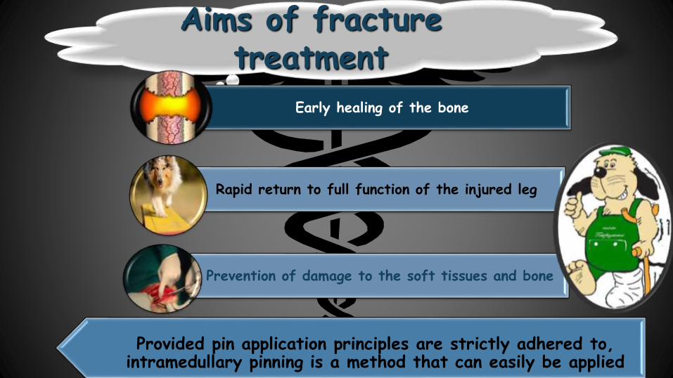
Coetzee (2000) and Hach (2000)- Rotational stability can also be increasedby cerclage wire, polyglycolic suture, external fixation, interlocking pins andtrilam nails, or by using a C-clamp on the plate
Lanz and others (1999) - Stabilisation of a Salter Harris type IVphyseal fracture of the humeral condyle in a miniature pinscher wassimplified by using orthofix partially threaded Kirschner wire, withexcellent clinical results.
Olmstead and others (1995), Denny and Butterworth (2000)- Partiallythreaded pins having a negative profile ending creates a weak point in thepin-thread junction, so if these pins are to be used, the junction must notbe near the fracture line

Many veterinary practitioners in field conditions
• Do not have access to the specialized and costly equipment to undertake complex orthopaedic procedures
• Or lack the technical assistance required to perform such operations
However, many of them have access to, and are skilled in using the simpler, more affordable equipment necessary to place an intramedullary pin and cerclage wires
That’s why a slight innovation and modification has been made with provision of threads at one end of the existing Steinman pin

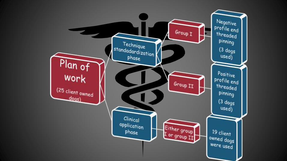
To standardize the technique of application of end threaded intramedullary pin for management of long bone fractures in
canines
To evaluate the efficacy of end threaded intramedullary pin in management of long bone fractures in canines


• Will overcome the potential post operative complications of intramedullary pinning
• Will be an efficient and cost-effective technique in managing various long bone fractures in canines encountered by veterinarians in field conditions
Intramedullary compression and
fixation device with a threaded end
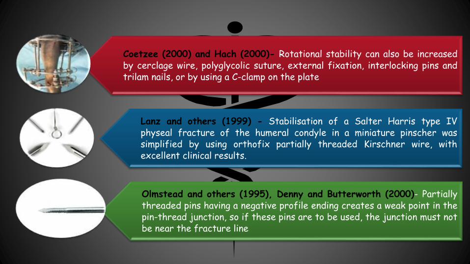
To assess the feasibility of using end threaded intramedullary pins which:
• Provides and , & to an extent
• But devoid of some complications like accurate proficiency as in plate application and its contouring with bone interface
• As well as
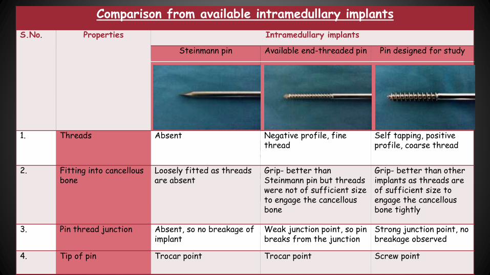
Comparison from available intramedullary implants
S.No. Properties Intramedullary implants
Steinmann pin Available end-threaded pin Pin designed for study
1. Threads Absent Negative profile, fine thread
Self tapping, positive profile, coarse thread
2. Fitting into cancellous bone
Loosely fitted as threads are absent
Grip- better than Steinmann pin but threads were not of sufficient size to engage the cancellous bone
Grip- better than other implants as threads are of sufficient size to engage the cancellous bone tightly
3. Pin thread junction Absent, so no breakage of implant
Weak junction point, so pin breaks from the junction
Strong junction point, no breakage observed
4. Tip of pin Trocar point Trocar point Screw point

Description of implant

Preparation of implantImplants used in fracture repair bear all or part of the load normally carried by the bone (Coetzee 2002; Ness 2006).
The implant used over here, in the management of long bone fractures in small animals was manufactured from iron-based alloys, specially 316L stainless steel
Composition of 316L stainless steel (%)(Mears and Rothwell 1982):
Iron :55-60%
Chromium :17-20%
Nickel :10-14%
Molybdenum :2.8%
Manganese :1.7%
Silicone :0.57%
Copper :0.1%
Nitrogen :0.095%
Phosphorous :0.025%
Carbon :0.024%
Sulphur :0.003%
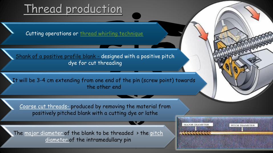
Thread production
Cutting operations or thread whirling technique
Shank of a positive profile blank - designed with a positive pitch dye for cut threading
It will be 3-4 cm extending from one end of the pin (screw point) towards the other end
Coarse cut threads- produced by removing the material from positively pitched blank with a cutting dye or lathe
The major diameter of the blank to be threaded > the pitch diameter of the intramedullary pin

End threaded negative profile
trocar point
End threaded positive profile screw point

• Surgical approach
Introduction of pin from the fracture site in proximal
part of bone
Withdrawl of pin in upward
direction till the last thread reach the
fracture site
Reduction of fractured
segments and introduction of
threaded end into distal part of bone
Seating of the threaded end in distal cancellous bone followed by cutting of extra pin over the skin


Postoperative Care: Antibiotics -Amoxirum Forte 300mg @5-20 mg/kg b.w. I.M b.i.d X5 days
postoperatively
Anti-inflammatory -Meloxicam (MELONEX) 5mg/ml @ 0.2-0.5mg/kg b.w. S/C -o.d X 3 days postoperatively
Syrp. Osteopet (Calcium supplement) @ 5ml b.i.d for two months and
Syrp.Sharkoferol or Multistar pet @ 5ml b.i.d for two months
The owner was strictly advised to provide complete rest to the animal and its restrained movement till 1month post-operatively. Skins sutures were removed
10 days post-operatively

Pin removal
Pin was then gently pulled out by rotating in anti-clockwise direction followed by suturing the skin incision
Removed using a pair of pliers and a Jacob’s chuck by means of a stab incision through the skin
The site of pin insertion over the bone was felt, shaved and scrubbed thoroughly with an antiseptic solution.
Removed under general anaesthesia after radiographic evidence of periostealbridging and moderate callus formation

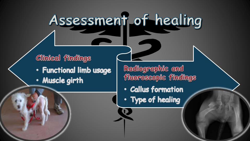
Parameters evaluated
Type of fracture
Immediate weight bearing
Assessment of healing
Post fixation physiotherapy
Time taken in recovery
Overall functional recovery
Post implant removal
evaluation

Grades 0 1 2 3 4
Limb use description
Non-use Slight use limping Slight limping Normal
Weight bearingand gait
No weight bearing; carrying the limb while walking
Slight weight bearing; touching the toe while walking
Moderate weight bearing, touching the sole while walking with limping
Good weight bearing; with slight limping
Complete weight bearing; with no sign of limping
Functional limb usage
Grades 0 1 2 3
Amount of Callus formation
None(no visible callus)
Small (<10% increase in the bone diameter)
Moderate(10-20% increase in the bone diameter)
Large (>20% increase in the bone diameter)
Callus formation
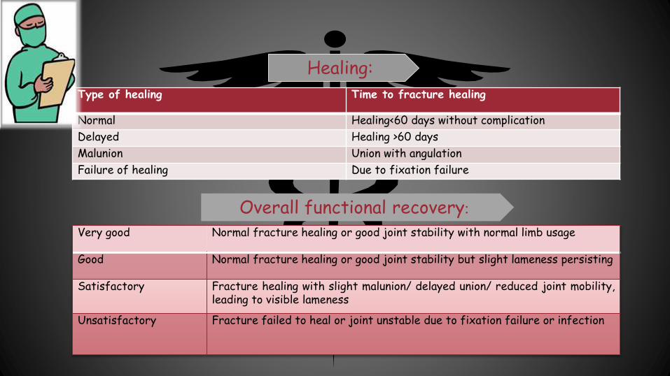
Very good Normal fracture healing or good joint stability with normal limb usage
Good Normal fracture healing or good joint stability but slight lameness persisting
Satisfactory Fracture healing with slight malunion/ delayed union/ reduced joint mobility,leading to visible lameness
Unsatisfactory Fracture failed to heal or joint unstable due to fixation failure or infection
Overall functional recovery:
Healing:
Type of healing Time to fracture healing
Normal Healing<60 days without complication
Delayed Healing >60 days
Malunion Union with angulation
Failure of healing Due to fixation failure

S.No.
Parameters Situation Score
1 Pain None 25When walking on irregular surface/ ground 20When walking on metalled road 10When walking on indoor cemented floor 5Constant and severe 0
2 Stiffness None 10Stiff 0
3 Swelling None 10Mild 5Constant 0
4 Climbing stairs Not a problem 10Asymmetrically 5Impossible 0
5 Running Possible 5Impossible 0
6 Jumping Possible 5Impossible 0
7 Squatting while defecation Possible 5Impossible 0
8 Walking assistance None 25Manual support by owner 0
9 Physiotherapeuticassistance
None 15Needed 0
Post implant removal ---- Rehabilitation evaluation

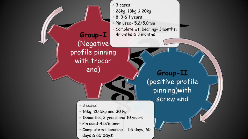
Group-II (positive profile
pinning)with screw end
• 3 cases
• 16kg, 20.5kg and 30 kg
• 18months, 3 years and 10 years
• Pin used-4.5/6.5mm
• Complete wt. bearing- 55 days, 60
days & 60 days
Group-I(Negative
profile pinning with trocar
end)
• 3 cases
• 26kg, 18kg & 20kg
• 8, 3 & 1 years
• Pin used- 5.2/5.0mm
• Complete wt. bearing- 3months, 4months & 3 months

Group-I
Case reported : Complete proximal diaphyseal comminuted fracture of left femur
Technique used: End threaded negative profile pinning with a trocar end
Pre-opPost-op
42 days Post-op
Post-pin removal

Group-II
Case reported : Complete mid diaphyseal simple spiral fracture of right femur
Technique used: End threaded positive profile pinning with a screw end
Pre-op Post-op
21 days Post-op
Post-pin removal


0
10
20
case-1case-2
case-3
Mus
cleGirth
Group-I pre-op
immediate post-op
21 day post-op
42nd day post-op
0
10
20
case-4case-5
case-6
Mus
cle G
irth
Group-II pre-op
immediate post-op
21 day post-op
42nd day post-op

Weight bearing and functional limb usage
00.20.40.60.8
11.21.41.61.8
2
Group-I
0
0.5
1
1.5
2
2.5
3
3.5
4
Day-21 Day-42
Case-4 2 4
Case-5 2 3
Case-6 1 3W
eight
bearing
sco
re
Group-II
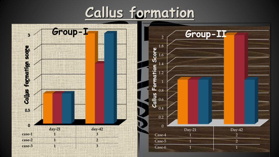
Callus formation
day-21 day-42
case-1 1 3
case-2 1 2
case-3 1 3
Group-I
0
0.2
0.4
0.6
0.8
1
1.2
1.4
1.6
1.8
2
Day-21 Day-42
Case-4 1 2
Case-5 1 2
Case-6 1 1
Callus
For
mation
Sco
re
Group-II

Pin migration
• Slight pin migration observed in all the cases
• In one case, pin broke at thread-pin interface
• No pin migration observed
• No incidence of pin breakage
Blood supply and healing
• Pin was snuggly fitted into the medulla of long bone, covering maximum diameter thus obstructing medullary circulation
• Thus delayed healing
• Only 60-70% medullary occupancy
• Thus early healing
Implant application and removal
• Application was same in both the groups
• Removal- always with rotation in whereas sometimes with rotation in

Radiograph representing medullary occupancy by the implant in a cadaver bone
Fig: a-craniocaudal view; b-mediolateral view; c-cross-sectional view of cortical bone along with
implant(60% occupancy); d-cross-sectional view of cancellous bone with implant (snuggly fitted)
a b
c
d
Medullary cavity
Cortical bone
Implant
Implant
Cortical bone
(Cr-cd)

S.no. Properties Technique of end threaded I.M.pinning
Negative profile Positive profile
1 Weight bearing Late Early
2 Callus formation Large Normal
3 Pin migration Yes No
4 Rotational forces Not overcome overcome
5 Blood supply Impaired Not affected
6 Healing time Late Early
7 Method of introduction Mild rotational resistance Moderate rotational
resistance
8 Removal of implant Easy (sometimes with
rotation)
Easy (always with rotation)
9 Muscle girth Reduced Normal


Bone type Fracture localisation Fracture line Degree of bone
fragmentation
Tot
al n
o.
% f
rom
tot
al
no. o
f fr
actu
res
% f
rom
tot
al
no. o
f sp
ecif
ic
bon
e fr
actu
res
Tot
al n
o.
% f
rom
tot
al
no. o
f fr
actu
res
% f
rom
tot
al
no. o
f sp
ecif
ic
bon
e fr
actu
res
Tot
al n
o.
% f
rom
tot
al
no. o
f fr
actu
res
% f
rom
tot
al
no. o
f sp
ecif
ic
bon
e fr
actu
res
Humerus D 3 15.79 75 T 2 13.34 50 S 3 15.79 75
M 0 0 0 O 1 6.66 25 C 1 5.26 25
E 1 5.26 25 S 0 0 0
I 0 0 0
Femur D 10 52.63 71.42 T 4 26.67 28.57 S 11 57.89 78.57
M 3 15.79 21.43 O 5 33.34 35.71 C 3 15.79 21.43E 1 5.26 7.15 S 1 6.66 7.14
I 1 6.66 7.14
Tibia D 1 5.26 100 T 0 0 0 S 1 5.26 100
M 0 0 0 O 1 6.66 100 C 0 0 0
E 0 0 0 S 0 0 0
I 0 0 0
Key: D- diaphyseal, M- metaphyseal, E- epiphyseal (fracture localisation); T- transverse, O- oblique, S- spiral, I-impacted (fracture line); S- simple, C- comminuted (degree of bone fragmentation)
Depending on bone type, fracture localisation, fracture line and degree of bone fragmentation

Bone type Patient age
Category Total no. % from total
no. of
fractures
% from total
no. of specific
bone fractures
Humerus Under 6months 1 5.26 25
6-12 months 1 5.26 25
1-6 years 1 5.26 25
6-12 years 1 5.26 25
Femur Under 6months 9 47.37 64.29
6-12 months 4 21.05 28.57
1-6 years 1 5.26 7.14
6-12 years 0 0 0
Tibia Under 6months 0 0 0
6-12 months 1 5.26 100
1-6 years 0 0 0
6-12 years 0 0 0
Depending upon bone type and patient age

Bone type Patient weight
Weight
category
Total no. % from total
no. of
fractures
% from total
no. of specific
bone fractures
Humerus I 2 10.53 50
II 1 5.26 25
III 1 5.26 25
Femur I 8 42.11 57.14
II 5 26.32 35.71
III 1 5.26 7.14
Tibia I 1 5.26 100
II 0 0 0
III 0 0 0
Depending upon bone type and patient weight
Key: I-III (weight categories: 0-10, 10-20, 20-30)
0
2
4
6
8
10
12
14
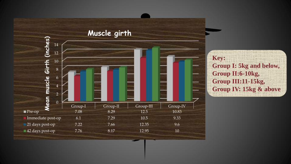
Group-I Group-II Group-III Group-IV
Pre-op 7.08 8.29 12.5 10.83
Immediate post-op 6.1 7.29 10.5 9.33
21 days post-op 7.22 7.66 12.35 9.6
42 days post-op 7.76 8.17 12.95 10
Mean
mus
cle G
irth
(inch
es)
Muscle girth
Key:
Group I: 5kg and below,
Group II:6-10kg,
Group III:11-15kg,
Group IV: 15kg & above

Pin size Cases Age Body weight (kgs)
3.5/4.0mm 1 1 and ½ month 3.40
2 2 months 3.70
3 2 months 3.70
4 3 months 4.70
5 4 months 4.50
6 4 months 4.30
7 18 months 11.50
4.5/5.0mm 8 3 and ½ month 8.50
9 4 months 9.75
10 4 months 11.00
11 6 months 7
12 6 months 13
13 6 months 13
14 9 years 15
4.5/6.5mm 15 8 month 9.40
16 11 month 21
17 1 year 21
18 4 months 7.5
19 4 months 9.80

0
0.5
1
1.5
2
2.5
3
3.5
4
Group-I Group-II Group-III Group-IV
21 day 1.6 1.57 2 1.67
42 day 3.8 3.71 4 3.67
Mean
scor
e
Post operative weight bearing
Key:
Group I: 5kg and below,
Group II:6-10kg,
Group III:11-15kg,
Group IV: 15kg & above

0
0.2
0.4
0.6
0.8
1
1.2
1.4
1.6
1.8
2
Group-I Group-II Group-III Group-IV
21st day 2 2 1.75 2
42nd day 1.6 1.57 1.5 1.67
Mean
scor
ePost operative callus formation
Key:
Group I: 5kg and below,
Group II:6-10kg,
Group III:11-15kg,
Group IV: 15kg & above

Pre-op Post-op 21 days Post-op Post-pin removal
Case-1
Case-2
Callus formation
Bone healing and recovery
• Metaphyseal/epiphyseal (n=3) and distal or proximal 3rd diaphyseal (n=6) fractures healed early (within 21 days), as compared to mid-diaphyseal (n=4) fractures because cortical bone is much slower to heal than cancellous bone
• In all the 19 cases direct bone healing occurred through contact healing, where osteonal remodeling occurred rapidly across a fracture in areas of apposed cortical bone
• In some of the cases, where small gaps remained at the fracture site but the fixation was rigid, bone healing occurred through gap healing.


Bilateral femur fracture- Rt. : simple complete distal metaphyseal oblique and lft. : simple complete proximal metaphyseo-diaphyseal long oblique

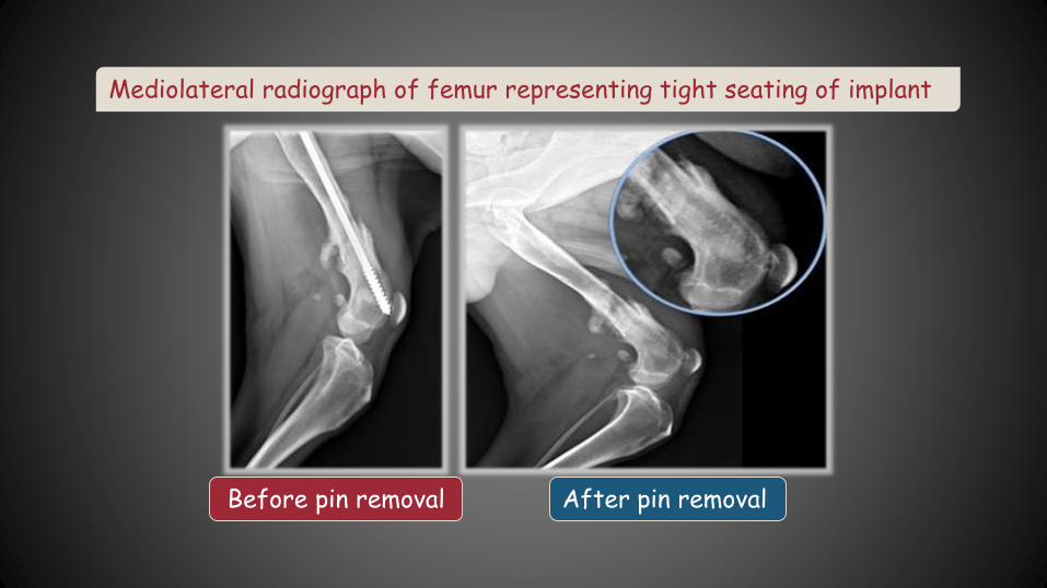
Before pin removal After pin removal
Mediolateral radiograph of femur representing tight seating of implant
Other advantages
• The end threaded intramedullary positive profile pin is also a cost-effective option
• No damage to the nerve was encountered either during application or removal of the intramedullary end-threaded pin.
• Post-recovery removal is easy and does not require a major surgical procedure

The end threaded intramedullary positive profile screw ended self tapping pin used for fixation of long bone fractures in canines can resist pin migration, pin breakage and all loads acting on the bone i.e. rotation, compression, tension, bending and also shearing to an extent with no post-operative complications.
The end threaded intramedullary positive profile screw ended pin is inexpensive and can be easily used in field conditions in managing long bone fractures in canines, as compared to other orthopaedic implants.
Thankyou…