
Semi-supervised Learning for Multi-label...
6
Semi-supervised Learning for Multi-label Classification * Liyue Shen Stanford University [email protected] Ruiyang Song Stanford University [email protected] Abstract In this report we consider the semi-supervised learning problem for multi-label image classification, aiming at ef- fectively taking advantage of both labeled and unlabeled training data in the training process. In particular, we im- plement and analyze various semi-supervised learning ap- proaches including a support vector machine (SVM) method facilitated by principal component analysis (PCA), and a self-training method that iteratively conducts supervised learning and enlarges the set of training labels on the go. We compare the performances of semi-supervised learning methods with supervised learning benchmarks, and intro- duce a heuristic performance analysis for the training pro- cess. In addition, we analyze the impact of different training parameters for the PCA-SVM and the self-training method on the prediction performance. The algorithms are imple- mented for the ChestX-ray14 [32] medical image dataset. 1. Introduction The recent progress in deep learning research has sig- nificantly improved the performance of various computer vision tasks for natural images including image classifica- tion [16, 29, 30, 12], object detection [9, 27] and instance segmentation [19] benefited from the availability of a large volume of labeled natural image datasets. It is expected that computer vision methods based on supervised learning will also contribute to medical image applications such as early- stage cancer detection and image diagnosis. However, chal- lenges emerge as it is difficult to construct large densely labeled medical datasets since manually annotating medi- cal images requires medical and clinical expertise. In other words, labeled medical data are often much more expensive * This is a joint project with CS331B. The two projects focus on differ- ent methods and algorithms to solve the semi-supervised learning problem. In particular, the CS229 project primarily concentrates on the PCA-SVM and the self-training method, while the CS331B project concentrates on the supervised deep learning approaches and the CNN-ladder networks. Some materials and experiment results (e.g. for baseline and comparison purpose) are shared in the two project reports. The projects focus on fun- damentally different major approaches and provide independent analyses. Figure 1: Two multi-label chest X-ray image samples from ChestX-ray14 dataset [32] with radiology report, disease key- words extraction and localization results. than unlabeled data. Two primary approaches have been introduced to tackle the medical image recognition task on a dataset with few la- beled samples: semi-supervised learning and transfer learn- ing. Semi-supervised learning considers a prediction prob- lem with only a small number of labeled training data by exploiting the information provided by both labeled and un- labeled data [2]. The labeled data will provide information for joint distribution of samples and labels, while the unla- beled data provides information of the distribution of sam- ples [35]. Multiple approaches have been developed and widely applied in computer vision, and a comprehensive lit- erature review can be found in [35]. One of the notable methods for semi-supervised learn- ing is the self-training technique, which is first introduced in [20]. The main idea of self-training is to iteratively ap- ply a supervised learning algorithm based on the currently available training labels and include the predicted examples with high confidence scores into the updated training set. In this manner, more information of the originally unlabeled data is incorporated into the classifier after each iteration as the training label set is enlarged in each step. Transfer learning is another method widely used in com- puter vision scenarios to overcome the challenge of insuf- 1
Transcript of Semi-supervised Learning for Multi-label...
Semi-supervised Learning for Multi-label Classification∗
Liyue ShenStanford [email protected]
Ruiyang SongStanford University
Abstract
In this report we consider the semi-supervised learningproblem for multi-label image classification, aiming at ef-fectively taking advantage of both labeled and unlabeledtraining data in the training process. In particular, we im-plement and analyze various semi-supervised learning ap-proaches including a support vector machine (SVM) methodfacilitated by principal component analysis (PCA), and aself-training method that iteratively conducts supervisedlearning and enlarges the set of training labels on the go.We compare the performances of semi-supervised learningmethods with supervised learning benchmarks, and intro-duce a heuristic performance analysis for the training pro-cess. In addition, we analyze the impact of different trainingparameters for the PCA-SVM and the self-training methodon the prediction performance. The algorithms are imple-mented for the ChestX-ray14 [32] medical image dataset.
1. IntroductionThe recent progress in deep learning research has sig-
nificantly improved the performance of various computervision tasks for natural images including image classifica-tion [16, 29, 30, 12], object detection [9, 27] and instancesegmentation [19] benefited from the availability of a largevolume of labeled natural image datasets. It is expected thatcomputer vision methods based on supervised learning willalso contribute to medical image applications such as early-stage cancer detection and image diagnosis. However, chal-lenges emerge as it is difficult to construct large denselylabeled medical datasets since manually annotating medi-cal images requires medical and clinical expertise. In otherwords, labeled medical data are often much more expensive
∗This is a joint project with CS331B. The two projects focus on differ-ent methods and algorithms to solve the semi-supervised learning problem.In particular, the CS229 project primarily concentrates on the PCA-SVMand the self-training method, while the CS331B project concentrates onthe supervised deep learning approaches and the CNN-ladder networks.Some materials and experiment results (e.g. for baseline and comparisonpurpose) are shared in the two project reports. The projects focus on fun-damentally different major approaches and provide independent analyses.
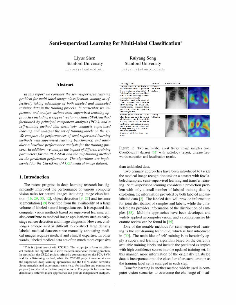
Figure 1: Two multi-label chest X-ray image samples fromChestX-ray14 dataset [32] with radiology report, disease key-words extraction and localization results.
than unlabeled data.Two primary approaches have been introduced to tackle
the medical image recognition task on a dataset with few la-beled samples: semi-supervised learning and transfer learn-ing. Semi-supervised learning considers a prediction prob-lem with only a small number of labeled training data byexploiting the information provided by both labeled and un-labeled data [2]. The labeled data will provide informationfor joint distribution of samples and labels, while the unla-beled data provides information of the distribution of sam-ples [35]. Multiple approaches have been developed andwidely applied in computer vision, and a comprehensive lit-erature review can be found in [35].
One of the notable methods for semi-supervised learn-ing is the self-training technique, which is first introducedin [20]. The main idea of self-training is to iteratively ap-ply a supervised learning algorithm based on the currentlyavailable training labels and include the predicted exampleswith high confidence scores into the updated training set. Inthis manner, more information of the originally unlabeleddata is incorporated into the classifier after each iteration asthe training label set is enlarged in each step.
Transfer learning is another method widely used in com-puter vision scenarios to overcome the challenge of insuf-
1
ficient labeled training data. When the unlabeled trainingdataset is small, transfer learning methods extract informa-tion from the trained model of a different large dataset tofacilitate the current task. A literature survey on transferlearning is presented in [21].
This project studies a medical image classification prob-lem based on the ChestX-ray14 [32] dataset that containsover 100 thousand front-view X-ray images with annota-tions of 14 thoracic diseases. We first propose a deep learn-ing approach with supervised learning. Then we introducesemi-supervised methods using machine learning based onprincipal component analysis (PCA) and support vector ma-chine (SVM). In addition, we study a self-training approachwhere in each iteration we perform supervised deep learn-ing algorithms and enlarge the labeled training sets. Wecompare their performances with the supervised learningbenchmarks and the performance of the semi-supervisedladder network approach.
2. Related WorkDeep Learning in Medical Imaging. One of the firstsuccessful approaches of applying deep neural networks tobiomedical imaging is [4]. Recent works studied the appli-cations including skin cancer classification [8], breast can-cer diagnosis [22], brain tumor segmentation [31], and lungnodule detection [3], where deep learning methods haveshown good experimental performance. An overview of therecent progress is summarized in [34].Semi-supervised Learning. Empirical results show thatsemi-supervised learning improves the performance com-pared to supervised learning that only exploits labeleddata [10]. Primary methods for solving semi-supervisedlearning include generative models [13], self-training [28],transductive SVMs [1], entropy regularization [10], andgraph-based models [36]. [11] and [14] consider a semi-supervised image classification problem with a variationalinference algorithm based on deep generative models. An-other deep neural network based method is the ladder net-work [24]. In [18], a self-training support vector machine(SVM) algorithm is studied. In [28], an object detectionproblem is studied with the self training expecation maxi-mization (EM) method.
3. Methods & ExperimentsThis project primarily focuses on the thoracic disease
classification problem based on X-ray image data, whichcan be formulated as a multi-label problem since each sam-ple possibly has multiple diseases simultaneously.
In this section, we first briefly introduce the ChestX-ray14 dataset, then describe the methods we apply: theResNet [12] model which is a supervised baseline, theSVM-PCA method, the self-training approach, and the lad-
der network for comparison.
3.1. ChestX-ray14 Dataset
The ChestX-ray14 [32] dataset illustrated in Figure 1is currently the largest chest X-ray database that contains112,120 frontal-view X-ray images from 32,717 patientswith 14 common thoracic disease categories labeled by textmining radiology reports. In ChestX-ray14, 60,412 sam-ples are healthy and 51,708 samples have (possibly mul-tiple) thoracic diseases. Following the experiment settingin [32], we randomly choose 78,484 images (70%) used fortraining, 11,212 images (10%) for validation and 22,424 im-ages (20%) for testing. In [32], a multi-label classificationbenchmark is also presented.
3.2. Supervised Learning
3.2.1 ResNet model for transfer learning
To begin with, we apply the deep residual network (ResNet)model for transfer learning and particularly choose theResNet-18 and ResNet-50 models inspired by [12].
For our multi-label classification setting, we adjust theoriginal ResNet model that is supposed for single-label clas-sification using the multi-label soft margin loss when train-ing the network.
Denote by C the collection of categories. Let T ∈{0, 1}|C| be the actual image label and Y ∈ R|C| be thenetwork prediction. The training loss can be formulated asfollows:
L(Y, T ) = −|C|∑i=1
[ti log
1
1 + e−yi+ (1− ti) log
e−yi
1 + e−yi
].
The final predicted probability is then derived by applyingthe sigmoid activation function on the confidence score vec-tor.
We use the ImageNet pre-trained model as the initial-ization to train the ResNet model with the idea of trans-fer learning. The experiment results of supervised learningand transfer learning are shown in Table 1. Here, the area-under-curve (AUC) score is applied as the metric. comparedwith benchmark results [32] and previous work [33, 23],our method gets the state-of-the-art results in average AUCscores as well as most disease classes. We refer to [5] forthe implementation of the ResNets.
3.3. Semi-supervised Learning
3.3.1 PCA-SVM baseline model
We consider a baseline machine learning model combiningPCA and SVM. First, we preprocess the X-ray images oforiginal size 1024× 1024 by resizing them into 128× 128.We apply the PCA approach to reduce the data dimensionof the flattened image vector to a dimension of 2500. Then
2
Method Benchmark [32] DenseNet-LSTM [33] CheXNet [23] Ours - ResNet-18 (Fine-tune) Ours - ResNet-50 (Fine-tune)
Atelectasis 0.7158 0.772 0.8209 0.8190 0.8276Cardiomegaly 0.8065 0.904 0.9048 0.8998 0.9013Effusion 0.7843 0.859 0.8831 0.8881 0.8903Infiltration 0.6089 0.695 0.7204 0.7165 0.7229Mass 0.7057 0.792 0.8618 0.8534 0.8690Nodule 0.6706 0.717 0.7766 0.7738 0.7884Pneumonia 0.6326 0.713 0.7632 0.7593 0.7588Pneumothorax 0.8055 0.841 0.8932 0.8934 0.9033Consolidation 0.7078 0.788 0.7939 0.8116 0.8178Edema 0.8345 0.882 0.8932 0.9061 0.9106Emphysema 0.8149 0.829 0.926 0.9083 0.9198Fibrosis 0.7688 0.767 0.8044 0.8149 0.8197PT 0.7082 0.765 0.8138 0.8007 0.8048Hernia 0.7667 0.914 0.9387 0.8822 0.8700NoFinding - 0.762 - 0.7229 0.7894
Average 0.7379 0.798 0.8424 0.8377 0.8432
Table 1: Per-class AUC scores of ROC curves for multi-label classification on ChestX-ray14 dataset, which present the quantitativeperformance of different models with or without transfer learning in fully-supervised learning setting.
we use the SVM model to classify the image vectors intonormal and abnormal categories. In this way, we train abinary classifier for each of the 14 disease categories.
The number of components selected for the PCA is cru-cial for the classifier performance. We begin with a low-dimensional PCA and found that the subsequently trainedSVM classifier is not able to discriminate images of dif-ferent classes, and all the prediction outputs are the same.This might be because of the fact that in a low-dimensionalspace, the images of different classes are mixed togetherand can not be separated by a SVM model effectively. Be-sides, we realized that it is important to balance the trainingsamples. Recall that in the semi-supervised learning, thenumber of training samples is very small. If only a smallamount of positive examples are randomly chosen for train-ing, it is hard for the SVM to give a good boundary sincethe chosen positive examples only represent a partial distri-bution of the positive samples.
We apply the PCA-SVM method trained on a training setcomprising of 2000 labeled samples and evaluate the modelin the testing dataset (22,424 images, which is 20% of thewhole ChestX-ray database). We use different PCA dimen-sions ranging from 1000, 2000, and 5000. The testing per-formance of ROC AUC scores are presented in Table 2. Weconsult [6] for the implementation of the PCA-SVM algo-rithm.
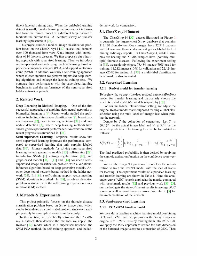
In order to explore the impact of the number oflabeled training samples and the number of com-ponents in the PCA on the testing performance, wecompare the performances with the number of traininglabels ranging from {0.1, 1, 2, 5, 10, 15, 20, 25, 50} ×103, and the PCA dimension ranging from{20, 100, 500, 1000, 1500, 2000, 2500, 5000}. The ex-periment result is shown in Fig. 2. We see that the
Figure 2: Semi-supervised learning performance of PCA-SVMmodel on the test dataset for classification of disease class ”Fi-brosis” with different number of training labels and various PCAreduction dimensions.
performance is improved as the number of active dimen-sions in the PCA step increases. The classifier performspoorly when the PCA dimension is as low as 20 and 100.We also observe that when the number of active PCAcomponents is relatively low at 20, 100, and 500, theAUC scores can decrease as the number of training labelsincreases. Note that when the PCA dimension is low,a large number of training labels may cause overfittingand jeopardize the testing performance. Approximately2000 PCA components and 2000 training labels will besufficient for a good performance, above which increasingthe number of training labels and the number of PCAcomponents does not improve the scores significantly.
3
3.3.2 Self-training
In this subsection we study the self-training approach thatextracts information from both the labeled and unlabeledtraining data through sequentially training a network withnew labels added based on previous iterations. Whilethe general theoretic analysis of the performance of self-training algorithms can be difficult [35], here we try to pro-vide a heuristic analysis.
Suppose the training data are from a set X and the un-derlying correct classification labels are from a set Y . Wehave m0 labeled training samples in X0 ⊂ X with la-bels Y0 and m1 = M − m0 unlabeled training samplesin X1 ⊂ X . We start with m0 training labels and imple-ment a supervised learning algorithm with a particular net-work architecture and derive confidence scores S for theM − m0 unlabeled samples. Afterwards, we select c un-labeled samples with the highest confidence measures de-noted by Sc = {p1, . . . , pc} respectively, and incorporatethem into the labeled training set together with their pre-dicted labels Yc. This training process continues sequen-tially as the label set gets enlarged gradually. In this ap-plication, we implement a fine tuned ResNet-50 network ineach iteration as the supervised learning method. The selftraining approach is summarized in Algorithm 1.
Suppose the relationship between the samples and theunderlying classification result can be described with amapping f : X 7→ Y . If we are given correct labels forall the training data, we will arrive at a learnt mapping f .However, in the self-training approach, the c picked pre-dictions in each step may bias the learning result from fdue to possible incorrect predictions, which we roughlyview as noise. If we approximate the probability that theselected predictions are correct with their respective con-fidence scores p1, . . . , pc, for stochastic gradient descent,each newly selected label will incur noise with mean in theorder of εi = (1− pi)α, i = 1, . . . , c.
Algorithm 1: Self-training method with ResNet
1 initialize Xlabel := X0, Ylabel := Y0, α, c;2 repeat3 (Ypredict,Spredict) := ResNet(Xlabel, Ylabel, α);4 (Xc, Yc,Sc) := findmaxc(Spredict);5 Xlabel := Xlabel
⋃Xc, Ylabel := Ylabel
⋃Yc.;
6 until iterations finished;
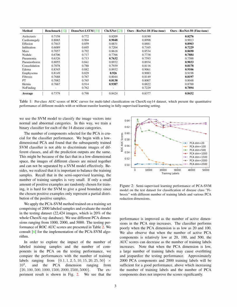
One of the most important parameters besides the learn-ing rate α is the selection parameter c, i.e. the number ofpredicted labels added to the training label set in each round.In Figure 3, we present the performances of the self-trainingmethod with c ranging in {1, 2, 3, 4, 5} with learning rateα = 0.01. We see that empirically for this application,
Figure 3: Performance of the self-training approach with differentselection parameter c’s. The model is based on fine-tuning theResNet-50 network and trained with 2000 images.
c = 3 or 4 will be good choices.
The performance of the self-training method is presentedin Table 2 and 3. In Table 2, we compare the per-class AUCscores of self-training based on ResNet18 and ResNet50using 2000 training labels with other approaches. In Ta-ble 3, we compare the performances of self-training with1000, 2000, and 5000 labels respectively with other meth-ods. Both empirical examples show that self-training canbe applied to improve the classification performances. Itis shown to outperform the CNN ladder network for mostclasses and is usually better than or close to the benchmark.
3.3.3 CNN-based ladder network
Another approach for semi-supervised learning is the lad-der network [24] proposed based on the stacked denoisingautoencoders [25, 26], where a noisy encoder feed forwardpath and a corresponding denoising decoder path are addedfor learning unlabeled training data to the normal feed for-ward network through additional lateral connections. Theperformance of ladder networks for the MNIST dataset [17]and the CIFAR-10 dataset [15] have been evaluated in pre-vious work.
To construct ladder network for chest X-ray image clas-sification, we propose a 11-layer CNN model with increas-ing feature map channels and decreasing convolution ker-nels in the feedforward encoder path. And the final layer isa global mean pooling layer to aggregate the spatial imageinformation. The experiment results for CNN-based laddernetwork is shown in Table 2 for comparison purpose. Werefer to the github repository in [7] for the implementationof ladder networks.
4

Method Bench-mark [32]
PCASVM(1000)
PCASVM(2000)
PCASVM(5000)
ResNet18 ResNet50 ResNet18ST ResNet50ST CNN-Ladder
ATE 0.7158 0.605 0.619 0.629 0.7273 0.7084 0.7312 0.7120 0.4941CARD 0.8065 0.655 0.689 0.704 0.7501 0.7334 0.7453 0.7404 0.7939EFF 0.7843 0.673 0.674 0.686 0.8104 0.8118 0.8123 0.8043 0.7395INFI 0.6089 0.602 0.598 0.609 0.6593 0.6500 0.6624 0.6552 0.5803Mass 0.7057 0.554 0.558 0.579 0.6758 0.6729 0.6687 0.6646 0.6108NOD 0.6706 0.529 0.539 0.551 0.6511 0.6386 0.6364 0.6436 0.5312PNA 0.6326 0.592 0.590 0.612 0.6792 0.6711 0.6823 0.6861 0.5907PTX 0.8055 0.610 0.616 0.625 0.7803 0.7735 0.7755 0.7821 0.6630CON 0.7078 0.667 0.672 0.685 0.7699 0.7598 0.7616 0.7645 0.6711Edema 0.8345 0.754 0.761 0.780 0.8573 0.8559 0.8594 0.8659 0.8170EMPH 0.8149 0.582 0.596 0.616 0.7570 0.7789 0.7806 0.7677 0.6879FIB 0.6158 0.614 0.618 0.632 0.7263 0.7285 0.7307 0.7377 0.6158PT 0.7082 0.575 0.584 0.600 0.6971 0.6982 0.7088 0.6964 0.6411Hernia 0.7667 0.677 0.661 0.649 0.8179 0.8145 0.8219 0.8497 0.7007
Avg. 0.7379 0.6206 0.6268 0.6398 0.7399 0.7354 0.7412 0.7407 0.6527
Table 2: Per-class AUC scores of different semi-supervised methods with the same 2000 labeled training data for multi-label classificationon ChestX-ray14 dataset. ”ST” denotes self-learning approach. The per-class scores for the PCA-SVM method with 1000 and 5000 labelsare also included for comparison.
Average AUC score with # of used labels 1000 2000 5000 All (78484)
Benchmark [32] - - - 0.7379DenseNet-LSTM [33] - - - 0.798CheXNet [23] - - - 0.8424
Baseline (PCA+SVM) 0.6206 0.6268 0.6398 -ResNet-18 (Fine-tune) 0.7015 0.7399 0.7744 0.8377ResNet-18 (Fine-tune) + self-training 0.7062 0.7412 0.7721 -ResNet-50 (Fine-tune) 0.7052 0.7354 0.7752 0.8432ResNet-50 (Fine-tune) + self-training 0.7088 0.7407 0.7783 -
Table 3: Average AUC scores of all 14 thoracic categories for multi-label classification on ChestX-ray14 dataset demonstrating thequantitative performance of different semi-supervised learning approaches including self-training.
4. Conclusion
In this paper, we study the problem of multi-label imageclassification based on the frontal chest X-ray image datasetChestXray14. We conduct a machine learning approachbased on PCA-SVM, and a self-training method based onResNet in each iteration, and compare them with the per-formances of several benchmarks and the CNN networks.In particular, we studied the impact of PCA dimension inthe PCA-SVM method and the influence of the selectionparameter in self-training approach. We find that in the par-ticular classification problem considered in this report, self-training usually exhibits good performance scores compar-ing to other benchmarks and methods. Besides, we intro-duce an intuitive understanding of the performance analysisof self-training. Future works include the rigorous theo-retic study of the performance of self-training, and improve-ments to the network structures for better capturing infor-mation from unlabeled data.
5. ContributionShen is responsible for the introduction and literature re-
view of computer vision and medical imaging with deeplearning, and Song is responsible for the introduction andsurvey of semi-supervised learning and transfer learning.The two authors worked jointly on implementing and an-alyzing the machine learning and self-training methods.Shen also contributed to the supervised baselines and theladder network as a joint work with her CS331B project.
References[1] K. P. Bennett and A. Demiriz. Semi-supervised support vec-
tor machines. In Advances in Neural Information processingsystems, pages 368–374, 1999. 2
[2] O. Chapelle, B. Scholkopf, and A. Zien. Semi-supervisedlearning (chapelle, o. et al., eds.; 2006)[book reviews]. IEEETransactions on Neural Networks, 20(3):542–542, 2009. 1
[3] J.-Z. Cheng, D. Ni, Y.-H. Chou, J. Qin, C.-M. Tiu, Y.-C.Chang, C.-S. Huang, D. Shen, and C.-M. Chen. Computer-
5

aided diagnosis with deep learning architecture: Applica-tions to breast lesions in us images and pulmonary nodulesin ct scans. Nature, (6):24454 EP –, 2016. 2
[4] D. Ciresan, A. Giusti, L. Gambardella, and J. Schmidhu-ber. Deep Neural Networks Segment Neuronal Membranesin Electron Microscopy Images. Nips, pages 1–9, 2012. 2
[5] Contributors. Github repository. https://github.com/pytorch/vision/blob/master/torchvision/models/resnet.py. 2
[6] Contributors. Github repository. https://github.com/bjlkeng/sandbox/tree/master/notebooks/vae-semi_supervised_learning. 3
[7] CuriousAI. Github repository. https://github.com/CuriousAI/ladder. 4
[8] A. Esteva, B. Kuprel, R. A. Novoa, J. Ko, S. M. Swetter,H. M. Blau, and S. Thrun. Dermatologist-level classifi-cation of skin cancer with deep neural networks. Nature,542(7639):115–118, 2017. 2
[9] R. Girshick. Fast r-cnn. In International Conference on Com-puter Vision (ICCV), 2015. 1
[10] Y. Grandvalet and Y. Bengio. Semi-supervised learning byentropy minimization. In Advances in neural informationprocessing systems, pages 529–536, 2005. 2
[11] M. Guillaumin, J. Verbeek, and C. Schmid. Multimodalsemi-supervised learning for image classification. In Com-puter Vision and Pattern Recognition (CVPR), 2010 IEEEConference on, pages 902–909. IEEE, 2010. 2
[12] K. He, X. Zhang, S. Ren, and J. Sun. Deep residual learn-ing for image recognition. In CVPR, pages 770–778. IEEEComputer Society, 2016. 1, 2
[13] A. Holub, M. Welling, and P. Perona. Exploiting unlabelleddata for hybrid object classification. In Proc. Neural Infor-mation Processing Systems, Workshop Inter-Class Transfer,volume 7, 2005. 2
[14] D. Kingma, D. Rezende, S. Mohamed, and M. Welling.Semi-supervised learning with deep generative models. InNIPS, 2014. 2
[15] A. Krizhevsky, V. Nair, and G. Hinton. Cifar-10 (canadianinstitute for advanced research). 4
[16] A. Krizhevsky, I. Sutskever, and G. E. Hinton. Imagenetclassification with deep convolutional neural networks. InF. Pereira, C. J. C. Burges, L. Bottou, and K. Q. Weinberger,editors, Advances in Neural Information Processing Systems25, pages 1097–1105. Curran Associates, Inc., 2012. 1
[17] Y. LeCun, L. Bottou, Y. Bengio, and P. Haffner. Gradient-based learning applied to document recognition. Proceed-ings of the IEEE, 86:2278–2324, 1998. 4
[18] Y. Li, C. Guan, H. Li, and Z. Chin. A self-training semi-supervised svm algorithm and its application in an eeg-basedbrain computer interface speller system. Pattern RecognitionLetters, 29(9):1285–1294, 2008. 2
[19] J. Long, E. Shelhamer, and T. Darrell. Fully convolutionalnetworks for semantic segmentation. CVPR, Nov. 2015. 1
[20] K. Nigam and R. Ghani. Analyzing the effectiveness andapplicability of co-training. In Proceedings of the ninth in-ternational conference on Information and knowledge man-agement, pages 86–93. ACM, 2000. 1
[21] S. J. Pan and Q. Yang. A survey on transfer learning.IEEE Transactions on knowledge and data engineering,22(10):1345–1359, 2010. 2
[22] R. Platania, S. Shams, S. Yang, J. Zhang, K. Lee, and S.-J.Park. Automated breast cancer diagnosis using deep learningand region of interest detection (bc-droid). In Proceedingsof the 8th ACM International Conference on Bioinformatics,Computational Biology,and Health Informatics, ACM-BCB’17, pages 536–543, New York, NY, USA, 2017. ACM. 2
[23] P. Rajpurkar, J. Irvin, K. Zhu, B. Yang, H. Mehta, T. Duan,D. Ding, A. Bagul, C. Langlotz, K. Shpanskaya, M. P. Lun-gren, and A. Y. Ng. Chexnet: Radiologist-level pneumo-nia detection on chest x-rays with deep learning. CoRR,abs/1711.05225, 2017. 2, 3, 5
[24] A. Rasmus, H. Valpola, M. Honkala, M. Berglund, andT. Raiko. Semi-supervised learning with ladder network.CoRR, abs/1507.02672, 2015. 2, 4
[25] R. T. Rasmus, A. and H. Valpola. Denoising autoencoderwith modulated lateral connections learns invariant represen-tations of natural images. CoRR, 2016. 4
[26] R. T. Rasmus, A. and H. Valpola. Lateral connections indenoising autoencoders support supervised learning. CoRR,2016. 4
[27] S. Ren, K. He, R. Girshick, and J. Sun. Faster R-CNN: To-wards real-time object detection with region proposal net-works. In Advances in Neural Information Processing Sys-tems (NIPS), 2015. 1
[28] C. Rosenberg, M. Hebert, and H. Schneiderman. Semi-supervised self-training of object detection models. 2005.2
[29] K. Simonyan and A. Zisserman. Very deep convolutionalnetworks for large-scale image recognition. ICLR, 2015. 1
[30] C. Szegedy, W. Liu, Y. Jia, P. Sermanet, S. Reed,D. Anguelov, D. Erhan, V. Vanhoucke, and A. Rabinovich.Going deeper with convolutions. In 2015 IEEE Conferenceon Computer Vision and Pattern Recognition (CVPR), pages1–9, June 2015. 1
[31] K. Tseng, Y. Lin, W. H. Hsu, and C. Huang. Joint sequencelearning and cross-modality convolution for 3d biomedicalsegmentation. CVPR, 2017. 2
[32] L. L. Z. L. M. B. R. S. Xiaosong Wang*, Yifan Peng*.Chestx-ray8: Hospital-scale chest x-ray database and bench-marks on weakly-supervised classification and localizationof common thorax diseases. In CVPR, 2017. 1, 2, 3, 5
[33] P. E. D. D. C. B. B. D. Yao, Li and K. Lyman. Learning todiagnose from scratch by exploiting dependen- cies amonglabels. arXiv preprint, 2017. 2, 3, 5
[34] S. Zhou, H. Greenspan, and D. Shen. Deep Learning forMedical Image Analysis. Elsevier Science, 2017. 2
[35] X. Zhu. Semi-supervised learning literature survey. Tech-nical Report 1530, Computer Sciences, University ofWisconsin-Madison, 2005. 1, 4
[36] X. Zhu, J. Lafferty, and R. Rosenfeld. Semi-supervisedlearning with graphs. PhD thesis, Carnegie Mellon Univer-sity, language technologies institute, school of computer sci-ence, 2005. 2
6


![Surface & Coatings Technology - COnnecting … · nificantly lower wear volumes resulted after this surface hardening treatment. ... [1,2] . γ′-Fe 4N and α ... on AISI 4140 steel](https://static.fdocuments.us/doc/165x107/5b7870f37f8b9a331e8ba42f/surface-coatings-technology-connecting-nicantly-lower-wear-volumes-resulted.jpg)
