Self‐healing encapsulation and controlled release of ...
13
RESEARCH REPORT Self-healing encapsulation and controlled release of vaccine antigens from PLGA microparticles delivered by microneedle patches J. Maxwell Mazzara 1,2 | Lukasz J. Ochyl 1,2 | Justin K. Y. Hong 1,2 | James J. Moon 1,2,3 | Mark R. Prausnitz 4 | Steven P. Schwendeman 1,2,3 1 Department of Pharmaceutical Sciences, University of Michigan, Ann Arbor, MI 2 Biointerfaces Institute, University of Michigan, Ann Arbor, MI 3 Department of Biomedical Engineering, University of Michigan, Ann Arbor, MI 4 School of Chemical and Biomolecular Engineering, Georgia Institute of Technology, Atlanta, GA Correspondence Steven P. Schwendeman, University of Michigan Biointerfaces Institute, 2800 Plymouth Rd., Ann Arbor, MI 48109. Email: [email protected] Funding information Pharmaceutical Research and Manufacturers of America Foundation Abstract There is an urgent need to reduce reliance on hypodermic injections for many vaccines to increase vaccination safety and coverage. Alternative approaches include controlled release for- mulations, which reduce dosing frequencies, and utilizing alternative delivery devices such as microneedle patches (MNPs). This work explores development of controlled release microparti- cles made of poly (lactic-co-glycolic acid) (PLGA) that stably encapsulate various antigens though aqueous active self-healing encapsulation (ASE). These microparticles are incorporated into rapid-dissolving MNPs for intradermal vaccination. PLGA microparticles containing Alhydrogel are loaded with antigens separate from micro- particle fabrication using ASE. This avoids antigen expsoure to many stressors. The microparti- cles demonstrate bi-phasic release, with initial burst of soluble antigen, followed by delayed release of Alhydrogel-complexed antigen over approximately 2 months in vitro. For delivery, the microparticles are incorporated into MNPs designed with pedestals to extend functional micro- needle length. These microneedles readily penetrate skin and rapidly dissolve to deposit micro- particles intradermally. Microparticles remain in the tissue for extended residence, with MNP- induced micropores resealing readily. In animal models, these patches generate robust immune responses that are comparable to conventional administration techniques. This lays the frame- work for a versatile vaccine delivery system that could be self-applied with important logistical advantages over hypodermic injections. KEYWORDS controlled release, microneedles, PLGA, vaccine delivery 1 | INTRODUCTION While vaccines represent our strongest weapons against contagious disease, several obstacles still limit their maximum potential. For example, most vaccines require booster doses to induce protective levels of immunity. Besides, the large molecular size of vaccine anti- gens often prevents oral administration, and hypodermic injections are necessary to elicit the desired immune response. The reliance on repeated hypodermic injections creates many logistical challenges, such as difficulties with storage, disposal, and administration via healthcare professional. This not only increases costs, but also decreases availability, particularly in developing nations. If the full booster schedule is not administered, an individual may not develop protective immunity. Furthermore, hypodermic needles are designed for intramuscular (i.m.) delivery. However, the muscle has a low level of resident antigen-presenting cells (APCs), thus requiring higher Received: 30 March 2018 Revised: 31 May 2018 Accepted: 10 June 2018 DOI: 10.1002/btm2.10103 © 2018 The Authors. Bioengineering & Translational Medicine published by Wiley Periodicals, Inc. on behalf of The American Institute of Chemical Engineers. This is an open access article under the terms of the Creative Commons Attribution License, which permits use, distribution and reproduction in any medium, provided the original work is properly cited. Bioengineering & Translational Medicine 2018;1–13 wileyonlinelibrary.com/journal/btm2 1
Transcript of Self‐healing encapsulation and controlled release of ...

R E S E A R CH R E POR T
Self-healing encapsulation and controlled release of vaccineantigens from PLGA microparticles delivered by microneedlepatches
J. Maxwell Mazzara1,2 | Lukasz J. Ochyl1,2 | Justin K. Y. Hong1,2 | James J. Moon1,2,3 |
Mark R. Prausnitz4 | Steven P. Schwendeman1,2,3
1Department of Pharmaceutical Sciences,
University of Michigan, Ann Arbor, MI
2Biointerfaces Institute, University of
Michigan, Ann Arbor, MI
3Department of Biomedical Engineering,
University of Michigan, Ann Arbor, MI
4School of Chemical and Biomolecular
Engineering, Georgia Institute of Technology,
Atlanta, GA
Correspondence
Steven P. Schwendeman, University of
Michigan Biointerfaces Institute, 2800
Plymouth Rd., Ann Arbor, MI 48109.
Email: [email protected]
Funding information
Pharmaceutical Research and Manufacturers of
America Foundation
AbstractThere is an urgent need to reduce reliance on hypodermic injections for many vaccines to
increase vaccination safety and coverage. Alternative approaches include controlled release for-
mulations, which reduce dosing frequencies, and utilizing alternative delivery devices such as
microneedle patches (MNPs). This work explores development of controlled release microparti-
cles made of poly (lactic-co-glycolic acid) (PLGA) that stably encapsulate various antigens though
aqueous active self-healing encapsulation (ASE). These microparticles are incorporated into
rapid-dissolving MNPs for intradermal vaccination.
PLGA microparticles containing Alhydrogel are loaded with antigens separate from micro-
particle fabrication using ASE. This avoids antigen expsoure to many stressors. The microparti-
cles demonstrate bi-phasic release, with initial burst of soluble antigen, followed by delayed
release of Alhydrogel-complexed antigen over approximately 2 months in vitro. For delivery, the
microparticles are incorporated into MNPs designed with pedestals to extend functional micro-
needle length. These microneedles readily penetrate skin and rapidly dissolve to deposit micro-
particles intradermally. Microparticles remain in the tissue for extended residence, with MNP-
induced micropores resealing readily. In animal models, these patches generate robust immune
responses that are comparable to conventional administration techniques. This lays the frame-
work for a versatile vaccine delivery system that could be self-applied with important logistical
advantages over hypodermic injections.
KEYWORDS
controlled release, microneedles, PLGA, vaccine delivery
1 | INTRODUCTION
While vaccines represent our strongest weapons against contagious
disease, several obstacles still limit their maximum potential. For
example, most vaccines require booster doses to induce protective
levels of immunity. Besides, the large molecular size of vaccine anti-
gens often prevents oral administration, and hypodermic injections
are necessary to elicit the desired immune response. The reliance on
repeated hypodermic injections creates many logistical challenges,
such as difficulties with storage, disposal, and administration via
healthcare professional. This not only increases costs, but also
decreases availability, particularly in developing nations. If the full
booster schedule is not administered, an individual may not develop
protective immunity. Furthermore, hypodermic needles are designed
for intramuscular (i.m.) delivery. However, the muscle has a low level
of resident antigen-presenting cells (APCs), thus requiring higher
Received: 30 March 2018 Revised: 31 May 2018 Accepted: 10 June 2018
DOI: 10.1002/btm2.10103
© 2018 The Authors. Bioengineering & Translational Medicine published by Wiley Periodicals, Inc. on behalf of The American Institute of Chemical Engineers.This is an open access article under the terms of the Creative Commons Attribution License, which permits use, distribution and reproduction in any medium, providedthe original work is properly cited.
Bioengineering & Translational Medicine 2018;1–13 wileyonlinelibrary.com/journal/btm2 1

doses than would be needed when compared to more APC-dense tis-
sue such as the skin.1–4
In an effort to increase the availability of vaccines and improve
worldwide vaccination coverage, the next generation of vaccines
should reduce reliance on hypodermic injections. This could be
achieved through multiple approaches. One option is developing
single-administration vaccines, which may offer protective immunity
from a single dose.5 A second concept, which could be accomplished
separately or in tandem, is to utilize alternative delivery devices such
as microneedle patches (MNPs) that avoid the logistical hurdles of
hypodermic needles and are also more patient-friendly.4,6–9
A promising route to developing safe single-administration vac-
cines is through controlled antigen release.5,10,11 This can be pulsatile
to mimic current prime-boost paradigms,12,13 or continuous to mimic
a naturally developing infection.14,15 In either case, the goal is to
develop protective immunity via extended/delayed antigen exposure
from a single administration. A common approach for controlled
release is to encapsulate the active ingredient in a bio-erodible poly-
mer such as a poly(lactic-co-glycolic acid) (PLGA).16–18 While this
approach has generated commercial success with various small mole-
cules and peptides, it has not historically translated well to biomacro-
molecules such as protein antigens.19 This is primarily due to the
harsh stresses experienced during fabrication and sterilization of, and
release from, the microparticles, which are known to damage sensitive
proteins.20–24
Newer approaches however allow for the separation of micropar-
ticle fabrication from the act of protein loading, thus allowing stable
protein to be encapsulated.24–26 This method, termed active self-
healing encapsulation (ASE), employs a protein-trapping agent inside
the microparticles, which draws protein into the microparticles from
an aqueous solution at high efficiency, followed by a dynamic self-
healing process of microparticles' surface pores that trap protein
inside the microparticles (Figure 1a).24,26,27 This method is well suited
for protein antigens, and has great potential for the development of a
single-administration controlled release vaccine delivery system.
Whereas these PLGA controlled release systems have potential
for reducing overall dosing requirements, they still rely on hypodermic
needles for administration, which are often disliked by patients,
require serious storage and disposal considerations, and generally
must be administered by a healthcare professional.1,6,8,28 MNPs are
an attractive alternative, as they do not suffer from many of the
obstacles mentioned above. In brief, MNPs are typically patches con-
taining small sharp projections (�100–1,000 μm) that penetrate into
superficial layers of the skin and deliver a therapeutic payload intra-
dermally (i.d.).4,6,7,28,29 Due to their small size, the patches cause little
or no pain and generally no bleeding.30,31 They also have reduced
storage/disposal requirements, and may dissolve entirely after appli-
cation, leaving behind no biohazardous sharps waste, which reduces
risk of accidental stick or reuse.32 Furthermore, MNPs are generally
preferred by patients over traditional hypodermic injections, and can
be successfully self-administered without a healthcare professional.8
Lastly, by delivering the payload to the skin, they take advantage of
the potent intradermal immune system, which can generate stronger
responses than what is typical of the muscle, or can generate equiva-
lent responses from lower doses.1,4,33,34
Explored here is the combination of controlled protein antigen
release from PLGA microparticles loaded via ASE with the logistical
and immunological benefits of administration via microneedles. PLGA
microparticles are first fabricated without antigen present, containing
only the common vaccine adjuvant Alhydrogel, and trehalose as a sta-
bilizing and pore-forming excipient. A variety of different vaccine anti-
gens are then loaded into the same microparticle formulation using
the ASE loading paradigm. These microparticles are then incorporated
in a MNP, where the controlled antigen release behavior is evaluated
FIGURE 1 (a) Schematic of aqueous ASE loading method. Porous microparticles containing trehalose-stabilized Alhydrogel are fabricated and
freeze-dried. Microparticles are soaked in an antigen solution, antigen enters the pores and adsorbs to Alhydrogel. The solution is then mildlyheated, healing the pores and entrapping the antigen. Microparticles can then be collected, washed, and utilized. (b) SEM images of porous ASE
microparticles after fabrication and lyophilization (left), and after loading and partial self-healing (right). Scale = 20 μm
2 MAZZARA ET AL.

in vitro. These patches readily penetrate skin and then rapidly dissolve
to deliver the microparticles i.d. where they reside to release antigen.
This system has great potential as a self-applied and versatile con-
trolled release vaccine delivery system.
2 | RESULTS AND DISCUSSION
2.1 | Fabrication and evaluation of ASE-loadedPLGA microparticles
The formulation parameters of the ASE PLGA microparticles were
selected to produce spherical, porous microparticles within the
desired size range (10–60 μm) that demonstrated self-healing when
incubated in solution above the hydrated PLGA glass-transition tem-
perature (Tg).27,35 The Tg of the dry microparticles was 46.5 �C, while
after hydration this value dropped to 32.6 �C (Supporting Informa-
tion). The observed Tg depression of the hydrated microparticles is
expected because of the well-known plasticization effect of water on
polymers.36 The microparticles were well formed and highly porous as
observed via scanning electron microscopy (SEM; Figure 1b). The
hydrated microparticles had a volume-weighted mean diameter of
35.0 μm; larger than the limit up to which phagocytic cells can inter-
nalize a particle.37 Thus, encapsulated antigen will likely be hidden
from the immune system until it is released from the microparticles as
soluble or adjuvant-bound protein.
The major advantage of the ASE loading strategy is it allows for-
mulation optimization of the preformed microspheres in the absence
of protein. This reduces the amount of potentially expensive protein
wasted during pilot formulation studies. Any microparticles larger than
the desired size could be excluded from the final product with particle
sieves without wasting antigen.
To evaluate the microparticles' ability to load different antigens,
dry and unloaded microparticles were co-incubated with various anti-
gen solutions during a loading gamut that included 48 hr at 42 �C.
After this period, the pores on the microparticle surface had partially
or fully healed (Figure 1b). This serves to close off some diffusion
pathways for soluble antigen, and slows the inital burst release. While
many different loading conditions, including varying antigen concen-
tration, volume, or maximum temperature, successfully produced
antigen-loaded microparticles, it was found using ovalbumin (OVA) as
the model antigen that 0.5 ml of a 1 mg/ml OVA solution incubated
with 20 mg of microparticles for 2 days at 4 �C, followed by 1 day at
room temperature, and 2 days at 42 �C produced the best combina-
tion of wt/wt antigen loading and encapsulation efficiency (EE%) for
this formulation (Supporting Information). Also, a variety of antigens,
both model and clinically relevant, were successfully encapsulated into
the exact same formulation of microparticles—that is, alterations to
the microparticles were not needed to accommodate different anti-
gens. The changes in wt/wt loading generally correlated with the anti-
gens' affinity for the Alhydrogel that was included in the formulation
(Table 1). It should be noted that the time used for self-healing encap-
sulation in the above protocol is undesirably long. Means to accelerate
the loading via the use of plasticizers in the polymer, for example, are
currently under investigation.
Alhydrogel is a common vaccine adjuvant currently included in
many different vaccines.38,39 It was loaded into the microparticles at
3.5% (theoretical wt/wt). The adjuvant binds to antigens to create col-
loidal particles, thus extending their residence time and increasing
phagocytosis.38–40 Here, Alhydrogel also acts as an agent to preferen-
tially sequester antigen inside the microparticles. That is, during incu-
bation antigen diffuses into the microparticle pores where it binds
Alhydrogel to become trapped inside the microparticles (i.e., loading)
before the surface pores heal under elevated temperature. Because
Alhydrogel can bind to most proteins at pH above the protein's pI, it
offers a versatile system to work with many different antigens with-
out the need to change the microparticle formulation. The primary
additional consideration is for thermoliable antigens—in this case the
anthrax antigen rPA and the plague antigen F1-V. In order for these
antigens to remain stable during the loading conditions, an appropri-
ate stabilizer, such as 20% wt/wt trehalose added to the antigen solu-
tion was necessary, as has been previously reported.41 While the
addition of trehalose as an excipient stabilized the antigen, it also
interferes with loading. When 20% trehalose was added to OVA con-
trols, loading was reduced by 45% (data not shown). Thus, future stud-
ies focused on formulations with OVA and Hepatitis B surface antigen
(rHBsAg).
2.2 | Development of microneedle patchescontaining ASE microparticles
Microparticles were then incorporated into microneedles composed
of highly water-soluble materials (i.e., polyvinyl alcohol [PVA] and
sucrose). In this way, the microneedles dissolve quickly in the skin
(thereby allowing the MNP to be removed from the skin within a few
minutes), leaving the microparticles deposited as a depot within the
skin. MNPs were prepared using “standard” microneedles (Figure 2a)
and using “pedestal” microneedles that were mounted atop a pedastal
to improve microneedle insertion into deformable skin (Figure 2c).
The PVA/sucrose composition was selected as it maximized solid con-
tent of the filling solution, while also providing an acceptable
viscosity.
Previous work has explored incorporating nanoparticles into
microneedles,42–44 but incorporation of microparticles into micronee-
dles has received limited attention.29 In this study, the microparticles
are large and thus remain extracellular during release. Furthermore,
this is the first time microparticles loaded via the ASE loading
TABLE 1 Multiple antigens can be loaded into the same microparticle
formulation using the ASE technique
AntigenMicroparticleloading % (wAv)
Adsorption capacityto Alhydrogei (mg/mg)
OVA 1.64 � 0.03 1.25 � 0.02
rHBsAg 2.25 � 0.11 2.66 � 0.29
rPAa 0.90 � 0.02 1.21 � 0.05
Fl-Va 0.83 � 0.14 0.81 � 0.06
TTb 1.38 � 0.20 N/A
a Twenty percent trehalose added to the loading solution to improve tem-perature stability.
b Data from Ref. [24], which utilized similar microparticle formulations andan identical loading approach. �SEM.
MAZZARA ET AL. 3

technique have been utilized in a MNP, which is expected to improve
antigen stability.24
When making standard patches, microparticles could be readily
observed in the microneedles, with few particles in the backing
(Figure 2a,b). The process was also easily adapted to include a pedes-
tal design that increased the functional length of the microneedles
while keeping the microparticles localized to the microneedle portion
(Figure 2c,d).
The standard and pedestal MN patches contained approximately
244 and 208 μg of microparticles, respectively (Table 2). The differ-
ence was likely due to the extra manipulation required of the pedestal
patches. Using the model antigen OVA, which loads into the micropar-
ticles at 1.6% (wt/wt), this corresponded to a final antigen dose of 4.0
and 3.4 μg/patch for standard and pedestal patches, respectively
(Table 2). Also, each patch is expected to contain less than 10 μg of
alhydrogel—well below the FDA limit of 0.85 mg/dose, even if multi-
ple patches were administered. The antigen loading would be
expected to change when using different antigens. To adjust dosage,
several options are possible, such as changing the number of micro-
needles in the array, using multiple patches, or diluting the microparti-
cles with a packing excipient. It may be challenging to incorporate
additional microparticles in this size range into a microneedle without
changing the overall geometry.
Pedestal-based microneedles are helpful for overcoming the elas-
ticity of the skin and ensuring more full penetration/insertion of the
microneedles into the tissue. Using a standard pyramidal/conical
microneedle design, it is common for only 25% of the total micronee-
dle volume to be dissolved or deposited in the tissue.45–47 The pedes-
tal design utilized here was crafted using three-dimensional (3D)-
printed master parts that were re-cast using soluble materials. While
3D printing lacks the micron-scale precision and accuracy of photo-
lithograpy, presice dimensions and smooth surfaces are not generally
required of the pedestal part, so 3D printing was an effective means
of reducing fabrication costs and time. In addition, by creating a ped-
estal patch that is fully soluble, it eliminates considerations for dis-
posal of biohazardous waste versus other two-part systems.47,48
While the standard microneedles had a height of 600 μm, and the
pedestal part was 800 μm tall, the final tip-to-base height of the ped-
estal patches was 1,183 � 6 μm, suggesting roughly 200 μm of over-
lap between the pedestal and the microneedle, as confirmed by
confocal imaging (Figure 2d).
2.3 | In vitro controlled release
In vitro release was evaluated for both independent microparticles
and MNPs containing microparticles using both model (OVA) and clini-
cally relevant (rHBsAg) antigens. For MNPs, encapsulated microparti-
cles were first liberated from the PVA/sucrose microneedle matrix by
dissolving and rinsing with cold dI-H2O to avoid interference with the
antigen signal. Soluble antigen release from MNPs was observed to
occur over 2–4 weeks. This included an initial burst release followed
by a slight linear phase. After this period, no additional soluble antigen
was detectable. During this phase, �60% of encapsulated OVA, and
�10% of rHBsAg were released (Figure 3a). The difference between
the two antigens' release profiles is likely due to differences in their
predominant binding mechanism to the Alhydrogel inside the
FIGURE 2 Micrographs of microparticle-loaded microneedle patches. (a) Standard patch, (b) fluorescent micrograph of standard patch loaded
with fOVA-loaded microparticles, (c) pedestal patch with sulforhodamine B added to the first PVA/sucrose cast, and (d) confocal image ofindividual pedestal microneedle containing microparticles loaded with fOVA. Scale = 250 μm
TABLE 2 Microparticle and antigen mass contained within a single
standard or pedestal microneedle patch. % MPs delivered representsthe percent of microparticles delivered to the tissue after a 20-minapplication on live mice. �SEM
Patch designMicroparticlemass/patch
OVA mass/patch(μg)
% MPsdelivered
Standard 244 μg � 8 4.0 � 0.0 25% � 11
Pedestal 208 μg � 9 3.4 � 0.0 55% � 8
4 MAZZARA ET AL.

microparticles. Antigens can bind Alhydrogel through two dominant
mechanisms; reversibly through electrostatic interactions, and irre-
versibly through ligand exchange.49,50 OVA binds primarily through
electrostatic interactions,49 thus a larger percentage is expected to
desorb from the Alhydrogel and diffuse out of the microparticles dur-
ing this phase. rHBsAg, however, binds primarily through ligand
exchange.51 Thus, a lower percentage desorbs and more remains
inside the microparticles as a particulate complexed to Alhydro-
gel.49,52 It is noteworthy that in vivo, the dissolution and clearance of
the PVA/sucrose binding material would be anticipated to take addi-
tional time. Thus, the early stage of antigen release is expected to
occur slower in vivo than under the in vitro test described above.
The stability of the antigen released during this early phase was
evaluated by comparing antigen concentration as determined by size-
exclusion chromatography (SEC) to that determined via an enzyme-
linked immunosorbent assay (ELISA) method (Figure 3b). Stability, par-
ticularly at the early timepoints, was near 100%, with little or no
decreases at later timepoints. This suggests the combination of the
ASE technique along with the presence of stabilizing excipients in the
microparticles and MNPs successfully stabilized the antigen during
the loading, microneedle fabrication, and freeze-drying phases.
Release from microparticles not incorporated into MNPs was gener-
ally similar, but with a slightly larger burst release and higher total per-
cent soluble release (Supporting Information).
To confirm that the remaining fraction of antigen (antigen that
did not desorb from Alhydrogel and release from the microparticles as
soluble antigen) was still inside the microparticles and had not
released as a soluble aggregate or degradation product, microparticles
were subjected to total nitrogen analysis after 35 days of in vitro
release (Table 3). After day 35, the standalone microparticles had
released �70% of encapsulated OVA. As the polymer and other excip-
ients are nitrogen-free, the total nitrogen content can be correlated
back to protein content. Roughly 27% of encapsulated protein was
recovered (97% total recovery). This strongly suggests the fraction of
antigen that is not released during the soluble release phase is remain-
ing inside the microparticles as a ligand-bound particulate complexed
with Alhydrogel, although protein aggregation could not be ruled out.
To evaluate the release characteristics of this remaining antigen
fraction, a fluorescently-labeled OVA (fOVA) was encapsulated into
microparticles using the ASE technique. Figure 4a shows the colloidal
particles formed by adsorbing fOVA onto Alhydrogel—small particu-
lates no more than a few microns in diameter. Figure 4b,f show micro-
particles loaded with fOVA after 1, 2, 3, 4, and 6 weeks of in vitro
release (after washing away any soluble antigen released). After one
and 2 weeks, the microparticles showed no obvious signs of bulk deg-
radation, and the florescent signal was still localized to the microparti-
cles only. After 3 weeks, however, degradation of the polymer
microparticles was apparent both in confocal and SEM images
(Supporting Information). As this happened, mass loss of the polymer
occurred and larger pores began to form. This allowed the Alhydrogel-
fOVA complex to escape, which was visible outside the microparticles.
This was more apparent at week 4, where the complex was now more
visible, and heavy microparticle degradation was obvious. By week
6, the microparticles were fully degraded and the remaining fraction
of Alhydrogel-complexed antigen was released and available for pre-
sentation to the immune system. Again, because of the larger size of
these microparticles, antigen still encapsulated inside the microparti-
cles is hidden from the immune system until release.
2.4 | Skin penetration and microparticle delivery
To evaluate skin penetration, excised porcine inner ear tissue was
used. Standard and pedestal patches were pressed into taut skin with
the thumb. Standard patches produced a full 100 clearly identifiable
microchannels, while pedestal patches produced an average of
98 � 2 (n = 5, �SEM) microchannels (Figure 5a,b). This suggests the
patches possess the mechanical integrity necessary to penetrate skin
tissue.
FIGURE 3 Microneedle patches encapsulating microparticles demonstrate controlled release of stable soluble antigen over approximately
1 month in vitro. (a) Controlled release of soluble OVA and rHBsAg from MNPs (solid) and microparticles (for control, dashed).(b) Immunoreactivity of OVA after release from MN patches, defined as ratio of concentration as determined by ELISA to concentrationdetermined via SEC. n = 3, �SEM
TABLE 3 The fraction of antigen not released from microparticles
in vitro during the soluble release phase can be accounted for vianitrogen analysis. Approximately 70% of encapsulated OVA wasreleased as soluble antigen by day 35 from micropaticles. Theremaining samples mass was found to contain approximately 27% oftotal encapsulated OVA, for 97% total recovery. �SEM
% Soluble OVA release(day 35)
% Remaining(N2 analysis)
Total OVArecovery 1
69.9% � 0.0 26.9% � 0.0 96.8% � 3.5
MAZZARA ET AL. 5

To verify that after the microneedles penetrate skin they dissolve
i.d. to deliver microparticles, pedestal MNPs were fabricated with
microparticles loaded with fOVA. The resulting MNPs were applied as
above, but the patches were allowed to remain in the tissue for
20 min to dissolve. After removing the patches, the tissue was fluores-
cently imaged to visualize the microparticles (Figure 5c). The fluores-
cence was localized to the 10 × 10 grid pattern, strongly suggesting
the microneedles dissolve i.d. and release the microparticle payload,
and the microparticles do not spread out either on the surface of the
skin or within the tissue. Afterwards, the tissue was frozen and cryo-
sectioned to visualize cross-sections of the skin at the application site.
Figure 5d shows a representative cross-section of the tissue, and con-
firms that microparticles had been i.d. deposited via the MNPs.
Together, Figures 5c,d suggest that microparticles are not left on the
surface of the skin where they would be inactive, but rather are
deposited below the stratum corneum, mostly in the dermis.
FIGURE 4 Laser fluorescent confocal images of the microparticles and released Alhydrogel-fOVA complex during release of Alhydrogel-fOVA
complex from ASE microparticles in vitro. (a) Alhydrogel-fOVA complex without encapsulation. fOVA-loaded ASE microparticles after, (b) 7 days,(c) 14 days, (d) 21 days, (e) 28 days, and (f ) 42 days of in vitro release at 37 �C. Scale = 100 μm
FIGURE 5 Microneedle patches efficiently penetrate the skin and deposit microparticles intradermally. (Top) Micrographs of excised porcine skin
after application and staining of penetration sites. (a) Standard patch, (b) pedestal patch. (bottom) fluorescent micrographs of tissue afterapplication of pedestal patch containing fOVA-loaded microparticles. (c) Overhead, (d) cross-sectional. Scale = 1 mm
6 MAZZARA ET AL.

After removing the partially dissolved patches from the tissue, it
was apparent that some microparticles had not been deposited and
remained on the patch after administration (Supporting Infomation).
The fraction left in the patch could not be determined gravimetrically,
as the patches picked up a considerable amount of tissue and hair.
Rather, a GPC method was developed to quantify the mass of polymer
left on the patch after administration. As shown in Table 2, the stan-
dard patches only delivered 25% of the microparticles (consistent with
previous results, when presented),45,46 while the addition of the ped-
estal improved this significantly, to 55%. In addition to the elasticity
of the skin, insertion is likely limited by rapid dissolution of the micro-
needle tip, which could quickly become dull after insertion and pre-
vent further tissue penetration. To further improve delivery, slower
dissolving and materials could be investigated, possibly coupled with
more advanced microneedle-fabrication methods.
2.5 | Skin resealing and in vivo microparticle tracking
A potential concern for advancing MNP technologies is the submilli-
meter pores introduced in the skin by application of the patch. If these
micropores do not close quickly the potential for infection may exist,
although prior reserarch suggests this risk is small.53 While several
studies have investigated the kinetics of skin resealing, the existing lit-
erature focuses on solid, nondissolving-type MNPs that do not
deposit any material in or otherwise occlude the micropores.54–56
Thus, it was necessary to explore the skin resealing kinetics after
application of the MNPs used here to evaluate if the microparticles, or
the PVA/sucrose microneedle matrix, affected the skin's ability to
close the micropores. To evaluate skin resealing, trans-epithelial water
loss (TEWL) was measured, as it correlates well to the barrier proper-
ties of the skin.57
Four styles of patches were evaluated: (a) a non-dissolving MNP
of equivalent geometry that was made of high Mw poly(lactic acid)
(PLA) (fabricated as previously reported58) and did not deposit mate-
rial in the skin, (b) the microparticle-loaded dissolving pedestal MNPs
explored above, (c) dissolving pedestal MNPs that contained nanopar-
ticles rather than microparticles (median diameter = 7.1 μm),59 and
(d) a dissolving pedestal MNP that did not contain any microparticles
(vehicle).
Immediately after application, TEWL values for all test groups
rose significantly (Figure 6). The PLA patches generated a higher
response than the other groups, likely because the stronger, nondis-
solving material allowed for the creation of a larger/deeper wound
not filled with dissolved material or particles.
Within 6 hours of application, micropores introduced by the PLA
patches had largely resealed. This is consistent with previous literature
suggesting pores made from solid nondissolving MNPs reseal
quickly.54 Micropores from vehicle patches and from patches contain-
ing smaller nanoparticles mostly resealed by the end of the first day,
whereas those from the microparticle patches resealed between the
second and third day. These data suggest that material deposited in
the skin by dissolving microneedles acts as an occlusion and slows the
skin resealing process, and that resealing is further slowed by larger
particles as compared to smaller ones or only soluble material. How-
ever, the skin still resealed within a resonable timefrime, likely
encapsulating the microparticles in the dermal space. Additional stud-
ies are needed to determine the relationship between skin resealing
and possible infection or leakage of microparticles out of the skin.
While penetration and microparticle deposition studies are useful
to determine how well MNPs deposit their payload when applied, it is
also important to determine the behavior of the microparticles and
antigen in the skin over time. While it is generally understood that sol-
uble materials are readily delivered to the circulation and/or lym-
phatics, the behavior of larger biodegradable depots is less well
characterized. To evaluate this, microparticles were again loaded with
fOVA and fabricated into pedestal MNPs. Patches were applied to
shaved mice which were imaged over time to evaluate the strength
and localization of the fluorescent signal. Values were compared
against i.d. injected microparticles and soluble OVA.
After administration, the application site was highly visible
through fluorescent imaging, with individual micropores identifiable
(Figure 7). Over the next 3–10 days, the application site retained its
fluorescence for patches and injected microparticles. After only 1 day,
however, the soluble antigen signal was heavily attenuated and was
lost entirely by day 3. As this retention is longer than the time
required to reseal the skin, it suggests that microparticles deposited
by the MNPs are not quickly pushed out of the skin either by the
rapid turnover of the epidermal layer,60 or by the general movement
of the animals. The signal from MNP-deposited microparticles was
slightly attenuated compared with injected microparticles, possibly
due to some loss from the surface or from the animals cleaning the
application site, which may have removed additonal microparticles.
2.6 | Immunizations via ASE microparticles andmicroneedles
To determine if the microparticles delivered hypodermically or via a
MNP stimulate an immune response, mice were dosed with
FIGURE 6 Murine skin resealing as measured by TEWL after in vivo
application of various microneedle patches. Unoccluded microporesmade by nondissolving PLA patches reseal rapidly, while microporeswith deposited material reseal slower, with larger occlusions takingthe longest. All values are compaired against a needle-free applicationcontrol using Fisher's LSD test. ****p < .0001, ***p < .001, **p < .01,*p < .05. �SEM
MAZZARA ET AL. 7

microparticles containing OVA or rHBsAg by multiple routes of
administration. Two MNPs were applied to the shaved dorsal flank,
while an equivalent delivered dose of antigen-loaded microparticles
were hypodermically injected either i.d. or i.m. Control groups con-
sisted of equivalent delivered antigen doses of Alhydrogel-adsorbed
antigen (positive control), soluble antigen, or MNP containing antigen-
free microparticles (negative control/sham). Booster doses were given
21 days after the priming immunization.
On day 42, blood was drawn and analyzed for anti-OVA or anti-
rHBsAg total IgG serum levels, as well as IgG1 and IgG2c, which are
indicators of Th2 and Th1-type immunity, respectively.61 For both
antigens, all microparticle and/or MNP-dosed groups showed high
antigen-specific total IgG levels compared with the sham and soluble
OVA groups, and were as-good-as or better than the conventional
vaccine group with Alhydrogel-adsorbed antigen (Figure 8a,b). Similar
trends were observed for IgG1, whereas only i.d. and i.m. injected
OVA-loaded microparticles elicited weakly significantly elevated
IgG2c levels (Supporting Information). Given that Alhydrogel-
adsorbed antigens do not frequently generate Th1 responses, and
coupled with high levels of IL-10 levels produced by restimulated
splenocytes (Supporting Information), these data suggest that delivery
using microparticles and MNPs are capable of generating robust
Th2-type immune responses that perform as-well-as or better than
conventional vaccination approaches in mice. Furthermore, this
approach was readily translatable to different antigens without any
changes to the formulation or fabrication process, as evidenced by
high IgG levels for both OVA and rHBsAg.
Blood was also drawn and analyzed on day 20, 1 day before
booster doses). In this case, microparticles, but not MNPs, showed sig-
nificantly higher antigen-specific IgG levels compared to controls. The
FIGURE 7 fOVA-loaded ASE microparticles remain in the skin for
several days following intradermal administration from microneedlesor Mantoux injection (i.d.), with i.d. soluble OVA as control, asdetermined by normalized radiance quantification of fOVA signal atthe application site. n = 8, �SEM
FIGURE 8 ASE microparticles and MNPs generate robust antibody responses. Serum IgG levels at day 20 (left, prime) and day 42 (right, boost).
(a) OVA-immunized groups, (b) rHBsAg-immunized groups. (c) Concentrations were determined using an IgG1 standard, and may not be absolutefor other IgG isotypes. ****p < .0001, ***p < .001, **p < .01, *p < .05. n = 5, �SEM
8 MAZZARA ET AL.
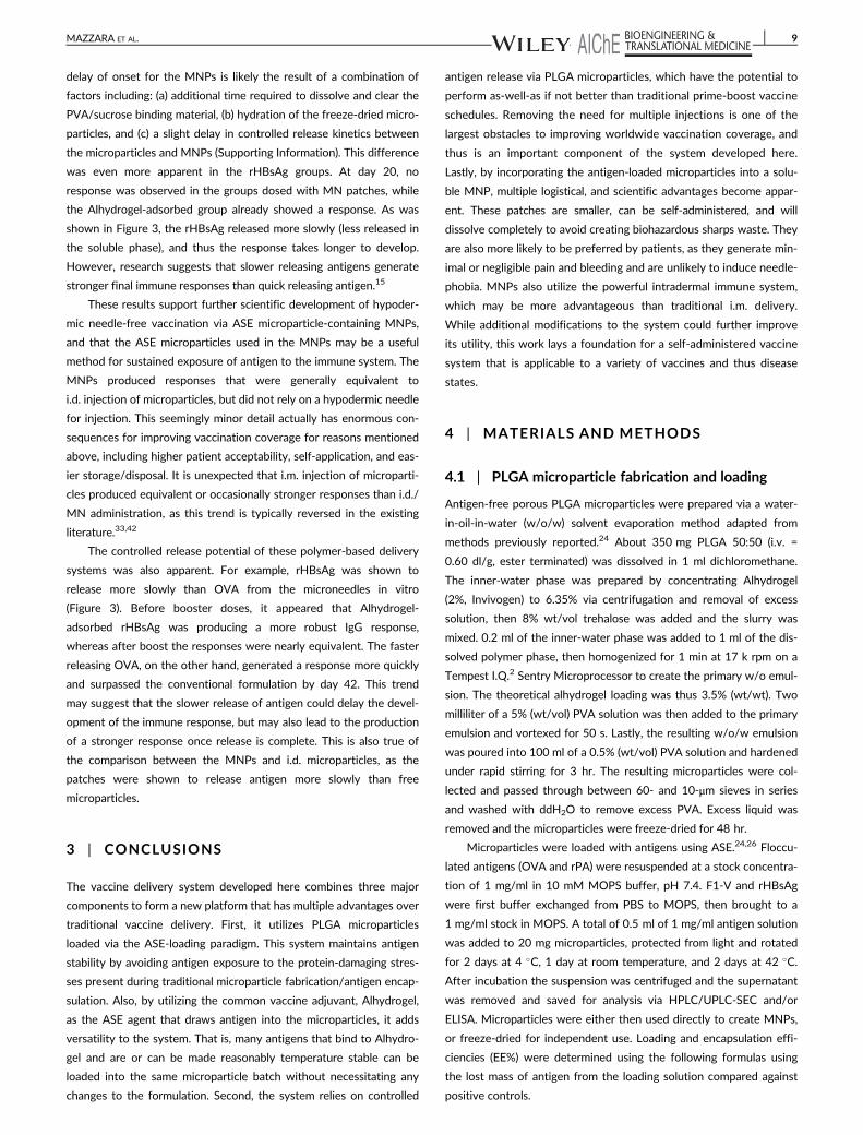
delay of onset for the MNPs is likely the result of a combination of
factors including: (a) additional time required to dissolve and clear the
PVA/sucrose binding material, (b) hydration of the freeze-dried micro-
particles, and (c) a slight delay in controlled release kinetics between
the microparticles and MNPs (Supporting Information). This difference
was even more apparent in the rHBsAg groups. At day 20, no
response was observed in the groups dosed with MN patches, while
the Alhydrogel-adsorbed group already showed a response. As was
shown in Figure 3, the rHBsAg released more slowly (less released in
the soluble phase), and thus the response takes longer to develop.
However, research suggests that slower releasing antigens generate
stronger final immune responses than quick releasing antigen.15
These results support further scientific development of hypoder-
mic needle-free vaccination via ASE microparticle-containing MNPs,
and that the ASE microparticles used in the MNPs may be a useful
method for sustained exposure of antigen to the immune system. The
MNPs produced responses that were generally equivalent to
i.d. injection of microparticles, but did not rely on a hypodermic needle
for injection. This seemingly minor detail actually has enormous con-
sequences for improving vaccination coverage for reasons mentioned
above, including higher patient acceptability, self-application, and eas-
ier storage/disposal. It is unexpected that i.m. injection of microparti-
cles produced equivalent or occasionally stronger responses than i.d./
MN administration, as this trend is typically reversed in the existing
literature.33,42
The controlled release potential of these polymer-based delivery
systems was also apparent. For example, rHBsAg was shown to
release more slowly than OVA from the microneedles in vitro
(Figure 3). Before booster doses, it appeared that Alhydrogel-
adsorbed rHBsAg was producing a more robust IgG response,
whereas after boost the responses were nearly equivalent. The faster
releasing OVA, on the other hand, generated a response more quickly
and surpassed the conventional formulation by day 42. This trend
may suggest that the slower release of antigen could delay the devel-
opment of the immune response, but may also lead to the production
of a stronger response once release is complete. This is also true of
the comparison between the MNPs and i.d. microparticles, as the
patches were shown to release antigen more slowly than free
microparticles.
3 | CONCLUSIONS
The vaccine delivery system developed here combines three major
components to form a new platform that has multiple advantages over
traditional vaccine delivery. First, it utilizes PLGA microparticles
loaded via the ASE-loading paradigm. This system maintains antigen
stability by avoiding antigen exposure to the protein-damaging stres-
ses present during traditional microparticle fabrication/antigen encap-
sulation. Also, by utilizing the common vaccine adjuvant, Alhydrogel,
as the ASE agent that draws antigen into the microparticles, it adds
versatility to the system. That is, many antigens that bind to Alhydro-
gel and are or can be made reasonably temperature stable can be
loaded into the same microparticle batch without necessitating any
changes to the formulation. Second, the system relies on controlled
antigen release via PLGA microparticles, which have the potential to
perform as-well-as if not better than traditional prime-boost vaccine
schedules. Removing the need for multiple injections is one of the
largest obstacles to improving worldwide vaccination coverage, and
thus is an important component of the system developed here.
Lastly, by incorporating the antigen-loaded microparticles into a solu-
ble MNP, multiple logistical, and scientific advantages become appar-
ent. These patches are smaller, can be self-administered, and will
dissolve completely to avoid creating biohazardous sharps waste. They
are also more likely to be preferred by patients, as they generate min-
imal or negligible pain and bleeding and are unlikely to induce needle-
phobia. MNPs also utilize the powerful intradermal immune system,
which may be more advantageous than traditional i.m. delivery.
While additional modifications to the system could further improve
its utility, this work lays a foundation for a self-administered vaccine
system that is applicable to a variety of vaccines and thus disease
states.
4 | MATERIALS AND METHODS
4.1 | PLGA microparticle fabrication and loading
Antigen-free porous PLGA microparticles were prepared via a water-
in-oil-in-water (w/o/w) solvent evaporation method adapted from
methods previously reported.24 About 350 mg PLGA 50:50 (i.v. =
0.60 dl/g, ester terminated) was dissolved in 1 ml dichloromethane.
The inner-water phase was prepared by concentrating Alhydrogel
(2%, Invivogen) to 6.35% via centrifugation and removal of excess
solution, then 8% wt/vol trehalose was added and the slurry was
mixed. 0.2 ml of the inner-water phase was added to 1 ml of the dis-
solved polymer phase, then homogenized for 1 min at 17 k rpm on a
Tempest I.Q.2 Sentry Microprocessor to create the primary w/o emul-
sion. The theoretical alhydrogel loading was thus 3.5% (wt/wt). Two
milliliter of a 5% (wt/vol) PVA solution was then added to the primary
emulsion and vortexed for 50 s. Lastly, the resulting w/o/w emulsion
was poured into 100 ml of a 0.5% (wt/vol) PVA solution and hardened
under rapid stirring for 3 hr. The resulting microparticles were col-
lected and passed through between 60- and 10-μm sieves in series
and washed with ddH2O to remove excess PVA. Excess liquid was
removed and the microparticles were freeze-dried for 48 hr.
Microparticles were loaded with antigens using ASE.24,26 Floccu-
lated antigens (OVA and rPA) were resuspended at a stock concentra-
tion of 1 mg/ml in 10 mM MOPS buffer, pH 7.4. F1-V and rHBsAg
were first buffer exchanged from PBS to MOPS, then brought to a
1 mg/ml stock in MOPS. A total of 0.5 ml of 1 mg/ml antigen solution
was added to 20 mg microparticles, protected from light and rotated
for 2 days at 4 �C, 1 day at room temperature, and 2 days at 42 �C.
After incubation the suspension was centrifuged and the supernatant
was removed and saved for analysis via HPLC/UPLC-SEC and/or
ELISA. Microparticles were either then used directly to create MNPs,
or freeze-dried for independent use. Loading and encapsulation effi-
ciencies (EE%) were determined using the following formulas using
the lost mass of antigen from the loading solution compared against
positive controls.
MAZZARA ET AL. 9

%wtwt
loading :mass of antigen encapsulated bymicroparticles
mass of microparticles×100%
EE%:mass of antigen encapsulated bymicroparticlesinitial mass of antigen in the loading solution
×100%
4.2 | Fabrication of microneedle patches
MNP fabrication methods have been described previously and their
adaptation to this study is summarized here45 The patches were fabri-
cated by casting onto polydimethtylsioxane (PDMS) molds to produce
MNPs containing a 10 × 10 array of pyramidal microneedles (300 ×
300 × 600 μm3) with tip-to-tip spacing at 640 μm.
To make standard MNPs (lacking a pedestal), antigen-loaded
microparticles were first washed 3× with MOPS, then resuspended in
cold ddH2O at an approximate concentration of 40 mg/ml and kept
on ice. About 25 μl of the microparticle suspension was pipetted onto
the surface of the PDMS mold, and the mold was pulled under vac-
uum for 10 min at approximately 25 in.Hg. Excess suspension was
then removed and returned to the stock for reuse. The mold was then
centrifuged for 10 min at 3,220 rcf at 4 �C. Excess microparticles were
removed from the surface of the mold via gentle tape-stripping.
Approximately 90 μl of a 40% PVA + 30% sucrose (wt/vol) solution
was then applied over the molds, and pulled under vacuum for
30 min. The patches were then allowed to dry in a fume hood over-
night before being demolded and trimmed of excess material around
the edges to form a �1 cm2 square patch. The patches were then
freeze-dried for >48 hr. Patches were stored under desiccation at
4 �C until use.
Plastic pedestal masters were 3D printed with assistance from
the University of Michigan 3D lab using a ProJet 3500 HD Max
printer. The pedestal was prepared based on lithography methods pre-
viously described.48 It consisted of a 10 × 10 array that could be over-
laid onto the MNP mold (equivalent center-to-center spacing of
640 μm), made of pyramidal trapezoids with a 300 μm wide square
base, 800 μm tall, and a 130 μm wide square top. After fabrication the
mold was cleaned of printing oil, then a PDMS mold was cast from
the structure. This new mold allowed the pedestal part to be recre-
ated using excipients with expected excellent biocompatibility
(PVA/sucrose).
To create pedestal patches, the aforementioned patch process
was carried out identically through the first centrifugation step. After
tape-stripping away excess surface microparticles, 25 μl of the PVA/-
sucrose mixture (as above) was vacuumed onto the mold for 10 min
while the mold was covered to prevent evaporation and premature
hardening of the patch. Surface PVA/sucrose was then removed using
a razor under a stereomicroscope (Nikon Olympus). The PVA/sucrose
pedestal part was then manually aligned with the microneedle cavities
(still in the mold) such that the tip of each pedestal aligned with the
tip of the microneedle molds. The pedestal was then gently pressed
into the mold and was allowed to dry in-place in a fume hood over-
night. Patches were then demolded and freeze-dried. Each patch used
in this study was visualized on a stereomicroscope to ensure micro-
needle quality. Malformed patches were occasionally formed, but
discarded.
4.3 | In vitro release and stability
For in vitro evaluation of antigen-loaded microparticles, microparticles
were suspended in 1 ml PBST (PBS + 0.02% Tween 80). For MNPs,
four patches were first dissolved in ddH2O and rinsed 5× to remove
PVA/sucrose microneedle matrix material, then the remaining micro-
particles were resuspended in 0.25 ml PBST. Release studies were
carried out at 37 �C while protected from light and shaken at
240 rpm. At each timepoint (1, 3, 7 days and weekly thereafter), sam-
ples were spun-down and the full release media was removed for anti-
gen analysis via HPLC/UPLC-SEC and/or ELISA.
4.4 | Size exclusion Chromatogaphy of antigens
Unless otherwise stated, antigen concentration was determined by
SEC using either high or ultra performance liquid chromatography
(HPLC/UPLC). In either case, the mobile phase consisted of PBS,
pH 7.4, flowed at 1 ml/min (HPLC) or 0.4 ml/min (UPLC). Injection
volumes were 50 or 10 μl for HPLC and UPLC, respectively. All sam-
ples were filtered through 0.45 μm filters prior to injection. A TSKgel
G3000SWxl column was used for HPLC and an Acquity BEH SEC (4.6
× 150 mm) column was used for UPLC. UV detection was done at
215 nm. All samples were carried out in triplicate or greater, and only
monomeric protein content was considered.
4.5 | Total nitrogen analysis
Total protein content was extrapolated from total nitrogen content
using a modified automated Dumas technique.62 Microparticle pellets
were washed 3× with ddH2O, then freeze-dried. About 1–4 mg of
microparticles were massed into tin pans, which were crimped to
remove excess air. Samples were run on a Leco TrueSpec Micro CHN.
The instrument was first blanked without samples to establish atmo-
spheric baselines. Carbon, hydrogen, and nitrogen standards were
then set in the anticipated range of nitrogen mass using USP-grade
EDTA. Lyophilized antigen standards were run to verify the percent
nitrogen in the protein and set a Protein Factor. Microparticle samples
were then dropped into the combustion chamber at 1,050 �C, which
converts all nitrogen to nitrogen gas, which is then quantified by a
thermal conductivity cell. Protein content was determined by multi-
plying the nitrogen mass by the protein factor after first subtracting
the nitrogen mass from negative controls (unloaded microparticles).
Percent protein could then be determined by dividing protein mass by
total sample mass.
4.6 | Confocal microscopy
To visualize the distribution of antigen inside the microparticles after
encapsulation, microparticles were loaded using an Ovalbumin-Alexa
Fluor 647 conjugate (fOVA) similar to as described above. After wash-
ing, the microparticles were resuspended in ddH2O and placed on a
glass slide with a coverslip and cross-sectional Z-stacked images were
taken on a Nikon A-1 spectral confocal laser scanning microscope
(CLSM) operating with a Cy5 filter and NIS Elements viewing and
analysis software.
10 MAZZARA ET AL.

To evaluate the particulate release fraction, fOVA-loaded micro-
particles were resuspended in PBST at 37 �C. At predetermined time
points, a sample of the suspension was removed and washed with
ddH2O before similarly imaging as above via CLSM. Images were com-
pared against Alhydrogel that had similarly been loaded with fOVA
and washed of unbound antigen.
4.7 | Microneedle insertion
For ex vivo evaluation of mechanical integrity, excised porcine ear tis-
sue was used. The shaved inner skin with cartilage attached was sepa-
rated from the outer skin and subcutaneous fat, and pinned taut.
MNPs were gently placed tip-down onto the skin, and pressed in
firmly with the thumb for 10 s. The patch was then removed and Gen-
tian Violet (Ricca Chemical Co.) was applied to the application site for
1 min before being wiped away with an alcohol pad. The application
site was then cut away and imaged on a stereomicroscope (n = 5 for
each patch type).
To evaluate depth of penetration/microparticle deposition, micro-
particles loaded with OVA-AlexaFluor 488 conjugate were fabricated
into MNPs and the experiment was performed similar to above,
except patches were held on the tissue for 5 min with pressure, then
placed in a 37 �C chamber at 98% humidity for 15 additional minutes
to allow the microneedless to dissolve. The backing of the patches
was gently removed and the application site tissue was cut out and
embedded in OCT compound, which was subsequently dipped in iso-
pentane chilled by surrounding LN2. The samples were then cut into
50 μm sections using a Leicia 3050S cryostat onto Superfrost+ micro-
scope slides. Slides were thawed and immediately imaged on an Olym-
pus fluorescent stereomicroscope.
To determine the mass of microparticles delivered upon applica-
tion of MNPs, male nude BALB/c mice were cleared of any light hair
using depilatory cream (Nair) 1 day in advance of patch application.
Mice were anesthetized and placed on a heated pad to maintain body
temperature. A fold of skin from the dorsal flank was pulled from the
body and held taut on a cutting board. A MNP was gently pressed into
the skin for 5 min. Pressure was then removed and the patch was kept
on the skin for an additional 15 min. The remaining portion of the
patch was then removed and placed in a microcentrifuge tube. Four
patches were used per sample (n = 3 samples). The patches were then
dissolved in ddH2O and washed 5×, then dried in a vacuum oven at
40 �C overnight. To account for residual animal tissue that was picked
up by the patches, the mass of microparticles remaining in the patches
after application was determined by gel permeation chromatography
(GPC). Briefly, the residual microparticles were dissolved in tetrahy-
drofuran, filtered, and ran on a GPC column against standard masses
of dissolved microparticles.
4.8 | In vivo microparticle tracking
The treatment of all experimental animals in these procedures were in
accordance with University committee on use and care of animals
(University of Michigan UCUCA), and all NIH guidelines for the care
and use of laboratory animals. Pedestal MNPs were made loaded with
fOVA and applied to male albino C57BL/6J mice as described above.
Two patches were applied per mouse, to the left anterior and right
posterior dorsal flank. At predetermined time-points, the whole animal
was anesthetized and imaged using a PerkinElmer IVIS Spectrum
imaging system. Fluorescence data was processed using a region-of-
interest analysis with background subtraction using Living Image 4.5
software. Other study groups included mice given an i.d. injection to
the same locations of an equivalent delivered dose of fOVA-loaded
microparticles or soluble fOVA. Mice were kept on an alfalfa-free diet
to reduce autofluorescence. Depilatory cream was not reapplied dur-
ing the study, but hair was kept trimmed using electric razors (n = 4
mice/group, two applications per mouse).
4.9 | Skin resealing
TEWL was measured using a Delfin Technologies VapoMeter with
DelfWin 4 capture software. Study groups consisted of application of:
(a) PLA master patches (no pedestal), (b) ASE microparticle-loaded
pedestal patches, (c) pedestal patches loaded with smaller nano-sized
PLGA particles,59 and (d) vehicle-only patches (pedestal MNPs made
of only PVA/sucrose, no microparticles). Three measurements were
taken per application site, per animal, at each timepoint, and the
TEWL chamber was allowed to re-equilibrate to environmental condi-
tions before each measurement. To measure TEWL, the VapoMeter
was gently pressed against the application site without manual ten-
sion applied to the skin. Data are presented as percent increase over
an application control using ANOVA with Fisher's LSD. The applica-
tion control consisted of a flat PVA/sucrose mock patch that did not
contain any microneedles, but was applied similarly to other groups.
4.10 | Immunizations
C57Bl/6 (for OVA groups) or BALB/c (for rHBsAg groups) mice,
5–6 weeks old, five mice/group, were purchased from Jackson Labo-
ratories. The choice of mouse strain was reliant on reagents available
for the different antigens. One day prior to priming and booster immu-
nization the application site for MN patches or i.d. administered
groups was shaved and depilatory cream was applied, or just shaved
for i.m. administered groups. On day zero mice were immunized with
either: (a) two pedestal MNPs, or equivalent delivered antigen dose
from, (b) i.d. microparticles, (c) i.m. microparticles, (d) Alhydrogel-
adsorbed antigen, or (e) soluble antigen. A sham group received
patches containing microparticles that did not contain antigen.
Booster doses were given 21 days after the priming dose.
To evaluate antibody titers, blood was drawn on days 20 and
42 via submandibular bleed. Serum was separated using Microvette
500 Zgel serum collection tubes centrifuged for 5 min at 10,000 rcf.
Serum was stored at −80 �C until analysis. Serum samples were ana-
lyzed by the University of Michigan Cancer Center Immunology Core
for IgG, IgG1, and IgG2c via ELISA. Due to reagent availability,
antigen-specific IgG1 isotype was used as a standard for all IgGs to
determine relative concentration. Data were compared using one-way
ANOVA with Tukey's post-test via GraphPad Prism software.
To evaluate the nature of the cytokine response produced after
restimulation of splenic lymphocytes, all mice were euthanized on day
42 and spleens were collected under sterile conditions. Splenocytes
MAZZARA ET AL. 11

were collected by grinding each spleen through a 70 μm nylon
strainer. Red blood cells were lysed with ACK lysing buffer and the
cells were washed 3× with sterile PBS before being resuspended in
RPMI 1640 media supplemented with glutamine, 10% FBS (10%),
1 U/ml penicillin + 1 μg/ml streptomycin, 55 μM 2-mercaptoethanol,
MEM nonessential amino acids (1%), 1 mM sodium pyruvate, and
10 mM HEPES. Cells were then plated at 5 × 10⁵ cells/well in a
96-well plate and stimulated with media (negative control) or
25 μg/ml whole antigen (OVA or rHBsAg). Positive controls were
pooled from each spleen within a group and stimulated with 2 μl/ml
PMA/ionomycin (cell stimulation cocktail). Cells were incubated for
96 hr at 37 �C with 5% CO2 before collecting the supernatant and
storing at −80 �C. Concentrations of IL2, IL6, IL10, and TNFα were
analyzed via ELISA through the University of Michigan Cancer Center
Immunology Core. Stimulated cell supernatants were compared
against negative controls using Student's t-test.
ACKNOWLEDGMENTS
This study was funded in part by a Paul Professorship to SPS. JMM
was funded by a pre-doctoral fellowship from the Pharmaceutical
Research and Manufacturers of America Foundation. Mark Prausnitz
is an inventor of patents that have been or may be licensed to compa-
nies developing MNP-based products, a paid advisor to companies
developing MNP-based products, and is a founder/shareholder of
companies developing MNP-based products (e.g., Micro Biomedical).
The resulting potential conflict of interest has been disclosed and is
managed by the Georgia Institute of Technology and Emory
University.
LITERATURE CITED
1. Glenn GM, Kenney RT. Mass vaccination: solutions in the skin. CurrTop Microbiol. 2006;304:247-268.
2. Belshe RB, Newman FK, Wilkins K, et al. Comparative immunogenicityof trivalent influenza vaccine administered by intradermal or intramus-cular route in healthy adults. Vaccine. 2007;25(37–38):6755-6763.
3. Koutsonanos DG, Compans RW, Skountzou I. Targeting the skin formicroneedle delivery of influenza vaccine. Adv Exp Med Biol. 2013;785:121-132.
4. Marshall S, Sahm LJ, Moore AC. The success of microneedle-mediatedvaccine delivery into skin. Hum Vaccin Immunother. 2016;12(11):2975-2983.
5. Cleland JL. Single-administration vaccines: controlled-release technol-ogy to mimic repeated immunizations. Trends Biotechnol. 1999;17(1):25-29.
6. Kim YC, Park JH, Prausnitz MR. Microneedles for drug and vaccinedelivery. Adv Drug Deliv Rev. 2012;64(14):1547-1568.
7. Prausnitz MR, Langer R. Transdermal drug delivery. Nat Biotechnol.2008;26(11):1261-1268.
8. Norman JJ, Arya JM, McClain MA, Frew PM, Meltzer MI,Prausnitz MR. Microneedle patches: usability and acceptability forself-vaccination against influenza. Vaccine. 2014;32:1856-1862.
9. Indermun S, Luttge R, Choonara YE, et al. Current advances in the fab-rication of microneedles for transdermal delivery. J Control Release.2014;185:130-138.
10. Lofthouse S. Immunological aspects of controlled antigen delivery.Adv Drug Deliv Rev. 2002;54(6):863-870.
11. O'Hagan DT, Singh M, Gupta RK. Poly(lactide-co-glycolide) microparti-cles for the development of single-dose controlled-release vaccines.Adv Drug Deliv Rev. 1998;32(3):225-246.
12. Kohn J, Niemi SM, Albert EC, Murphy JC, Langer R, Fox JG.
Single-step immunization using a controlled release, biodegradable
polymer with sustained adjuvant activity. J Immunol Methods. 1986;
95(1):31-38.13. Sanchez A, Gupta RK, Alonso MJ, Siber GR, Langer R. Pulsed
controlled-release system for potential use in vaccine delivery. J
Pharm Sci. 1996;85(6):547-552.14. Kemp JM, Kajihara M, Nagahara S, Sano A, Brandon M, Lofthouse S.
Continuous antigen delivery from controlled release implants induces
significant and anamnestic immune responses. Vaccine. 2002;20(7–8):1089-1098.
15. Johansen P, Storni T, Rettig L, et al. Antigen kinetics determines
immune reactivity. Proc Natl Acad Sci USA. 2008;105(13):5189-5194.16. Wischke C, Schwendeman SP. Degradable polymeric carriers for par-
enteral controlled drug delivery. In: Siepmann J, Siegel RA, Rathbone
MJ, eds. Fundamentals and Applications of Controlled Release Drug
Delivery. Boston, MA: Springer US; 2012:171-228.17. Schwendeman SP, Costanitino HR, Gupta RK, Langer R. Peptide, pro-
tein, and vaccine delivery from implantable polymeric systems. In:
Controlled Drug Delivery, Challenges and Strategies. Washington, DC:
American Chemical Society; 1997:229-267.18. Hoffman AS. The origins and evolution of "controlled" drug delivery
systems. J Control Release. 2008;132(3):153-163.19. Schwendeman SP, Costantino HR, Gupta RK, Peptide LR. Protein, and
vaccine delivery from implantable polymeric systems progress and
challenges. In: Park K, ed. Controlled Drug delivery: Challenges and
Strategies. Washington DC: American Chemical Society; 1997:
229-267.20. Schwendeman SP. Recent advances in the stabilization of proteins
encapsulated in injectable PLGA delivery systems. Crit Rev Ther Drug
Carrier Syst. 2002;19(1):73-98.21. Maa YF, Hsu CC. Protein denaturation by combined effect of shear
and air-liquid interface. Biotechnol Bioeng. 1997;54(6):503-512.22. Sah H. Protein behavior at the water/methylene chloride interface. J
Pharm Sci. 1999;88(12):1320-1325.23. Alexander P, Hamilton LD, Stacey KA. Irradiation of proteins in the
solid state. I. Aggregation and disorganization of secondary structure
in bovine serum albumin. Radiat Res. 1960;12:510-525.24. Desai KG, Schwendeman SP. Active self-healing encapsulation of vac-
cine antigens in PLGA microspheres. J Control Release. 2013;165(1):
62-74.25. Shah RB, Schwendeman SP. A biomimetic approach to active
self-microencapsulation of proteins in PLGA. J Control Release. 2014;
196:60-70.26. Reinhold SE, Desai KG, Zhang L, Olsen KF, Schwendeman SP.
Self-healing microencapsulation of biomacromolecules without
organic solvents. Angew Chem Int Ed Engl. 2012;51(43):10800-10803.27. Huang J, Mazzara JM, Schwendeman SP, Thouless MD. Self-healing
of pores in PLGAs. J Control Release. 2015;206:20-29.28. Donnelly RF, Raj Singh TR, Woolfson AD. Microneedle-based drug
delivery systems: microfabrication, drug delivery, and safety. Drug
Deliv. 2010;17(4):187-207.29. Park JH, Allen MG, Prausnitz MR. Polymer microneedles for
controlled-release drug delivery. Pharm Res. 2006;23(5):1008-1019.30. Gill HS, Denson DD, Burris BA, Prausnitz MR. Effect of microneedle
design on pain in human volunteers. Clin J Pain. 2008;24(7):585-594.31. Haq MI, Smith E, John DN, et al. Clinical administration of micronee-
dles: skin puncture, pain and sensation. Biomed Microdevices. 2009;
11(1):35-47.32. Arya J, Prausnitz MR. Microneedle patches for vaccination in develop-
ing countries. J Control Release. 2016;240:135-141.33. Quan FS, Kim YC, Vunnava A, et al. Intradermal vaccination with influ-
enza virus-like particles by using microneedles induces protection
superior to that with intramuscular immunization. J Virol. 2010;84(15):
7760-7769.34. Kim YC, Quan FS, Compans RW, Kang SM, Prausnitz MR. Formulation
and coating of microneedles with inactivated influenza virus to
improve vaccine stability and immunogenicity. J Control Release. 2010;
142(2):187-195.
12 MAZZARA ET AL.

35. Mazzara JM, Balagna MA, Thouless MD, Schwendeman SP. Healingkinetics of microneedle-formed pores in PLGA films. J Control Release.2013;171(2):172-177.
36. Blasi P, D'Souza SS, Selmin F, DeLuca PP. Plasticizing effect ofwater on poly(lactide-co-glycolide). J Control Release. 2005;108(1):1-9.
37. Champion JA, Walker A, Mitragotri S. Role of particle size in phagocy-tosis of polymeric microspheres. Pharm Res. 2008;25(8):1815-1821.
38. Baylor NW, Egan W, Richman P. Aluminum salts in vaccines—US per-spective. Vaccine. 2002;20(suppl 3):S18-S23.
39. Marrack P, McKee AS, Munks MW. Towards an understanding of theadjuvant action of aluminium. Nat Rev Immunol. 2009;9(4):287-293.
40. Shirodkar S, Hutchinson RL, Perry DL, White JL, Hem SL. Aluminumcompounds used as adjuvants in vaccines. Pharm Res. 1990;7(12):1282-1288.
41. Jiang G, Joshi SB, Peek LJ, et al. Anthrax vaccine powder formulationsfor nasal mucosal delivery. J Pharm Sci. 2006;95(1):80-96.
42. Demuth PC, Garcia-Beltran WF, Ai-Ling ML, Hammond PT, Irvine DJ.Composite dissolving microneedles for coordinated control of antigenand adjuvant delivery kinetics in transcutaneous vaccination. AdvFunct Mater. 2013;23(2):161-172.
43. Zaric M, Lyubomska O, Poux C, et al. Dissolving microneedle deliveryof nanoparticle-encapsulated antigen elicits efficient cross-primingand Th1 immune responses by murine Langerhans cells. J Invest Der-matol. 2015;135(2):425-434.
44. Larraneta E, McCrudden MT, Courtenay AJ, Donnelly RF. Micronee-dles: a new frontier in nanomedicine delivery. Pharm Res. 2016;33(5):1055-1073.
45. Chu LY, Choi SO, Prausnitz MR. Fabrication of dissolving polymermicroneedles for controlled drug encapsulation and delivery: bubbleand pedestal microneedle designs. J Pharm Sci. 2010;99(10):4228-4238.
46. Naito S, Ito Y, Kiyohara T, Kataoka M, Ochiai M, Takada K.Antigen-loaded dissolving microneedle array as a novel tool for percu-taneous vaccination. Vaccine. 2012;30(6):1191-1197.
47. Chu LY, Prausnitz MR. Separable arrowhead microneedles. J ControlRelease. 2011;149(3):242-249.
48. Chen MC, Huang SF, Lai KY, Ling MH. Fully embeddable chitosanmicroneedles as a sustained release depot for intradermal vaccination.Biomaterials. 2013;34(12):3077-3086.
49. Morefield GL, Jiang D, Romero-Mendez IZ, Geahlen RL, Hogenesch H,Hem SL. Effect of phosphorylation of ovalbumin on adsorption byaluminum-containing adjuvants and elution upon exposure to intersti-tial fluid. Vaccine. 2005;23(12):1502-1506.
50. Hem SL, Hogen EH. Aluminum-containing adjuvants: properties, for-mulation, and use. In: Singh M, ed. Vaccine Adjuvants and Delivery Sys-tems. Hoboken, NJ: John Wiley & Sons; 2007:81-114.
51. Hansen B, Belfast M, Soung G, et al. Effect of the strength of adsorp-tion of hepatitis B surface antigen to aluminum hydroxide adjuvant onthe immune response. Vaccine. 2009;27(6):888-892.
52. Iyer S, Robinett RS, HogenEsch H, Hem SL. Mechanism of adsorptionof hepatitis B surface antigen by aluminum hydroxide adjuvant. Vac-cine. 2004;22(11–12):1475-1479.
53. Lee JW, Choi SO, Felner EI, Prausnitz MR. Dissolving microneedlepatch for transdermal delivery of human growth hormone. Small.2011;7(4):531-539.
54. Kalluri H, Kolli CS, Banga AK. Characterization of microchannels cre-ated by metal microneedles: formation and closure. AAPS J. 2011;13(3):473-481.
55. Donnelly RF, Singh TR, Tunney MM, et al. Microneedle arrays allowlower microbial penetration than hypodermic needles in vitro. PharmRes. 2009;26(11):2513-2522.
56. Gupta J, Gill HS, Andrews SN, Prausnitz MR. Kinetics of skin resealingafter insertion of microneedles in human subjects. J Control Release.2011;154(2):148-155.
57. Rovee DT, Maibach HI. The epidermis in wound healing. Boca Raton,FL: CRC Press; 2004.
58. Park JH, Allen MG, Prausnitz MR. Biodegradable polymer micronee-dles: fabrication, mechanics and transdermal drug delivery. J ControlRelease. 2005;104(1):51-66.
59. Bailey B. Development and Characterization of Novel Self-EncapsulatingPoly(lactic-co-glycolic acid) Microspheres for Vaccine Delivery. Ph.D.thesis, University of Michigan, Ann Arbor, MI; 2016.
60. Clausen OP, Schjolberg AR. Turnover and maturation kinetics inregenerating mouse epidermis. Apmis. 1988;96:111-119.
61. Storni T, Kundig TM, Senti G, Johansen P. Immunity in response toparticulate antigen-delivery systems. Adv Drug Deliv Rev. 2005;57(3):333-355.
62. Etheridge RD, Pesti GM, Foster EH. A comparison of nitrogen valuesobtained utilizing the Kjeldahl nitrogen and Dumas combustionmethodologies (Leco CNS 2000) on samples typical of an animalnutrition analytical laboratory. Anim Feed Sci Tech. 1998;73(1–2):21-28.
SUPPORTING INFORMATION
Additional supporting information may be found online in the Sup-
porting Information section at the end of the article.
How to cite this article: Mazzara JM, Ochyl LJ, Hong JKY,
Moon JJ, Prausnitz MR, Schwendeman SP. Self-healing encap-
sulation and controlled release of vaccine antigens from PLGA
microparticles delivered by microneedle patches. Bioengineer-
ing & Translational Medicine. 2018;1–13. https://doi.org/10.
1002/btm2.10103
MAZZARA ET AL. 13