![Review Article Current Perspectives in NSAID-Induced ... · reducing the NSAID-induced gastropathy [ ].... Acid Suppressants. Acid increases NSAID-induced mucosal injury and gastric](https://static.fdocuments.us/doc/165x107/60b703eed9b43379f63b197c/review-article-current-perspectives-in-nsaid-induced-reducing-the-nsaid-induced.jpg)
Self-assembling Ultrashort NSAID-Peptide Nanosponges ... · with under-lying disease. Wound...
47
Self-assembling Ultrashort NSAID-Peptide Nanosponges: Multifunctional Antimicrobial and Anti-inflammatory Materials McCloskey, A. P., Gilmore, S. M., Zhou, J., Draper, E. R., Porter, S., Gilmore, B., Xu, B., & Laverty, G. (2016). Self-assembling Ultrashort NSAID-Peptide Nanosponges: Multifunctional Antimicrobial and Anti-inflammatory Materials. RSC Advances, 6(115), 114738-114749. https://doi.org/10.1039/C6RA20282A Published in: RSC Advances Document Version: Peer reviewed version Queen's University Belfast - Research Portal: Link to publication record in Queen's University Belfast Research Portal Publisher rights © Royal Society of Chemistry 2016. This work is made available online in accordance with the publisher’s policies. General rights Copyright for the publications made accessible via the Queen's University Belfast Research Portal is retained by the author(s) and / or other copyright owners and it is a condition of accessing these publications that users recognise and abide by the legal requirements associated with these rights. Take down policy The Research Portal is Queen's institutional repository that provides access to Queen's research output. Every effort has been made to ensure that content in the Research Portal does not infringe any person's rights, or applicable UK laws. If you discover content in the Research Portal that you believe breaches copyright or violates any law, please contact [email protected]. Download date:26. Jun. 2020
Transcript of Self-assembling Ultrashort NSAID-Peptide Nanosponges ... · with under-lying disease. Wound...
Self-assembling Ultrashort NSAID-Peptide Nanosponges:Multifunctional Antimicrobial and Anti-inflammatory Materials
McCloskey, A. P., Gilmore, S. M., Zhou, J., Draper, E. R., Porter, S., Gilmore, B., Xu, B., & Laverty, G. (2016).Self-assembling Ultrashort NSAID-Peptide Nanosponges: Multifunctional Antimicrobial and Anti-inflammatoryMaterials. RSC Advances, 6(115), 114738-114749. https://doi.org/10.1039/C6RA20282A
Published in:RSC Advances
Document Version:Peer reviewed version
Queen's University Belfast - Research Portal:Link to publication record in Queen's University Belfast Research Portal
Publisher rights© Royal Society of Chemistry 2016. This work is made available online in accordance with the publisher’s policies.
General rightsCopyright for the publications made accessible via the Queen's University Belfast Research Portal is retained by the author(s) and / or othercopyright owners and it is a condition of accessing these publications that users recognise and abide by the legal requirements associatedwith these rights.
Take down policyThe Research Portal is Queen's institutional repository that provides access to Queen's research output. Every effort has been made toensure that content in the Research Portal does not infringe any person's rights, or applicable UK laws. If you discover content in theResearch Portal that you believe breaches copyright or violates any law, please contact [email protected].
Download date:26. Jun. 2020
1
Self-assembling Ultrashort NSAID-Peptide Nanosponges: Multifunctional Antimicrobial
and Anti-inflammatory Materials
A. P. McCloskey,a S. M. Gilmore,a J. Zhou,b E. R. Draper,c S. Porter,a B. F. Gilmore,a Bing
Xub and G. Laverty,*a
aSchool of Pharmacy, Queen’s University, Belfast, BT9 7BL, N. Ireland.
bDepartment of Chemistry, Brandeis University, Waltham, Massachusetts, MA 02454, USA.
cDepartment of Chemistry, University of Liverpool, Liverpool, L69 7ZD, UK..
Abstract
Peptide-based materials are receiving significant attention for use within biomedicine due to
their high chemical and functional versatility enabling tailoring of their structure to replicate
the properties of host tissue and the extracellular matrix. This paper studies the design,
synthesis and characterization of NSAID-peptide conjugates. Attachment of NSAIDs to a
diphenylalanine-dilysine (FFKK-OH) peptide sequence generates supramolecular hydrogel
forming molecules with antimicrobial and anti-inflammatory properties. NSAID-peptides
demonstrate broad-spectrum antimicrobial activity against both Gram-positive and Gram-
negative bacteria implicated in a variety of antimicrobial resistant nosocomial infections
including Staphylococcus aureus and Pseudomonas aeruginosa. Naproxen-peptides show
particular promise, forming biocompatible nanofibrous viscoelastic hydrogel nanosponges
composed of β-sheet secondary structures at low concentrations (0.4% w/v). Conjugation of
the peptide motif FFKK-OH to naproxen increases selectivity for COX-2 enzyme, implicated
in chronic wound scar-tissue formation. Our findings suggest that ultrashort NSAID-peptides
have potential use as multifunctional materials for a range of biomedical applications. This
includes as topical agents for treatment of chronic wounds, where a profile of persistent
inflammation, pain and the presence of infection has been proven to be detrimental to
2
successful wound repair. This work may also serve as a template for the design of future
medical device coatings with tailored antimicrobial and anti-inflammatory properties.
Introduction
Antimicrobial resistance is becoming an increasingly menacing threat to society and is
currently attributed to at least 700,000 deaths worldwide annually. A U.K Government review
in 2014 concluded that without significant investment in new therapies this total would
increase to more than 10 million deaths by 2050, a figure greater than predicted for cancer.1
Hospital-acquired infections, especially those related to implantation of medical devices and
skin wounds, are major contributors to this threat demonstrating an increased prevalence of
microorganisms that display resistance to standardly employed antimicrobial therapies. These
are responsible for increased patient morbidity and mortality, and significant economic cost
due to extended hospital stay/sick days. Infections are particularly problematic among those
with increased susceptibility and compromised immunity, for example the elderly and those
with under-lying disease.
Wound infections can be categorized into a variety of forms; most problematic being surgical
site infections (SSIs), diabetic ulcers and major trauma, for example burns. SSIs are one of the
most frequently reported hospital acquired infections accounting for 31% of nosocomial
infections and are a reservoir for so-called hospital superbugs including Staphylococcus
aureus and Pseudomonas aeruginosa.2 SSIs are present in nearly 5% of patients who undergo
a surgical procedure and over a third of postoperative deaths are related to SSIs.3 It is clear
SSIs remain a significant burden to healthcare. Failure of antimicrobial therapy and/or
unresolved healing can result in the development of a chronic wound.
Chronic infected wounds are particularly problematic with a profile of exaggerated
inflammation and resistant infection severely limiting wound healing.4 They are responsible
3
for multiple treatment failures and commonly require a minimum of eight weeks to fully
resolve, due to increased microbial resistance to standard drug regimens and impaired healing.
Nonhealing wounds costs the US healthcare system $3billion annually and affects over 2% of
the population. 5,6 Acute and chronic wounds are resolved through similar biomolecular
pathways, however chronic wounds commonly stall in the inflammatory phase partly due to
the presence of pathogenic biofilm infection.7 Implanted biomaterials are also associated with
an exaggerated host immune response, termed the foreign body response. Sustained
inflammation, present primarily within chronic wounds and biomaterial infections, has been
proven to be detrimental to wound repair.8 This has commonly been minimized and controlled
using steroidal and non-steroidal anti-inflammatory drugs (NSAIDs), limiting the action of
inflammatory mediators such as prostaglandins. Long-term systemic use of such drugs leads
to undesirable side effects including adverse gastrointestinal, renal and cardiovascular risks.
Debate still exists regarding the use of systemic NSAIDs to aid wound repair due to possible
anti-proliferative effects demonstrated in animal studies and their possible contribution to
increasing the severity of group A streptococcal infections.9,10 Despite reservations with
systemic NSAID use, localized sustained release systems are commonly employed and
NSAID topical applications demonstrate benefit in the treatment of chronic wounds.11 For
example an ibuprofen foam formulation provides localized delivery, reduced pain, a moist
environment and has been proven to be advantageous for the resolution of chronic venous leg
ulcers.12
There has been increased interest in the therapeutic potential of antimicrobial peptides.
Displaying multiple modes of intracellular and extracellular action, antimicrobial peptides
demonstrate a reduced propensity for developing antimicrobial resistance and are a significant
prospect for solving the current shortage of antimicrobials in pharmaceutical development.13
Several research groups have studied their incorporation into synthetic hydrogel matrices for
4
the prevention of biomaterial and wound infections with promising results thus far.14-16
Hydrogels possessing inherent antimicrobial properties represent a significant benefit by
reducing microbial contamination within the matrix and at the wound/implantation site.17
Self-assembling biomimetic hydrogels, including peptide-based strategies, have received
attention as biofunctional materials and drug delivery platforms due to their similarity to the
extracellular tissue matrix. Peptide-based materials possess: increased chemical functionality
and versatility; improved biocompatibility; increased water capacity; moisture vapor
transmission and gaseous exchange; biodegradability and tailored immunogenicity. Hydrogels
provide improved healing at wound sites by enabling rapid absorption of wound exudate and
protection of newly formed skin. Researchers have begun to employ a strategy whereby
peptides are optimized to obtain multi-functionality, both pharmacological (antimicrobial,
anti-biofilm, anti-inflammatory, analgesia) and physical (self-assembly, hydrogelation),
within a single molecule. Most recently our group developed an ultrashort cationic peptide
motif with selective activity against resistant pathogens implicated in medical device related
infections.18 Ultrashort peptides are of particular interest to the pharmaceutical industry as
they are generally more cost-effective to synthesize relative to their larger peptide/protein
counterparts and therefore more realistic molecules for clinical translation.
Self-assembling hydrogel systems based on drug molecules eliminates the necessity for a drug
delivery vehicle, typically a synthetic polymer and may be tailored to respond to
environmental and physiological stimuli, for example pH change or the presence of specific
enzymes.19,20 Synthetic-based polymers demonstrate limited drug loading, are difficult to
functionalize and acidic degradation products can induce an unwanted inflammatory
response.21 The presence of aromatic groups, such as those present within NSAIDs, facilitates
assembly of NSAID-peptide conjugates where the NSAID moiety replaces more traditional
aromatic groups found in self-assembling systems such as carboxybenzyl,22 9-
5
fluorenylmethyloxycarbonyl (Fmoc),23 and 2-naphthoyl (Nap).24 Work thus far has focused
on NSAIDs clinically used as topical preparations for acute pain, for example ibuprofen and
naproxen.25 Recently a multifunctional approach has been successful in creating an anti-
inflammatory and anti-HIV hydrogel using naproxen conjugated to reverse transcriptase
inhibitors via a peptide linker. 26 The idea of a multifunctional antimicrobial and anti-
inflammatory platform is adopted from nature where herb extracts curcumin (Curuma longa),
dayflower (Commelina diffusa) and bark (Spathodea campanulata) have demonstrated benefit
in wound healing.27,28 Building on previously investigated NSAID conjugated peptides and
antimicrobial peptides we have developed novel self-assembling NSAID-peptides utilizing
racemic (±)-ibuprofen (Ibu), indomethacin (Ind) and (S)-(+)-naproxen (Npx) (Fig. 1).
This report examines the synthesis and characterization of NSAID conjugated ultrashort
cationic self-assembled peptide hydrogels as potential antimicrobial and anti-inflammatory
biomaterials. Incorporation of an NSAID into the ultrashort carboxylic acid terminated motif
(FFKK-OH) was hypothesized to confer improved hydrogel strength and cyclooxygenase
(COX) inhibitory properties to the molecule. The development of a NSAID conjugated
hydrogelator may also serve as a carrier for other therapeutic agents since the arrest of the
inflammatory response is one of the most important factors in successful application of
biomaterials.29
Experimental methods
Materials
Wang resin preloaded with Fmoc-Lys(Boc) (mesh size 100-200, 0.65 mmol/g substitution),
(2-(1H-benzotriazol-1-yl)-1,1,3,3-tetramethyluronium hexafluorophosphate) (HBTU),
diisopropyl ethylamine (DIEA), Fmoc and Boc protected amino acids were obtained from
Novabiochem, Merck KGaA (Dramstadt, Germany). 37% hydrochloric acid (HCl), sodium
6
hydroxide anhydrous pellets (≥99%), acetonitrile (HPLC grade, ≥99.93%), (±)-ibuprofen
(≥98%), indomethacin (≥99%), (S)-(+)-naproxen (≥99%), deuterated dimethyl sulfoxide (d6-
DMSO), deuterium oxide (D2O), deuterium chloride (DCl), sodium deuteroxide (NaOD),
trifluoracetic acid, triisopropylsilane, thioanisole and Whatman pH indicator paper (pH 1-14)
were purchased from Sigma-Aldrich (Dorset, U.K.). NCTC Clone 929 (ATCC CCL 1)
murine fibroblast subcutaneous connective tissue cells, S. epidermidis (ATCC 25984), S.
aureus (ATCC 6584), P. aeruginosa (PAO1) and E. coli (ATCC 11303) supplied by LGC
Standards (London, U.K). AlamarBlue® was obtained from AbD Serotec (Oxford, U.K.).
Sterile Nunc™ 96-well microtiter plates supplied by VWR International (Leicestershire,
U.K.). Fresh defibrinated equine erythrocytes were purchased from Laboratory Supplies and
Instruments Ltd (Antrim, U.K.). Cayman’s COX fluorescent inhibitor screening assay kit was
obtained from Cambridge Bioscience Ltd (Cambridge, U.K.).
Synthesis, purification and identification
NSAID-peptide hybrids (IbuFFKK, IndFFKK and NpxFFKK) were synthesized as per
standard Fmoc-based solid-phase peptide methods using a nitrogen bubbler apparatus.18 Wang
resin preloaded with Fmoc-Lys(Boc) was utilized to produce carboxylic acid terminated
peptides, upon cleavage with 95% v/v trifluoracetic acid, 2.5% v/v triisopropylsilane and
2.5% v/v thioanisole for 3 hours at room temperature. The peptide amine terminus was
conjugated to the corresponding carboxylic acid of the NSAID before cleavage. Standard
HBTU coupling was performed in dimethylformamide (DMF) with 4-fold molar excess of
DIEA and 3-fold excess of Fmoc-protected amino acid or NSAID used for coupling for 3
hours at room temperature. Precipitation was achieved using cold diethyl ether (-20 °C).
Crude product was dissolved in ethyl acetate and subjected to a series of washes with 1 mM
HCl (3 x 50 ml) and water (3 x 50 ml and dried over anhydrous magnesium sulfate (MgSO4).
7
NSAID-peptide identities were confirmed using 1H NMR analysis (Varian Unity Inova 400
spectrometer, Varian systems, Palo Alto, California, U.S.A.) in d6-DMSO and electrospray
mass spectroscopy (Thermo Finnigan LCQ Deca ion trap, Thermo Fisher Scientific, Waltham,
Massachusetts, U.S.A.). NSAID-peptide purity was determined by reverse-phase-HPLC
(Agilent 1260 series, Agilent Technologies Ltd, Cork, Ireland), using a Gemini C18 column
(250 mm x 4.6 mm) with a flow rate of 1.5 mL/min and gradient of 2-60% acetonitrile (30
minutes) in 0.05% TFA-water. All NSAID-peptides were found to have purity greater than
95%.
Self-assembling pH-triggered gelation
NSAID-peptide hydrogels prepared by a process of pH-triggered induction.18 The method of
gelation followed the sequence of steps outlined for of 2% w/v NSAID-peptide formulations
in Table 1. A stock solution was prepared by suspending each NSAID-peptide in deionized
water. Complete dissolution was achieved at pH 9 via titration with 1 M NaOH due to
deprotonation of the terminal carboxylic acid moiety. Changes in pH were monitored using
Whatman pH paper. Protonation of the terminal carboxylate ion, by titration with 0.5 M HCl
to pH 7, enabled formation of homogeneous hydrogels at concentrations above the minimum
gelation concentration (% w/v) for each molecule. Minimum gelation concentration (% w/v)
was defined as the lowest concentration of NSAID-peptide that formed a self-supporting
hydrogel, observed via a gel inversion assay after 24 hours development.30 Gels and solutions
were differentiated based on flow characteristics with gels remaining suspended and solutions
demonstrating flow.
8
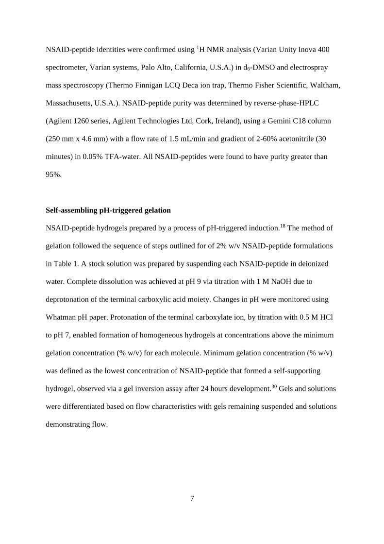
Table 1 Stepwise formulation of a self-assembling pH-triggered 2% w/v NSAID-peptide (500
µL)
Formulation step Constituent a) Quantity
1 NSAID-peptide 10mg pre-weighed
2 Deionized H2O or D2O 200 µL (in 50 µL aliquots)
3 1M NaOH or NaOD 50 µL (in 10 µL aliquots)
4 Deionized H2O or D2O 200 µL (in 50 µL aliquots)
5 0.5M HCl or DCl 20 µL (in 10 µL aliquots)
6 Deionized H2O or D2O to 500 µL
a) Deuterated solvents employed for FTIR purposes and IbuFFKK where relevant in text
Cryo-scanning electron and transmission electron microscopy
NSAID-peptides were prepared via pH induction as described and analyzed for 3D
morphology using a Hitachi Analytical Benchtop SEM TM3030 (Hitachi High-Technologies
Europe, Berkshire, U.K.) with Deben Cool Stage (Deben, Suffolk, U.K.). Cryo-SEM samples
were freeze-dried by vacuum lyophilization at 2 °C. Images were obtained at -10 °C, using an
accelerating voltage of 15 kV and a range of magnifications (15-30 000x).31 TEM was
performed using a FEI Morgagni 268 transmission electron microscope (FEI electronics,
Burlington, Massachusetts, U.S.A.). A negative staining technique was employed.31 TEM
samples were placed on 400 mesh copper grids, glow discharged and coated with a carbon
film (35nm), rinsed thrice with double distilled water, then stained trice with a 2% w/v
solution of uranyl acetate. Excess stain was removed by blotting with filter paper. The grids
were left to air dry prior to examination of the 3D architecture of the NSAID-peptide samples.

9
Fourier transform infrared spectroscopy
FTIR spectra were obtained at a resolution of 2 cm-1 and over wavelengths 4000-400 cm-1
(128 scans) using a Jasco 4000 series FTIR spectrometer (Jasco Inc. Tokyo, Japan). Samples
were prepared as described above but using deuterated solvents (D2O, DCl, NaOD).
Hydrogels were sandwiched between two calcium fluoride discs (0.05 mm spacer). A D2O,
DCl, NaOD mixture, prepared to the same concentrations as NSAID-peptide containing
samples, was used as a background and subtracted from all spectra.
Oscillatory rheology
Dynamic rheological measurements were performed using an Anton Paar Physica MCR301
rheometer. A cup and vane measuring system was used to perform frequency sweeps. For
frequency 2 mL of the gels were prepared in 7 mL Sterilin vials as described previously.30 All
experiments were performed at 25 °C from 1-100 rad s-1 at a strain of 0.0003 %.
Bacterial susceptibility assay
The ability of NSAID-peptides to reduce bacterial viability were tested using a colony
counting method previously outlined by Mateescu and Jiang.32,33 S. epidermidis (ATCC
25984), S. aureus (ATCC 6584), P. aeruginosa (PAO1) and E. coli (ATCC 11303) were
subcultured for 24 hours at 37 °C in Müller Hinton broth (MHB), optically adjusted to an
optical density reading of 0.3 at 550 nm (1 × 10⁸ colony forming units per milliter (CFU/mL))
in phosphate buffered saline (PBS) and further diluted in MHB (equivalent to 2 × 10⁶
CFU/mL) prior to plating 100 μL into each well of a microtiter plate containing 100 μL of
self-assembled NSAID-peptide (2-0.5% w/v) prepared as described above. Control wells
included bacteria treated with PBS, as the negative control (100% survival) and 2% w/v
HPMC as an inert hydrogel to study the effect of gelation on bacterial viability. Inoculated
10
microtiter plates were incubated for 24 hours in a Gallenkamp gyrorotary incubator (37 °C)
and 20 μL samples were taken from each well, serially diluted in PBS and transferred onto
Müller Hinton agar plates for colony counting via the Miles and Misra method. Results were
displayed as the mean (Log10 CFU/mL) of four replicates.
Cyclooxygenase enzyme inhibition assay
The anti-inflammatory activity of the NSAID-peptides (1- 2000 µM) were determined against
both COX-1 and COX-2 enzymes using a COX Fluorescent Inhibitor Screening Assay Kit.
Reagents used were supplied and prepared according to the kit protocol. Potassium hydroxide
(KOH), DMSO, COX-1 (SC-560) and COX-2 (DuP-697) inhibitors were supplied ready for
use. Assay buffer (x10) (final formulation: 100 mM Tris-HCl, pH 8.0), heme, arachadonic
acid (final concentration: 2 mM) were prepared as per kit instructions. The test plates (one
plate per COX enzyme) were set up according to the kit protocol with triplicates for each
NSAID-peptide concentration. The inhibitor/sample was incubated with the enzyme for 5
minutes at room temperature. 10 μL of 10-acetyl-3, 7-dihydroxyphenoxazine (ADHP) was
added followed by 10 μL of arachidonic acid (reaction initiator). The plates were incubated
for 2 minutes at room temperature and then read at an excitation wavelength of 530 nm and an
emission wavelength of 585 nm using a FLUOstar Omega Fluorometer (BMG Labtech,
Ortenberg, Germany) and Gen5 data analysis software (BioTek, Swindon, U.K.). IC50
(concentration needed to inhibit COX activity by half) values were interpreted from activity
curves (Log10 inhibitor vs normalized response) using GraphPad Prism 6 (GraphPad Software
Inc, California, USA). The percentage inhibition of the COX enzymes was calculated by
measurement of respective emission wavelengths (585nm) and using Equation 1.
11
% COX inhibition = (COX activity no inhibitor – NSAID/peptide activity)/(COX activity no
inhibitor) x 100
(1)
In each case, the background fluorescence was subtracted from each fluorescence value
obtained. The percentage inhibition of the test inhibitors SC-560 and DuP-697 enabled a
direct comparison to be made between the NSAID-peptide activity and that of a known
inhibitor. NSAID only wells ((±)-ibuprofen, indomethacin, (S)-(+)-naproxen: 1-50 µM) were
tested to determine whether or not conjugation of the NSAID to the peptide altered anti-
inflammatory activity/selectivity. All compounds were assayed in triplicate.
Hemolysis assay
The ability of NSAID-peptides to induce hemogolobin release from fresh equine erythrocytes
was tested using a method previously outlined by our group.34 100 μL of equine erythrocytes
were treated with 100 μL of NSAID-peptides for 1 hour at 37 °C. Control wells included
0.1% v/v Triton X-100 (100% hemolysis, positive control) and PBS (0% hemolysis, negative
control). Following incubation the erythrocytes were centrifuged at 1000 g and aliquots of the
supernatant used to determine hemoglobin released in a fresh 96-well microtiter plate read at
405 nm using a Tecan Sunrise plate reader (Tecan UK Ltd, Reading, U.K.) and Equation 2
below. Results are reported as the mean of six replicates.
% hemolysis = (Abs405nm peptide – Abs405nm PBS)/(Abs405nm 0.1% triton X - Abs405nm PBS) x
100
(2)
12
Cell Viability Assay
Cell cytotoxicity was assessed using murine fibroblast subcutaneous connective tissue NCTC
clone 929 (ATCC CCL 1). Cells were cultured in Minimum Essential Medium (MEM)
containing phenol red with Earle’s Salts and L-glutamine, supplemented with 10% horse
serum and 1% penicillin and streptomycin (Invitrogen, Paisley, U.K.). Cells were grown at
37 °C and 5% CO2 and subcultured at 80-90% confluency. Subculturing involved removal of
spent media, washing with sterile PBS and detachment of cell monolayers with 0.05% trypsin/
0.53 mM EDTA.4Na solution (Invitrogen, Paisley, U.K.). Cells were cultured until at least
third passage and inoculated at 1 x 10⁴ cells per well in 96-well microtiter plate and incubated
for 24 hours. The media was then removed and the cells exposed to 100 μL of a range of
NSAID-peptide samples for 24 hours. Control wells included PBS (100% viability, negative
control) and 70% ethanol treated cells (100% kill, positive control). Following exposure to
NSAID-peptides, cell viability was assessed using a 10% v/v solution of alamarBlue® diluted
in supplemented MEM and allowed to develop for 10 hours. Absorption was measured at 570
nm using a Tecan Sunrise plate reader. Cell viability was calculated using Equation 3 below
and reported as the mean of six replicates.
% cell viability = 100 - [(Abs570nm peptide – Abs570nm PBS)/(Abs570nm 70% ethanol - Abs570nm
PBS) x 100]
(3)
Cell cytotoxicity was also analysed using a Cytotoxicity Detection KitPLUS assay (Sigma-
Aldrich, Dorset, U.K.) that quantifies lactate dehydrogenase (LDH) release. LDH acts as a
marker for toxicity and is rapidly released from the cell cytosol when the plasma membrane is
damaged. After 24 hours incubation with NSAID-peptides 100 µL of the prepared
Cytotoxicity Detection KitPLUS test reagent was added to each well and incubated for 5
13
minutes after which 50 µL of the stop reagent was added to each well and absorbance
measured at 490 nm. A high LDL release control, corresponding to 100% toxicity, was
created via addition of 5 µL of lysis solution to non-peptide treated wells 15 minutes before
the addition of prepared Cytotoxicity Detection KitPLUS test reagent. Aside from background
absorbance reading, three additional experimental controls were used (as per manufacturer’s
protocol). These were: a) the peptide at the highest concentration used in media alone (500
µM), this is compared to the background to assess possible additional absorbance caused by
the treatment; b) media and LDH standard; c) peptide at highest concentration (500 µM) used
in media with an equal volume of LDH standard as a control to examine the possible effect of
the peptide on an equal quantity of LDH standard (checking if peptide is quenching or
enhancing the LDH release). No significant addition or reduction was observed in the
absorbance for each of these three controls. Cell viability was calculated using Equation 4
below and reported as the mean of six replicates.
% toxicity = (Abs490nm peptide – Abs490nm PBS)/(Abs490nm High LDL control- Abs490nm PBS) x
100
(4)
A LIVE/DEAD® Viability/Cytotoxicity fluorescent assay (Thermo Fisher Scientific,
Waltham, MA, USA) was used to quantify the ratio of viable to non-viable NCTC 929 cells
alongside fluorescence microscopy (EVOS FL microscope, Thermo Fisher Scientific,
Waltham, MA, USA). Following 24 hours incubation with NSAID-peptides, NCTC 929 cells
were incubated for 20 minutes with a mixture of 4 µM ethidium homodimer-1 and 2 µM
calcein AM in PBS. Viable cells stained green due to the conversion of calcein AM to calcein,
whilst non-viable cells stained red due to ethidium homodimer-1. Three randomly chosen
areas were selected for quantitative analysis with 200 cells counted for the presence of viable
and non-viable cells.
14
Statistical analysis
Statistical analyses were performed using Microsoft Excel 2013 and GraphPad Prism 6.
Standard deviations were obtained at each concentration of NSAID-peptide tested based on
six replicates for quantitative bacterial viability assays and mean values obtained. For cell
cytotoxicity assays standard deviations and mean values were also obtained from six
replicates at each concentration. Statistical analyses were employed using a Kruskal-Wallis
test, with a Dunn’s multiple comparisons test used to identify individual differences between
the reduction in bacterial viability for each NSAID-peptide hydrogel relative to the negative
PBS control. A Kruskal-Wallis test, followed by a Dunn’s multiple comparisons test, was also
utilized for statistical analysis of tissue culture cytotoxicity data by comparison of percentage
viability (alamarBlue®) and toxicity (LDH assay) for the NSAID-peptides employed to the
PBS negative control (100% viability). Hemolysis data was compared by the same statistical
method with percentage hemolysis compared to the PBS, negative, non-hemolytic, control
(0% hemolysis). Non-parametric Kruskal-Wallis tests were employed rather than parametric
Analysis of Variance (ANOVA) as data was shown to be non-normally distributed using the
Kolmogorov and Smirnov method. In all cases a probability of p < 0.05 denoted significance.
Results and discussion
Gel inversion assay
A variety of methods exist to successfully induce peptide hydrogelation including pH, thermal
(heat-cool) and enzymatic means. In order to ensure the conditions that mediate hydrogelation
were replicated as closely as possible we decided to utilize a pH-triggered approach, to a final
physiological pH of 7.4, at room temperature and using L-amino acid isomers.
Diastereoisomers of NSAID peptides have previously demonstrated different behavior in
relation to self-assembly and hydrogelation.25 All NSAID-peptides formed clear solutions at
15
pH 9 and above. Hydrogelation is driven by subsequent titration of acid, highlighting the role
of the terminal carboxylic acid moiety in self-assembly. 18
To enable hydrogelation a critical balance is required between molecular hydrophilicity and
hydrophobicity with the surrounding medium, primarily water, providing excellent capacity
for hydrogen bonding.35,36 NpxFFKK demonstrated the greatest capacity to form stable
hydrogels possessing a critical gelation concentration of 0.4% w/v (Table 2, Figs. 2c and S7).
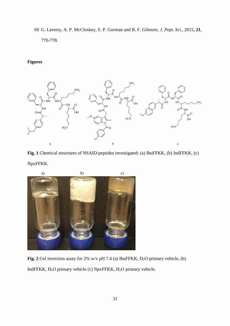
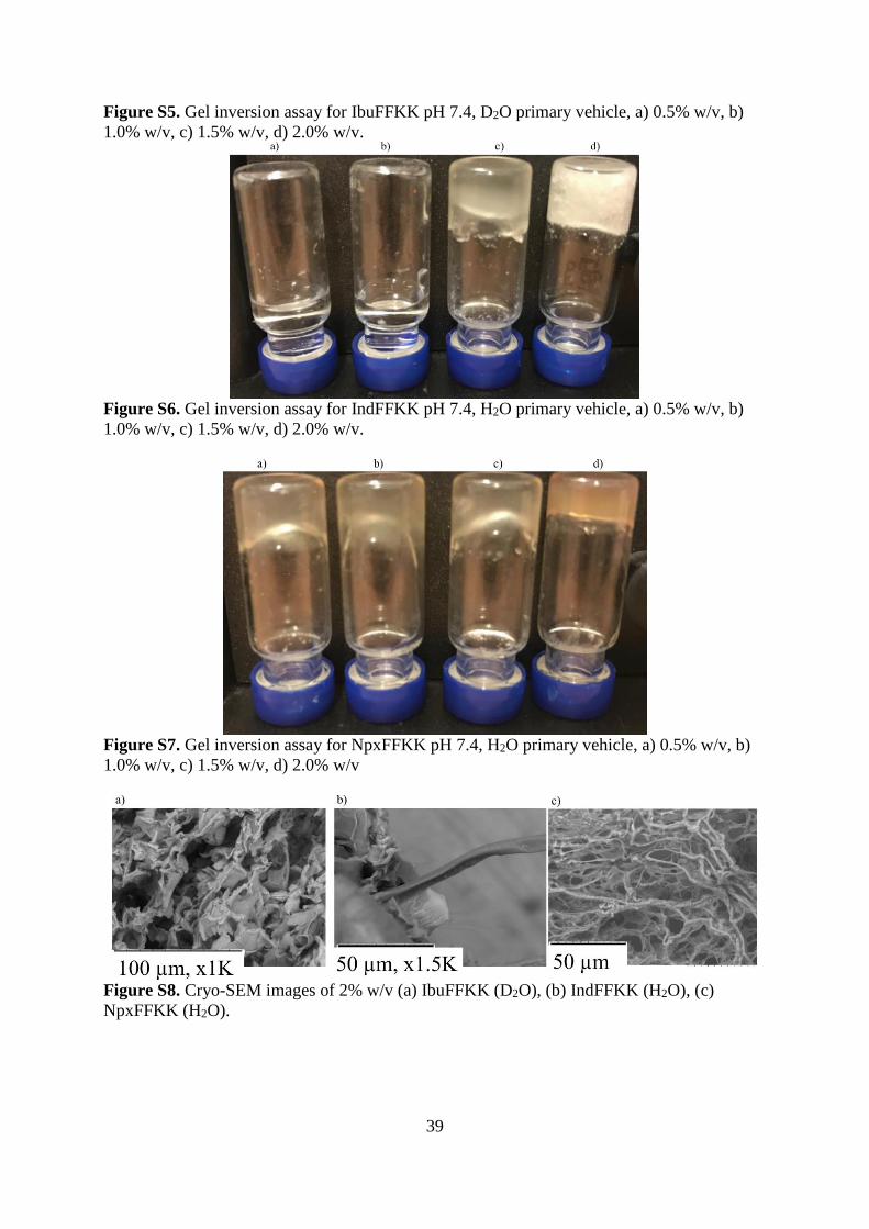
IbuFFKK was unable to form hydrogels in standard deionized water (H2O) instead forming an
opaque, white precipitate (Fig. S4). Interestingly IbuFFKK formed a transparent soft gel at
2% w/v using deuterated water (D2O) (Figs. 2a and S5). This phenomenon may be due to
increased hydrogen bond strength between deuterated hydrogen and electron donors (amines,
carbonyl and carboxylic acid groups) present within the NSAID-peptide primary structure.37,38
The number of hydrogen bonds per molecule of water is also higher for deuterated water
compared to standard water.38 As hydrogel formation is a delicate balance between
solubilization and precipitation the use of deuterated water may favor increased hydrogen
bond formation and improved solubilization for IbuFFKK resulting in formation of a clear
hydrogel at 2% w/v. IndFFKK was able to form a self-supporting supramolecular hydrogel at
1.5% w/v (Fig. S6) becoming an increasingly white and opaque hydrogel as the concentration
increased to 2% w/v (Fig. 2b).
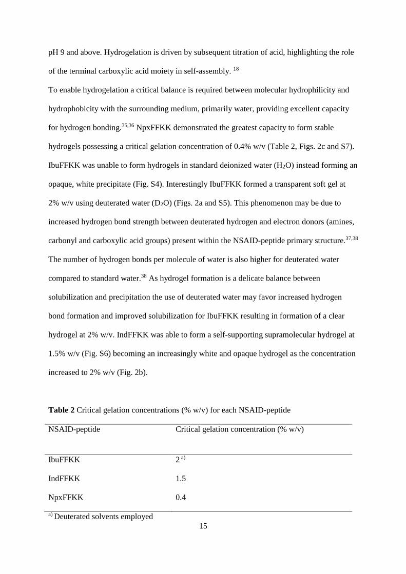
Table 2 Critical gelation concentrations (% w/v) for each NSAID-peptide
NSAID-peptide Critical gelation concentration (% w/v)
IbuFFKK 2 a)
IndFFKK 1.5
NpxFFKK 0.4
a) Deuterated solvents employed
16
Microscopy
Transmission electron microscopy (TEM) and cryogenic scanning electron microscopy (cryo-
SEM) were utilized to examine the nanoscale architecture of the molecular assemblies.
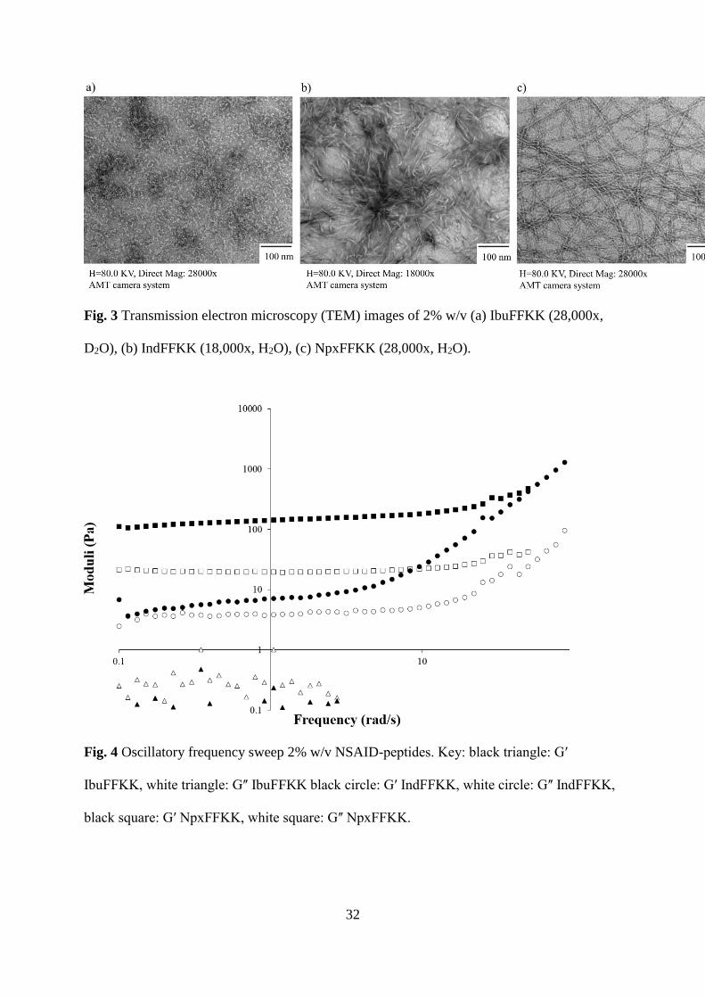
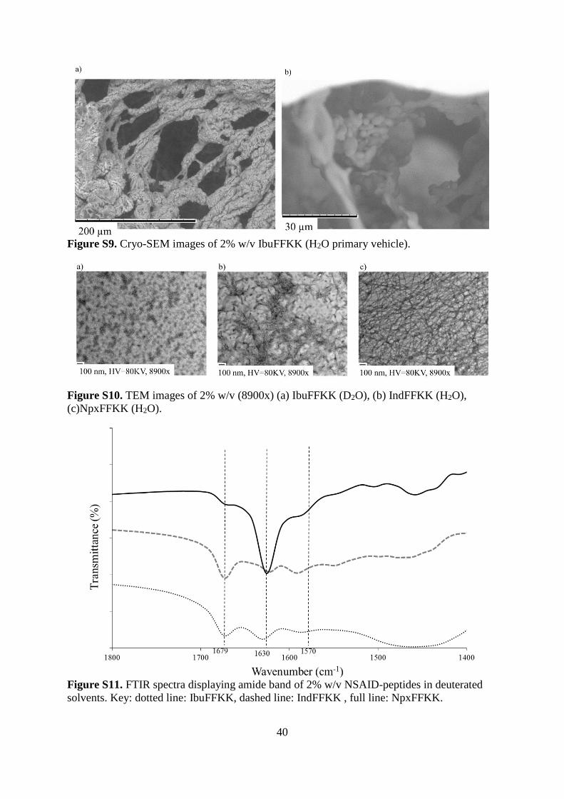
NpxFFKK hydrogels were found to be composed of nanofibers that entangled to form an
entangled network (Figs. 3c and S8c and S10c). Naproxen has previously demonstrated
ability to self-assemble into supramolecular hydrogels with nanofibrous architecture when
attached to a variety of peptidomimetic molecules including peptide amphiphiles and β-
peptides.21,39 By contrast 2% w/v IbuFFKK forms less uniform, non-fibrous structures in the
presence of deuterated water (Figs. 3a and S8a and S10a). Interactions between these
nanoparticles and the surrounding solvent are sufficient to form a self-supporting
supramolecular hydrogel as defined by the vial inversion assay.30 However rheological
analysis, detailed below, confirms that the resulting material more closely resemble a viscous
liquid with surfactant-like properties. Cryo-SEM images for 2% w/v IbuFFKK in standard
deionized water show the presence of a non-uniform fibers (Fig. S9a) and particles (Fig. S9b)
that result in formation of a white precipitate. IndFFKK formed short nanotape-like structures
(Figs. 3b and S8b and S10b) where hydrophobic segments are packed tightly away from the
aqueous interface. These appear less uniform, forming areas of dense pockets, compared to
the nanofibrous architecture of NpxFFKK. These networks of, varying morphologies, are
responsible for the immobilization of surrounding solvent molecules resulting in gel
formation. 21 The structural morphologies correlate well to the oscillatory rheological profiles,
most notably the storage moduli, as discussed further below.
However, the molecular-level understanding of the exact mechanism of peptide gelation
kinetics remains unclear despite extensive research and warrants further investigation within
the field.40 It should also be noted that analysis of TEM images of gels, such as morphology
of fibers, may not accurately represent the gel network and so should be done with caution.
17
TEM images are of dried gels, whereas tests such as rheology are carried out on wet gels.
Therefore, the structures observed by SEM could be due to drying effects.41,42
Fourier transform infrared spectroscopy
The secondary structures of NSAID-peptide nanostructures were determined by Fourier
Transform Infrared (FTIR) spectroscopy. All FTIR studies were conducted in deuterated
solvents, acknowledging the difficulty encountered using standard water due to strong
absorbance within the amide I region (1700-1600 cm-1).21 The amide I band is generated
through stretching of the C=O bond and its participation in hydrogen bonding provides an
insight into peptide secondary structures. All NSAID-peptides were studied at or above
critical gelation concentrations (2% w/v). NpxFFKK demonstrated the most predominant β-
sheet secondary structure characterized by a strong reduction in transmittance at 1630 cm-1
(Fig. S11).43 This trough is less pronounced for IndFFKK and particularly IbuFFKK owing to
less uniform nanoparticle and nanotape structures respectively. However, they both possess a
shoulder-like reduction in transmittance at 1679 cm-1 possibly linked to the presence of
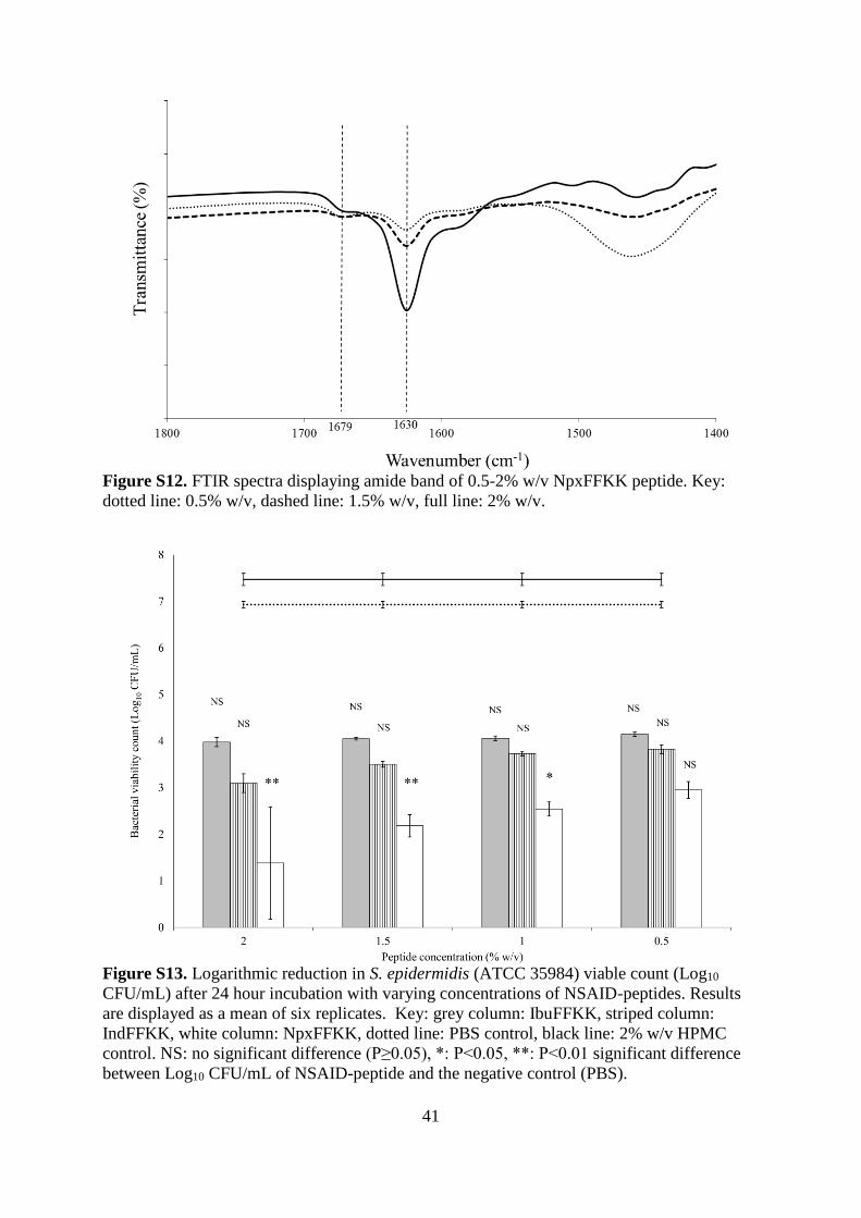
antiparallel β-sheets (Fig. S11). Increased concentration of NpxFFKK, from 0.5 to 2.0% w/v,
correlated to a respective reduction in transmittance at 1630 and 1679 cm-1 owing to a greater
presence of β-sheet secondary structures and more rigid hydrogels at 2% w/v NpxFFKK (Fig.
S12). However, interpreting this data for short dipeptide molecules is often difficult as has
been seen with other ultrashort peptide hydrogels.
Oscillatory Rheology
Oscillatory rheology was utilized to study the viscoelastic properties of 2% w/v self-
assembled NSAID-peptide hydrogels (Fig. 4). The frequency dependence of their storage (Gʹ)
and loss moduli (Gʺ) were measured using a dynamic frequency sweep, varying the
18
oscillation frequency (1-100 rad s−1) at a constant oscillation amplitude (0.0003%) and
temperature (25 °C).
NpxFFKK exhibits viscoelastic properties of a solid-like material as observed by the maximal
storage modulus (Gʹ: ~398 Pa) being ten times larger than the maximal loss modulus (Gʺ:
~39.8 Pa). They are independent of frequency and demonstrate good tolerance to external
shear force. NpxFFKK has the highest mean storage modulus and greatest gel strength of the
NSAID-peptides (Gʹ: ~398). Its Gʹ and Gʺ are comparable to that of the widely reported
FmocFF hydrogels.44 A larger conjugate system, conferred by the presence of naproxen in
NpxFFKK (Fig. 1c), enables stronger π- π and van der Waals’ intermolecular interactions
between NpxFFKK molecules and allows longer fibers and more entangled arrangement of
nanofibers as observed via microscopy (Fig. 3c). Similarly IndFFKK is capable of forming a
stable hydrogel at 2% w/v possessing a mean Gʹ of 25.1 Pa and a mean Gʺ of 6.6 Pa. Reduced
Gʹ and Gʺ in comparison to NpxFFKK is explained by formation of shorter nanotape
architectures rather than longer nanofiber structures observed in NpxFFKK gels (Figs. 3b and
S8b and S10b). Despite forming what appears to be a self-supporting hydrogel upon inversion
at a concentration of 2% w/v in deuterated water (Fig. 2a), rheology confirms IbuFFKK does
not form a hydrogel with significant mechanical rigidity. A high dependence on frequency
shows that it is behaving as a liquid rather than a gel, as gels show no dependence on
frequency. This is due to reduced aromatic-aromatic (π- π) interactions provided by the
terminal isopropyl-substituted phenyl group of IbuFFKK (Fig. 1a) that are only sufficient to
allow formation of non-uniform nanoparticle structures. This result is similar to that observed
previously by the Xu group where IbuFF demonstrated a low Gʹ value of only 13 Pa.25
19
Bacterial Susceptibility
The antibacterial activity of NSAID-peptides was tested using a viable count assay after 24
hour exposure to varying concentrations of NSAID-peptides. Clinically relevant bacterial
strains, implicated in a variety of antibiotic resistant nosocomial infections (biomaterial and
wounds), were selected, namely: methicillin resistant S. epidermidis (ATCC 25984), S. aureus
(ATCC 6584), P. aeruginosa (PAO1) and E. coli (ATCC 11303). All NSAID-peptides
demonstrated broad-spectrum antibacterial activity (Gram-positive and Gram-negative) (Figs.
5, 6, S13 and S14). The mechanism of action of our NSAID-peptides is likely to follow the
key parameters of antimicrobial peptide activity, namely hydrophobic bulk and cationic
charge enabling interaction with bacterial membranes.13 The addition of two units of cationic
charge, in this case lysine, to an ultrashort (less than seven amino acid units) peptidomimetic
sequence is sufficient to confer antimicrobial activity.34 As previously demonstrated by our
group, the ε-amino group of lysine has the ability to interact with negatively charged bacterial
membranes and their anionic hydroxylated phospholipids resulting in detergent-like effects,
cell lysis and death.18
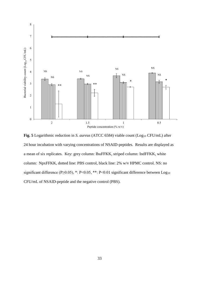
At least a three Log10 CFU/mL (99.9%) reduction in viable bacteria, commonly employed as a
threshold for bactericidal efficacy, was observed for each NSAID-peptide at concentrations of
0.5% w/v and above.45 Statistical analysis, however, shows NpxFFKK to be the only NSAID-
peptide that is significantly bactericidal against all four isolates with improved activity
correlating to increased concentration. For example, significant bacterial kill was achieved at
concentrations of 1.5% w/v and above for Gram-negative P. aeruginosa and E. coli; 1.0% and
above for Gram-positive S. epidermidis and 0.5% w/v and above for S. aureus. A reduction
greater than 5.5 Log10 CFU/mL was obtained for 2% w/v NpxFFKK against S. epidermidis
and S. aureus. The improved antimicrobial efficacy of NpxFFKK may be related to its ability
to form a viscoelastic hydrogel of uniform nanofibrous architecture. Recent studies have
20
suggested that that molecular folding, structural conformation, assembly state and bulk
mechanical properties are important considerations for the rational design of antimicrobial
selective hydrogels.17,46,47 We propose our NpxFFKK hydrogel acts similarly to the
dimethyldecylammonium chitosan-graft-poly(ethylene glycol) methacrylate (DMDC-Q-g-
EM) ‘anion sponge’ developed by the Chan-Park group.48 Based on this theory, the
nanoporous architecture of cationic NpxFFKK allows increased interactions, termed
‘suctioning,’ with anionic constituents of the bacterial membrane. Therefore the hydrogel acts
as a molecular sponge resulting in bacterial membrane disruption and cell death.
The inclusion of cationic charge density within the hydrogel matrix is a key parameter for
selective antimicrobial activity as demonstrated by the range of β-hairpin hydrogels (MAX,
PEP6R and MARG) developed by the Schneider group.49,50,51 Hydrogelation alone is not
sufficient to bestow antimicrobial activity, for example by restricting the diffusion of
chemical messengers and nutrients. This is confirmed by the observed lack of efficacy of non-
ionic 2% w/v hydroxypropyl methylcellulose (HPMC) against all bacterial isolates. This is an
unsurprising observation given that bacteria commonly prefer to exist and successfully
survive within a surface-attached, extracellular polymeric matrix granted by the biofilm
phenotype. As the Dong group hypothesized for their multi-domain peptides, the combined
effect of localized cationic charge on the hydrogel surface and the porous network of
crosslinked nanofibers (Fig. 3c, S8c and S10c) are likely to be responsible for the improved
antimicrobial efficacy of NpxFFKK.32,47 Our ultrashort peptide motif possesses improved
antimicrobial efficacy compared to related peptide-based strategies in the literature at a
reduced polymer size and increased ease of synthesis.
Chronic wounds fail to heal and are characterized by persistent inflammation due in part to
the presence of bacterial biofilms.52 Research by Wolcott and Rhodes also demonstrated that
the presence of bacterial infection in chronic wounds perpetuates a destructive level of
21
inflammation.53 Treatments active against resistant bacteria allowed non-healable, chronic
wounds to heal. They concluded that topical agents with the ability to disrupt biofilm forming
microorganisms should be central to the treatment of chronic wounds.54 Previous work by our
group demonstrates the significant efficacy of the FFKK-OH peptide motif against bacterial
isolates. They may have a significant clinical benefit for the treatment of chronic infected
wounds.18 Whilst NSAIDs have recently been demonstrated to possess antibacterial activity
alone by specifically targeting DNA Clamp, a key bacterial protein involved in multiplication,
its inclusion within our motif is primarily to provide potential anti-inflammatory, analgesic
and self-assembly characteristics.55 The benefits of NSAIDs as antibiotic therapy in their own
right have to be verified clinically. Such studies provide hope to extend the currently available
antibiotic formulary utilizing a readily available and licensed group of drugs.
These results may inform future strategies whereby the host immune response is controlled in
combination with antimicrobial and antibiofilm activity. For example IDR-1018, a synthetic
variant of the host defense peptide LL-37, has an ability to inhibit biofilm formation in
combination with immunomodulatory effects which prevent tissue damage.56 Although IDR-
1018 did not display an ability to self-assemble into supramolecular structures the Hancock
group did demonstrate antibiofilm peptides could be used to control mediators of the immune
response, including stimulating monocyte chemoattractant protein (MCP-1) and inhibiting
lipopolysaccharide induced interleukin-1β (IL-1β) production in peripheral blood
mononuclear cells.57
Cyclooxygenase enzyme inhibition
We performed in vitro inhibition assays for both COX-1 and COX-2 in the presence of
varying concentrations of NSAID-peptides. Molecular modeling, utilizing the crystal structure
of COX enzymes, has previously shown that the NSAID carboxylate end is available for
22
peptide modification due to the large open space in the structure of COX.46,58 Addition of the
peptide sequence FFKK-OH to the NSAID motif increases respective IC50 values relative to
NSAID only values. However, the NSAID-peptide motifs retain significant inhibitory activity
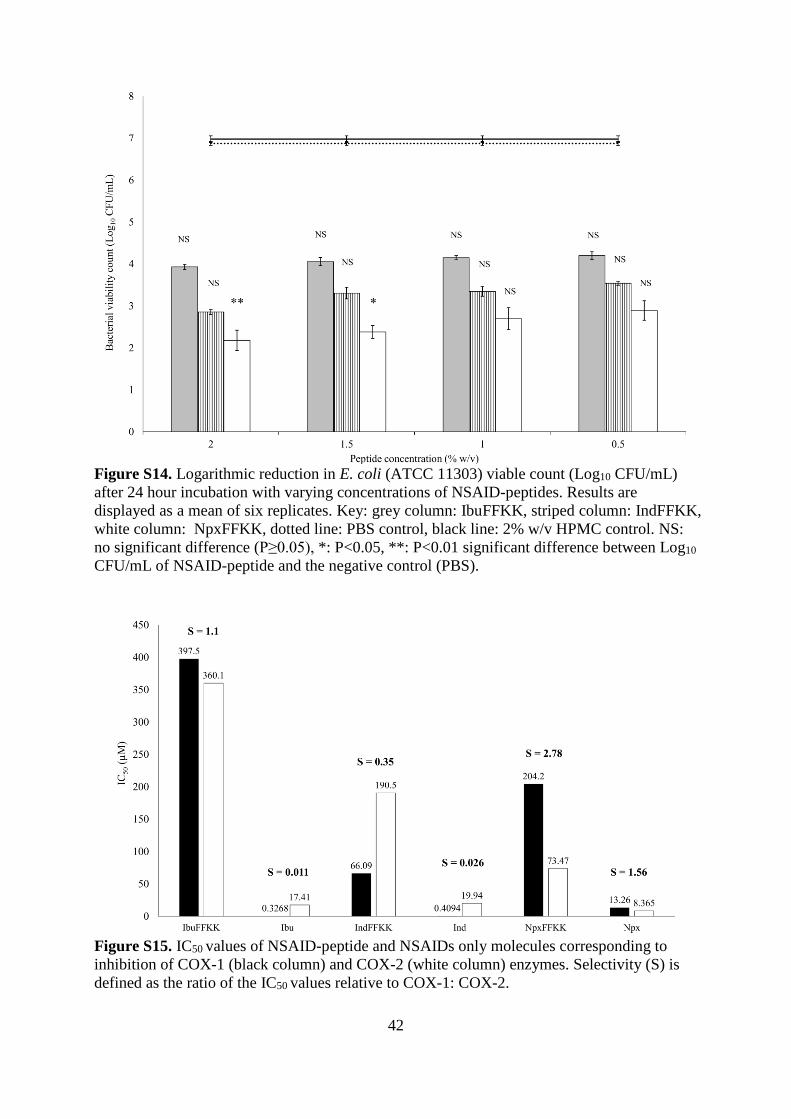
with IC50 values within the μM range. For example, NpxFFKK exhibits IC50 values of 204.20
μM and 73.47 μM against COX-1 and COX-2 respectively compared with naproxen only
values of 13.26 μM (COX-1) and 8.365 μM (COX-2) (Fig. S15). NSAID-peptides also
demonstrate increased selectivity for inhibiting COX-2 compared to NSAID alone. Selectivity
(S) values, related to the ratio of IC50 COX-1: IC50 COX-2, are highest for NpxFFKK (S =
2.78) compared to naproxen only (S = 0.19). This correlates to previous work on NSAID-
peptide hydrogelators where it was proven that addition of a peptide moiety increased COX-2
selectivity.25,26 Replacing L-amino acid enantiomers with their respective D-forms may
increase inhibition of COX-2 further.46 Selective COX-2 inhibition is preferred clinically,
especially for systemic administration of NSAIDs, due to a reduction in renal and
gastrointestinal side effects linked to COX-1 inhibition but may also have potential value in
the chronic wound environment.59 Studies have shown that upon degradation, the
pharmaceutical efficacy of NSAIDs in theory should be maintained and pharmacological
activity exhibited until the hydrogel has completely degraded. Therefore an extended profile
of anti-inflammatory activity should be possible.60
COX-2 and its enzymatic product prostaglandin E2 (PGE2) demonstrate an important role in
the early acute host response to stimuli such as wounds and are responsible for the
upregulation of inflammatory mediators. Separate studies in murine and rat wound models by
Blomme and Futagmi showed a significant induction of COX-2 expression after 12 hours,
peaking three days after injury.61,62 However within chronic wounds, COX-2 has recently
been associated with the unwanted development of scar tissue in the latter stages of adult
wound repair.63,64 Therefore NSAID-peptide conjugates with increased selectivity for COX-2
23
inhibition may be of benefit in the chronic stages of wound healing, replicating fetal wound
healing where scarless healing is linked to a reduced inflammatory response.64 Despite this
there is an appreciation that wound healing is a complex pathway and the exact role of
multiple inflammatory mediators (cytokines, macrophages, matrix metalloproteinases) has not
yet been fully elucidated.6 In particular, the diverse nature of the immune and inflammatory
response to foreign medical implants favors the use of corticosteroids, rather than NSAIDs,
due to their broad-spectrum of activity against inflammatory mediators such as leukotrienes.65
NSAID-peptides may serve a greater purpose within chronic wound healing, providing
localized pain relief, antimicrobial and anti-inflammatory activity. Removal of an avascular
fibrous capsule that surrounds implanted medical devices may be promoted by inclusion of
pro-angiogenic factors including vascular endothelial growth factor (VEGF). Our
nanomaterials have the potential to act as a drug delivery platform for the design of future
medical device coatings incorporating such factors.11 The effect of NSAID-peptides
nanostructures on mediators of the immune response. The overall wound healing pathway and
foreign body response to medical implants warrants further clinical investigation. A more
obvious therapeutic benefit for NSAID-peptides is provided via inhibition of nociception and
pain receptors linked to COX.66 Reduction in pain can be linked to improvement in wound
healing and patient prognosis.67
Hemolysis and cell viability
To evaluate cell biocompatibility of NSAID-peptides, they were incubated with an
International Standard (ISO) cell line utilized for biomaterial testing (NCTC 929 murine
fibroblast subcutaneous connective tissue). Fibroblasts are appropriate as they are one of the
major cell types involved both in wound healing and adherence to implanted medical
devices.11 Four separate assays were performed using alamarBlue® cell viability, hemolysis,
24
LDH quantification and LIVE/DEAD® staining. The results suggest that NSAID-peptide
nanomaterials are biocompatible and may be suitable for use within a range of biomaterial
and topical applications. No significant reduction was observed in cell viability after 24 hour
exposure to varying concentrations of NSAID-peptides (20-500 µM) utilizing an
alamarBlue® cell viability assay (Fig. 7). Cationic NSAID-peptides demonstrate reduced
toxicity against mammalian cells due to inherent differences in the membrane potential
gradient and lipid composition of bacterial and mammalian cell membranes. Eukaryotic cells
are composed of zwitterionic lipids (sterols, cholesterol, phosphatidylcholine, sphingomyelin)
whereas bacterial cells are derived from anionic phospholipids.68 Positively charged NSAID-
peptide therefore interact preferentially with negatively charged bacterial cell membranes as
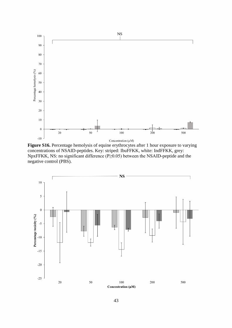
confirmed by a hemolysis assay (Fig. S16), commonly utilized to determine the membrane
selectivity of antimicrobial peptides.18,32,69 No significant hemolysis was observed upon
NSAID-peptide (20-500 µM) exposure to equine erythrocytes relative to a PBS negative
control. Cell cytocompatibility was confirmed via quantification of the toxicity marker LDH
(Figure S17). In all instances the mean level of LDH was less than the negative control (PBS)
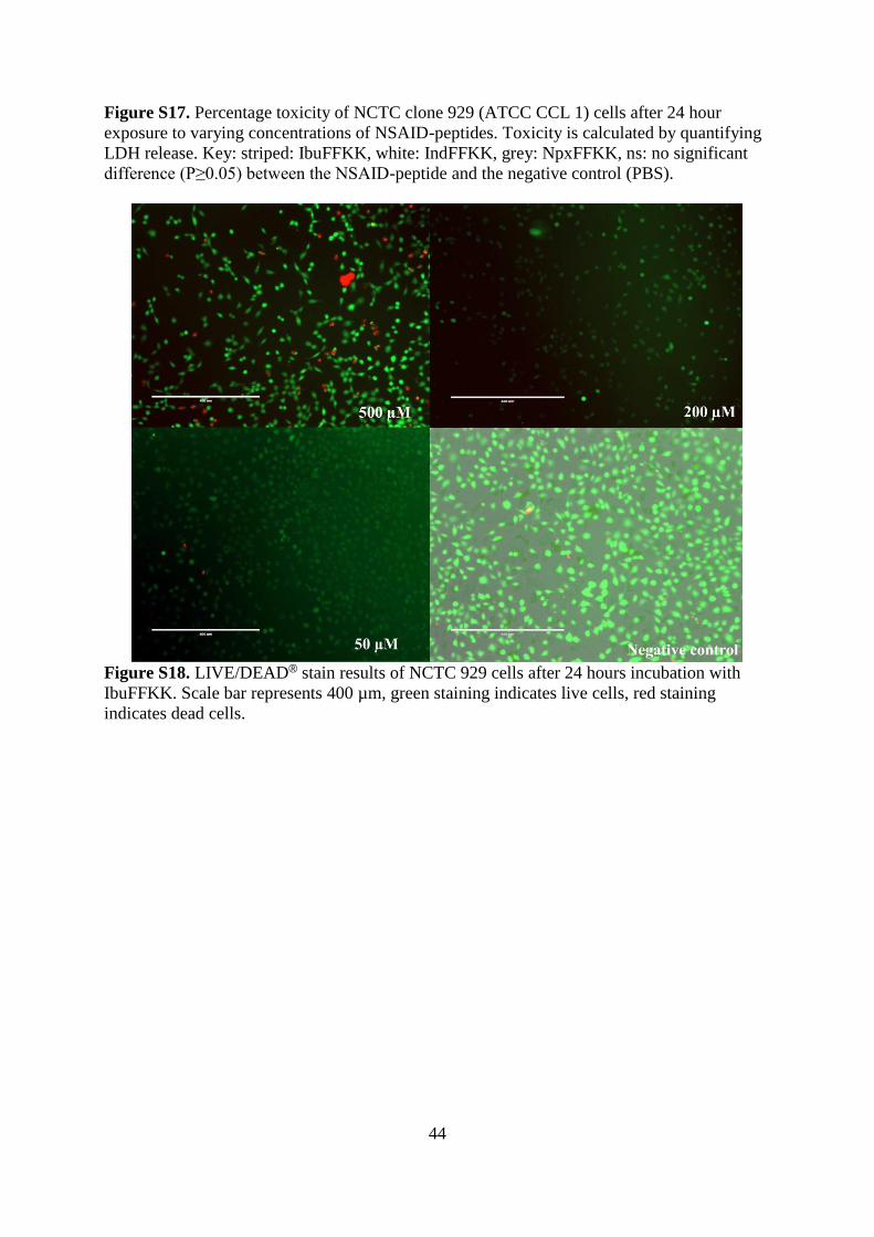
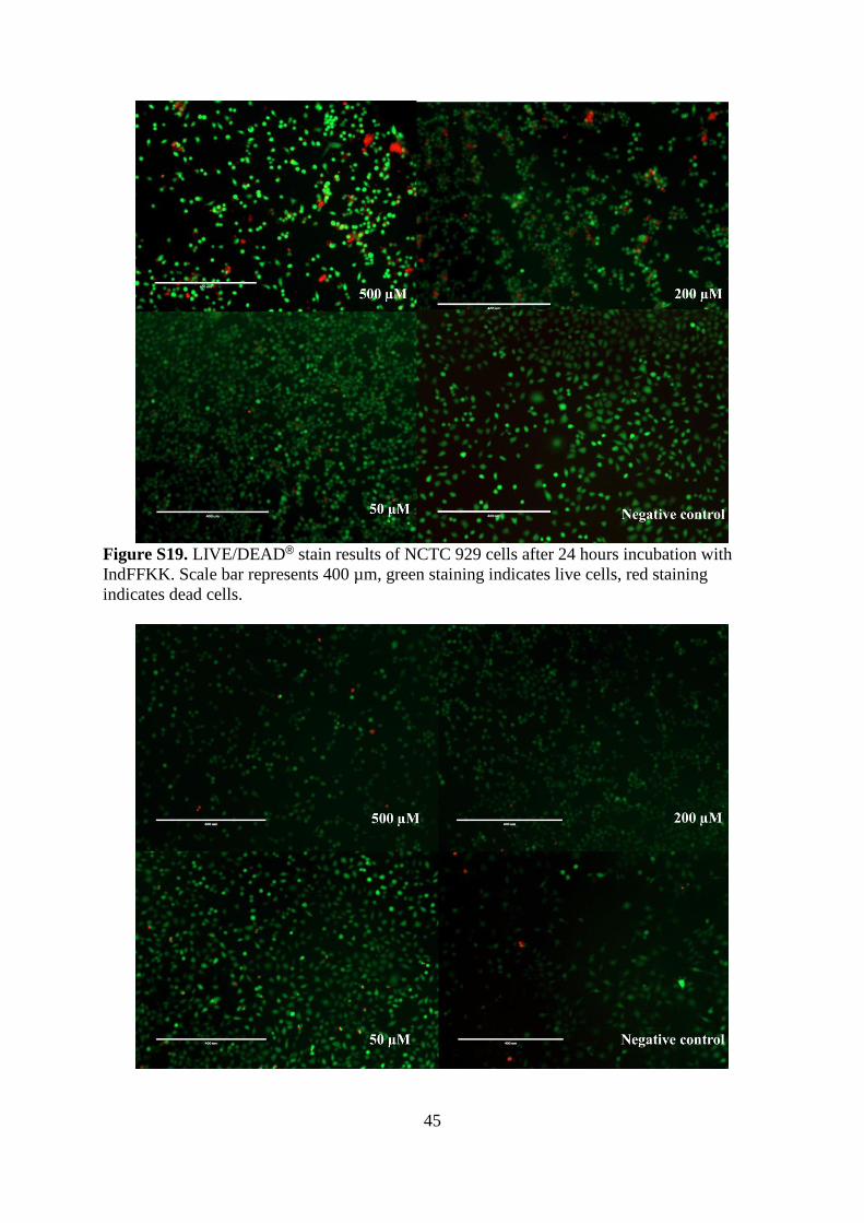
resulting in negative percentage toxicity values. A LIVE/DEAD® Viability/Cytotoxicity
assay provided qualitative evidence of NSAID-peptide via fluorescent microscopy imaging
(Figures S18-S20). The majority of cells stained green with calcein after 24 hours exposure
across all concentrations (20-500 µM) as confirmed by quantitative counting of viable and
non-viable cells (Figure S21). The results for each cytotoxicity assay correlate strongly with
each other. For example LIVE/DEAD® counting for 500 µM NpxFFKK resulted in 97%
viability which is in good agreement with 95% viability obtained at the same concentration
for the alamarBlue® cell viability assay.
25
Conclusions
In summary, we have created a new class of NSAID-peptides with the ability to form defined
nanostructures with multiple biofunctional properties (antimicrobial, anti-inflammatory,
hydrogel forming). NpxFFKK displays particular promising forming a viscoelastic
biocompatible hydrogel with improved COX-2 selectivity and the ability to target
antimicrobial resistant bacteria implicated in the most severe nosocomial infections. These
hydrogel formulations may be beneficial in the treatment of chronic infected wounds, where a
heightened inflammatory response to infection leads to impaired healing. The next stage in
development would be to demonstrate their beneficial effects in an in vivo, clinical wound
environment closely monitoring the effect of NSAID-peptides on inflammatory mediators.
Their ultrashort, low molecular weight structure makes their synthesis more amenable to cost-
effective upscale by the pharmaceutical industry compared to larger peptides and proteins.
This work provides an example of multifunctional peptide hydrogelators that will contribute
to the development of future biofunctional nanomaterial therapies, especially within
biomaterial applications (wound dressings, medical implants, prostheses), thereby increasing
the available treatment options to clinicians and patients and limiting the increasing threat of
antimicrobial resistance.
Acknowledgements
This work was supported by the Queen’s University Research Support Package for New
Academic Staff for and a Royal Society Research Grant (RG150171) for GL. APM
acknowledges funding provided by a N. Ireland Department of Employment and Learning
PhD studentship grant. The manuscript was written through contributions of all authors. All
authors have given approval to the final version of the manuscript. APM, SG, ERD, JZ, SP,
BFG and BX contributed equally. We acknowledge help from Prof. Dave Adams (University
26
of Liverpool) for allowing us access to his Anton Paar Physica MCR301 rheometer for
rheological studies.
References
1 J. O'Neill, The Review on Antimicrobial Resistance: UK Government Report, London,
2015.
2 Centre for Disease control (CDC), Data from the National Hospital Discharge Survey.
Available:
http://www.cdc.gov/nchs/data/nhds/4procedures/2010pro_numberpercentage.pdf,
2010.
3 P. Astagneau, C. Rioux, F. Golliot, G. Brucker and INCISO Network Study Group, J.
Hosp. Infect., 2001, 48, 267-274.
4 S. Guo and L. A. Dipietro, J. Dent. Res., 2010, 89, 219-229.
5 F. Gottrup, Am. J. Surg., 2004, 187, 38S-43S.
6 N. B. Menke, K. R. Ward, T. M. Witten, D. G. Bonchev and R. F. Diegelmann, Clin.
Dermatol., 2007, 25, 19-25.
7 P. C. Konturek, T. Brzozowski, S. J. Konturek, S. Kwiecien, A. Dembinski and E. G.
Hahn, Scand. J. Gastroenterol., 2001, 36, 1239-1247.
8 M. Ståhle, in Antimicrobial Peptides and Innate Immunity, ed. P. S. Hiemstra and S. A.
J. Zaat, Springer, Basel, 2013, p. 123-129.
9 G. D. Krischak, P. Augat, L. Claes, L. Kinzl and A. Beck, J. Wound Care, 2007, 16,
76-78.
10 S. M. Hamilton, C. R. Bayer, D. L. Stevens and A. E. Bryant, J. Infect. Dis., 2014, 209,
1429-1435.
11 J. M. Morais, F. Papadimitrakopoulos and D. J. Burgess, AAPS J., 2010, 12, 188-196.
27
12 P. Price, K. Fogh, C. Glynn, D. L. Krasner, J. Osterbrink and R. G. Sibbald, Int.
Wound. J., 2007, 4 Suppl 1, 1-3.
13 G. Laverty, S. P. Gorman and B. F. Gilmore, Int. J. Mol. Sci., 2011, 12, 6566-6596.
14 G. Laverty, S. P. Gorman and B. F. Gilmore, J. Biomed. Mater. Res. A., 2012, 100,
1803-1814.
15 R. T. Cleophas, J. Sjollema, H. J. Busscher, J. A. Kruijtzer and R. M. Liskamp,
Biomacromolecules, 2014, 15, 3390-3395.
16 Z. Xie, N. V. Aphale, T. D. Kadapure, A. S. Wadajkar, S. Orr, D. Gyawali, G. Qian, K.
T. Nguyen and J. Yang, J. Biomed. Mater. Res. A., 2015, 103, 3907-3918.
17 V. W. Ng, J. M. Chan, H. Sardon, R. J. Ono, J. M. Garcia, Y. Y. Yang and J. L.
Hedrick, Adv. Drug Deliv. Rev., 2014, 78, 46-62.
18 G. Laverty, A. P. McCloskey, B. F. Gilmore, D. S. Jones, J. Zhou and B. Xu,
Biomacromolecules, 2014, .
19 M. J. Webber, J. B. Matson, V. K. Tamboli and S. I. Stupp, Biomaterials, 2012, 33,
6823-6832.
20 F. Zhao, M. L. Ma and B. Xu, Chem. Soc. Rev., 2009, 38, 883-891.
21 J. Majumder, M. R. Das, J. Deb, S. S. Jana and P. Dastidar, Langmuir, 2013, 29,
10254-10263.
22 D. M. Ryan, S. B. Anderson, F. T. Senguen, R. E. Youngman and B. L. Nilsson, Soft
Matter, 2010, 6, 475-479.
23 E. R. Draper, K. L. Morris, M. A. Little, J. Raeburn, C. Colquhoun, E. R. Cross, T. O.
McDonald, L. C. Serpell and D. J. Adams, Cryst. Eng. Comm., 2015, 17, 8047-8057.
24 Z. Yang, G. Liang, M. Ma, Y. Gao and B. Xu, J. Mater. Chem, 2007, 17, 850-854.
25 J. Y. Li, Y. Kuang, J. F. Shi, Y. Gao, J. Zhou and B. Xu, Beilstein J. Org. Chem., 2013,
9, 908-917.
28
26 J. Y. Li, X. Li, Y. Kuang, Y. Gao, X. Du, J. Shi and B. Xu, Adv. Healthc. Mater., 2013,
2, 1586-1590.
27 A. Y. Mensah, P. J. Houghton, R. A. Dickson, T. C. Fleischer, M. Heinrich and P.
Bremner, Phytother. Res., 2006, 20, 941-944.
28 C. Gong, Q. Wu, Y. Wang, D. Zhang, F. Luo, X. Zhao, Y. Wei and Z. Qian,
Biomaterials, 2013, 34, 6377-6387.
29 S. Bhuniya, Y. J. Seo and B. H. Kim, Tetrahedron Lett, 2006, 47, 7153-7156.
30 D. J. Adams, M. F. Butler, W. J. Frith, M. Kirkland, L. Mullen and P. Sanderson, Soft
Matter, 2009, 5, 1856-1862.
31 A. G. Pogorelov and I. I. Selezneva, Bull. Exp. Biol. Med., 2010, 150, 153-156.
32 L. Jiang, D. Xu, T. J. Sellati and H. Dong, Nanoscale, 2015, 7, 19160-19169.
33 M. Mateescu, S. Baixe, T. Garnier, L. Jierry, V. Ball, Y. Haikel, M. H. Metz-Boutigue,
M. Nardin, P. Schaaf, O. Etienne and P. Lavalle, PLoS One, 2015, 10, e0145143.
34 G. Laverty, M. McLaughlin, C. Shaw, S. P. Gorman and B. F. Gilmore, Chem. Biol.
Drug Des., 2010, 75, 563-569.
35 L. Chen, S. Revel, K. Morris, L. C Serpell and D. J. Adams, Langmuir, 2010, 26,
13466-13471.
36 A. P. McCloskey, B. F. Gilmore and G. Laverty, Pathogens, 2014, 3, 791-821.
37 P. Cioni and G. B. Strambini, Biophys. J., 2002, 82, 3246-3253.
38 A. K. Soper and C. J. Benmore, Phys. Rev. Lett., 2008, 101, 065502.
39 M. Conda-Sheridan, S. S. Lee, A. T. Preslar and S. I. Stupp, Chem. Commun. (Camb),
2014, 50, 13757-13760.
40 M. L. Muro-Small, J. Chen and A. J. McNeil, Langmuir, 2011, 27, 13248-13253.
41 M. Kolbel and F. M. Menger, Chem. Commun., 2001, , 275-276.
42 L. A. Estroff and A. D. Hamilton, Chem. Rev., 2004, 104, 1201-1218.
29
43 V. Jayawarna, S. M. Richardson, A. R. Hirst, N. W. Hodson, A. Saiani, J. E. Gough
and R. V. Ulijn, Acta Biomater., 2009, 5, 934-943.
44 C. Tang, A. M. Smith, R. F. Collins, R. V. Ulijn and A. Saiani, Langmuir, 2009, 25,
9447-9453.
45 G. A. Pankey and L. D. Sabath, Clin. Infect. Dis., 2004, 38, 864-870.
46 J. Li, Y. Kuang, Y. Gao, X. Du, J. Shi and B. Xu, J. Am. Chem. Soc., 2013, 135, 542-
545.
47 D. Xu, L. Jiang, A. Singh, D. Dustin, M. Yang, L. Liu, R. Lund, T. J. Sellati and H.
Dong, Chem. Commun. (Camb), 2015, 51, 1289-1292.
48 P. Li, Y. F. Poon, W. Li, H. Y. Zhu, S. H. Yeap, Y. Cao, X. Qi, C. Zhou, M. Lamrani,
R. W. Beuerman, E. T. Kang, Y. Mu, C. M. Li, M. W. Chang, S. S. Leong and M. B.
Chan-Park, Nat. Mater., 2011, 10, 149-156.
49 D. A. Salick, J. K. Kretsinger, D. J. Pochan and J. P. Schneider, J. Am. Chem. Soc.,
2007, 129, 14793-14799.
50 D. A. Salick, D. J. Pochan and J. P. Schneider, Adv. Mater., 2009, 21, 4120-4123.
51 A. S. Veiga, C. Sinthuvanich, D. Gaspar, H. G. Franquelim, M. A. Castanho and J. P.
Schneider, Biomaterials, 2012, 33, 8907-8916.
52 E. A. Grice and J. A. Segre, Adv. Exp. Med. Biol., 2012, 946, 55-68.
53 R. D. Wolcott, D. D. Rhoads, M. E. Bennett, B. M. Wolcott, L. Gogokhia, J. W.
Costerton and S. E. Dowd, J. Wound Care, 2010, 19, 45-6, 48-50, 52-3.
54 R. D. Wolcott and D. D. Rhoads, J. Wound Care, 2008, 17, 145-8, 150-2, 154-5.
55 Z. Yin, Y. Wang, L. R. Whittell, S. Jergic, M. Liu, E. Harry, N. E. Dixon, M. J. Kelso,
J. L. Beck and A. J. Oakley, Chem. Biol., 2014, 21, 481-487.
56 S. C. Mansour, C. de la Fuente-Nunez and R. E. Hancock, J. Pept. Sci., 2015, 21, 323-
329.
30
57 E. F. Haney, S. C. Mansour, A. L. Hilchie, C. de la Fuente-Nunez and R. E. Hancock,
Peptides, 2015, 71, 276-285.
58 K. C. Duggan, M. J. Walters, J. Musee, J. M. Harp, J. R. Kiefer, J. A. Oates and L. J.
Marnett, J. Biol. Chem., 2010, 285, 34950-34959.
59 F. E. Silverstein, G. Faich, J. L. Goldstein, L. S. Simon, T. Pincus, A. Whelton, R.
Makuch, G. Eisen, N. M. Agrawal, W. F. Stenson, A. M. Burr, W. W. Zhao, J. D.
Kent, J. B. Lefkowith, K. M. Verburg and G. S. Geis, JAMA, 2000, 284, 1247-1255.
60 P. K. Vemula, J. Li and G. John, J. Am. Chem. Soc., 2006, 128, 8932-8938.
61 A. Futagami, M. Ishizaki, Y. Fukuda, S. Kawana and N. Yamanaka, Lab. Invest., 2002,
82, 1503-1513.
62 E. A. Blomme, K. S. Chinn, M. M. Hardy, J. J. Casler, S. H. Kim, A. C. Opsahl, W. A.
Hall, D. Trajkovic, K. N. Khan and C. S. Tripp, Br. J. Dermatol., 2003, 148, 211-223.
63 T. A. Wilgus, Y. Vodovotz, E. Vittadini, E. A. Clubbs and T. M. Oberyszyn, Wound
Repair Regen., 2003, 11, 25-34.
64 T. A. Wilgus, V. K. Bergdall, K. L. Tober, K. J. Hill, S. Mitra, N. A. Flavahan and T.
M. Oberyszyn, Am. J. Pathol., 2004, 165, 753-761.
65 M. Kastellorizios, N. Tipnis and D. J. Burgess, in Immune Responses to Biosurfaces,
ed. J. D. Lambris, K. N. Ekdahl, D. Ricklin and D. Nilsson, Springer International
Publishing, Switzerland, 2015, p. 93-108.
66 S. Bingham, P. J. Beswick, D. E. Blum, N. M. Gray and I. P. Chessell, Semin. Cell
Dev. Biol., 2006, 17, 544-554.
67 L. McGuire, K. Heffner, R. Glaser, B. Needleman, W. Malarkey, S. Dickinson, S.
Lemeshow, C. Cook, P. Muscarella, W. S. Melvin, E. C. Ellison and J. K. Kiecolt-
Glaser, Ann. Behav. Med., 2006, 31, 165-172.
68 A. J. Mason, A. Marquette and B. Bechinger, Biophys. J., 2007, 93, 4289-4299.
31
69 G. Laverty, A. P. McCloskey, S. P. Gorman and B. F. Gilmore, J. Pept. Sci., 2015, 21,
770-778.
Figures
Fig. 1 Chemical structures of NSAID-peptides investigated: (a) IbuFFKK, (b) IndFFKK, (c)
NpxFFKK.
Fig. 2 Gel inversion assay for 2% w/v pH 7.4 (a) IbuFFKK, D2O primary vehicle, (b)
IndFFKK, H2O primary vehicle (c) NpxFFKK, H2O primary vehicle.
32
Fig. 3 Transmission electron microscopy (TEM) images of 2% w/v (a) IbuFFKK (28,000x,
D2O), (b) IndFFKK (18,000x, H2O), (c) NpxFFKK (28,000x, H2O).
Fig. 4 Oscillatory frequency sweep 2% w/v NSAID-peptides. Key: black triangle: Gʹ
IbuFFKK, white triangle: Gʺ IbuFFKK black circle: Gʹ IndFFKK, white circle: Gʺ IndFFKK,
black square: Gʹ NpxFFKK, white square: Gʺ NpxFFKK.
33
Fig. 5 Logarithmic reduction in S. aureus (ATCC 6584) viable count (Log10 CFU/mL) after
24 hour incubation with varying concentrations of NSAID-peptides. Results are displayed as
a mean of six replicates. Key: grey column: IbuFFKK, striped column: IndFFKK, white
column: NpxFFKK, dotted line: PBS control, black line: 2% w/v HPMC control. NS: no
significant difference (P≥0.05), *: P<0.05, **: P<0.01 significant difference between Log10
CFU/mL of NSAID-peptide and the negative control (PBS).
34
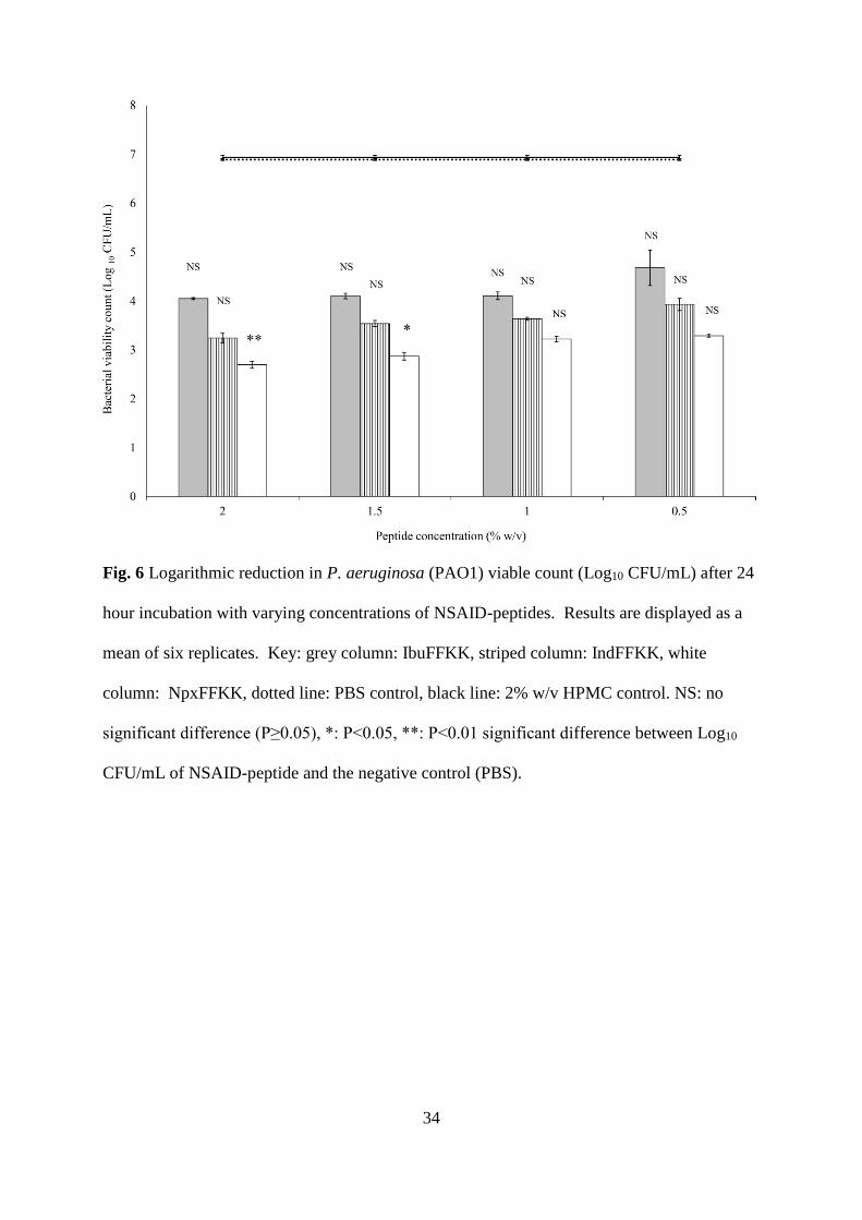
Fig. 6 Logarithmic reduction in P. aeruginosa (PAO1) viable count (Log10 CFU/mL) after 24
hour incubation with varying concentrations of NSAID-peptides. Results are displayed as a
mean of six replicates. Key: grey column: IbuFFKK, striped column: IndFFKK, white
column: NpxFFKK, dotted line: PBS control, black line: 2% w/v HPMC control. NS: no
significant difference (P≥0.05), *: P<0.05, **: P<0.01 significant difference between Log10
CFU/mL of NSAID-peptide and the negative control (PBS).
35
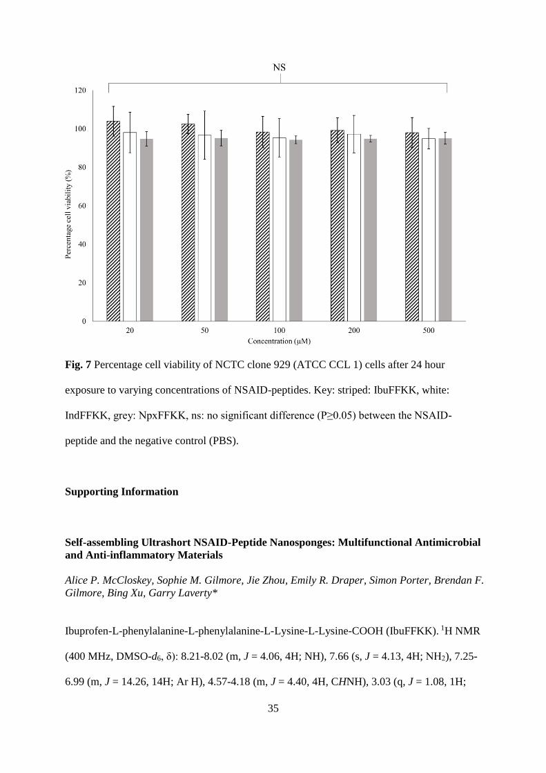
Fig. 7 Percentage cell viability of NCTC clone 929 (ATCC CCL 1) cells after 24 hour
exposure to varying concentrations of NSAID-peptides. Key: striped: IbuFFKK, white:
IndFFKK, grey: NpxFFKK, ns: no significant difference (P≥0.05) between the NSAID-
peptide and the negative control (PBS).
Supporting Information
Self-assembling Ultrashort NSAID-Peptide Nanosponges: Multifunctional Antimicrobial
and Anti-inflammatory Materials
Alice P. McCloskey, Sophie M. Gilmore, Jie Zhou, Emily R. Draper, Simon Porter, Brendan F.
Gilmore, Bing Xu, Garry Laverty*
Ibuprofen-L-phenylalanine-L-phenylalanine-L-Lysine-L-Lysine-COOH (IbuFFKK). 1H NMR
(400 MHz, DMSO-d6, δ): 8.21-8.02 (m, J = 4.06, 4H; NH), 7.66 (s, J = 4.13, 4H; NH2), 7.25-
6.99 (m, J = 14.26, 14H; Ar H), 4.57-4.18 (m, J = 4.40, 4H, CHNH), 3.03 (q, J = 1.08, 1H;
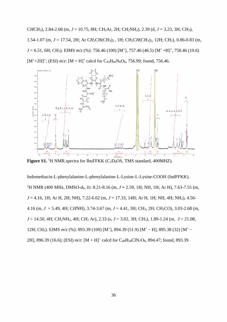
36
CHCH3), 2.84-2.68 (m, J = 10.75, 8H; CH2Ar, 2H; CH2NH2), 2.39 (d, J = 3.23, 3H; CH3),
1.54-1.07 (m, J = 17.54, 2H; Ar CH2CH(CH3)2 , 1H; CH2CH(CH3)2, 12H; CH2), 0.86-0.83 (m,
J = 6.51, 6H; CH3). EIMS m/z (%): 756.46 (100) [M+], 757.46 (46.5) [M+ +H]+, 758.46 (10.6)
[M++2H]+; (ESI) m/z: [M + H]+ calcd for C43H60N6O6, 756.99; found, 756.46.
Figure S1. 1H NMR.spectra for IbuFFKK (C2D6OS, TMS standard, 400MHZ).
Indomethacin-L-phenylalanine-L-phenylalanine-L-Lysine-L-Lysine-COOH (IndFFKK).
1H NMR (400 MHz, DMSO-d6, δ): 8.21-8.16 (m, J = 2.59, 1H; NH, 1H; Ar H), 7.63-7.55 (m,
J = 4.16, 1H; Ar H, 2H; NH), 7.22-6.62 (m, J = 17.33, 14H; Ar H, 1H; NH, 4H; NH2), 4.56-
4.16 (m, J = 5.49, 4H; CHNH), 3.74-3.67 (m, J = 4.41, 3H; CH3, 2H; CH2CO), 3.03-2.68 (m,
J = 14.50, 4H; CH2NH2, 4H; CH2 Ar), 2.33 (s, J = 3.02, 3H; CH3), 1.89-1.24 (m, J = 21.08,
12H; CH2). EIMS m/z (%): 893.39 (100) [M+], 894.39 (51.9) [M+ − H], 895.38 (32) [M+ −
2H], 896.39 (16.6); (ESI) m/z: [M + H]+ calcd for C49H58ClN7O8, 894.47; found, 893.39.
37
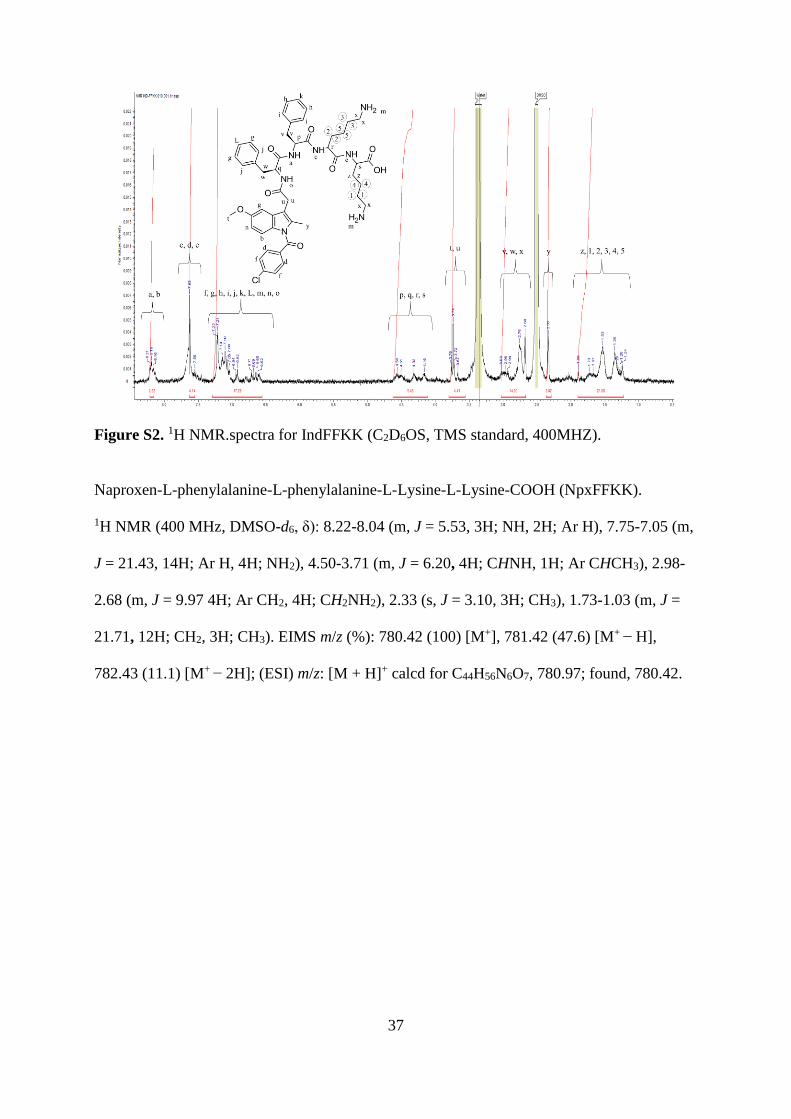
Figure S2. 1H NMR.spectra for IndFFKK (C2D6OS, TMS standard, 400MHZ).
Naproxen-L-phenylalanine-L-phenylalanine-L-Lysine-L-Lysine-COOH (NpxFFKK).
1H NMR (400 MHz, DMSO-d6, δ): 8.22-8.04 (m, J = 5.53, 3H; NH, 2H; Ar H), 7.75-7.05 (m,
J = 21.43, 14H; Ar H, 4H; NH2), 4.50-3.71 (m, J = 6.20, 4H; CHNH, 1H; Ar CHCH3), 2.98-
2.68 (m, J = 9.97 4H; Ar CH2, 4H; CH2NH2), 2.33 (s, J = 3.10, 3H; CH3), 1.73-1.03 (m, J =
21.71, 12H; CH2, 3H; CH3). EIMS m/z (%): 780.42 (100) [M+], 781.42 (47.6) [M+ − H],
782.43 (11.1) [M+ − 2H]; (ESI) m/z: [M + H]+ calcd for C44H56N6O7, 780.97; found, 780.42.
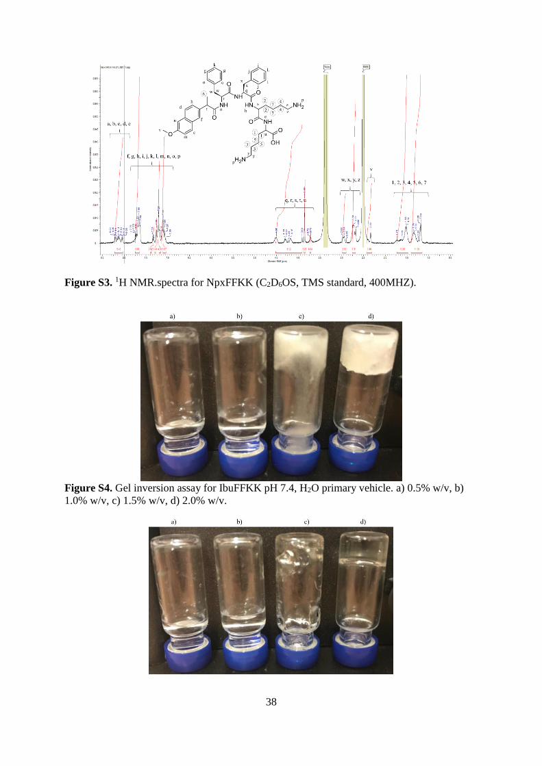
38
Figure S3. 1H NMR.spectra for NpxFFKK (C2D6OS, TMS standard, 400MHZ).
Figure S4. Gel inversion assay for IbuFFKK pH 7.4, H2O primary vehicle. a) 0.5% w/v, b)
1.0% w/v, c) 1.5% w/v, d) 2.0% w/v.
39
Figure S5. Gel inversion assay for IbuFFKK pH 7.4, D2O primary vehicle, a) 0.5% w/v, b)
1.0% w/v, c) 1.5% w/v, d) 2.0% w/v.
Figure S6. Gel inversion assay for IndFFKK pH 7.4, H2O primary vehicle, a) 0.5% w/v, b)
1.0% w/v, c) 1.5% w/v, d) 2.0% w/v.
Figure S7. Gel inversion assay for NpxFFKK pH 7.4, H2O primary vehicle, a) 0.5% w/v, b)
1.0% w/v, c) 1.5% w/v, d) 2.0% w/v
Figure S8. Cryo-SEM images of 2% w/v (a) IbuFFKK (D2O), (b) IndFFKK (H2O), (c)
NpxFFKK (H2O).
40
Figure S9. Cryo-SEM images of 2% w/v IbuFFKK (H2O primary vehicle).
Figure S10. TEM images of 2% w/v (8900x) (a) IbuFFKK (D2O), (b) IndFFKK (H2O),
(c)NpxFFKK (H2O).
Figure S11. FTIR spectra displaying amide band of 2% w/v NSAID-peptides in deuterated
solvents. Key: dotted line: IbuFFKK, dashed line: IndFFKK , full line: NpxFFKK.
41
Figure S12. FTIR spectra displaying amide band of 0.5-2% w/v NpxFFKK peptide. Key:
dotted line: 0.5% w/v, dashed line: 1.5% w/v, full line: 2% w/v.
Figure S13. Logarithmic reduction in S. epidermidis (ATCC 35984) viable count (Log10
CFU/mL) after 24 hour incubation with varying concentrations of NSAID-peptides. Results
are displayed as a mean of six replicates. Key: grey column: IbuFFKK, striped column:
IndFFKK, white column: NpxFFKK, dotted line: PBS control, black line: 2% w/v HPMC
control. NS: no significant difference (P≥0.05), *: P<0.05, **: P<0.01 significant difference
between Log10 CFU/mL of NSAID-peptide and the negative control (PBS).
42
Figure S14. Logarithmic reduction in E. coli (ATCC 11303) viable count (Log10 CFU/mL)
after 24 hour incubation with varying concentrations of NSAID-peptides. Results are
displayed as a mean of six replicates. Key: grey column: IbuFFKK, striped column: IndFFKK,
white column: NpxFFKK, dotted line: PBS control, black line: 2% w/v HPMC control. NS:
no significant difference (P≥0.05), *: P<0.05, **: P<0.01 significant difference between Log10
CFU/mL of NSAID-peptide and the negative control (PBS).
Figure S15. IC50 values of NSAID-peptide and NSAIDs only molecules corresponding to
inhibition of COX-1 (black column) and COX-2 (white column) enzymes. Selectivity (S) is
defined as the ratio of the IC50 values relative to COX-1: COX-2.
43
Figure S16. Percentage hemolysis of equine erythrocytes after 1 hour exposure to varying
concentrations of NSAID-peptides. Key: striped: IbuFFKK, white: IndFFKK, grey:
NpxFFKK, NS: no significant difference (P≥0.05) between the NSAID-peptide and the
negative control (PBS).
44
Figure S17. Percentage toxicity of NCTC clone 929 (ATCC CCL 1) cells after 24 hour
exposure to varying concentrations of NSAID-peptides. Toxicity is calculated by quantifying
LDH release. Key: striped: IbuFFKK, white: IndFFKK, grey: NpxFFKK, ns: no significant
difference (P≥0.05) between the NSAID-peptide and the negative control (PBS).
Figure S18. LIVE/DEAD® stain results of NCTC 929 cells after 24 hours incubation with
IbuFFKK. Scale bar represents 400 µm, green staining indicates live cells, red staining
indicates dead cells.
45
Figure S19. LIVE/DEAD® stain results of NCTC 929 cells after 24 hours incubation with
IndFFKK. Scale bar represents 400 µm, green staining indicates live cells, red staining
indicates dead cells.
46
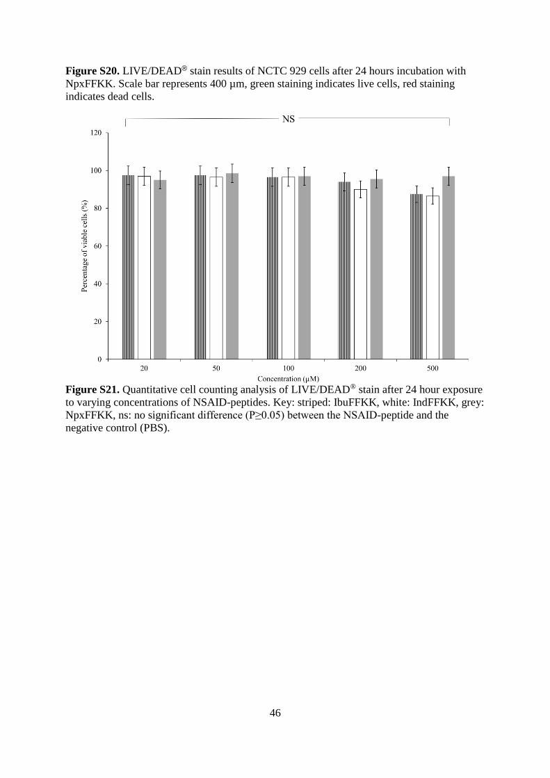
Figure S20. LIVE/DEAD® stain results of NCTC 929 cells after 24 hours incubation with
NpxFFKK. Scale bar represents 400 µm, green staining indicates live cells, red staining
indicates dead cells.
Figure S21. Quantitative cell counting analysis of LIVE/DEAD® stain after 24 hour exposure
to varying concentrations of NSAID-peptides. Key: striped: IbuFFKK, white: IndFFKK, grey:
NpxFFKK, ns: no significant difference (P≥0.05) between the NSAID-peptide and the
negative control (PBS).