Self-Assembling Micelles Based on an ... - Kumar Lab
9
Self-Assembling Micelles Based on an Intrinsically Disordered Protein Domain Sarah H. Klass, † Matthew J. Smith, † Tahoe A. Fiala, † Jess P. Lee, † Anthony O. Omole, † Bong-Gyoon Han, ‡ Kenneth H. Downing, ‡,∥ Sanjay Kumar, ⊥ and Matthew B. Francis* ,†,‡,§ † Department of Chemistry, University of California, Berkeley, California 94720, United States ⊥ Department of Bioengineering, University of California, Berkeley, California 94720, United States ‡ Molecular Biophysics and Integrated Bioimaging Division, Structural Biology and Imaging Department, and § Materials Sciences Division, Lawrence Berkeley National Laboratory, Berkeley, California 94720, United States * S Supporting Information ABSTRACT: The self-assembly of micellar structures from diblock polymers that contain hydrophilic and hydrophobic domains has been of great interest for the encapsulation of drugs and other hydrophobic molecules. While most commercially used surfactants are derived from hydrocarbon sources, there have been recent efforts to replace these with biodegradable, nontoxic, biologically synthesized alternatives. Previous examples have primarily examined naturally occur- ring self-assembling proteins, such as silk and elastin-like sequences. Herein, we describe a new series of fusion proteins that have been developed to self-assemble spontaneously into stable micelles that are 27 nm in diameter after enzymatic cleavage of a solubilizing protein tag. The sequences of the proteins are based on a human intrinsically disordered protein, which has been appended with a hydrophobic segment. The micelles were found to form across a broad range of pH, ionic strength, and temperature conditions, with critical micelle concentration (CMC) values in the low micromolar range, 3 orders of magnitude lower than the CMC of commonly used surfactant sodium dodecyl sulfate (SDS). The reported micelles were found to solubilize hydrophobic metal complexes and organic molecules, suggesting their potential suitability for catalysis and drug delivery applications. Furthermore, the inherent flexibility in the design of these protein sequences enables the encoding of additional functionalities for many future applications. Overall, this work represents a new biomolecular alternative to traditional surfactants that are based on nonrenewable and poorly biodegradable hydrocarbon sources. ■ INTRODUCTION It has been estimated that greater than 30% of eukaryotic proteins are disordered or contain disordered regions of 50 consecutive amino acids or more. 1 These intrinsically disordered proteins (IDPs) and intrinsically disordered protein regions (IDRs) lack the traditional secondary and tertiary structural motifs found in conventional proteins. Algorithms and data μ bases developed for the purpose of identifying IDPs and IDRs have accelerated our understanding of the various roles this class of proteins can play in cellular regulation and disease. 2−5 Furthermore, the disordered nature of certain IDPs have been shown to be vital to the phase separation that is implicated in the formation of membrane-less organelles. 6−9 IDPs based on elastin-like protein (ELP) sequences that exhibit the ability to undergo thermoresponsive phase transitions have been exploited to develop genetically tailorable protein-based polymers for applications in drug delivery and protein-based materials that are naturally biodegradable. 10−12 In contrast to the abundant examples of synthetically produced self-assembling peptides, 13−17 the recombinant expression of protein-based materials is more amenable to scale up and allows for precise genetic manipulation. To date, successful examples of using this approach to prepare micellular materials have started with naturally occurring motifs that are already known to self-assemble, including the formation of vesicles from the sunflower protein oleosin 18 and the formation of hydrogels by leucine zipper proteins. 19 Most notably, the self-assembly of the naturally phase separating elastin-based IDPs can be controlled by appending these sequences with hydrophobic-rich, 20−23 or post-translationally modified hydrocarbon tails. 11 These proteins have been shown to form a variety of temperature-sensitive structures (including micelles), 21,24 and they have been evaluated for their drug delivery potential in vivo. 25,26 However, given their great abundance and chemical diversity, IDPs remain an underutilized source of biologically derived and biodegradable polymers, with the majority of research being conducted on a small number of sequences that have shown utility in the development of protein-based self- Received: October 8, 2018 Published: February 11, 2019 Article pubs.acs.org/JACS Cite This: J. Am. Chem. Soc. 2019, 141, 4291-4299 © 2019 American Chemical Society 4291 DOI: 10.1021/jacs.8b10688 J. Am. Chem. Soc. 2019, 141, 4291−4299 Downloaded via UNIV OF CALIFORNIA BERKELEY on May 7, 2019 at 05:55:09 (UTC). See https://pubs.acs.org/sharingguidelines for options on how to legitimately share published articles.
Transcript of Self-Assembling Micelles Based on an ... - Kumar Lab

Self-Assembling Micelles Based on an Intrinsically DisorderedProtein DomainSarah H. Klass,† Matthew J. Smith,† Tahoe A. Fiala,† Jess P. Lee,† Anthony O. Omole,†
Bong-Gyoon Han,‡ Kenneth H. Downing,‡,∥ Sanjay Kumar,⊥ and Matthew B. Francis*,†,‡,§
†Department of Chemistry, University of California, Berkeley, California 94720, United States⊥Department of Bioengineering, University of California, Berkeley, California 94720, United States‡Molecular Biophysics and Integrated Bioimaging Division, Structural Biology and Imaging Department, and §Materials SciencesDivision, Lawrence Berkeley National Laboratory, Berkeley, California 94720, United States
*S Supporting Information
ABSTRACT: The self-assembly of micellar structures fromdiblock polymers that contain hydrophilic and hydrophobicdomains has been of great interest for the encapsulation ofdrugs and other hydrophobic molecules. While mostcommercially used surfactants are derived from hydrocarbonsources, there have been recent efforts to replace these withbiodegradable, nontoxic, biologically synthesized alternatives.Previous examples have primarily examined naturally occur-ring self-assembling proteins, such as silk and elastin-likesequences. Herein, we describe a new series of fusion proteinsthat have been developed to self-assemble spontaneously into stable micelles that are 27 nm in diameter after enzymaticcleavage of a solubilizing protein tag. The sequences of the proteins are based on a human intrinsically disordered protein,which has been appended with a hydrophobic segment. The micelles were found to form across a broad range of pH, ionicstrength, and temperature conditions, with critical micelle concentration (CMC) values in the low micromolar range, 3 ordersof magnitude lower than the CMC of commonly used surfactant sodium dodecyl sulfate (SDS). The reported micelles werefound to solubilize hydrophobic metal complexes and organic molecules, suggesting their potential suitability for catalysis anddrug delivery applications. Furthermore, the inherent flexibility in the design of these protein sequences enables the encoding ofadditional functionalities for many future applications. Overall, this work represents a new biomolecular alternative to traditionalsurfactants that are based on nonrenewable and poorly biodegradable hydrocarbon sources.
■ INTRODUCTION
It has been estimated that greater than 30% of eukaryoticproteins are disordered or contain disordered regions of 50consecutive amino acids or more.1 These intrinsicallydisordered proteins (IDPs) and intrinsically disordered proteinregions (IDRs) lack the traditional secondary and tertiarystructural motifs found in conventional proteins. Algorithmsand data μ bases developed for the purpose of identifying IDPsand IDRs have accelerated our understanding of the variousroles this class of proteins can play in cellular regulation anddisease.2−5 Furthermore, the disordered nature of certain IDPshave been shown to be vital to the phase separation that isimplicated in the formation of membrane-less organelles.6−9
IDPs based on elastin-like protein (ELP) sequences thatexhibit the ability to undergo thermoresponsive phasetransitions have been exploited to develop genetically tailorableprotein-based polymers for applications in drug delivery andprotein-based materials that are naturally biodegradable.10−12
In contrast to the abundant examples of syntheticallyproduced self-assembling peptides,13−17 the recombinantexpression of protein-based materials is more amenable to
scale up and allows for precise genetic manipulation. To date,successful examples of using this approach to preparemicellular materials have started with naturally occurringmotifs that are already known to self-assemble, including theformation of vesicles from the sunflower protein oleosin18 andthe formation of hydrogels by leucine zipper proteins.19 Mostnotably, the self-assembly of the naturally phase separatingelastin-based IDPs can be controlled by appending thesesequences with hydrophobic-rich,20−23 or post-translationallymodified hydrocarbon tails.11 These proteins have been shownto form a variety of temperature-sensitive structures (includingmicelles),21,24 and they have been evaluated for their drugdelivery potential in vivo.25,26
However, given their great abundance and chemicaldiversity, IDPs remain an underutilized source of biologicallyderived and biodegradable polymers, with the majority ofresearch being conducted on a small number of sequences thathave shown utility in the development of protein-based self-
Received: October 8, 2018Published: February 11, 2019
Article
pubs.acs.org/JACSCite This: J. Am. Chem. Soc. 2019, 141, 4291−4299
© 2019 American Chemical Society 4291 DOI: 10.1021/jacs.8b10688J. Am. Chem. Soc. 2019, 141, 4291−4299
Dow
nloa
ded
via
UN
IV O
F C
AL
IFO
RN
IA B
ER
KE
LE
Y o
n M
ay 7
, 201
9 at
05:
55:0
9 (U
TC
).
See
http
s://p
ubs.
acs.
org/
shar
ingg
uide
lines
for
opt
ions
on
how
to le
gitim
atel
y sh
are
publ
ishe
d ar
ticle
s.
assembled materials.9,12,23,27 In an effort to expand thesequence space of self-assembling IDPs, we have exploredmethods to introduce self-assembly, through genetic mod-ification, to a novel IDP sequence that normally shows nophase separation or aggregation behavior.Herein, we describe the construction and characterization of
a new class of self-assembling proteins based on an intrinsicallydisordered sequence derived from the human neurofilamentheavy arm side chain (NF-H) protein.28 In vivo the NF-Hdomain of interest, referred to as IDP herein, forms extendedprotrusions that flare out from the core regions of cylindricalfibers, providing mechanical integrity to the axons of neuronsand acting as a regulator of neurofilament transport.29−32
These disordered protein regions involve a characteristicrepeat sequence that can be generalized as “SPAE(A/V)K”,which has been found to expand and contract in response topH, phosphorylation, and ionic strength conditions.33,34 Thesesequences do not assemble or aggregate on their own, insteadbehaving more like unstructured organic polymers. As a result,there is significant interest in using this sequence to create“smart biomaterials” in the form of protein-based environ-mentally sensitive polymer brushes.24,35 By designing andadding a hydrophobic domain to the C-terminus of the IDPsequence, we sought to generate an IDP-based diblockcopolymer that, through entropic and enthalpic forces,undergoes self-assembly in aqueous solutions, Figure 1.
In an effort to develop and characterize a new class of self-assembling proteins, we report an engineered protein constructthat allows facile expression and purification, with the potentialfor scalable production. Upon cleavage of an initial solubledomain, the construct self-assembles into micelles that arehighly stable with respect to temperature, pH, and ionicstrength conditions. Micelle structures were observed atsubmicromolar concentrations, and the ability of theseconstructs to solubilize hydrophobic cargo molecules in theircore regions was demonstrated. These IDP-based micellesprovide tunable, biorenewable, biodegradable, protein-basedsurfactants with properties similar to those of commonly usedsurfactants found in detergents, drug delivery systems, andencapsulating agents that are traditionally derived fromchemically modified hydrocarbons, which pose a great health
and environmental impact.36,37 Furthermore, this approachshows promise for the development of new protein-basedmaterials from previously overlooked sequences that may posenew or interesting biomaterial properties.
■ RESULTS AND DISCUSSIONDesign of Sequences. A major challenge in the
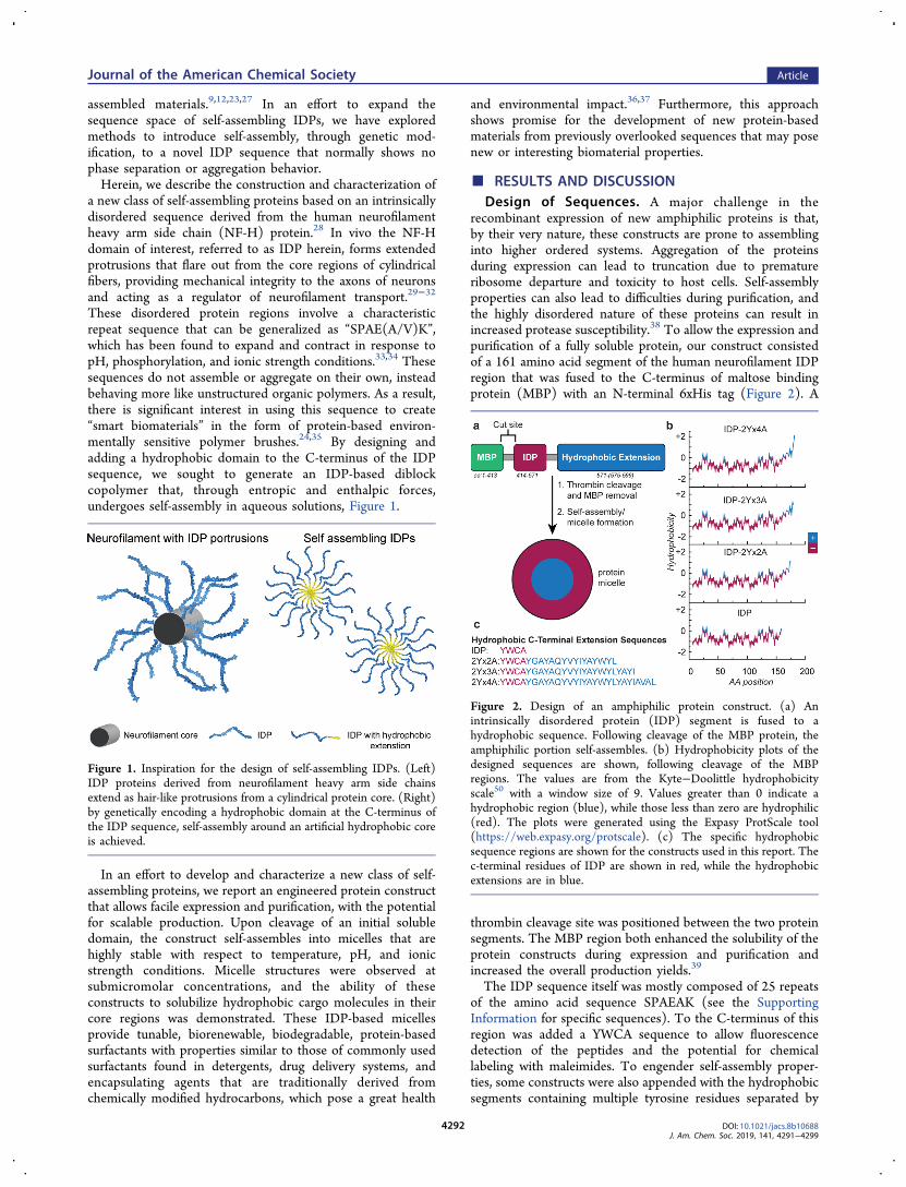
recombinant expression of new amphiphilic proteins is that,by their very nature, these constructs are prone to assemblinginto higher ordered systems. Aggregation of the proteinsduring expression can lead to truncation due to prematureribosome departure and toxicity to host cells. Self-assemblyproperties can also lead to difficulties during purification, andthe highly disordered nature of these proteins can result inincreased protease susceptibility.38 To allow the expression andpurification of a fully soluble protein, our construct consistedof a 161 amino acid segment of the human neurofilament IDPregion that was fused to the C-terminus of maltose bindingprotein (MBP) with an N-terminal 6xHis tag (Figure 2). A
thrombin cleavage site was positioned between the two proteinsegments. The MBP region both enhanced the solubility of theprotein constructs during expression and purification andincreased the overall production yields.39
The IDP sequence itself was mostly composed of 25 repeatsof the amino acid sequence SPAEAK (see the SupportingInformation for specific sequences). To the C-terminus of thisregion was added a YWCA sequence to allow fluorescencedetection of the peptides and the potential for chemicallabeling with maleimides. To engender self-assembly proper-ties, some constructs were also appended with the hydrophobicsegments containing multiple tyrosine residues separated by
Figure 1. Inspiration for the design of self-assembling IDPs. (Left)IDP proteins derived from neurofilament heavy arm side chainsextend as hair-like protrusions from a cylindrical protein core. (Right)by genetically encoding a hydrophobic domain at the C-terminus ofthe IDP sequence, self-assembly around an artificial hydrophobic coreis achieved.
Figure 2. Design of an amphiphilic protein construct. (a) Anintrinsically disordered protein (IDP) segment is fused to ahydrophobic sequence. Following cleavage of the MBP protein, theamphiphilic portion self-assembles. (b) Hydrophobicity plots of thedesigned sequences are shown, following cleavage of the MBPregions. The values are from the Kyte−Doolittle hydrophobicityscale50 with a window size of 9. Values greater than 0 indicate ahydrophobic region (blue), while those less than zero are hydrophilic(red). The plots were generated using the Expasy ProtScale tool(https://web.expasy.org/protscale). (c) The specific hydrophobicsequence regions are shown for the constructs used in this report. Thec-terminal residues of IDP are shown in red, while the hydrophobicextensions are in blue.
Journal of the American Chemical Society Article
DOI: 10.1021/jacs.8b10688J. Am. Chem. Soc. 2019, 141, 4291−4299
4292

small hydrophobic amino acids due to their hydrophobicity,propensity to π-stack, and H-bonding capabilities, as shown inFigure 2b,c.The self-assembling regions of these peptides are referred to
as “2Yx#A” herein, where # = 2,3,4. A higher number indicatesa longer hydrophobic sequence.Construction of Plasmids and Expression of Proteins.
The IDP segment was initially prepared as a fusion to a 6xHis-MBP protein and was constructed using two gene blocks dueto cloning difficulties arising from its repetitive sequence. Thegenerated MBP-IDP plasmid served as the basis for alladditional constructs. The MBP-IDP-2Yx(2−4)A plasmidswere constructed by performing overhang PCR on the MBP-IDP (or subsequent) plasmid.Pure monomeric IDP protein was obtained in a manner
similar to IDP-2Yx(2−4)A proteins, as reported in theSupporting Information. Characterization using DLS revealedan average diameter of 11.3 ± 0.8 nm, which was consistentwith literature reports.35 The MBP-IDP-2Yx(2−4)A constructswere expressed in Rosetta (DE3) pLysS competent cells, asthis strain has been shown to stabilize pET recombinants thatencode proteins that can affect cell growth and viability. Thiscell line also improves the performance of rarely used E. colicodons through the addition of non-native tRNA genes on aseparate plasmid.40
Following cell lysis, the constructs were purified by nickelnitroloacetic acid (Ni-NTA) affinity chromatography due tothe binding affinity of the 6xHis group encoded on the MBPportion of the fusion protein. By LC−MS and SDS-PAGEanalysis, the MBP-IDP-2Yx2A constructs were found toexpress well and with high purity following affinity purification(Figure S1).By DLS, MBP-IDP-2Yx2A showed a diameter similar to that
of IDP (11.1 ± 1.3 nm), indicating that it was still monomericat this stage. Constructs MBP-IDP-2Yx3A and MBP-IDP-2Yx4A, which only differ from MBP-IDP-2Yx2A in theaddition of 4 and 8 hydrophobic amino acids, respectively,resulted in lower bacterial titers. Additionally, for MBP-IDP-2Yx3A and MBP-IDP-2Yx4A, smaller cell pellets wereobserved after expression, soapy foams formed uponsonication, and truncations were observed in the LC−MStraces following NiNTA purification (Figure S2). Additionally,a DLS hydrodynamic diameter of 36 ± 15 nm was observedfor MBP-IDP-2Yx3A, and a diameter of 115 ± 6 nm wasobserved for MBP-IDP-2Yx4A (Figure S3). Taken together, itappears that the MBP-IDP-2Yx3A and MBP-IDP-2Yx4Aconstructs are aggregation-prone prior to MBP cleavage,making them more difficult to express and purify.MBP Cleavage and Purification of the IDP-2Yx2A
Constructs. Following Ni-NTA purification, MBP-IDP-2Yx2A was cleaved using thrombin and purified by preparativescale reversed phase chromatography to ensure high purity forthis study; however, ion exchange chromatography could alsobe used to purify the final construct on a larger scale (albeit atslightly lower purity; see Figure S1c). The resulting proteinwas stored as a solid following lyophilization. For use, the drypowder was resuspended in Milli-Q water by vortexing andsonicating prior to the addition of the desired buffer.Following MBP cleavage and protein purification, a
hydrodynamic diameter of 26.9 ± 4.6 nm was observed inMilli-Q water (Figure 3b). This size correlates with micelle-likestructures with an 11 nm hydrophilic IDP portion and a 2.5nm hydrophobic interior, as would be expected given the
apparent size of the IDP segment alone by DLS (11.1 nm) plusa 16 amino acid hydrophobic extension if an α-helicalconfiguration is assumed (2.4 nm, 1.5 Å per residue). Micelleformation in the size regime of 27 nm was shown to bereproducible across multiple expression batches (Figure S5).To confirm that the cysteine residue in the hydrophobicdomain was not responsible for micelle formation, weconstructed and expressed an IDP-2Yx2A-C → S mutant
Figure 3. Analysis of the protein constructs. (a) LC−MS analyses areshown for MBP-IDP-2Yx2A (red), IDP-2Yx2A after MBP removalwith thrombin (green), and IDP-2Yx2A after HPLC purification(blue). The MBP-IDP-2Yx2A expected molecular weight is 63 605Da. In the IDP-2Yx2A spectra, the cysteine residue is capped as adisulfide with β-mercaptoethanol, corresponding to an expectedmolecular weight of 18 366 Da. The IDP-2Yx2A disulfide dimerappears at 36 580 Da in the bottom spectrum. (b) DLS analysis ofIDP-2Yx2A throughout the purification process shows changes in thediameters of the constructs. Prior to cleavage, the MBP-IDP-2Yx2Aprotein has a diameter similar to that of IDP itself, and is presumablymonomeric. Following thrombin cleavage, a diameter between that ofthe MBP monomer and IDP is observed. After removal of the MBP, asharp increase in the average diameter is observed, indicating self-assembly in both pure water and buffer (phosphate, pH 5.7). Thepolydispersity indices (PDIs) of the IDP-2Yx2A samples above theCMC are generally between 0.2 and 0.5, depending on the bufferconditions (see Figure S4 for full DLS traces for IDP and IDP-2Yx2A). (c) A Coomassie-stained SDS page gel of HPLC purifiedIDP-2Yx2A is shown, indicating >95% purity. (d) MD simulations ofIDP and IDP-2Yx2A at 50 ns. Residues in red indicate the IDPportion of each construct, while blue residues indicate thehydrophobic c-terminal residues.
Journal of the American Chemical Society Article
DOI: 10.1021/jacs.8b10688J. Am. Chem. Soc. 2019, 141, 4291−4299
4293

that, when analyzed by DLS, showed micelles with an averagediameter of 26.5 ± 4.9 nm in Milli-Q H2O (Figure S6).The two constructs were further analyzed using molecular
dynamic (MD) simulations. After running the simulations for50 ns, both constructs were similar to the sizes measured byDLS for a single monomer. Between 40 and 50 ns ofsimulation, the radius of gyration (Rg) fluctuated between 3and 4 nm for IDP and between 4 and 5 nm for 2Yx2A (FigureS7). As the Rg for a single monomer of 2Yx2A would equate to1/4 of the total diameter of an assembled micelle, the MDresults indicate an expected gyrational diameter of 16−20 nmfor the assembled structure. This results in a hydrodynamicdiameter (Dh) for a spherical particle of 20.6−25.8 nm, whereDh × 0.775 = Rg. In both constructs, the IDP portion showedno indication of collapse over the 50 ns simulation time(Supporting Information movies 1 and 2).Effects of Salt, pH, and Temperature on IDP-2Yx2A
Micelles. The effects of changes to salt concentration and pHwere evaluated for the protein assemblies using DLS.Structures consistent with micelles were observed at pH valuesranging from 3.7−9.7 and salt concentrations of 0−200 mMKxPO4 (Figure 4a), NaCl, and CaCl2 (Figure S8). At most pH
values, the micelle sizes were consistent at ∼27 nm with lowpolydispersity. At pH 7.9, significant heterogeneity andpolydispersity in the micellar diameters were observed (Figure4a,b). Interestingly, this pH corresponded to the mostcollapsed state of the hydrophilic IDP portion, as determinedin previous studies.33,35 This may affect the self-assemblybehavior of IDP-2Yx2A and was a property we sought toexplore further.35
To examine the thermal stability of the micelles, DLSmeasurements of IDP-2Yx2A were taken upon heating from 25
to 70 °C. As the temperature increased, the average diameterand polydispersity of the sample decreased, Figure 4c. Noprecipitation was observed, and the size change was found tobe reversible upon cooling. Similar trends are reported in theliterature for conventional SDS micelles and casein micelles,where the micellar volume decreases with increasing temper-ature.41,42 This is likely due to factors including thehydrophobic tail packing and the interactions between waterand the hydrophilic headgroups.
Determination of the Critical Micelle Concentration(CMC). As polymeric micelles often find use in biomedical43
and aquatic environments,44 we sought to explore theirstability in buffers that reflect the pH of blood (1xPBS, pH7.4) and rainwater (phosphate buffer, pH 5.7, 100 mM; seeSupporting Information S1 for specific buffer compositions).45
Herein these buffers will be referred to as pH 7.4 buffer andpH 5.7 buffer, respectively.The CMC values of IDP-2Yx2A in these buffers were
determined using fluorescence spectroscopy. The emissionratios of the first and third vibronic bands of pyrene weremeasured at varying concentrations of protein at roomtemperature. The I1/I3 ratio is known to increase withincreasing polarity of the probe environment.14,46 At highconcentrations, an I1/I3 ratio of 0.9−1.0 was observed in theIDP-2Yx2A samples for both buffers. This indicated that themedium surrounding the pyrene was more hydrophobic thanwhen in phosphate buffer alone (I1/I3 = 1.33). In contrast, nochange in the I1/I3 band ratio was observed for the IDP proteinlacking the 2Yx2A sequence at any concentration in the pH 5.7buffer (I1/I3 = 1.33−1.36 over a range of 1−100 μM).The I1/I3 ratios of the IDP-2Yx2A samples in both buffers
were fit to a logistic function (R2 for pH 7.4 = 0.97; R2 for pH5.7 = 0.97), with EC50 values of 26 μM for pH 7.4 buffer and13 μM in pH 5.7 buffer. However, because the CMC was solow, and the final concentration of pyrene must be maintainedat 2 μM to obtain good quality fluorescence spectra, this assaycan only provide an upper limit of the CMC.47 A moreaccurate determination of CMC may be simply the emergenceof the I3 band, which indicates a CMC of 2.5 μM (the lowestconcentration at which a distinct I3 band was observed)(Figure 5). This value is reflected by the EC20 of the logistic fit.From the EC20, IDP-2Yx2A CMC values were determined tobe 17.5 μM in pH 7.4 buffer and 2.6 μM in pH 5.7 buffer.These results again agree with the reported behavior of theIDP sequence at near-neutral pH, where the sequence isknown to be in its most collapsed state.35 This can berationalized by a balancing of the glutamic acid and lysineresidue charges, causing an overall decrease in the charge of thehydrophilic domain (and therefore a decrease in hydro-philicity). This results in a less stable assembly. At pH 5.7, sidechain protonation results in a net positive charge (increasedhydrophilicity) for the hydrophilic domains and thus promotesa more stable and extended assembly.These results were also compared to a dilution study
performed using dynamic light scattering under the samebuffer conditions. By DLS, the micelle size (∼27 nm) and lowpolydispersity were maintained at concentrations above 10 μMin both buffers. As the concentration decreased further, themicelles swelled and became less uniform at pH 7.4. Inagreement with the fluorescence data, the samples in pH 5.7buffer showed consistent diameters and low polydispersitydown to 0.5 μM. It should be noted that the data point at 0.1μM in pH 5.7 buffer, where a dramatic increase in size is
Figure 4. pH and temperature effects on micelle size, as measured byDLS. (a) DLS measurements of IDP-2Yx2A micelles were performedunder varying pH and buffer conditions. Over all pH values and saltconcentrations examined, the average diameter was 26.2 ± 4.3 nm.(b) The diameter changes occurring upon pH and salt variation aresummarized as a topographical plot. The largest variability in diameteris observed as the pH changes from 7 to 8, but disassembly was notobserved. (c) The influence of temperature on micelle size isdetermined by DLS. As the temperature increases, the micellediameter decreases from 27.0 ± 1.1 to 16.5 ± 0.5 nm.
Journal of the American Chemical Society Article
DOI: 10.1021/jacs.8b10688J. Am. Chem. Soc. 2019, 141, 4291−4299
4294

observed, occurs near the detection limit of the DLSinstrument.Validation of Size, Shape, and CMC by Cryo-TEM and
SAXS. Cryogenic transmission electron microscopy (cryo-TEM) and small-angle X-ray scattering (SAXS) analysisconfirmed the size, shape, and CMC of the micelles. SAXSanalysis was conducted on IDP-2Yx2A in pH 7.4 buffer and inpH 5.3 buffer, as compared to IDP itself as a nonassemblingcontrol. The SAXS intensity curves showed a clear differencebetween the IDP-2Yx2A samples and the IDP sample, withboth IDP-2Yx2A samples showing a dramatic increase in thelow q region. This indicated the presence of large particles.Unfortunately, due to the high concentration of proteinrequired for SAXS analysis, a monodisperse core−shell modelcannot be applied to the data. Although not observed by cryo-TEM (see below), we hypothesize that this could be due tointermicelle interactions, leading to a small fraction of largerand elongated particles in the sample at higher concentrationsthat also contribute to the larger intensity profiles seen by DLS(Figure S9). However, a lower limit (excluding the presence oflarger aggregates) radius of gyration (Rg) and radius of thecross-section (Rc) can be calculated. For the IDP-2Yx2Asamples, Rg values obtained from the Guinier analysis weregreater than 9.0 nm, while the Rc values were between 3.6 and5.2 nm. For nonassembling IDP, the Rg was calculated to begreater than 4.0 nm, and a Rc value was not observed. This
indicates a clear difference in the assembly states of IDP andIDP-2Yx2A (Figure S10).The spherical shape and low CMC of the micelles were
confirmed using cryo-TEM on two unique expression batchesof the IDP-2Yx2A micelles and under different cryo-TEMsample preparations. First, a 6.5 μM solution of IDP-2Yx2Amicelles in 10 mM phosphate buffer pH 5.7 was imaged on aholey gold grid covered by a thin sheet of continuous carbon.Many spherical micelles were observed with an averagediameter of 24.8 ± 4.6 nm. (Figure 6a). To obtain a higher
contrast image and confirm the observed increase in size at lowconcentrations by DLS, a 0.4 μM solution of IDP-2Yx2A wasexamined on uncoated holey carbon grids. Spherical micellarstructures were again observed (Figure 6b). Consistent withthe DLS results, the low concentration of particles resulted inlarger diameters of 50.5 ± 12.1 nm and greater polydispersity.To obtain directly comparative data, each cryo-TEM samplewas analyzed by DLS prior to cryogenic freezing. Both the 6.5μM and the 0.4 μM samples showed a good correlationbetween the diameters derived from DLS and the cryo-TEManalyses (Figure 6c). Overall, this indicates that the sizesindicated by DLS are accurate, the micelles swell with dilutionbeyond the CMC, and the CMC of these IDP-2Yx2A micelleslies in the low-micromolar range.
Solubilization of Hydrophobic Molecules. A keyapplication of surfactants and micelles stems from their abilityto solubilize hydrophobic molecules in aqueous solution. Toevaluate the ability of IDP-2Yx2A micelles to do this, threemolecules with unique functions were chosen. The antifungalagent pyraclostrobin, with a solubility of 1.9 mg/L in water,
Figure 5. CMC evaluations at pH 5.7 and pH 7.4. (a) Fluorescencetraces of pyrene with 0−100 μM IDP-2Yx2A in pH 5.7 buffer areshown. An enhanced I3 fluorescence band of pyrene is observed atconcentrations as low as 2.5 μM. (b) Pyrene fluorescence wassimilarly evaluated for IDP-2Yx2A in pH 7.4 buffer and for thenonassembling IDP in pH 5.7 buffer. The ratios of the first and thirdvibronic bands (I1/I3) are plotted and fit to logistic functions todetermine upper limits of the CMC values. Both IDP-2Yx2A samplesshow enhancement of the pyrene I3 band, indicating micelleformation. IDP-2Yx2A has a lower CMC value in pH 5.7 bufferthan in pH 7.4 buffer, with EC20 values of 2.6 and 17.5 μM,respectively. No I3 band enhancement was observed for IDP at anyconcentration. (c) DLS analysis of IDP-2Yx2A micelles is shown atdecreasing concentrations in pH 5.7 buffer and pH 7.4 buffer.
Figure 6. Cryo-TEM analysis of IDP-2Yx2A micelles. (a) A 6.5 μMsolution of IDP-2Yx2A micelles (10 mM phosphate buffer pH 5.7)was imaged on holey gold grids coated with thin carbon film. Thepresence of many spherical particles is observed, confirming the shapeand low CMC of IDP-2Yx2A micelles. (b) A more dilute sample (0.4μM solution of 2Yx2A micelles in 100 mM phosphate buffer pH 5.3)was also examined embedded in vitrified ice on uncoated carbongrids. The micelles show an increased size and greater polydispersity,consistent with observations in Figure 5c. (c) The hydrodynamicdiameter as measured by DLS for each sample shows diameterscomparable to those obtained by cryo-TEM image analysis usingImageJ (n = 500, 6.5 μM) (n = 20, 0.4 μM). This confirms that thediameter observed by DLS is an accurate representation of themicelles’ true size.
Journal of the American Chemical Society Article
DOI: 10.1021/jacs.8b10688J. Am. Chem. Soc. 2019, 141, 4291−4299
4295

was first loaded into the micelle structure by combining bothpyraclostrobin and lyophilized IDP-2Yx2A in a small volume ofTHF, followed by dilution with pH 5.7 buffer. Any remaininginsoluble pyraclostrobin was removed by centrifugation, andthe THF was removed under vacuum. Following an additionalround of resuspension in water and subsequent centrifugation,the supernatant was diluted 1:1 with acetonitrile before HPLCanalysis to quantify the amount of pyraclostrobin that had beensolubilized (Figure S11). In the presence of IDP-2Yx2A,substantially more pyraclostrobin was solubilized thanoccurred in buffer alone (Figure 7a). On the basis of UVmeasurements, 15.2 ± 8 molecules of pyraclostrobin wereloaded per protein monomer. Assuming each IDP-2Yx2Amolecule is a cone with a length of 13.5 nm and a base radius
of 1.25 nm, it can be estimated that each 27 nm micelle spherecontains about 470 protein molecules. This suggests thatthousands of pyraclostrobin molecules are contained withineach micelle.As a second cargo type, a singlet oxygen-generating
tetraphenyl-porphyrin (TPP) molecule used for photodynamictherapy (PDT) was evaluated similarly. TPP is completelyinsoluble in water and thus is nonfluorescent. However, uponincubation with IDP-2Yx2A, characteristic emission peaksaround 650 and 715 nm were observed (Figure 7b).As the degradation profile of a solubilizing or delivery agent
would be of particular importance for both antifungal and PDTcompounds, the degradation profile of IDP-2Yx2A micelleswas examined at room temperature and exposed to air. After aperiod of 50 d, no evidence of IDP-2Yx2A was observed byLC−MS or SDS PAGE gel (Figure S12), presumably due todegradation from the proteases secreted by adventitiousbacteria.Finally, a Suzuki coupling catalyst (Pd(dppf)Cl2) with poor
water solubility was added to IDP-2Yx2A micelles. Loadingwas then analyzed by TEM without additional staining.Consistent with the catalyst being concentrated in the interiorof the micelle structures, small, spherical concentrations ofmetal complexes were observed at the expected sizes of 15 ± 8nm (Figure 7c). This result indicates that, upon the loading ofthis hydrophobic catalyst precursor, the spherical micelle-likeshape of the particles is maintained and does not undergoaggregation. Furthermore, this demonstrates potential applica-tions in the micellar catalyst and the templating of metalnanoparticles.
■ CONCLUSIONDeveloping biologically derived and biodegradable surfactantscould help to alleviate the environmental effects of detergentsthat have been traditionally hydrocarbon-based. The move toreplace these compounds with synthetic peptide-basedamphiphiles has resulted in many highly successful self-assembling systems; however, their syntheses typically involvethe use of organic solvents, and the costs associated with theirproduction can limit scale-up potential. The new IDP-basedproteins described herein offer high yields using inexpensivebacterial expression systems, and exhibit CMC values that arelower than many other types of surfactants, such as sodiumdodecyl sulfate (SDS), which has a CMC of 8 mM.48 Theyoffer excellent thermostability and remain assembled across abroad range of pH, ionic strength, and temperature conditions.These features suggest that, even in their current form, theycan find use in a number of industrial applications. The abilityto house hydrophobic cargo molecules, along with the overallnanoscale dimensions, open the door to applications intargeted drug delivery. Perhaps the greatest opportunity isthe potential for tuning the sequences to obtain desiredsolubilizing, assembling, and response properties, as everyposition along the polypeptide chain can be specified on thegenetic level. We are currently exploring the potential of thispromising class of surfactants for use as paint emulsifiers,antifungal dispersants, and bioremediation materials.
■ EXPERIMENTAL PROCEDURESExpression and Purification of MBP-IDP. MBP-IDP plasmids
were prepared as described in the Supporting Information andtransformed into E. coli BL21(DE3) competent cells. Starter cultures(20 mL of Lauria Broth (LB, VWR Life Sciences), including 50 mg/L
Figure 7. Evaluation of small molecule loading into IDP-2Yx2Aprotein micelles. (a) Pyraclostrobin shows substantially greatersolubility (0.6 ± 0.3 nmol) in 3 μM IDP-2Yx2A solution versuswater alone (0.01 ± 0.01 nmol). The molar ratio of pyraclostrobin toIDP-2Yx2A protein monomers is 15.2 ± 8. (b) The loading oftetraphenyl-porphyrin (TPP) for singlet oxygen generation wasevaluated by fluorescence emission. When solubilized in thehydrophobic environment of IDP-2Yx2A, the characteristic emissionspectrum of TPP was observed. No material dissolved in water alone.(c) Unstained TEM images of hydrophobic Suzuki coupling reagentPd(dppf)Cl2 show loading into IDP-2Yx2A micelles. The heavymetals Pd and Fe provide sufficient contrast to image the micelleinterior, which have a measured diameter averaging 15 ± 8 nm (over4000 particles analyzed).
Journal of the American Chemical Society Article
DOI: 10.1021/jacs.8b10688J. Am. Chem. Soc. 2019, 141, 4291−4299
4296

kanamycin, were grown from single colonies, grown overnight at 37°C, and used to inoculate 1 L of Terrific Broth (TB, Sigma) mediathat contained 50 mg/L kanamycin. Cultures were grown to an OD ≈0.5, cooled for 20 min at 16 °C, induced with 0.5 mM IPTG, andexpressed (∼18 h) at 25 °C. Cells were harvested by centrifugationfor 15 min at 4000g at 4 °C. The resulting pellet was lysed in 30 mLof lysis buffer (20 mM HEPES, pH = 7.5, 300 mM NaCl, 10 mM β-mercaptoethanol, 10 mM imidazole = buffer A) supplemented withone tablet of EDTA-free SigmaFast Protease Inhibitor (Sigma-Aldrich), 2 mM PMSF, and 10 mg of lysozyme. The resuspendedsample was lysed by sonication (amplitude 50%, 2:4 s on:off for 10min), followed by 20 min of centrifugation at 24 000g at 4 °C. Thesupernatant was filtered through a 40 μm Steriflip filter (Millipore),and loaded onto a 5 mL NiNTA column (Protino, Machery Nagel)connected to an Akta purifier that was pre-equilibrated with buffer A.The column was washed with 50 mL (10 column volumes) of 20 mMHEPES (pH = 7.5), 300 mM NaCl, 10 mM βMe, and 10 mMimidazole. The protein was eluted with 20 mM HEPES (pH = 7.5),300 mM NaCl, 10 mM β-mercaptoethanol, and 250 mM of imidazole.Imidazole was removed by spin concentration with an Amicon Ultra15 mL 30 kDaA MWCO spin filter (Millipore Sigma) and 20 mMHEPES (pH = 7.5) buffer containing 100 mM NaCl.Expression and Purification of MBP-IDP-2Yx(2−4)A and
MBP-IDP-2Yx2A-C>S. Plasmids were transformed into E. coliRosetta 2 pLys competent cells. Starter cultures in 20 mL of LBwith 50 mg/L kanamycin were grown overnight at 37 °C from singlecolonies and used to inoculate 1 L of TB media containing 50 mg/Lkanamycin. Cultures were grown to an OD ≈ 0.5, cooled for 20 minat 16 °C, induced with 0.1 mM IPTG, and expressed for about 6 h at16 °C. Cells were harvested by centrifugation for 15 min at 4000g at 4°C. The pellet was transferred to a 50 mL Falcon tube in PBS buffer,and spun down for 10 min at 4000 rcf (g). The resulting pellet (∼5 g)was lysed in 30 mL of lysis buffer (20 mM HEPES, pH = 7.5, 300 mMNaCl, 10 mM imidazole = buffer A) supplemented with one tablet ofEDTA-free SigmaFast Protease Inhibitor (Sigma-Aldrich), 2 mMPMSF, and 10 mg lysozyme. The resuspended sample was lysed bysonication (amplitude 50%, 2:4 s on:off for 10 min) followed by 20min of centrifugation at 24 000 rcf (g) at 4 °C. The supernatant wasfiltered through a 40 μm Steriflip filter (Millipore), and loaded onto a5 mL NiNTA column (Protino, Machery Nagel) connected to anAkta purifier that was pre-equilibrated with buffer A. The column waswashed with 50 mL (10 CV) of 20 mM HEPES (pH = 7.5), 300 mMNaCl, 10 mM imidazole. The protein was eluted with 20 mM HEPES(pH = 7.5), 300 mM NaCl, 250 mM imidazole. Purity and identity ofthe recovered protein were analyzed by gel and LC−MS. Imidazolewas removed by spin concentration with 20 mM HEPES (pH = 7.5),100 mM NaCl.MBP Cleavage Procedure. MBP-IDP, MBP-IDP-2Yx(2−4)A,
and MBP-IDP-2Yx2A-C>S samples were subsequently digested with1 mg of thrombin protease (high purity from Bovine, MPBiomedicals). Complete digestion was achieved at room temperatureafter 1 h, as confirmed by ESI-TOF LC−MS.Molecular Dynamic Simulations. MD simulations were carried
out using the Desmond software package and run with theOPLS_2005 force field49 available through Maestro. Both 2Yx2Aand IDP were modeled in an alpha helical conformation at time zero.Simulations were carried out at 300 K and at a constant pressure of1.01325 bar. Each structure was fully solvated with SPC water in acube with an edge length of 20 Å. Each simulation was run for a totalof 50 ns.Protein Purification. The IDP sample was diluted to a final
volume of 50 mL with 20 mM HEPES (pH = 7.5) containing 10 mMβ-mercaptoethanol and loaded onto a 1 mL HiTrap HP cationexchange column connected to an Akta purifier (GE Healthcare). Thecolumn was washed with 10 column volumes of 20 mM HEPES (pH= 7.5) buffer containing 10 mM β-mercaptoethanol and eluted with20 mM HEPES (pH = 7.5) buffer containing 10 mM β-mercaptoethanol and 1 M NaCl. The protein was >95% pure bySDS-PAGE gel electrophoresis and LC−MS. The IDP-2Yx(2−4)Aand IDP-2Yx2A-C>S samples were purified using reversed phase
purification on a Biotage chromatography system. To these sampleswas added 10% acetonitrile (ACN), and the resulting solutions wereloaded onto a 10 g C18 Biotage SNAP Bio 300A reversed phasecolumn that had been equilibrated with 10% ACN in H2O + 0.1%TFA. The column eluted using a gradient of 10−100% ACN, with thedesired product eluting around 40% ACN. The fractions containingIDP-2Yx2A were analyzed by ESI/TOF LC−MS for purity. Purefractions were collected and lyophilized to dryness, yielding theprotein product as a white powder. SDS-PAGE analysis withCoomassie staining indicated >95% purity.
DLS Analysis. DLS analysis was performed on a MalvernInstruments Zetasizer Nano ZS. Data plots and standard deviationswere calculated from an average of three measurements, each of whichconsisted of 13 runs. Measurement data are presented as a diameterdetermined by the % number distribution. The % number was used inthis analysis because it shows the size of the majority population in asample. We believe that heterogeneity is evident in the micellesamples as evidenced by the polydispersity. It is likely that largerstructures or intermicelle interactions occur and form a small numberof larger particles that contribute to larger diameters when analyzedby % intensity, which heavily weights large particles in a sample asopposed to the most abundant. Unless otherwise stated, allmeasurements are the average % number reported and were takenusing 0.2 μM spin filtered samples at 25 °C after a 2 min period oftemperature equilibration.
CMC Determination by Pyrene Fluorescence. To each samplewas added a 2 μM solution of pyrene in the appropriate buffer, andthe solutions were allowed to equilibrate for 5 min. Each proteinsolution was then diluted with an additional portion of a 2 μM pyrenesolution in the appropriate buffer to keep the pyrene and saltconcentrations constant, but decrease the protein concentration.Fluorescence emission spectra were collected on a Fluoromax-4spectrofluorometer (HORIBA Scientific) exciting at 335 nm with a 5nm window and monitoring emission from 350 to 600 nm. Theflorescence emission intensities of the first and third vibrionic bandsof pyrene were recorded.
Cryogenic Transmission Electron Microscopy (Cryo-TEM).Cryo-TEM samples were prepared by blotting EM grids using aVitrobot (FEI company) and plunging into liquid ethane. Cryo-TEMimages were recorded at −180 °C using a liquid-nitrogen-cooled cryo-holder under low-dose conditions. These experiments were run on aFEI CM200 electron microscope, equipped with a Gatan UltrascanCCD camera, and a JEM 3100FSC electron microscope (JEOL,Tokyo, Japan), equipped with a F224HD CCD camera (TVIPS,Gauting, Germany). Both microscopes were equipped with a fieldemission gun and operated at a 200 kV accelerating voltage.
Resuspension of Protein Micelles with HydrophobicCompounds. To load hydrophobic molecules into the IDP-2Yx2Amicelles, the lyophilized protein power and a hydrophobic moleculeof interest were first suspended in THF. The sample was then dilutedwith the appropriate aqueous buffer to the desired proteinconcentration. To remove hydrophobic molecules that were notsolubilized, the samples were centrifuged at 13.1g for 1 min. Thesupernatants were then recovered, transferred to clean 1.5 mLEppendorf tubes, and centrifuged again. The supernatants wererecovered and transferred to another set of clean 1.5 mL tubes, andthe process was repeated for a total of three rounds of centrifugation.The sample was then lyophilized. The lyophilized samples were thenreconstituted in Milli-Q water, resulting in clear solutions. Eventhrough hydrophobic molecules were observed to precipitate fromsolution, three rounds of centrifugation and supernatant removal wereagain performed to ensure that all insoluble material was removedfrom each solution. The amount of solubilized pyraclostrobin wasthen determined using HPLC, as detailed in the SupportingInformation.
Journal of the American Chemical Society Article
DOI: 10.1021/jacs.8b10688J. Am. Chem. Soc. 2019, 141, 4291−4299
4297

■ ASSOCIATED CONTENT*S Supporting InformationThe Supporting Information is available free of charge on theACS Publications website at DOI: 10.1021/jacs.8b10688.
Movie showing the first construct; the IDP portionshowed no indication of collapse over the 50 nssimulation time (AVI)
Movie showing the second construct; the IDP portionshowed no indication of collapse over the 50 nssimulation time (AVI)
Experimental details and further characterization experi-ments (PDF)
■ AUTHOR INFORMATIONCorresponding Author*[email protected] H. Downing: 0000-0002-3543-7013Matthew B. Francis: 0000-0003-2837-2538NotesThe authors declare the following competing financialinterest(s): This work has been submitted as part of aprovisional patent by the authors.∥K.H.D.: deceased 8/2/2018.
■ ACKNOWLEDGMENTSWe would like to thank Dr. Robert Glasser for his input andexpertise in discussing our cryo-TEM images. This work wassupported by the BASF CARA program. The SAXS dataanalysis benefited from the use of the SasView application,originally developed under NSF award DMR-0520547. Sas-View contains code developed with funding from the EuropeanUnion’s Horizon 2020 research and innovation programmedunder the SINE2020 project, grant agreement no. 654000,http://www.sasview.org/. A portion of this work wasconducted at the Advanced Light Source (ALS), a nationaluser facility operated by Lawrence Berkeley NationalLaboratory on behalf of the Department of Energy, Office ofBasic Energy Sciences, through the Integrated DiffractionAnalysis Technologies (IDAT) program, supported by theDOE Office of Biological and Environmental Research.Additional support comes from the National Institute ofHealth project MINOS (R01GM105404) and a High-EndInstrumentation Grant S10OD018483.
■ REFERENCES(1) Dunker, A. K.; Lawson, J. D.; Brown, C. J.; Williams, R. M.;Romero, P.; Oh, J. S.; Oldfield, C. J.; Campen, A. M.; Ratliff, C. M.;Hipps, K. W.; Ausio, J.; Nissen, M. S.; Reeves, R.; Kang, C.; Kissinger,C. R.; Bailey, R. W.; Griswold, M. D.; Chiu, W.; Garner, E. C.;Obradovic, Z. Intrinsically Disordered Protein. J. Mol. GraphicsModell. 2001, 19 (1), 26−59.(2) Vucetic, S.; Obradovic, Z.; Vacic, V.; Radivojac, P.; Peng, K.;Iakoucheva, L. M.; Cortese, M. S.; Lawson, J. D.; Brown, C. J.; Sikes,J. G.; Newton, C. D.; DUnker, A.K. DisProt: A Database of ProteinDisorder. Bioinformatics 2005, 21 (1), 137−140.(3) Varadi, M.; Vranken, W.; Guharoy, M.; Tompa, P. Computa-tional Approaches for Inferring the Functions of IntrinsicallyDisordered Proteins. Front. Mol. Biosci. 2015, 2, 45.(4) Tompa, P. Intrinsically Disordered Proteins: A 10-Year Recap.Trends Biochem. Sci. 2012, 37 (12), 509−516.
(5) Monastyrskyy, B.; Fidelis, K.; Moult, J.; Tramontano, A.;Kryshtafovych, A. Evaluation of Disorder Predictions in CASP9.Proteins: Struct., Funct., Genet. 2011, 79 (S10), 107−118.(6) Wright, P. E.; Dyson, H. J. Intrinsically Disordered Proteins inCellular Signalling and Regulation. Nat. Rev. Mol. Cell Biol. 2015, 16(1), 18−29.(7) Uversky, V. N. Intrinsically Disordered Proteins in OvercrowdedMilieu: Membrane-Less Organelles, Phase Separation, and IntrinsicDisorder. Curr. Opin. Struct. Biol. 2017, 44, 18−30.(8) Uversky, V. N.; Kuznetsova, I. M.; Turoverov, K. K.; Zaslavsky,B. Intrinsically Disordered Proteins as Crucial Constituents ofCellular Aqueous Two Phase Systems and Coacervates. FEBS Lett.2015, 589 (1), 15−22.(9) Dzuricky, M.; Roberts, S.; Chilkoti, A. Convergence of ArtificialProtein Polymers and Intrinsically Disordered Proteins. Biochemistry2018, 57 (17), 2405−2414.(10) MacEwan, S. R.; Weitzhandler, I.; Hoffmann, I.; Genzer, J.;Gradzielski, M.; Chilkoti, A. Phase Behavior and Self-Assembly ofPerfectly Sequence-Defined and Monodisperse Multiblock Copoly-peptides. Biomacromolecules 2017, 18 (2), 599−609.(11) Mozhdehi, D.; Luginbuhl, K. M.; Simon, J. R.; Dzuricky, M.;Berger, R.; Varol, H. S.; Huang, F. C.; Buehne, K. L.; Mayne, N. R.;Weitzhandler, I.; Bonn, M.; Parekh, S. H.; Chilkoti, A. GeneticallyEncoded Lipid−polypeptide Hybrid Biomaterials That ExhibitTemperature-Triggered Hierarchical Self-Assembly. Nat. Chem.2018, 10 (5), 496−505.(12) Simon, J. R.; Carroll, N. J.; Rubinstein, M.; Chilkoti, A.; Lopez,G. P. Programming Molecular Self-Assembly of IntrinsicallyDisordered Proteins Containing Sequences of Low Complexity.Nat. Chem. 2017, 9 (6), 509−515.(13) Vauthey, S.; Santoso, S.; Gong, H.; Watson, N.; Zhang, S.Molecular Self-Assembly of Surfactant-like Peptides to Form Nano-tubes and Nanovesicles. Proc. Natl. Acad. Sci. U. S. A. 2002, 99 (8),5355−5360.(14) Guler, M. O.; Claussen, R. C.; Stupp, S. I. Encapsulation ofPyrene within Self-Assembled Peptide Amphiphile Nanofibers. J.Mater. Chem. 2005, 15 (42), 4507.(15) Hartgerink, J. D.; Beniash, E.; Stupp, S. I. Self-Assembly andMineralization of Peptide-Amphiphile Nanofibers. Science 2001, 294(5547), 1684−1688.(16) Zimenkov, Y.; Dublin, S. N.; Ni, R.; Tu, R. S.; Breedveld, V.;Apkarian, R. P.; Conticello, V. P. Rational Design of a Reversible PH-Responsive Switch for Peptide Self-Assembly. J. Am. Chem. Soc. 2006,128 (21), 6770−6771.(17) Magnotti, E. L.; Hughes, S. A.; Dillard, R. S.; Wang, S.; Hough,L.; Karumbamkandathil, A.; Lian, T.; Wall, J. S.; Zuo, X.; Wright, E.R.; Conticello, V. P. Self-Assembly of an α-Helical Peptide into aCrystalline Two-Dimensional Nanoporous Framework. J. Am. Chem.Soc. 2016, 138 (50), 16274−16282.(18) Vargo, K. B.; Parthasarathy, R.; Hammer, D. A. Self-Assemblyof Tunable Protein Suprastructures from Recombinant Oleosin. Proc.Natl. Acad. Sci. U. S. A. 2012, 109 (29), 11657−11662.(19) Petka, W. A.; Harden, J. L.; McGrath, K. P.; Wirtz, D.; Tirrell,D. A. Reversible Hydrogels from Self-Assembling Artificial Proteins.Science (80-.) 1998, 281 (5375), 389 LP−392.(20) Hassouneh, W.; Zhulina, E. B.; Chilkoti, A.; Rubinstein, M.Elastin-like Polypeptide Diblock Copolymers Self-Assemble intoWeak Micelles. Macromolecules 2015, 48 (12), 4183−4195.(21) Park, W. M.; Champion, J. A. Thermally Triggered Self-Assembly of Folded Proteins into Vesicles. J. Am. Chem. Soc. 2014,136 (52), 17906−17909.(22) Weitzhandler, I.; Dzuricky, M.; Hoffmann, I.; Garcia Quiroz, F.;Gradzielski, M.; Chilkoti, A. Micellar Self-Assembly of RecombinantResilin-/Elastin-Like Block Copolypeptides. Biomacromolecules 2017,18 (8), 2419−2426.(23) Wright, E. R.; Conticello, V. P. Self-Assembly of BlockCopolymers Derived from Elastin-Mimetic Polypeptide Sequences.Adv. Drug Delivery Rev. 2002, 54 (8), 1057−1073.
Journal of the American Chemical Society Article
DOI: 10.1021/jacs.8b10688J. Am. Chem. Soc. 2019, 141, 4291−4299
4298

(24) Srinivasan, N.; Kumar, S. Ordered and Disordered Proteins asNanomaterial Building Blocks. Wiley Interdiscip. Rev. NanomedicineNanobiotechnology 2012, 4 (2), 204−218.(25) MacEwan, S. R.; Chilkoti, A. Applications of Elastin-likePolypeptides in Drug Delivery. J. Controlled Release 2014, 190, 314−330.(26) Koria, P.; Yagi, H.; Kitagawa, Y.; Megeed, Z.; Nahmias, Y.;Sheridan, R.; Yarmush, M. L. Self-Assembling Elastin-like PeptidesGrowth Factor Chimeric Nanoparticles for the Treatment of ChronicWounds. Proc. Natl. Acad. Sci. U. S. A. 2011, 108 (3), 1034−1039.(27) Keeley, F. W.; Bellingham, C. M.; Woodhouse, K. A. Elastin asa Self-Organizing Biomaterial: Use of Recombinantly ExpressedHuman Elastin Polypeptides as a Model for Investigations ofStructure and Self-Assembly of Elastin. Philos. Trans. R. Soc. LondonB. Biol. Sci. 2002, 357 (1418), 185−189.(28) Lees, J. F.; Shneidman, P. S.; Skuntz, S. F.; Carden, M. J.;Lazzarini, R. A. The Structure and Organization of the Human HeavyNeurofilament Subunit (NF-H) and the Gene Encoding It. EMBO J.1988, 7 (7), 1947−1955.(29) Adiga, S. P.; Brenner, D. W. Molecular Basis for NeurofilamentHeavy Chain Side Arm Structure Modulation by Phosphorylation. J.Phys. Chem. C 2010, 114 (12), 5410−5416.(30) Chang, R.; Kwak, Y.; Gebremichael, Y. Structural Properties ofNeurofilament Sidearms: Sequence-Based Modeling of NeurofilamentArchitecture. J. Mol. Biol. 2009, 391 (3), 648−660.(31) Bhagawati, M.; Rubashkin, M. G.; Lee, J. P.; Ananthanarayanan,B.; Weaver, V. M.; Kumar, S. Site-Specific Modulation of ChargeControls the Structure and Stimulus Responsiveness of IntrinsicallyDisordered Peptide Brushes. Langmuir 2016, 32 (23), 5990−5996.(32) Ackerley, S.; Thornhill, P.; Grierson, A. J.; Brownlees, J.;Anderton, B. H.; Leigh, P. N.; Shaw, C. E.; Miller, C. C. J.Neurofilament Heavy Chain Side Arm Phosphorylation RegulatesAxonal Transport of Neurofilaments. J. Cell Biol. 2003, 161 (3), 489−495.(33) Lei, R.; Lee, J. P.; Francis, M. B.; Kumar, S. StructuralRegulation of a Neurofilament-Inspired Intrinsically DisorderedProtein Brush by Multisite Phosphorylation. Biochemistry 2018, 57(27), 4019−4028.(34) Lee, H.; Akers, W.; Bhushan, K.; Bloch, S.; Sudlow, G.; Tang,R.; Achilefu, S. Near-Infrared PH-Activatable Fluorescent Probes forImaging Primary and Metastatic Breast Tumors. Bioconjugate Chem.2011, 22 (4), 777−784.(35) Srinivasan, N.; Bhagawati, M.; Ananthanarayanan, B.; Kumar, S.Stimuli-Sensitive Intrinsically Disordered Protein Brushes. Nat.Commun. 2014, 5 (1), 5145.(36) Lechuga, M.; Fernandez-Serrano, M.; Jurado, E.; Nunez-Olea,J.; Ríos, F. Acute Toxicity of Anionic and Non-Ionic Surfactants toAquatic Organisms. Ecotoxicol. Environ. Saf. 2016, 125, 1−8.(37) Cowan-Ellsberry, C.; Belanger, S.; Dorn, P.; Dyer, S.; McAvoy,D.; Sanderson, H.; Versteeg, D.; Ferrer, D.; Stanton, K. Environ-mental Safety of the Use of Major Surfactant Classes in NorthAmerica. Crit. Rev. Environ. Sci. Technol. 2014, 44 (17), 1893−1993.(38) Baneyx, F. Recombinant Protein Expression in EscherichiaColi. Curr. Opin. Biotechnol. 1999, 10, 411−421.(39) Raran-Kurussi, S.; Waugh, D. S. Unrelated Solubility-EnhancingFusion Partners MBP and NusA Utilize a Similar Mode of Action.Biotechnol. Bioeng. 2014, 111 (12), 2407−2411.(40) Tegel, H.; Tourle, S.; Ottosson, J.; Persson, A. Increased Levelsof Recombinant Human Proteins with the Escherichia Coli StrainRosetta(DE3). Protein Expression Purif. 2010, 69 (2), 159−167.(41) Hammouda, B. Temperature Effect on the Nanostructure ofSDS Micelles in Water. J. Res. Natl. Inst. Stand. Technol. 2013, 118,151−167.(42) Beliciu, C. M.; Moraru, C. I. Effect of Solvent and Temperatureon the Size Distribution of Casein Micelles Measured by DynamicLight Scattering. J. Dairy Sci. 2009, 92 (5), 1829−1839.(43) Kwon, G.; Naito, M.; Yokoyama, M.; Okano, T.; Sakurai, Y.;Kataoka, K. Block Copolymer Micelles for Drug Delivery: Loading
and Release of Doxorubicin. J. Controlled Release 1997, 48 (2), 195−201.(44) Kaczerewska, O.; Brycki, B.; Ribosa, I.; Comelles, F.; Garcia, M.T. Cationic Gemini Surfactants Containing an O-Substituted Spacerand Hydroxyethyl Moiety in the Polar Heads: Self-Assembly,Biodegradability and Aquatic Toxicity. J. Ind. Eng. Chem. 2018, 59,141−148.(45) Charlson, R. J.; Rodhe, H. Factors Controlling the Acidity ofNatural Rainwater. Nature 1982, 295 (5851), 683−685.(46) Pineiro, L.; Novo, M.; Al-Soufi, W. Fluorescence Emission ofPyrene in Surfactant Solutions. Adv. Colloid Interface Sci. 2015, 215,1−12.(47) Wen, Y.; Li, J. Ultrastable Micelles Boost Chemotherapy. Nat.Biomed. Eng. 2018, 2 (5), 273−274.(48) Fuguet, E.; Rafols, C.; Roses, M.; Bosch, E. Critical MicelleConcentration of Surfactants in Aqueous Buffered and UnbufferedSystems. Anal. Chim. Acta 2005, 548 (1−2), 95−100.(49) Association for Computing Machinery; IEEE ComputerSociety. Proceedings: SC 06: Powerful beyond Imagination: Proceedings:November 11−17, 2006, Tampa Convention Center, Tampa, FL;Association for Computing Machinery, 2006.(50) Kyte, J.; Doolittle, R. F. A Simple Method for Displaying theHydropathic Character of a Protein. J. Mol. Biol. 1982, 157 (1), 105−132.
Journal of the American Chemical Society Article
DOI: 10.1021/jacs.8b10688J. Am. Chem. Soc. 2019, 141, 4291−4299
4299