Self-assembled peptide-based nanostructures: Smart...
20
Nano Today (2016) 11, 41—60 Available online at www.sciencedirect.com ScienceDirect journal homepage: www.elsevier.com/locate/nanotoday REVIEW Self-assembled peptide-based nanostructures: Smart nanomaterials toward targeted drug delivery Neda Habibi a , Nazila Kamaly b , Adnan Memic c , Hadi Shafiee a,∗ a Division of Biomedical Engineering, Division of Renal Medicine, Brigham and Women’s Hospital, Harvard Medical School, Boston, MA 02139, USA b Laboratory of Nanomedicine and Biomaterials, Department of Anesthesiology, Brigham and Women’s Hospital, Harvard Medical School, Boston, MA 02115, USA c Center for Nanotechnology, King Abdulaziz University, Jeddah 21589, Saudi Arabia Received 7 January 2016; received in revised form 12 February 2016; accepted 15 February 2016 Available online 11 March 2016 KEYWORDS Self-assembled; Peptide; Nanostructure; Drug delivery; Smart nanomaterials Summary Self-assembly of peptides can yield an array of well-defined nanostructures that are highly attractive nanomaterials for many biomedical applications such as drug delivery. Some of the advantages of self-assembled peptide nanostructures over other delivery platforms include their chemical diversity, biocompatibility, high loading capacity for both hydrophobic and hydrophilic drugs, and their ability to target molecular recognition sites. Furthermore, these self-assembled nanostructures could be designed with novel peptide motifs, making them stimuli-responsive and achieving triggered drug delivery at disease sites. The goal of this work is to present a comprehensive review of the most recent studies on self-assembled peptides with a focus on their ‘‘smart’’ activity for formation of targeted and responsive drug-delivery carriers. © 2016 Elsevier Ltd. All rights reserved. Introduction Molecular self-assembly is the spontaneous formation of ordered structures. These processes occur under ∗ Corresponding author. E-mail address: hshafi[email protected] (H. Shafiee). thermodynamic and kinetic conditions that are a conse- quence of specific and local molecular interactions [1]. Hydrogen bonding, hydrophobic interactions, electrostatic interactions, and van der Waals forces combine to main- tain molecules at a stable low-energy state. Self-association to form hierarchical structures at both nano and/or microscales occurs in order to achieve minimal energy state [2]. Self-assembly occurs spontaneously in nature during pro- tein folding, DNA double-helix formation, and the formation http://dx.doi.org/10.1016/j.nantod.2016.02.004 1748-0132/© 2016 Elsevier Ltd. All rights reserved.
Transcript of Self-assembled peptide-based nanostructures: Smart...

Nano Today (2016) 11, 41—60
Available online at www.sciencedirect.com
ScienceDirect
journa l homepage: www.e lsev ier .com/ locate /nanotoday
REVIEW
Self-assembled peptide-basednanostructures: Smart nanomaterialstoward targeted drug delivery
Neda Habibi a, Nazila Kamalyb, Adnan Memicc, Hadi Shafieea,∗
a Division of Biomedical Engineering, Division of Renal Medicine, Brigham and Women’s Hospital,Harvard Medical School, Boston, MA 02139, USAb Laboratory of Nanomedicine and Biomaterials, Department of Anesthesiology,Brigham and Women’s Hospital, Harvard Medical School, Boston, MA 02115, USAc Center for Nanotechnology, King Abdulaziz University, Jeddah 21589, Saudi Arabia
Received 7 January 2016; received in revised form 12 February 2016; accepted 15 February 2016Available online 11 March 2016
KEYWORDSSelf-assembled;Peptide;Nanostructure;Drug delivery;Smart nanomaterials
Summary Self-assembly of peptides can yield an array of well-defined nanostructures thatare highly attractive nanomaterials for many biomedical applications such as drug delivery.Some of the advantages of self-assembled peptide nanostructures over other delivery platformsinclude their chemical diversity, biocompatibility, high loading capacity for both hydrophobicand hydrophilic drugs, and their ability to target molecular recognition sites. Furthermore,these self-assembled nanostructures could be designed with novel peptide motifs, making themstimuli-responsive and achieving triggered drug delivery at disease sites. The goal of this work
is to present a comprehensive review of the most recent studies on self-assembled peptideswith a focus on their ‘‘smart’’ activity for formation of targeted and responsive drug-deliverycarriers.© 2016 Elsevier Ltd. All rights reserved.t
IntroductionMolecular self-assembly is the spontaneous formationof ordered structures. These processes occur under
∗ Corresponding author.E-mail address: [email protected]
(H. Shafiee).
qHittm[
t
http://dx.doi.org/10.1016/j.nantod.2016.02.0041748-0132/© 2016 Elsevier Ltd. All rights reserved.
hermodynamic and kinetic conditions that are a conse-uence of specific and local molecular interactions [1].ydrogen bonding, hydrophobic interactions, electrostatic
nteractions, and van der Waals forces combine to main-ain molecules at a stable low-energy state. Self-associationo form hierarchical structures at both nano and/or
icroscales occurs in order to achieve minimal energy state2].Self-assembly occurs spontaneously in nature during pro-
ein folding, DNA double-helix formation, and the formation

4
orasttipf
V
teTupgttaipaaaptfkc
S
Spt(ts
itf
P
�FpHht
bwoahiaa
otcaoSctha
��bcab�ssof[
�OipVphii‘
2
f cell membranes [3]. Self-assembling nanostructures fab-icated from natural biomolecular building blocks suchs amino acids are highly preferable to their syntheticelf-assembled monolayer (SAMs) alternatives [4] due toheir biocompatibility and ease of ‘‘bottom-up’’ fabrica-ion [5]. Although several self-assembly platforms have beenntroduced for biomedical applications, self-assemblingeptides remain the most attractive soft biomaterial optionor several reasons:
I. Peptides are easily synthesized using solid-phase meth-ods, which allows for sequence-specific modifications atthe molecular level [6].
II. Additional peptide functionalization can easily be per-formed by introducing compounds such as antibodies,enzymes, magnetic particles, or fluorescent compoundsto the peptide structure [7].
III. Custom supramolecular structures can be designedthrough engineering of self-assembled peptide buildingblocks [6].
IV. Naturally occurring self-assembly motifs present in pro-teins such as �-helices, �-sheets, and coiled-coils canbe used to drive the self-assembly process [8].
V. Peptides are the most attractive biomaterials for regen-erative scaffolds, since the main ‘‘signaling language’’in the extracellular matrix (ECM) is mediated via peptideepitopes [9].
I. Self-assembly is important in cell-penetrating peptide(CPP) mechanisms, which play a major role in introduc-ing drugs inside cell membranes and translocating genesinside a nucleus [10].
Though there have been several reviews on peptides andheir self-assembling properties, most are focused on tissuengineering rather than drug-delivery applications [11—13].herefore, there is a lack of comprehensive review on these of self-assembled peptides as ‘‘smart’’ drug-deliverylatforms that are capable of specific tissue or cellular tar-eting, and release of therapeutic components in responseo environmental cues. The focus of this review is on factorshat govern self-assembled peptide (SAP) targeting activitynd controlled-release properties. We aim to provide insightnto how SAPs can be engineered into smart drug-deliverylatforms that exhibit enhanced biological functions, suchs intracellular and targeting uptake, controlled release,nd reversible enzymatic hydrogel formation. Finally, welso cover a broad range of self-assembled peptides andeptide derivatives. We believe that this review highlightshe importance of self-assembled peptide nanostructuresor nanomedicine applications and can facilitate furthernowledge and understanding of these nanosystems towardslinical translation of such therapeutic materials.
elf-assembled peptides
elf-assembled peptides were categorized into peptides andeptide derivatives according to their building blocks. In
he section on peptides, naturally occurring peptide motifs�-helical, �-sheets, �-hairpins, etc.) are discussed and inhe section on peptide derivatives, self-assembled peptidesuch as peptides amphiphiles (PAs) with alkyl chains arehprs
N. Habibi et al.
ntroduced. Relevant examples are presented to highlighthe importance of these structures as basic building blocksor targeted drug-delivery carriers.
eptides
-Helical peptide/coiled coilor decades it has been known that physical and biologicalroperties can promote the formation of helical structures.owever, with the advent of material design, only recentlyave key molecules been discovered in order to incorporatehese helical structures into biomaterials.
The �-helical structure results from hydrogen bondingetween backbone amides that form right-handed �-helicesith a periodicity of 3.6 residues per turn. Interaction withther helices are possible through the side chains of themino acids involved, as they protrude outwards from theelix. However, it is challenging to produce these structuresn practice, in part because longer lengths (20—30 aminocid) are usually required to establish stable �-helical inter-ctions.
Coiled-coil structures are formed through the assemblyf �-helices into higher ordered structures. These architec-ures form due to the repeated pattern of hydrophobic andharged amino acid residues. In 2009 Smith et al. reportedself-assembled hydrogel [14]. This novel design consisted
f two complementary leucine-zipper peptides (SAF-p1 andAF-p2), which co-assembled into a sticky-ended dimer withomplementary overhanging ends. Multiple weak interac-ions between the fibers prompted the formation of theydrogel, which was shown to support PC12 cell adhesionnd proliferation into neurons [14].
-Sheet-Sheets are the most common natural motifs that cane used in driving the self-assembly of peptides. �-Sheetsonsist of sequences that possess alternating hydrophobicnd hydrophilic amino acids, providing the peptide back-one an amphiphilic property that directs formation of-sheets. Fishwick et al. proposed that the P11-II peptideequence QQRFQWQFEQQ and its derivatives form twisted �-heet tapes, naturally reinforced by the amphiphilic naturef the sequence. These �-sheet tapes are triggered toorm hydrogels by screening the charges between fibers15].
-Hairpinrientation of two �-sheets in anti-parallel directions results
n formation of �-hairpins in proteins. Shneider et al. [16]roposed a �-hairpin structure design with the sequenceKVKVKVKVDPPTKVKVKV, where DP is an enantiomer ofroline. This peptide possesses an alternating hydrophilic-ydrophobic motif and intermittent tetrapeptide (-VDPPT-)ntended to form a type II′ turn structure. This design allowsntra-molecular folding and �-sheet formation, yielding a‘hairpin’’ structure that can subsequently associate into
igher-order fibers and self-supporting hydrogels when theH is raised. These structural designs were used to formesponsive hydrogels, linking pH intra-molecular folding toelf-assembly.
Self-assembled peptide-based nanostructures 43
o fibix [12
DDao�tSnainae
fIsptThbTbtFF structures together. The interactions between the side-chain aromatic rings create well-ordered tubular structuresof significant length (100 �m) [5,24].
Figure 1 Design of a collagen peptide that self-assembles intbetween Lys and Asp at the ‘sticky end’ of a collagen triple hel
The �-hairpin MAX1 presented by Schneider et al.exhibited inherent antibacterial activity against both Gram-positive and -negative bacteria, likely due to the materialdisruption of the bacterial cell membrane [17]. Analogously,most cationic peptides that form �-helices or �-sheet-likestructures could be inserted into and thus disintegrate neg-atively charged bacterial cell membranes [18].
Triple helical peptides (collagen mimetic peptides)Collagen is the most abundant protein in the human ECM,interacting with cell-surface integrins for adhesion [6]. Col-lagen isolated from animals has been extensively used forengineering numerous tissue-mimicking scaffolds in vitro.However, due to the complex characteristics of collagenfabrication, creating hybrid scaffolds that combine colla-gen with other biomaterials is difficult. Other challengesincluding immunogenicity, poor mechanical properties, andthe lack of tissue-specific adhesion ultimately limit the uti-lization of purified collagen for making tissue-engineeredscaffolds. To recapitulate native tissue architecture andchemical composition, studies have been carried out usinga ‘‘bottom-up’’ approach to mimic the structure and func-tionality of collagen proteins with shorter peptide sequences[6].
Earlier studies of collagen mimetic peptides (CMPs)based on the sequence -(Pro-Hyp-Gly)x- or -(Pro-Pro-Gly)x-(where Hyp = hydroxyproline) have used models exploringthe structure and stability of collagen triple helices [19].A collagen fiber is constructed when three peptide chainscome together to form a triple helix. Building new bioma-terials is possible once the subunits assemble into largerfibers that form crosslinks, which generate 3D scaffolds.To construct 3D scaffolds, CMPs have been incorporated inhydrogels such as photo-polymerizable poly (ethylene oxide)diacrylate PEODA or PA molecules [20]. Hartgerink et al.developed hydrogels purely based on peptide interactions.They initially developed these structures using trimers withsticky ends capable of directing the assembly of larger fibersinto scaffolds (Fig. 1) [21]. These gels had mechanical prop-erties similar to those of native collagen gels, and couldeven be degraded by collagenase enzymes. These mimeticpeptides have tremendous applications in drug-delivery sys-
tems, since they can be combined with therapeutic drugsor cells and then injected into tissues, where they can self-assemble into stable gels capable of delivering therapeuticcomponents.Ffp
rous hydrogels by the aim of electrostatic interaction initiated] Printed with permission from ACS.
i-phenylalanine (FF)i-phenylalanine (FF), with the structure L-Phe-L-Phe, ispeptide building block associated with the pathogenesis
f Alzheimer’s disease. It was identified from Alzheimer’s-amyloid polypeptide studies, where it was proposed ashe core recognition motif able to guide self-assembly [22].ince Reches et al. reported that di-phenylalanine producesanotubes [23], many studies have been carried out to formvariety of functional nanostructures from FF-based build-
ng blocks such as nanotubes, spherical vesicles, nanofibrils,anowires, and ordered molecular chains (Fig. 2). Thepplication of FFs includes bioimaging, biosensors, guestncapsulation, nanofabrication as well as drug delivery.
FF is an aromatic dipeptide that displays interestingeatures as a building block for tubular nanostructures.n its self-assembly, cyclic hexamers are formed withix FF units (Fig. 3). Subsequently, stacking of hexamersroduces narrow channels, which leads to the forma-ion of self-assembled sheets due to hexagonal packing.he coiling of the sheets produces nanoscale tubes withydrophobic external walls. Finally, these nanotubes cane self-assembled on even larger scales, forming bundles.he self-assembly processes are illustrated in Fig. 3. Theackbone hydrogen bonds and � � bond interactions fromhe aromatic peptide side-chains hold the self-assembled
igure 2 Schematic representation of various nanostructuresormed by self-assembly of FF-based building blocks and theirotential applications [5] Printed with permission from RCS.

44 N. Habibi et al.
Figure 3 Schematic representation of the multiscale self-assembly of the FF-microtubes and their conjugation to RhB. FF hexamersself-associate to form narrow channel arrays, which give rise to nanotubes. Subsequently, these nanotubes cluster into largerm dropp
cbbtcntTgoshdptd
taadhceRddircp
cwRamratagnTetTcaadt
P
PTpi
icrotubes. The inner surfaces of the nanotubes exhibit both hyolar species [24] Printed with permission from ACS.
FF nanotubes have been transformed into spherical vesi-le structures by diluting the solution of FF in pH 7. It shoulde emphasized that different morphologies can be createdy simple modification of the solution’s parameters, such ashe solvent used and its concentration level. In fact, con-entration of FF plays a critical role in defining the finalanostructure morphology. The transition from FF nanotubeso vesicles is reversible and dependent on FF concentration.his is attributed to the sufficient free energy associationained by intermolecular interaction at high concentrationsf FF (100 mg/ml), while decreased concentrations disas-emble the organized arrangement (Fig. 4) [25]. Yan et al.as described, in more detail, the various nanostructureserived from FF, their self-assembly mechanisms, and theirotential applications [5]. Despite the tremendous advan-ages of FFs as basic building blocks, research on FFs asrug-delivery systems has been scarce.
In 2013, Silva et al. reported utilizing FF peptide nano-ubes for loading rhodamine. This platform was intendeds a model system conjugating a hydrophilic drug to self-ssembled peptide arrays [24]. The aim was to obtainata related to the location of the drug based on itsydrophilic characteristics on peptide arrays. There waslear formation of microtubular needles with average diam-ter of 2.2 �m (Fig. 5a). The homogeneous distribution ofhB fluorescence across the needles suggests uniformity ofrug conjugation with the peptide structures (Fig. 5a). Foretailed analysis, colocalization was performed by label-
ng the nanotubes with both ZcPc (a highly hydrophobiceagent colored green) and rhodamine (a hydrophilic drugolored red). Typical drug distribution images are dis-layed in Fig. 5b and c. The surface of the nanotubes wasPatr
hobic and hydrophilic groups, with the latter being able to trap
overed with the non-polar dye (green) and was co-locatedith RhB at various points. These results suggest that thehB was conjugated not only to the external surfaces of therrays but also on the inside of the structures. Atomic forceicroscopy (AFM) results showed FF microtubes (FFMTs) car-
ying cargo in either of two conformations: either as smallggregates located on the surface or homogeneously dis-ributed within the structure (Fig. 5e and f). These resultsre attributed to the arrangement of polar and non-polarroups in the organization of nanotube arrays. The exter-al and internal walls of the FFMTs are highly hydrophobic.he walls are composed of aromatic rings that make upach of the two FF side chains. Alternatively, in the pep-ide matrix, additional sites could host hydrophilic moieties.herefore, both hydrophilic and hydrophobic compoundsould be hosted in these FF nanotubes, giving them andvantage over other platforms [24]. However, FF peptidesre still better overall carriers for hydrophobic componentsue to their large number of hydrophobic aromatic func-ional groups [24].
eptide derivatives
As with alkyl tailhe most well-known PAs are molecules with hydrophiliceptide sequences linked to a hydrophobic tail. Accord-ng to a definition proposed by Kunitake in 1992, a typical
A consists of a hydrophobic tail, a linker, a spacer, andhydrophilic head [26]. These kinds of amphiphilic pep-ide molecules have four regions: (1) amino acids (chargedesidue) or hydrophilic peptides; linked to (2) hydrophobic

Self-assembled peptide-based nanostructures 45
Figure 4 AFM height image of the reversible transition from: (a) peptide nanotubes to (b) vesicle-like structures by diluting FFsolution; (c) Fluorescently-labeled ss-DNA joined necklace-like structures; (d) TEM image of the joined necklace-like structures[5,25,108] Printed with permission from RCS and Wiley-VCH.
Figure 5 Micrographs of the FF-MT/RhB samples: (a) fluorescence image representing peptide needles with high aspect ratios;(b) colocalization micrograph exhibiting the location of fluorescence RhB (in red) and ZcPc (false-colored in green); (c) detail ofthe region marked by a white dashed square in panel b; (d) SEM images of open ended nanotubes; (e) and (f) AFM images of RhBaggregates on nanotube surfaces [24] Printed with permission from ACS.
46 N. Habibi et al.
Figure 6 Chemical structure and space filling model of PA molecule, and Illustration of a self-assembled PA nanofiber (red spheresrepresenting water molecules). The four regions represent the unbranched alkyl (usually palmitic acid) tail (region I), (a) �-sheetamino acid sequence to promote formation of fibril structures through H-bonding (region II), charged amino acids to induce solubilityand fiber crosslinking (region III) and a peptide signaling epitope for biological response (region IV) [8,131] Printed with permissionf
al
sfsppos[uwsgbt
idjfaf
teaonHwacfhwmaapTh
ifim
cst(ap(aaotestTpcs
tTaactipmttitd
p
rom ELSEVIER.
lkyl chains or lipid tail; (3) �-sheet sequence; and (4) bio-ogical epitope for desired function (Fig. 6) [6].
Once a PA with an alkyl chain is exposed to an aqueousolution, the hydrophobic tail of the peptide shields itselfrom water to induce and/or stabilize a three-dimensionaltructure of a peptide head group, driving the self-assemblyrocess to form protein-like molecular architectures. Thisrocess is similar to spontaneous protein folding. As a resultf this self-assembly process, various types of structuresuch as nanofibers [8], micelles and vesicles [27], nanotapes28], nanotubes, and ribbons are constructed to reduce thenfavorable interactions of their hydrophobic componentsith their surroundings. For example, introducing �-sheet
equences into the structure of PAs often results in theeneration of cylindrical nanofibers with diameters rangingetween 6 and 10 nm and lengths of up to several microme-ers.
One of the first PAs was described by Bertranda et al.n 1995 [29]. The design consisted of a peptide sequenceerived from collagen (a1(IV) 1263—1277 (called IV-H1), con-ugated onto C12, C14, and C16 mono- and dialkyl tails toorm amphiphilic collagen-like peptides. These structuresre usually generated as a monolayer at the air—water inter-ace.
The morphology, functionality, and surface characteris-ics of PAs can be altered simply by changing the structurallements of the amphiphilic molecules. Stupp’s group playedsignificant role in pioneering various PA molecules based
n their understanding of the self-assembly mechanism ofanofibers for tissue-regeneration applications [30]. In 2001artgerink et al. developed PAs that allowed mineralizationith hydroxyapatite, which mimics the bone ECM to cre-te bone-interfacing biomaterials [21]. The PA moleculesonsist of five regions: (1) alkyl tail; (2) cysteine residueor polymerization; (3) glycine residue for the hydrophilicead group; (4) phosphorylated serine residues to interactith the calcium of hydroxyapatite; and (5) a cell-adhesionotif consisting of Arg-Gly-Asp RGD. The design of the PA
llows formation of nanofibers with reversible cross-linked
ctivity. In this case, the phosphorylated residue of the PArovided the possibility of interaction with hydroxyapatite.herefore, the fibers were able to guide mineralization ofydroxyapatite. This alignment was shown to be similar toiiaf
nteractions between hydroxyapatite crystals and collagenbrils in natural bone, demonstrating the potential of PAolecules for mimicking the architecture of ECM fibers [21].In 2010 Zhang et al. discovered another remarkable appli-
ation of PA, which allows cell encapsulation for applicationsuch as blood-vessel engineering [31]. The PA molecules inheir study were made from a C16 alkyl tail and a V3A3E3CO2H) peptide sequence and were able to self-assemblend form a gel. When sheared gently (e.g., as by the force ofipetting), these PA solutions formed aligned monodomainsFig. 7a); and when divalent cations like calcium weredded, the fibers formed string-like ‘‘noodle’’ gels with fiberlignment over macroscopic distances (Fig. 7c). The stringsf aligned nanofiber gels were used to direct the orienta-ion and alignment of mesenchymal stem cells (hMSCs) andncapsulate them in 3D culture (Fig. 7b). The process oftring formation did not affect cell viability, and within 12 hhe cells started to elongate in direction of the string axis.hus, these PAs could eventually support advanced thera-ies that require guided cell growth, migration, or spatialell interconnections in numerous tissues that include thepinal cord, heart, and brain.
The utility of PA for drug delivery has also been inves-igated by Fernandez-Carneado et al. from Giralt’s group.hey were able to combine a proline-rich sequence (i.e.,family of cell-penetrating peptides or CPPs) with fatty
cid moieties for internalization in HeLa cells [32]. Flowytometry and confocal laser scanning microscopy indicatedhat including fatty acids into the CPP structure significantlymproved internalization. Studying the interaction of theseeptides with model dioleoylphosphatidylcholine (DOPC)embranes demonstrated that the identity and length of
he fatty acyl moieties are crucial for cell membrane pene-ration. Therefore, using C6 fatty acids did not lead to anymprovement in internalization. This highlights the impor-ance of the structural design of PAs for their targeted drugelivery [32].
Sardan et al. designed a cell-penetrating arginine-richeptide with a lauryl structure (C12)—–PPPPRRRR-NH2,
ntegrated non-covalently into liposomal structures tomprove bilayer penetration of MCF7 cancer cells. Themphipathic properties of the peptide eliminated the needor crosslinking methods used for integration into liposome
Self-assembled peptide-based nanostructures 47
Figure 7 (a) Alignment of encapsulated human mesenchymal stem cells (hMSCs) along the string axis; (b) Calcein-labeled alignedcells cultured in string PA; (c) solution colored with trypan blue injected into phosphate buffered saline after heat treatment [31]
aictrup
SIliaomosgape
tluditeascssiao
thydrogels. These small molecule-based hydrogels can be
Printed with permission from NPG.
formulations [33]. The effectiveness of peptide integratedliposomal system as drug delivery platforms was investi-gated by using the anticancer drugs doxorubicin-HCl andpaclitaxel. Compared to free drugs, peptide-functionalizedliposomes enhanced cell uptake.
In 2012 Matson et al. designed four PAs with hydra-zone groups linked to ketone-containing model drugs suchas prodan, via hydrazine linkages [34]. The peptide-basedC16-V2A2E2 sequence self-assembled into hydrogels. Forexample, by controlling hydrazone hydrolysis, sustaineddrug-release profiles could be attained in an aqueousmedium at physiological pH. However, based on their resultsit was concluded that the packing density of the assemblednanofibers of PAs was the most significant factor controllingdrug-release kinetics. Furthermore, as the hydrophobic PAcore and �-sheet activity and order decreased, the releaserates increased.
In 2008 Cui et al. demonstrated an interesting morpho-logic transformation of PAs consisting of a 2-nitrobenzylgroup, a palmitoyl tail, and an oligopeptide GV3A3E3. TheN-terminal amide of the glycine residue was covalentlyattached to the 2-nitrobenzyl group and was susceptible tocleavage by 350 nm irradiation. Interestingly, the quadruplehelical structure was transformed to a typical cylindricalnanofiber upon irradiation at 350 nm, indicating its poten-tial use as a responsive system for a broad range of targeteddelivery medicine [13].
PAs with only amino-acids (self-complementarity)PAs composed solely of amino acids contain both hydrophilicand hydrophobic domains organized in an amphiphaticsequence. In PAs with an alkyl chain, the self-assembly pro-cess is governed by helices, while ionic interactions playa secondary role. In these types of peptides, however,electrostatic interaction plays a major role. The struc-ture of these peptides is based on the self-complementarynature of ionic bonds within alternative repeating units ofnegatively charged amino acids (i.e. Glu and Asp) and pos-itively charged amino acids (i.e., Lys and Arg) [35] Onceexposed to aqueous solutions, the hydrophobic part shields
itself from water, driving the process of self-assembly. Thisapproach was first demonstrated by Zhang et al. in 1993 withthe sequence (AEAEAKAK)2 derived from the yeast proteinzuotin [36]. The alternating hydrophobic region and chargedusfic
mino acid region forms a �-sheet structure together withonic bonds between positively charged K and negativelyharged A side chains. Later, a new structure was introducedermed (RARADADA)2 or RADA16, which replaced the Eesidues with D and the K residues with R and was widelysed to create hydrogel scaffolds for controlled release ofroteins and small molecules.
mall-molecule peptides with aromatic tailn 2009, Xu proposed formation of supramolecular hydroge-ators from small molecules. Through several non-covalentnteractions that include �—� stacking, hydrophobic inter-ctions, hydrogen bonding and van der Waals forces, smallrganic molecules can form gels. These are referred to asolecular hydrogelators since they can interact with each
ther to produce gels [37]. Recently, several groups havetarted to develop ‘‘self-delivery’’ systems based on hydro-elator principles, where the goal is to use therapeuticgents [31,38] to form gels with superior pharmacokineticrofiles. Currently, one of these hydrogelator systems wasven approved for clinical use by the FDA [39].
In addition to commercial drug formulations, severalherapeutic agents that include drug candidates such asanreotide, Ganirelix, Biotin, and F-moc L-lysin have beensed to form hydrogelators [37]. Some of these candi-ates can by themselves undergo spontaneous self-assemblynto supramolecular hydrogels, while others require addi-ional functionalization or structural modifications. Usingnzymes such as �-lactamases and phosphatase/kinasess models, Yang et al. illustrated the formation ofupramolecular hydrogels. The design and tuning of enzyme-atalyzed hydrogelators could offer novel strategies forcreening enzyme inhibitors and developing drug-deliveryystems. Yang et al. introduced the concept of produc-ng antibacterial effects from within the cell itself. Theyccomplished this through enzyme-regulated self-assemblyf small molecules inside cells [40].
Similarly, using Fmoc-FF and Fmoc-RGD, co-assembly ofhese aromatic peptide amphiphiles can generate bioactive
sed to mimic ECM. Ultimately, when combined with cellsuch as dermal fibroblasts, these hydrogels can form densebrous networks through the robust secretion of ECM fromells even within these simple Fmoc-FF/RGD scaffolds [41].
4
P
FsstNibitpQsssOhcspsati
Bp
Cs
Botsdap
atmsmtip[
tpvsmat
pat
chc
tfptrrdo
oisaso
SeTncsbcmmpte
hstopstLiio
Risorltwdaa
8
eptide synthesis routes
or more than a century, peptide synthesis has had aignificant impact in several fields including pure organicynthesis and protein chemistry. The formation of pep-ides usually occurs via repetitive amidation reactions. The-terminal amino group and the carboxylic group of an
ncoming amino acid are coupled to produce the peptideond. Bruce Merrifield was one of the pioneers of utilizingnsoluble resins as support during the synthesis of pep-ides that could then be cleaved off at the end of therocess to isolate the final peptide in solution [42,43].uickly these strategies were adopted in solid-phase peptideynthesis. However, remaining challenges include matrixwelling, limited selection of linker groups, and the neces-ity of using protecting groups for some amino acids.ver the last 15 years, advances in the peptide industryave lowered the cost of raw materials and improved thehromatographic methods used for peptide purification. Ithould be mentioned that the number of animal-derivedeptides on the market is decreasing implying that theolid phase methods are becoming more economics. Inddition, the use of enzymatic procedures (reversed pro-eolysis) to synthesize specific sequences is gaining renewednterest [44].
iomedical applications of self-assemblingeptide-based nanostructures
ontrolled and triggered-release fromtimuli-responsive hydrogels
ioactive peptide molecules capable of self-assembly intordered supramolecular structures are ideal building blockso form hydrogels, scaffolds, and nanofiber gels for use in tis-ue engineering, regenerative medicine [45], and injectablerug formulations [46,47]. The interest in utilizing self-ssembled systems stems from their advantages over otherolymers in forming hydrogels.
Peptide-based hydrogels can be fully biocompatiblend capable of incorporating functional molecules intoheir scaffolds [48]. Manipulating these structures at theolecular scale offers the remarkable capability of tuning
ome features such as chemical material, viscoelastic andechanical properties. Incorporating bio-functionality into
he material ligand and metal recognition, biodegradabil-ty and biocompatibility is now possible through employingeptide-based molecular building blocks for self-assembly49].
This technology also allows specific sequence modifica-ions at the molecular level that eventually affects the bulkroperties of the hydrogel. Thus small amino-acid sequenceariations can be used to tune material properties througholid-phase methodology. Thoughtful design of individualolecules used in preparation of biological scaffolds via self-
ssembly makes it possible to determine the properties ofhe larger material [47].
Preparation of self-assembled hydrogels from shorteptides relies completely on self-assembly, which takesdvantage of the intra-molecular folding of specific pep-ides. This process eliminates the need for the toxic
Cskp
N. Habibi et al.
ross-linker reagents commonly necessary for buildingydrogels from high-molecular-weight polymers thoroughhemical crosslinking techniques.
Importantly, these types of peptides are designed suchhat release properties are responsive to intramolecularolding events. Advanced drug carriers need to be biocom-atible, robust, and capable of slow release at specific timeshrough the entire path of delivery, and must have triggeredelease based on the requirements of the therapy. In thisegard, increased therapeutic efficacy could be achieved byesigning peptides that are stimuli responsive and capablef controlled release [50].
Self-assembling peptide hydrogels are an important classf hydrogels. They are potentially good choices for prepar-ng a strong drug delivery system able to respond to externaltimuli under variable physiological conditions of temper-ture and pH [50]. Discussed here are the capabilities ofelf-assembling peptide molecule designs amenable to tail-ring for the specific needs of therapy.
timuli-responsive hydrogels with physicallyncapsulated drugshe KLD12 peptide (Ac-KLDLKLDLKLDL) is known for its alter-ating hydrophobic and ionic hydrophilic amino acids, whichan form established �-sheet hydrogel structures in aqueousolutions [51]. Based on this, Law et al. designed a peptide-ased hydrogel KLD12 that is sensitive to protease andontained a protease-cleavable region in the self-assemblingotif [52]. The peptide hydrogel, when exposed to enzy-atic treatment, released its drug via a model therapeuticro-apoptotic peptide. Further use of self-assembled pep-ide sequences could allow release of therapeutics uponxplicit interaction with proteases associated with disease.
Yishay-Safranchik et al. examined the design of a peptideydrogel formed from self-assembling KLD motifs with twoeparate �-sheets linked with three or four glycine spacerso prolong the release of doxorubicin (DOX) and Smac (sec-nd mitochondria-derived activator of caspases)-derivedro-apoptotic peptide (SDPP) [47]. The two separate �-heets act as a cross-linker for the hydrogel, which prolongshe release rate, while a penetrating sequence (harboringys, KRRMKWKKK (FITC)) was added to the C terminus toncrease uptake of hydrogel by cells. The hydrogel could benjected in situ, advancing both the safety and effectivenessf cancer chemotherapy.
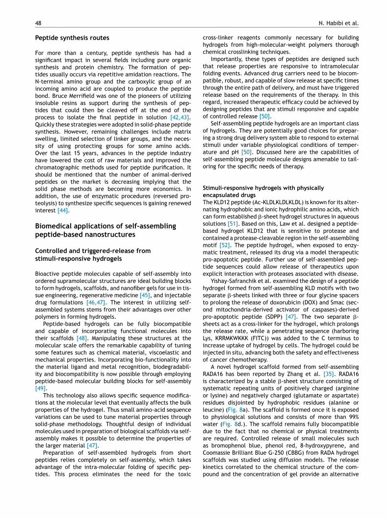
A novel hydrogel scaffold formed from self-assemblingADA16 has been reported by Zhang et al. [35]. RADA16
s characterized by a stable �-sheet structure consisting ofystematic repeating units of positively charged (argininer lysine) and negatively charged (glutamate or aspartate)esidues disjointed by hydrophobic residues (alanine oreucine) (Fig. 8a). The scaffold is formed once it is exposedo physiological solutions and consists of more than 99%ater (Fig. 8d.). The scaffold remains fully biocompatibleue to the fact that no chemical or physical treatmentsre required. Controlled release of small molecules suchs bromophenol blue, phenol red, 8-hydroxypyrene, and
oomassie Brilliant Blue G-250 (CBBG) from RADA hydrogelcaffolds was studied using diffusion models. The releaseinetics correlated to the chemical structure of the com-ound and the concentration of gel provide an alternative
Self-assembled peptide-based nanostructures 49
Figure 8 (a) Schematic illustration of the RADA16 molecule composed of negative (Asp) and positive (Arg) residues driving theassembly of the peptide by means of electrostatic interactions into (b) and (c) fibers and (d) macroscopic hydrogels in buffer [50]
fiwtt
pdswsipaaaTrt
adatt
SdI
Printed with permission from ELSEVIER.
means of controlling release kinetics. The results indicatedthat release profiles can be tailored by controlling nanofiber-diffusing molecular-level interactions [50].
A variety of proteins, such as lysozymes, trypsininhibitors, BSA, and IgG were also encapsulated withinthe Ac-(RADA)4-CONH2 peptide hydrogel and releasedslowly [53]. The combination of the peptide solution andtherapeutic-based proteins could be used for local drugrelease in tissue.
Elastin is a highly elastic protein in connective tissuesthat helps the tissues to resume its shape after exten-tion or contraction. Recombinant techniques allow one tocreate protein-based materials that exhibit some featurefound in natural proteins together with other properties oftechnological/medical interest. One of the most interestingprotein-based materials is the family of elastin-like recom-binamers (ELRs), which can form a variety of structures suchas nanoparticles, nanofibers, films, and hydrogels.
Alicia et al. designed a silk-elastin-based injectablemultiblock corecombinamer that spontaneously forms aphysically stable nanofibrillar hydrogel under physiologicalconditions. The idea was to create a system as the basis fordeveloping injectable fibrillar biomaterial platforms towarda fully functional, biomimetic, artificial ECM, and cell niches[54].
Machado et al. investigated the use of ELRs for the trans-plantation of autologous retinal pigment epithelial (RPE)cells for treatment of age-related macular degeneration,
since they were shown to maintain the functional featuresof (RPE) cells [55].Inspired by the cells’ structure, Torre et al. [56] pre-sented compartmentalized capsules responsive to magnetic
nopp
eld and temperature. The microcapsules were coatedith a temperature-responsive chitosan/ELR nanostruc-
ured shell. These engineered systems can be applied forransportation of bioactive agents and cells.
The effects of pH on intramolecular folding of �-hairpineptide self-assembled hydrogels were studied by Schnei-er et al. [49]. Infrared spectroscopy and circular dichroismhowed that at low pH, individual peptides in the hydrogelere unstructured, resulting in a low viscosity and aqueous
olution. Thus, varying the intramolecular folding of smallndividual peptides on the nanoscale can alter macroscopicroperties [49]. �-hairpin self-assembled hydrogels werelso used to encapsulate curcumin; an anti-inflammatoryntioxidant and anti-tumorigenic polyphenol derived fromplant, whose therapeutic use is limited by poor solubility.he peptide hydrogel has been proven to be an effective car-ier for the localized, consistent delivery of curcumin overime [57].
One challenge to the controlled-release use of self-ssembled nanofibers is their extreme resistance toegradation by protease. Collier et al. designed self-ssembling depsipeptides that contain ester bonds withinhe peptide backbone; these degraded in a period of dayso weeks by ester hydrolysis [58].
timuli-responsive hydrogels with chemically conjugatedrugs
n the past decade, supramolecular hydrogels, with
etworks of nanofibers formed through the self-assemblyf small molecules (i.e., hydrogelators), have proven to beromising biomaterials. Peptide-based hydrogelators haveroven their effectiveness due to their ability to produce a
5
losta
Xow
aacpaa
nrounbib
rcDapaathcucotattgo
gawi[
E
ObbrAba
o(mStsvapSiftaarpc
TDbbmipnpg
sdatpertfbatcytspecifically recognizes and binds to cells expressing �5�1.Functionalizing stealth PEG-liposomes decorated with PR bpeptide-amphiphile [69] were found to function better than
0
arge set of various functional molecules from a small arrayf residues. The combination of enzymatic reactions withelf-assembly of small molecules offers an effective methodo form nanofiber networks, which results in hydrogels undervariety of conditions.Studies have been undertaken by several groups including
u’s and Yang’s, who have been investigating developmentf supramolecular hydrogels from self-assembly of peptidesith chemically conjugated drugs [40,59—61].
Li et al. conjugated Taxol with a self-assembly motif andfunctional group cleavable by an enzyme. A precursor has
lso been designed for developing a Taxol hydrogel withoutompromising the activity of Taxol [59]. This approach holdsromise as a general method to generate nanofibers of ther-peutic molecules with a dual role: delivery carrier as wells drug.
In another study, tripeptide derivatives containing aaphthyl group, two phenylalanines, and one modified lysineesidue were conjugated to an olsalazine moiety (substratef azo reductase), which self-assembled into a hydrogelnder mildly acidic conditions. The reduction of olsalazineot only led to a transition from gel to solid phase,ut also released 5-aminosalicylic acid, which is an anti-nflammatory agent. This approach will eventually yield newiomaterials for site-specific drug delivery.
One of the first molecular hydrogelator systems waseported by Mao et al. in 2011; it was made of two anti-ancer drugs that complement each other. In this studyex-FFFK(Taxol/HCPT)E-ss-EE was designed and synthesizeds a precursor of molecular hydrogelators. The princi-le of this novel design included Dexamethasone regularlypplied to drive self-assembly (also an anti-inflammatorynd immunosuppressant used after chemotherapy); dipep-ide and tripeptide of phenylalanine (F) to form molecularydrogelators; and finally the disulfide bond, acting as aleavable linker connecting the hydrophilic parts and molec-lar hydrogelators. This structures could be applied in theavity after surgical tumor removal for the long-term releasef anti-cancer drugs [60]. This injectable self-delivery sys-em contains a high weight percentage of anti-cancer drugsnd can prolong their release [62]. Further, Wang et al. foundhat even the conjugates of Taxol with amino acids con-aining hydrophilic side chains such as serine, arginine, andlutamic acid were also efficient candidates for formationf molecular hydrogelators [61].
Ultra-short peptides with the ability to form hydro-els were functionalized with oxaliplatin (a platinum-basednticancer therapeutic). The oxaliplatin-peptide conjugateas tested for localized breast cancer therapy, and the
njectable gel showed significant tumor growth inhibition63].
nhanced cellular uptake with targeting activity
ne of the most desirable properties of a drug carrier is toind specifically with the target site [9], whether cell mem-ranes [64] or molecules of interest. To this regard, peptides
epresent the most important biological recognition motifs.s an example, NTFR and RGD PAs have been widely usedy researchers to bind fractalkine and a5�1 integrins. Self-ssembled peptides are used to enhance the functionalityFC
w
N. Habibi et al.
f common motifs and integrins. Cell-penetrating peptidesCPP) play an important role in introducing drugs inside cellembranes and translocation of genes inside the nucleus.
elf-assembly is key to CPP penetration mechanisms. Fur-hermore, the self-assembly process can produce varioustructures suitable for specific delivery and loading of aast array of drugs. In this regard, PAs (which contain anlkyl chain) mimic the capabilities of lipid systems, thusroviding improved drug-delivery through cell membranes.elf-assembled peptides have also been used to functional-ze conventional drug delivery carriers, such as liposomes,or enhanced cellular uptake and targeting [65]. In addi-ion, folate-targeting functions have been investigated fornticancer drug delivery [66]. Functional groups of self-ssembled structures act as a template for the alignment ofecognition molecules [67]. Self-assembled peptide motifslay a critical role in increasing targeting activities of drugarriers.
argeting function to improve uptakerug delivery carriers designed with peptide motifs haveeen extensively studied for targeted delivery. PAs haveeen designed to enhance the targeting activities of com-on recognition motifs. For example, researchers have
nitially applied RGD (the primary recognition site for �5�1)eptide-based techniques to target the integrin �5�1 (sig-ificantly up-regulated in tumor vasculature). However, thisrocess has limitations, since RGD recognizes several inte-rins, making specific targeting difficult.
Garg et al. designed a novel peptide-amphiphileequence namely PR b that closely mimics the cell adhesionomain of fibronectin [65]. The design structures includedC16 dialkyl ester tail, a glutamic acid (Glu) tail connec-
or, a — (CH2)2 — tail spacer, and a peptide headgroup. Theeptide headgroup contained a spacer KSS, PHSRN (the syn-rgy site for �5�1), a linker (SG)5, and RGDSP (the primaryecognition site for �5�1) (Fig. 9.). The KSS spacer was usedo keep the bioactive region further away from the inter-ace so the availability of the peptide could enhance theinding affinities to the targets [9,68]. The PR B peptide-mphiphiles were incorporated on stealth liposomes in ordero target integrin �5�1, which is expressed in colon cancerells. Functionalized liposomes were covered with polyeth-lene glycol (PEG). The role of PEG is to allow the liposomeso circulate in the blood, while the modified PR b peptide
igure 9 Structure of PR b peptide-amphiphile containing16 dialkyl ester tail, a glutamic acid (Glu) tail connector, a(CH2)2 tail spacer, and the peptide headgroup [65] Printedith permission from ELSEVIER and PUBMED.

rttsestt
saWHtad
[mass
si
Self-assembled peptide-based nanostructures
RGD targeting, providing better binding and greater uptakeof liposomes by colon cancer cells. The ability to design apeptide by sequence-specific modifications at the molecularlevel allows control of the activities of common motifs fordesired applications [9].
Membrane penetration function to improve uptakeCell-penetrating amphiphilic peptides such as HIV-Tat basedpeptides, oligoarginines, and chimeric cell-penetrating pep-tides have also been applied alone or in combination withother self-assembled peptides for the delivery of therapeu-tic cargos to their targets [70].
CPPs have been used as drug delivery vehicles, becausethey are able to translocate the cell membrane [71]. CPPs,which are cationic short peptides of fewer than 30 aminoacids, and oligoarginine-based CPPs with the length of 8—10arginine residues have shown the most efficient membranepenetration [72].
From the classic mechanism [72] the membrane pene-tration of CPPs is not associated with their self-assembly.Membrane penetration is based on the hydrogen bondinginteraction between the guanidinium groups of arginineresidues and the carboxyl, phosphoryl, or sulfuryl groupsof the carbohydrates and phospholipids on the cell sur-face (Fig. 10). The pathway of CPP translocation throughmembranes was initially explained via receptor- andendocytosis-independent mechanisms. Recently a new CPP
internalization mechanism has been demonstrated whichshows the role of self-assembly in ability of CPP peptidesin traversing cell membranes [10].Figure 10 Different steps in CPP-mediated intracellulardelivery: (1) Interaction of the CPP (represented as a green bar)with the cell-surface proteoglycans (in red). (2) Endocytic path-way. (3a) Degradative route to lysosomes in clathrin-mediatedendocytosis. (3b) CPP ultimately reach the Golgi apparatus (inpurple) or endoplasmic reticulum (ER, in grey) in caveolin-mediated endocytosis. (3c) Endosomal release [10] Printed withpermission from ELSEVIER.
gdaclcNt[
ua(loioMfdcli
(litadtircn
51
A third helix peptide (Antennapedia Homeodomain) waseported to translocate biological membranes. It was foundhat after internalization into E15 rat embryos, the pep-ides were recovered as a multimeric complex. The resultsuggest that CPP could internalize through cell membranesither in monomers or self-assembled aggregates. Moreover,ome important studies demonstrated that the retaining ofheir conformation by CPPs after self-assembly is the key toheir membrane-penetrating abilities.
The effect of self-assembled aggregates on DNA encap-ulation and delivery has also been studied. Peptides suchs 46�8 (LARLLARLLARLLARL) and Hel 11-7 (KLLKLLLKL-KKLLKLLK) form aggregates with DNA and transfer DNA.owever, transfection efficiency was shown to be poor inhe case of 46�12, when the peptide was not able to formggregates. Aggregate formation also prevents DNA degra-ation.
Tirrell et al. used an amphiphilic peptide (Gly-Pro Hyp)4-IV-H1] to modify PEG-coated liposomes in order to targetouse fibroblast cells. After incorporation of IV-H1 peptide-
mphiphile, they maintained their native triple-helicaltructure and enhanced uptake of the IV-H1 modified lipo-omes over non-targeted liposomes [73].
Dendrigraft poly-L-lysines (DGLs), a new kind ofelf-assembled peptide containing lysine, was recently mod-fied by a peptide (sequence: EPRNEEK) derived fromStreptococcus pneumoniae , a pathogen that causes menin-itis. The modified peptide was used for targeted geneelivery to a glioma (brain tumor). The aim was to takedvantage of the process by which certain pathogensan cross the blood-brain barrier (BBB) in a transcellu-ar or paracellular manner. In vivo imaging results andellular uptake indicated that EPRNEEK peptide-modifiedPs enhanced brain tumor-targeting activity comparedo a pentapeptide derived from endogenous laminin33,74].
Arginine-rich peptides have also been designed andsed for enhanced cellular uptake. Sardan et al. designedn arginine-rich cell-penetrating amphiphilic peptidelauryl—PPPPRRRR—Am), which was integrated into aiposome formulation (DOPG) in order to target the activityf anticancer agents (doxorubicin-HCl and paclitaxel)nside MCF7 breast cancer cells. Arginine PA integratedn liposomes enhanced the uptake of model reagents byCF7 breast cancer cells compared to native liposomes and
ree reagents [33]. Both hydrophilic (i.e., rhodamine andoxorubicin-HCl) and hydrophobic (i.e. paclitaxel) anti-ancer agents have been loaded in arginine-rich peptideiposomes in order to improve the uptake of both drugsnside cancer cells [33].
Stimuli-responsive supramolecular peptide-amphiphilesSPAs, dendritic poly(L-lysine) non-covalently linked poly(L-eucine)) were synthesized with the ability to reassemblen lower pH for intracellular drug delivery. SPAS are showno encapsulate guest molecules at pH 7.4 and release themt pH 6.2. In this way, SPAS can act as a smart platform toeliver their target to tumor cells with intracellular pH as arigger. The nanovehicle was loaded with DOX and delivered
nside HepG2 cancer cells. Confocal scanning microscopeesults indicated NVs can considerably enhance the endo-ytosis of DOX into HepG2 cells and deliver DOX to theiruclei [75].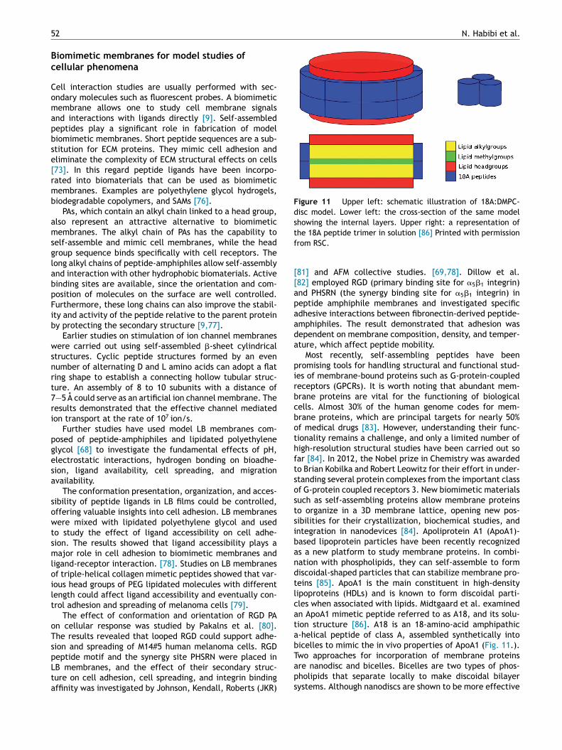
5 N. Habibi et al.
Bc
Comapbse[rmb
amsglabpFib
wsnrt7ri
pgesa
sowtsmloilt
oTspLta
Figure 11 Upper left: schematic illustration of 18A:DMPC-disc model. Lower left: the cross-section of the same modelshowing the internal layers. Upper right: a representation oftf
[[apaada
pirbcbothftsostsibandtlcatabT
2
iomimetic membranes for model studies ofellular phenomena
ell interaction studies are usually performed with sec-ndary molecules such as fluorescent probes. A biomimeticembrane allows one to study cell membrane signals
nd interactions with ligands directly [9]. Self-assembledeptides play a significant role in fabrication of modeliomimetic membranes. Short peptide sequences are a sub-titution for ECM proteins. They mimic cell adhesion andliminate the complexity of ECM structural effects on cells73]. In this regard peptide ligands have been incorpo-ated into biomaterials that can be used as biomimeticembranes. Examples are polyethylene glycol hydrogels,iodegradable copolymers, and SAMs [76].
PAs, which contain an alkyl chain linked to a head group,lso represent an attractive alternative to biomimeticembranes. The alkyl chain of PAs has the capability to
elf-assemble and mimic cell membranes, while the headroup sequence binds specifically with cell receptors. Theong alkyl chains of peptide-amphiphiles allow self-assemblynd interaction with other hydrophobic biomaterials. Activeinding sites are available, since the orientation and com-osition of molecules on the surface are well controlled.urthermore, these long chains can also improve the stabil-ty and activity of the peptide relative to the parent proteiny protecting the secondary structure [9,77].
Earlier studies on stimulation of ion channel membranesere carried out using self-assembled �-sheet cylindrical
tructures. Cyclic peptide structures formed by an evenumber of alternating D and L amino acids can adopt a flating shape to establish a connecting hollow tubular struc-ure. An assembly of 8 to 10 subunits with a distance of—5 A could serve as an artificial ion channel membrane. Theesults demonstrated that the effective channel mediatedon transport at the rate of 107 ion/s.
Further studies have used model LB membranes com-osed of peptide-amphiphiles and lipidated polyethylenelycol [68] to investigate the fundamental effects of pH,lectrostatic interactions, hydrogen bonding on bioadhe-ion, ligand availability, cell spreading, and migrationvailability.
The conformation presentation, organization, and acces-ibility of peptide ligands in LB films could be controlled,ffering valuable insights into cell adhesion. LB membranesere mixed with lipidated polyethylene glycol and used
o study the effect of ligand accessibility on cell adhe-ion. The results showed that ligand accessibility plays aajor role in cell adhesion to biomimetic membranes and
igand-receptor interaction. [78]. Studies on LB membranesf triple-helical collagen mimetic peptides showed that var-ous head groups of PEG lipidated molecules with differentength could affect ligand accessibility and eventually con-rol adhesion and spreading of melanoma cells [79].
The effect of conformation and orientation of RGD PAn cellular response was studied by Pakalns et al. [80].he results revealed that looped RGD could support adhe-ion and spreading of M14#5 human melanoma cells. RGDeptide motif and the synergy site PHSRN were placed in
B membranes, and the effect of their secondary struc-ure on cell adhesion, cell spreading, and integrin bindingffinity was investigated by Johnson, Kendall, Roberts (JKR)aps
he 18A peptide trimer in solution [86] Printed with permissionrom RSC.
81] and AFM collective studies. [69,78]. Dillow et al.82] employed RGD (primary binding site for �5�1 integrin)nd PHSRN (the synergy binding site for �5�1 integrin) ineptide amphiphile membranes and investigated specificdhesive interactions between fibronectin-derived peptide-mphiphiles. The result demonstrated that adhesion wasependent on membrane composition, density, and temper-ture, which affect peptide mobility.
Most recently, self-assembling peptides have beenromising tools for handling structural and functional stud-es of membrane-bound proteins such as G-protein-coupledeceptors (GPCRs). It is worth noting that abundant mem-rane proteins are vital for the functioning of biologicalells. Almost 30% of the human genome codes for mem-rane proteins, which are principal targets for nearly 50%f medical drugs [83]. However, understanding their func-ionality remains a challenge, and only a limited number ofigh-resolution structural studies have been carried out soar [84]. In 2012, the Nobel prize in Chemistry was awardedo Brian Kobilka and Robert Leowitz for their effort in under-tanding several protein complexes from the important classf G-protein coupled receptors 3. New biomimetic materialsuch as self-assembling proteins allow membrane proteinso organize in a 3D membrane lattice, opening new pos-ibilities for their crystallization, biochemical studies, andntegration in nanodevices [84]. Apoliprotein A1 (ApoA1)-ased lipoprotein particles have been recently recognizeds a new platform to study membrane proteins. In combi-ation with phospholipids, they can self-assemble to formiscoidal-shaped particles that can stabilize membrane pro-eins [85]. ApoA1 is the main constituent in high-densityipoproteins (HDLs) and is known to form discoidal parti-les when associated with lipids. Midtgaard et al. examinedn ApoA1 mimetic peptide referred to as A18, and its solu-ion structure [86]. A18 is an 18-amino-acid amphipathic-helical peptide of class A, assembled synthetically intoicelles to mimic the in vivo properties of ApoA1 (Fig. 11.).wo approaches for incorporation of membrane proteins
re nanodisc and bicelles. Bicelles are two types of phos-holipids that separate locally to make discoidal bilayerystems. Although nanodiscs are shown to be more effective
fvchtcaiuw[tSftaiat
bandfnrtwttfprttttttngc
eceEhtdcuctwc
Self-assembled peptide-based nanostructures
in the stabilization of membrane proteins, using bicelles iseasier when handling proteins.
Therefore, the focus of work by Midtgaard et al. [86]was to investigate whether A18 could provide the flexibilityof bicelles along with the native membrane-like environ-ment of nanodiscs. They observed that membrane proteinscould be easily incorporated in 18A discs and maintain theirstability.
Recent studies also showed that self-assembling pep-tides could interact with proteins or protein assembliesin cells and could be selectively formed in certain cells,thus providing novel strategies to manipulate the fate ofcells. Wang et al. developed self-assembling peptides withL- and D-amino acids (NBD-DFDFDYGLKKTETQ) and stud-ied their biological functions in cells. Interestingly, theresults demonstrated that the utilization of D-amino acidsin self-assembling peptides boosts their cellular membranelocalization [87]. D-amino acids were specially enriched incellular membranes at self-assembled stages. However theywere distributed uniformly in the cytoplasma of cells atunassembled stages. Wang et al. [87] demonstrated thepossibility of using such D-amino acid-based peptides to engi-neer cell surfaces.
Membrane interaction of a model �-sheet self-assembledpeptide, P11-2 (CH3CO-Gln-Gln-Arg-Phe-Gln-Trp-Gln-Phe-Glu-Gln-Gln-NH2), was studied by Salay et al. in order tomimic the toxicity and structural behavior of �-amyloid pep-tide (A�) [88]. In Alzheimer’s disease �-amyloid peptide (A�)is a main protein component of plaques. However its role inneurotoxicity is not fully understood. Model peptides couldhelp evaluate the possible mechanisms of toxicity, whichis common among self-assembling peptides associated withdisease. The results confirmed that transitional oligomershad higher toxicity against cells than monomeric forms andhigher aggregates of the peptide. Thus a rationally designedyet simple model �-sheet peptide recapitulates a variety ofthe important features of A� peptide function and structure.This supports the theory that toxicity arises from a commonmode of action on membranes regardless of specific aspectsof the sequence of amino acids.
Anticancer drug delivery
Cancer is among the most widespread causes of death, andchemotherapy is still the most common treatment. However,conventional chemotherapy agents have tremendous disad-vantages, as they can easily distribute nonspecifically inbody tissue, causing damage to normal in addition to tumorcells. This induces nonspecific drug distribution in the bodywith severe systemic side effects, and therefore chemother-apy often results in an unsatisfactory curative effect dueto the side effects of the drugs. Nanoparticles have beenextensively used for delivery of anticancer drugs, as theycan increase the efficacy of treatment and reduce undesir-able side effects. Nanosized carriers offer advantages suchas high drug-loading ability, improved stability by avoidingfast clearance by the reticuloendothelial and renal systems,and minimized drug loss during blood circulation [89].
Among various nanosized carriers, self-assembled pep-tide nanostructures have gained attention for the deliveryof anticancer agents and seem to be a promising approachfor cancer treatment. Self-assembly mechanisms allow
tdRn
53
ormation of various types of nanoparticles such as tubes,esicles, and hydrogels, each suitable for delivery of spe-ific types of anticancer agents. For example, injectableydrogel formulations can be placed in direct contact withumor tissues and therefore improve the safety and effi-acy of cancer treatment [47]. Nanotubes also have beenpplied to load doxorubicin with high efficiency in theirnner core [24]. Amphiphilic peptide nanovesicles have beensed for loading hydrophobic drugs in their alkyl core,hile the shell is modified to improve intracellular delivery
66]. Moreover, modifications have been applied to increaseargeting activity and intracellular delivery efficacy [33].everal self-assembled peptide structures have been usedor loading anticancer agents, such as doxorubicin, pacli-axel, curcumin, and fluorouracil and studied in preclinicalnd clinical trials, owing to their excellent biodegradabil-ty and biocompatibility. There have been several recentdvances in anticancer treatment using self-assembled pep-ides.
Standley et al. designed a PA with highly cationic mem-rane lytic (Lysin) sequence (KLAKLAK)2 conjugated to laurylcid (C16-A4G3(KLAKLAK)2) capable of forming cylindricalanofibers and disrupting cell membranes [90]. The resultsemonstrated that PA is more cytotoxic against the trans-ormed cell membranes of cancer cells due to increasedegative charge on these surfaces [91]. Moreover it wasevealed that in addition to passive permeation, KALA epi-ope mediated uptake targeting [92]. A limitation associatedith these kinds of PAs, however, is rapid degradation of pep-
ide after systematic delivery [93]. One tactic to enhancehe therapeutic-delivery vehicle is to functionalize the sur-ace with polymers such as PEG, which can protect fromroteolysis, improve circulation time, and lessen immuneesponse [94]. Furthermore, in 2012, Toft et al. introducedhe use of cytotoxic KLAK PA and a co-assembled combina-ion of pegylated PA (PEG PA) to make an effective antitumorherapy [95]. Addition of the pegylated PA to the nanostruc-ure limits degradation of the cytolytic PA by the proteaserypsin. Systemic administration of cytotoxic pegylated PAso an orthotopic mouse xenograft model of breast cancer sig-ificantly reduced tumor cell proliferation and overall tumorrowth, thus suggesting its effectiveness and versatility forancer treatment.
A study carried out in 2012 proposed molecular,lectrostatic interaction-based binding between EAK, a self-omplementary peptide and hydrophobic anticancer drugllipticine (EPT). EAK was able to increase the stability ofPT in aqueous solution [96,97] at a concentration muchigher than the drug’s solubility in water (∼0.62 �M at neu-ral pH) [98]. Furthermore, a fluorescence technique thatifferentiates the two molecular states (protonated andrystalline EPT in situ) was used to evaluate different molec-lar states of EPT for its anticancer efficacy. An in vitroytotoxicity assay has been applied to analyze the efficacy ofhe two molecular states of EPT, indicating that complexesith protonated EPT had higher cytotoxicity than those withrystalline EPT [99].
Researchers have also utilized micro- and nanopar-
icles containing self-assembled peptides conjugated tooxorubicin as an effective cancer treatment. In 2015,ubert pe rez et al. proposed hierarchical assembly of PAanofibers with alginate to shape micro-particles, in which
5
dsfwtes[
wpstfaPNtonf
wwcoc
tsaecrstFdawt
attnDmtetac
G
Dghvie
gisuctfppsb
rSfdibrutip
rec[tctaniabPba
Ctt
tpd1hsVfaact
s
4
oxorubicin was conjugated to an alginate core, and thehell was constructed of peptide amphiphile nanofibersunctionalized to target the folate receptor. Folate groupsere covalently linked to C16-V3A3K3 via a lysine linker
o target the doxorubicin-containing particles to cells over-xpressing the folate receptor. The PA shell increased theurface-over-volume ratio to its ideal amount for targeting6].
Furthermore, hybrid NPs were made of low-molecular-eight polylactide (PLA) [100] and self-assembling V6K2eptides (VVVVVVKK) [101], which self-assembled intopherical nanoparticles with 100 nm as an average diame-er. PLA-V6K2 were used to host doxorubicin and paclitaxelor cancer treatments, and their toxicity, release kinetics,nd cellular uptake were studied. The results revealed thatLA-V6K2 NPs had much higher tumor cell uptake than PLAPs that were stabilized with PEG (PLA-EG). This was dueo the electrostatic interactions between the lysine groupsf the peptide. The results indicated the greater efficacy ofanoparticles functionalized with self-assembled peptidesor cancer treatments [102].
Furthermore, different self-assembled dipeptide NPsith a non-protein amino acid, �, � dehydrophenylalanineere used for entrapment of curcumin. The di-peptide cur-umin NPs could be assembled in a mixture of aqueous andrganic phase and enhanced the biological activity of cur-umin [103].
Li et al. investigated dendrimer peptides with the struc-ure of glycyl-phenylalanyl-leucyl -glycine tetra-peptidepacer (Gly-Phe-Leu-Gly, GFLG) to host anti-cancer drugs assuitable substrate for protease cathepsin B, which is an
nzyme overexpressed in many tumor and tumor endothelialells. GFLG peptide could be used to design smart enzyme-esponsive drug-delivery vehicles. Fluorescence imagingtudies demonstrated that the nanoparticles can concen-rate in tumors and be retained for a long period [104].urthermore, multifunctional delivery platforms such asual-functional liposomes with active targeting hyaluroniccid (HA) and pH-responsive cell-penetrating peptide (CPP)ere fabricated for enhancing the efficacy of tumor-
argeted cancer treatments [105].Perhaps the most interesting application of self-
ssembled peptides for cancer treatment, however, ishe injectable gel formulation, in which chemotherapeu-ic agents (unlike traditional chemotherapy) can be locatedext to or into target tissues at higher local concentrations.ue to the slow rate of release, these formulations provideore prolonged and sustained cytotoxic action than conven-
ional chemotherapeutic agents, thus enhancing therapeuticfficacy. With this in mind, Yishay-Safranchik et al. verifiedhe possible use of in situ-forming hydrogels made of self-ssembled KLD motifs to tune or extend the release of theonventional cytotoxic drug (doxorubicin) [47].
ene delivery
evelopment of non-viral vehicles for efficient delivery ofenes is still requiring major improvements. Gene carriers
ave to be well organized to overcome the limitations ofector gene delivery, such as poor uptake into cells, tox-city and immunogenic response, and short-lived transgenexpression [106]. Research behind the designing of advancedcoam
N. Habibi et al.
ene delivery vehicles has focused on improving character-stic of efficient loading, enhanced cellular delivery, andpecific delivery. Cationic nanoparticles have been manip-lated and applied extensively due to their high loadingapacity for nucleic acids, their ability to transfer throughhe cell membrane, and their ability to protect nucleic acidsrom degradation. However, cationic formulations can haveroblems in some applications in vivo, as they deal withroteins in biological fluids and nonspecifically with cells tohape aggregates, which can lead to toxicity as well as pooriodistribution and immune responses [107].
Peptide-based self-assembled nanostructures also rep-esent a promising approach for efficient gene delivery.olid-phase methods enable precise control over peptideabrication at the molecular scale. Oligonucleotides can beelivered to the interior of cells through endocytosis follow-ng the transition of self-assembled nanotubes into vesiclesy changing the concentration of building blocks [108]. Thus,ecent developments in bio-inspired gene delivery vehiclesse a combination of viral genomes and self-assembled pep-ides [109]. This framework highlights some of the advancesn the development of viral vectors with self-assembly ofeptide-engineered nanostructure.
For efficient delivery the vector must pass four crite-ia. The vector must target specific cell-surface receptors,fficiently protect DNA from degradation, deliver the DNAargo to the nucleus, and disrupt the endosomal membrane110]. For this, cationic peptides could condense with DNAhrough electrostatic interactions. The most famous DNA-ondensing cationic peptide is L-lysine, which interacts withhe negative charge of DNA phosphate. Poly-L-Lysine (PLL) issynthetic peptide containing repeated lysine and a large
umber of surface amines. It is capable of electrostaticnteraction with polyanions. The length of the peptide has
strong effect on its properties, such as the size and sta-ility of DNA condensate. However, high-molecular-weightLLs exhibit high cytotoxicity, and PEGylation is known toe useful to increase circulation half-life of the complexnd avoid plasma protein binding.
CPPs enable cellular uptake of various molecular cargos.PPs are known to be powerful tools in overcoming limita-ions associated with gene delivery vehicles and are able toransfer genes with endosomal escape.
In 1997, Morris et al. [111] used MPG-designed peptideso deliver oligonucleotides. MPG is a synthesized 27-residueeptide vector that contains both a hydrophobic domain,erived from the fusion sequence of HIV gp41 (residues—17: G-A-L-F-L-G-FL-G-A-A-G-S-T-M-G-A)K-R-K-V′, and aydrophilic domain derived from the nuclear localizationequence of SV40 T-antigen (residues 21—27: P-K-S-K-R-K-). The hydrophobic domain is known to be essential bothor its membrane fusion activity and structural stabilization,nd the hydrophilic domain is useful for improving perme-tion of the nucleus. An acetyl group at the N-terminus and aysteamide group at the C-terminus were added to enhancehe ability to cross the cell membrane.
When MPG was combined with oligonucleotides inolution, they quickly shaped into a complex with tight non-
ovalent interactions. This implies that binding betweenligonucleotides and MPG occurs through electrostatic inter-ctions. Efficient delivery of this complex into the nucleus ofammalian cells happens in about an hour. MPG shows high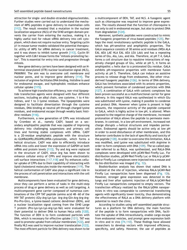
assnf
thwGEsfaaeapdGw[bdwaatepbcaibtwotcdRi
lFHlFttatp
tot
Self-assembled peptide-based nanostructures
attraction for single- and double-stranded oligonucleotides.Further studies were carried out to understand the mecha-nism of MPG peptides in gene delivery to mammalian cells[112]. The result revealed that a mutation in the nuclearlocalization sequence (NLS) of the SV40 antigen domain pre-vents the carrier from entering the nucleus, making it ahighly useful tool for robust siRNA delivery into cell cyto-plasm, which does not require nuclear entry. Targeting cyclin�1 in mouse tumor models validated the potential therapeu-tic utility of MPG for siRNA delivery in cancer treatment,and it was shown to inhibit tumor growth [113]. Cyclin B1together with Cdk1 kinase forms the ‘mitosis-promoting fac-tor’. This is essential for entry into and progression throughmitosis.
New gene delivery carriers have been designed with an N-terminal stearylated (STR) nuclear localization signal (NLS),PKKKRKV. The aim was to overcome cell membrane andnuclear pores, and to improve gene delivery [114]. Theinclusion of arginine facilitates DNA binding, histidine is usedfor endosomal escape, and hydrophobic residues enhancecellular uptake [115].
To achieve high transfection efficiency, non-viral lipopep-tide transfection agents were designed with four differentsections, including an alkyl chain, one cysteine, 1 to 4 his-tidines, and 1 to 3 lysine residues. The lipopeptides weredesigned to facilitate dimerization through the cysteineresidues, DNA binding at neutral pH through charged lysineresidues, and endosomal escape through weakly basic histi-dine residues [116].
Furthermore, a new generation of CPPs was introducedby Crombez et al., namely CADY, based on a sec-ondary amphipathic peptide that was capable of improvingdelivery into challenging suspensions and primary celllines and forming stable complexes with siRNA. CADYis a 20-residue amphiphatic peptide with the structureAc-GLWRALWRLLRSLWRLLWRA-cysteamide, which forms ahelical structure. CADY was able to efficiently introducesiRNA into cells and lower the expression of GAPDH at bothmRNA and protein levels [113]. Try and Arg were replacedin the structure of CADY, since Arg has been shown toenhance cellular entry of CPPs and improve electrostaticcell-surface interactions [117,118] and Try enhances cellu-lar uptake of CPPs due to their capability of interacting withlipid/cholesterol molecules inside the membrane [119]. Thehelical conformation of CADY also plays a significant role inthe process of cell penetration and interactions with the cellmembrane.
Multicomponents have been evaluated for gene delivery,since they can perform a series of tasks in the multi-stepprocess of drug or gene delivery as well as cell targeting. Amulticomponent gene carrier composed of numerous com-binations of the CPP TAT peptide (amino acid 48—60 of theHIV TAT protein) (Gly-Arg-Lys-Lys- Arg-Arg-Gln-Arg-Arg-Arg-Pro-Pro-Gln), a lysine-based cationic dendrimer (DEN), anda nuclear localization signal coming from the SV40 LargeT protein (Pro-Lys-Lys-Lys-Arg-Lys-Val) were evaluated fortheir potential to deliver DNA to human HeLa cells [120].The function of DEN is to form condensed particles with
DNA, which is necessary for effective uptake [121]. TAT wasused to promote uptake from cellular membranes [122], andfinally NLS was used to improve nuclear translocation [123].The most efficient particle for DNA delivery was shown to befirs
55
multicomponent of DEN, TAT, and NLS. A fusogenic agentuch as chloroquine was required to improve gene expres-ion. The results showed that the function of chloroquine isot only to aid in endosomal escape, but also to protect DNArom degradation [124].
Moreover, synthetic peptides were constructed to mimiche fusogenic properties of virus-based peptides [125]. Per-aps the most revolutionary synthetic peptide is the GALA,hich has pH-sensitive and amphiphilic properties. TheALA sequence consists of 30 amino acid residues (WEA ALAAL AEA LAE HLA EAL AEA LEA LAA) and has a repeatingeries of Glu, Ala, Leu, and Ala. Therefore, at pH 7.4, GALAorms a coil structure due to repulsive interactions of neg-tively charged groups of Glu, while at pH 5, it forms anmphipathic � helix due to a protonated Glu residue. GALAxhibits pH-responsive properties with membrane fusionctivity at pH 5. Therefore, GALA can induce an assistiverocess to release drugs from endosomes, like other virus-erived fusogenic peptides [126]. In terms of DNA delivery,ALA has limitations associated with its anionic properties,hich prevent formation of condensed particles with DNA
127]. A combination of GALA with cationic complexes haseen proven successful as a multifunctional carrier for geneelivery. In this regard KALA was synthesized in which Gluas substituted with Lysine, making it possible to condensend protect DNA. However when Lysine is present in highmounts, the responsive activity of the peptide comparedo GALA, which is highly active in low pH, is altered. Whenxposed to the negative charge of the membrane, increasedrotonation of KALA allows the peptide to permeate mem-ranes. In contrast, in a low-pH environment, increased netharge disrupts �-helical structures and impedes perme-tion. Endosomal agents should be active only at low pHn order to avoid disturbance of other membranes, and thisehavior contributes to the lack of specificity and toxicity ofhe peptide [128]. In order to overcome this limitation, KALAas modified, and Lysine was substituted with Arginine inrder to form complexes with DNA [109]. The so-called pep-ide referred to as RALA, was synthesized, and RALA-DNAomplexes were developed with pCMV-Red Firefly Luc. Foristribution studies, pCMV-Red Firefly Luc or RALA/p pCMV-ed or Firefly Luc complexes were injected into a mouse andts bio-distribution was imaged (Fig. 12).
Biodistribution analysis has shown that naked DNA isocalized at the site of injection, whereas RALA/pCMV-Redirefly Luc nanoparticles have been dispersed (Fig. 12i).owever, stronger gene expression was detected in the
ungs and liver after systemic injection of RALA/pCMV-Redirefly Luc nanoparticles compared to empty vectors. Theransfection efficacy realized by the RALA/pDNA nanopar-icles in vitro was comparable to commercial transfectinggents with significantly lower toxicity, thus demonstratinghe effectiveness of RALA as a DNA delivery platform withotential to reach the clinic.
According to studies using self-assembled peptide struc-ures as a platform for DNA delivery, the main functionf the peptide is to form nanoparticles with DNA, facili-ate the uptake of DNA intracellularly, enable cargo escape
rom endosomal vesicles, and prompt gene expression bothn vitro and in vivo [74,110]. These findings have allowedesearchers to develop vectors with improved efficiency,pecificity, and safety. However, the use of peptides as
56 N. Habibi et al.
Table 1 summarizes different types of self-assembled peptides with biomedical application.
Peptide type Nano structure Biomedical applications References
L-Phe-L-Phe (FF) Nanotubes, �-sheets,nanovesicles, nanofibrils,ordered chains,peptide-nanoparticlehybrids, dendritenanostructures
3D cell culture, gene delivery,anticancer drug delivery,bioimaging, biosensors, guestencapsulation
[5,24,129]
Peptide amphiphilic (PA)C16-Cysteine-Glycin-Serine-RGD Nanofibers Mineralization of
hydroxyapatite[21]
C16-V3A3F3 Nanofibers Cell encapsulation [31]C(16)V2A2E2-hydrazone Hydrogel Controlled release of prodan [130]C12-PPPPRRRR-NH2 Nanofibers Doxorubicin and paclitaxel
delivery with intracellularuptake
[33]
PA-IKVAV C16-(A4G3)(Isoleucine-lysine-valine-alanine-valine)
Nanofibers Artificial 3D scaffold,Encapsulation anddifferentiation of neuralprogenitor cells
[131]
C16-V3A3K3-lysin-folateAlginate core-PA shell
Micellar core—nanofibersShell
Doxorubicin delivery withfolate targeting
[66]
SC4 (KLFKRHLKWKII)-C16 Helical structures Anti-bacterial activity [77]RAD16-IIArg-Ala-Asp-Ala-Arg-Ala-Asp-Ala-Arg-Ala-Asp-Ala-Arg-Ala-Asp-Ala-NH2
Nanofibers Hydrophobic drug (pyrene)delivery
[35,64,132]
RADA16 I(AcRADARADARADARADA-CONH2)
Hydrogels Controlled release of smallmolecules, functional proteinsand anticancer drugs
[50,64]
EAK16-II Hydrogels Delivery of anticancer agents(Ellipticine)
[97,133]
(D-Ala-Glu-D-Ala-Gln)2 �-Sheets cylindersnanotubes
[134]
(Trp-D-Leu)4-Gln-D-Leu Cyclic nanotubes Delivery of anticancer drug(5-Fu)
[135]
mPEG-b-P(Glu-co-Phe) poly(ethyleneglycol)-b-poly(L-glutamicacid-co-Lphenylalanine)
Micellar-typenanoparticles
Anticancer drug doxorubicindelivery
[136]
Poly(L-lysine) poly(L-leucine) Dendritic vesicles pH-triggered responsive smartnano-vesicles for controlledintracellular drug delivery
[75]
(ELP) I Elastin-like polypeptide Inhibitor of tumor growth [137]KLDL3 (AcN-KLDLKLDLKLDL-CNH2) Hydrogels Controlled release of
doxorubicin and Smac-derivedpro-apoptotic peptide,Protease sensitive hydrogel
[47,51,52]
RGD-PANTFR-PA
Nanofibers Cell response studies,Melanoma and endothelial celladhesion, Enhanced liposomeuptake, Mineralization ofhydroxyapatite, Pyreneencapsulation
[9,21,80,138]
Collagen mimetic self-assembled peptidesIV-H1 peptide-amphiphiles(Gly-Pro-Hyp)4-[IV-H1]
Triple-helix structure Targeting fibroblast cell lines,Enhanced uptake of liposomes
[68,139]
Collagen IV—–a2�1 integrin andCD44/CSPG receptors
Triple-helix structure Melanoma cell activation [140]

Self-assembled peptide-based nanostructures 57
Figure 12 In vivo and ex vivo biodistribution of gene expression following administration of RALA/pCMV-Luc complexes. Repre-sentative images of pDNA (i) Dorsal; (ii) Harvested organs captured via IVIS imaging of C57BL/6 mice 2 and 7 days post administration
g 50 �
nd, t
Ptttbdbacaavbsto
of pCMV-Luc or RALA/pCMV-Luc N:P 10 nanoparticles containin(For interpretation of the references to color in this figure lege
delivery vectors is still in its infancy and has the potentialto grow exponentially (Table 1).
Conclusion and future outlook
The examples presented in this review highlight the poten-tial role of self-assembled peptides and their smart activityfor advanced drug delivery. A major advantage of using self-assembled peptides is that in many cases the carrier is thetherapeutic itself. Multiple functionalities could be includedin a single self-assembled carrier, including targeting, cellpermeation, endosomal escape motifs, release-responsivecharacteristics, and efficient ionic binding with therapeuticpayloads. The benefits of peptide-based carriers are many,since peptides make up the most important signaling lan-guage in the ECM [6], and once the peptides self-associate
into defined aggregates, they gain the other important func-tions of a self-assembled nanostructure (e.g., nanotubesand nanofibers, etc.). In recent years, peptides have pro-gressed from basic studies focused on introducing variousopro
g. (N = 3 ± SEM) Printed with permission from ELSEVIER [109].he reader is referred to the web version of this article.)
As and their self-assembly mechanisms to advanced studieshat test their function in in vitro systems and transla-ionally relevant in vivo models in order to develop novelherapies [6]. Based on these studies, many peptides haveeen studied or tested with in vivo model systems, and aiverse range of potential biological medical targets haveeen introduced. As building blocks, SAPs have significantdvantages in nanomedical applications due to their bio-ompatibility, responsiveness to environmental change, andbility to be functionalized with targeting ligands. Thesedvantages make SAPs essential elements for advanced in-ivo drug delivery applications. However, several issues muste addressed before clinical trials begin, including preciseize control during synthesis, stability of hydrophobic pep-ides in water-based solutions, and the long-term effectsf nanosized structures on cells. For improving control
ver the synthesized SAPs’ size, changes in the fabricationarameters or the use of templates could represent noveloutes in obtaining highly reproducible nanostructures. Inrder to enhance the stability of these self-assembled
5
nhsutaippMf1icwcatplsacpestspSachtdotitt
A
THBta
ab
R
8
anostructures in liquid-based solutions, studies in this fieldave shown that changes in pH most likely increase thetability of FF aromatic diphenylalanine peptides in liq-id solutions. However, future works should be directedowards a more complete study of the biocompatibilitynd immunogenicity of this biomaterial in order to clar-fy the consequences of exposure [7]. Two main routes foreptide synthesis are recombinant technology and solid-hase chemical methods. Even the smallest-scale Goodanufacturing Practices (GMP)-grade manufacturing lot by
ermentation of a recombinant organism can cost over US$million. The solid-phase method is still the manufactur-
ng method of choice for peptide synthesis [42—44]. Currenthallenges exist for manufacturing peptides at large scale,hich is essential for their clinical translation; additionalhallenges include peptide impurities and failure to detectnd remove them chromatographically. Lack of regularity inhe rules for defining peptide purity for therapeutic com-ounds and for synthesis of highly purified peptide mightead to loss of economic viability. The future outlook ofelf-assembled peptides in the area of drug delivery is thatgrowing number of peptide-based drugs will soon enter
linical trials. There will likely be a massive upsurge ineptide formulations with PEG, CPP for intracellular deliv-ry, and short-sequence peptides as carriers [141]. Similarly,everal self-assembled peptides with anti-bacterial proper-ies are expected to enter clinical trials [18]. Furthermore,elf-assembled membrane-penetrating peptides have theotential to increase the number of intracellular targets.upramolecular hydrogelators formed from small peptidesre already commercially available drugs that act both asarriers and the drug itself [37]. One of these hydrogelatorsas already been approved by FDA for clinical use [142]. Inhe future, peptides could act not only as the active ingre-ients of new drugs but as multifunctional ‘‘add-ons’’ tother pharmaceutical agents to direct them to their targets,o ferry them across cellular membranes, and/or to mod-fy their biological action. In conclusion, we anticipate thathe future for peptide-based self-assembled nanostructuredherapeutic compounds is bright and promising.
cknowledgments
his work was supported by the National Institute ofealth under award numbers R01AI118502 and F32AI102590;righam and Women’s Hospital and Harvard Medical Schoolhrough Bright Futures Prize, Innovation Evergreen Fund,nd Fund to Sustain Research Excellence.
This work was also supported by the Scientific WAQF Fundt King Abdulaziz University (KAU), Jeddah under grant num-er 17/1436.
eferences
[1] G. Whitesides, J. Kriebel, B. Mayers, in: WTS (Ed.), NanoscaleAssembly: Chemical Techniques, Springer, Berlin, 2005,pp. 217—238.
[2] T.H. Han, J. Kyun Oh, G.-J. Lee, S. Pyun, S. Kim, ColloidsSurf., B: Biointerfaces 79 (2010) 440.
[3] V.V. Korolkov, S. Allen, C.J. Roberts, Faraday Discuss 166(2013) 257.
N. Habibi et al.
[4] T. Zohrabi, N. Habibi, Int. J. Pept. Res. Ther. (2015) 1—9,http://dx.doi.org/10.1007/s10989-015-9468-6.
[5] X. Yan, P. Zhu, J. Li, Chem. Soc. Rev. 39 (6) (2010) 1877.[6] C.M. Rubert pe rez, N. Stephanopoulos, S. Sur, S. Lee, C.
Newcomb, S. Stupp, Ann. Biomed. Eng. 43 (2015) 501.[7] J. Castillo-León, K.B. Andersen, W.E. Svendsen, in: Rosario
Pignatello (Ed.), Biomaterials Science and engineering,INTECH, Shanghai, 2011, pp. 115—138, Vols.
[8] J.B. Matson, R. Helen Zha, S.I. Stupp, Curr. Opin. Solid StateMater. Sci. 15 (2011) 225—235.
[9] E. Kokkoli, A. Mardilovich, A. Wedekind, E.L. Rexeisen, A.Garga, Soft Mater. 2 (2006) 1015—1024.
[10] S. Pujals, J. Fernández-Carneado, C. López-Iglesias, Biochim.Biophys. Acta 1758 (2006) 264—279.
[11] S.-J. Rymer, S.J.B. Tendler, C. Bosquillon, C. Washington, C.J.Roberts, Ther. Delivery 2 (2011) 1043—1056.
[12] V.A. Kumar, N. Taylor, A. Jalan, L. Hwang, B. Wang, J. Hart-gerink, Biomacromolecules 15 (2014) 1484—1490.
[13] H. Cui, M. Webber, S. Stupp, Biopolymer 94 (2010) 1—18.[14] A.M. Smith, E.F. Banwell, W.R. Edwards, M.J. Pandya, D.N.
Woolfson, Adv. Funct. Mater. 16 (8) (2006) 1022—1030.[15] C. Fishwick, A.J. Beevers, L.M. Carrick, C.D. Whitehouse, A.
Aggeli, N. Boden, Nano Lett. 11 (3) (2003) 1475—1479.[16] J. Schneider, D. Pochan, B. Ozbas, J. Am. Chem. Soc. 124
(2002) 15030—15037.[17] D.A. Salick, J.K. Kretsinger, D.J. Pochan, J.P. Schneider, J.
Am. Chem. Soc. 129 (47) (2007) 14793—14799.[18] L. Liu, K. Xu, H. Wang, P.K. Jeremy Tan, W. Fan, S.S. Venka-
traman, et al., Nat. Nanotechnol. 4 (2009) 457—463.[19] G. Di Lullo, S. Sweeney, J. Korkko, L. Ala-Kokko, J. San Anto-
nio, J. Biol. Chem. 277 (6) (2002) 4223—4231.[20] H. Janice Leea, J. Leea, T. Chansakula, C. Yua, J. Elisseeffa,
S. Yu, Biomaterials 27 (30) (2006) 5268—5276.[21] J.D. Hartgerink, E. Beniash, S.I. Stupp, Science 294 (2001)
1684—1687.[22] C.H. Gorbitz, Chem. Commun. 22 (2006) 2332—2334.[23] M. Reches, E. Gazit, Phys. Biol. 3 (1) (2006) 10.[24] F.R. Silva, D.R. Araujo, E. Silva, R.A. Ando, W. Alves, Langmuir
29 (2013) 10205—10212, 2013.[25] X. Yan, Y. Cui, Q. He, K. Wang, J. Li, W. Mu, et al., Chem. Eur.
J. 14 (2008) 5974—5980.[26] T. Kunitake, Angew. Chem. Int. Ed. 31 (6) (1992) 709—726.[27] S. Santoso, W. Hwang, H. Hartman, S. Zhang, Nano Lett. 2 (7)
(2002) 687—691.[28] H. Cui, T. Muraoka, A. Cheetham, S. Stupp, Nano Lett. 9 (3)
(2009) 945—951.[29] N. Bertranda, J. Wub, X. Xua, N. Kamalyb, O.C. Farokhzad,
Adv. Drug Delivery Rev. 66 (2014) 2—25.[30] Y.S. Velichko, S. Stupp, M.O. De la Cruz, J. Phys. Chem. B 112
(8) (2008) 2326—2334.[31] S. Zhang, M.A. Greenfield, A. Mata, L.C. Palmer, R. Bitton,
J.R. Mantei, et al., Nat. Mater. 9 (7) (2010) 594—601.[32] J. Fernandez-Carneado, M. Kogan, N. Van Mau, S. Pujals,
S. pez-Iglesias, F. Heitz, et al., J. Pept. Res. 65 (6) (2005)580—590.
[33] M. Sardan, M. Kilinc, R. Genc, A.B. Tekinay, M.O. Guler, Fara-day Discuss 166 (2013) 269—283.
[34] J.B. Matson, C.J. Newcomb, R. Bitton, S.I. Stupp, Soft Mater.8 (13) (2012) 3586—3595.
[35] S. Zhang, F. Gelain, X. Zhao, Semin. Cancer Biol. 15 (2005)413—420.
[36] S. Zhang, T. Holmes, C. Lockshin, A. Rich, Proc. Natl. Acad.Sci. U.S.A. 90 (1993) 3334—3338.
[37] F. Zhao, M.L. Mab, B. Xu, Chem. Soc. Rev. 38 (2009)883—891.
[38] S. Fleming, R. Ulijn, Chem. Soc. Rev. 43 (2014) 8150—8177.[39] C. Valery, Proc. Natl. Acad. Sci. U.S.A. 100 (2003)
10258—10262.

[
[
[
[
[
[
[
[
[
Self-assembled peptide-based nanostructures
[40] Z. Yang, G. Liang, B. Xu, Acc. Chem. Res. 41 (2008) 315—326.[41] M. Zhou, R.V. Ulijn, J.E. Gough, J. Tissue Eng. (2014),
http://dx.doi.org/10.1177/2041731414531593 (in press).[42] J.M. Palomo, RSC Adv. 4 (2014) 32658—32672.[43] R.B. Merrifield, J. Am. Chem. Soc. 85 (1963) 2149.[44] R. Lax, Int. Pep. Rev. (2011) 1—15.[45] J. Elisseeff, Expert Opin. Biol. Ther. 4 (12) (2004) 1849—1859.[46] Q. Hou, P. De Bank, K. Shakesheff, J. Mater. Chem. 14 (2004)
1915—1923.[47] E. Yishay-Safranchik, M. Golana, A. David, Polym. Adv. Tech-
nol. 25 (2014) 539—544.[48] A. Hoffman, Adv. Drug Delivery Rev. 54 (2002) 3—12.[49] J. Schneider, D. Pochan, B. Ozbas, R. Karthikan, L. Pakstis, J.
krestsinger, J. Am. Chem. Soc. 124 (2002) 15030—15037.[50] Y. Nagai, L.D. Unsworth, S. Koutsopoulos, S. Zhang, J. Control.
Release. 115 (2006) 18—25.[51] J. Kisiday, M. Jin, B. Kurz, H. Hung, C. Semino, S. Zhang, et al.,
Proc. Natl. Acad. Sci. U.S.A. 99 (15) (2002) 9996—10001.[52] B. Law, R. Weissleder, C.H. Tung, Biomacromolecules 7 (4)
(2006) 1261—1265.[53] S. Koutsopoulosa, L. Unswortha, Y. Nagaia, S. Zhang, Proc.
Natl. Acad. Sci. U.S.A. 106 (12) (2008) 4623—4628.[54] F.-C. Alicia, F.J. Arias, M. Alonso, J.C. Rodríguez-Cabello,
Biomacromolecular 5 (2013) 1456—1468.[55] R. Machado, A.D. Costa, V. Sencadas, C. Garcia-Arévalo,
C.M. Costa, J.P. Gomes, et al., Biomed. Mater. 14 (2014)1587—1595.
[56] I.G.D. Torre, M. Santos, L. Quintanilla, A. Testera, M. Alonso,J.C.R. Cabello, Acta Biomater. 10 (6) (2014) 2495—2505.
[57] A. Altunbus, Biomaterials 32 (25) (2011) 5906—5914.[58] Y.F. Tian, G.A. Hudalla, H. Hana, J.H. Collier, Biomater. Sci.
1 (2013) 1037—1045.[59] X. Li, J. Li, Y. Gao, Y. Kuang, J. Shi, B. Xu, J. Am. Chem. Soc.
132 (50) (2010) 17707—17709.[60] L. Mao, H. Wang, M. Tan, L. Ou, D. Kongab, Z. Yang, Chem.
Commun. 48 (2012) 395—397.[61] H. Wang, L. Lv, G. Xu, C. Yang, J. Sun, Z. Yang, J. Mater. Chem.
22 (2012) 16933—16938.[62] H. Wangab, Z. Yang, Soft Matter 8 (2012) 2344—2347.[63] M.R. Reithofer, K.-H. Chan, A. Lakshmanan, D.H. Lam, A.
Mishra, B. Gopalan, et al., Chem. Sci. 5 (2014) 625—630.[64] M. Briuglia, A. Urquhart, D. Lamprou, Int. J. Pharm. 474
(2014) 103—111.[65] A. Garg, A.W. Tisdale, E. Haidari, E. Kokkoli, Int. J. Pharm.
366 (2009) 201—210.[66] B. Job, R.H. Zha, F. Tantakitti, E. Zhuang, R. Zandi, C.J. New-
comba, et al., RSC Adv. 5 (2015) 8753—8756.[67] N. Habibi, L. Pastorino, F. Soumetz, F. Sbrana, R. Raiteri, C.
Ruggiero, Colloids Surf., B: Biointerfaces 88 (2011) 366—372.[68] Y. Dori, H. Bianco-Peled, S.K. Satija, G.B. Fields, M. Tirrell,
J. Biomed. Mater. Res. 50 (2000) 75—81.[69] A. Mardilovich, J. Craig, M. McCammon, A. Garg, E. Kokkoli,
Langmuir 22 (2006) 3259—3264.[70] M. Magzoub, A. Graslund, Q. Rev. Biophys. 37 (2) (2004)
147—195.[71] B. Gupta, T. Levchenko, V. Torchilin, Adv. Drug Delivery Rev.
57 (2005) 637—651.[72] S.M. Fuchs, R.T. Raines, Cell. Mol. Life Sci. 63 (16) (2006)
1819—1822.[73] M. Tirrell, E. Kokkoli, M. Biesalski, Surf. Sci. 500 (1—3) (2002)
61—68.[74] Y. Liu, X. He, Y. Kuang, S. An, C. Wang, Y. Guo, et al., Mol.
Pharm. 11 (2014) 3330—3341.
[75] X. Xu, Y. Li, H. Li, R. Liu, M. Sheng, B. Bin He, Small 10 (6)(2014) 1133—1140.[76] L. Pastorino, N. Habibi, F. Soumetz, M. Giulianelli, C. Rug-
giero, Eur. Cell Mater. 22 (2011) 66.
[
59
[77] N.A. Lockwood, J.R. Haseman, M. Tirrell, K.H. Mayo,Biochem. J. 378 (2004) 93—103.
[78] A. Mardilovich, E. Kokkoli, Biomacromolecules 5 (2004)950—957.
[79] S.E. Ochsenhirt, E. Kokkoli, J.B. McCarthy, M.M. Tirrell, Bio-materials 27 (2006) 3863—3874.
[80] T. Pakalns, K.L. Haverstick, G.B. Fields, J.B. McCarthy, D.L.Mooradian, M. Tirrell, Biomaterials 20 (1999) 2265—2279.
[81] J.K. Loius, Mechanics of Contact Interaction, Press syndicateof the university of cambridge, Moscow, 1987.
[82] A.K. Dillow, S.E. Ochsenhirt, J.B. McCarthy, G.B. Fields, M.Tirrell, Biomaterials 22 (2001) 1493—1505.
[83] M.A. Yildirim, K.-I. Goh, M.E. Cusick, A.-L. Barab, M. Vidal,Nat. Biotechnol. 25 (10) (2007) 1119—1126.
[84] S. Zhang, Nat. Biotechnol. 21 (2003) 10—16.[85] T. Ritchie, Y. Grinkova, T. Bayburt, I. Denisov, J. Zolner-
ciks, W.M. Atkins, et al., Methods Enzymol. 464 (11) (2009)64011—64018.
[86] S.R. Midtgaard, M.C. Pedersen, J.J. Kain Kirkensgaard, K.K.Sørensen, K. Mortensen, K.J. Jensen, et al., Soft Matter 10(2014) 738—752.
[87] H. Wang, Y. Wang, A. Han, Y. Cai, N. Xiao, L. Wang, et al.,ACS Appl. Mater. Interfaces 6 (2014) 9815—9821.
[88] L.C. Salay, K.B. Wei Qi, L. Tamm, E. Fernandez Luiz, Biochim.Biophys. Acta 1788 (2009) 1714—1721.
[89] J. Shi, Z. Xiao, N. Kamaly, O.C. Farokhzad, Acc. Chem. Res.44 (10) (2011) 1123—1134.
[90] S.M. Standley, D.J. Toft, H. Cheng, S. Soukasene, J. Chen, S.M.Raja, V. Band, H. Band, V.L. Cryns, S.I. Stupp, Pept. CancerRes. 70 (2010) 3020—3026.
[91] N. Papo, Y. Shai, Biochemistry 42 (2003) 9346—9354.[92] N.R. Soman, S.L. Baldwin, G. Hu, J.N. Marsh, G.M.
Lanza, J.E. Heuser, et al., J. Clin. Invest. 119 (2009)2830—2842.
[93] D.P. McGregor, Curr. Opin. Pharmacol. 8 (2008) 616—619.[94] M.J. Joralemon, S. McRae, T. Emrick, Chem. Commun. 46
(2010) 1377—1393.[95] D.J. Toft, T.J. Moyer, S.M. Standley, Y. Ruff, A. Ugolkov, ASC
Nano 6 (9) (2012) 7956—7965.[96] S. Fung, H. Yang, P. Chen, Colloids Surf., B: Biointerfaces 55
(2) (2007) 200—211.[97] B.S. Yu Fung, H. Yang, P.T. Bhola, P. Sadatmousavi, E. Muzar,
M. Liu, P. Chen, Adv. Funct. Mater. 19 (2009) 74—83.[98] J. Liu, Y. Xiao, C. Allen, J. Pharm. Sci. 93 (1) (2004) 132—143.[99] S. Lu, H. Wang, Y. Sheng, M. Liu, P. Chen, J. Control. Release
160 (2012) 33—40.100] N. Nishiyama, K. Kataoka, Pharmacol. Ther. 112 (3) (2003)
630—648.101] G.V. Maltzahn, S. Vauthey, S. Santoso, S. Zhang, Langmuir 19
(10) (2003) 4332—4337.102] E. Jabbari, X. Yang, S. Moeinzadeh, X. He, Eur. J. Pharm.
Biopharm. 84 (2013) 49—62.103] S. Alam, J.J. Panda, V.S. Chauhan, Int. J. Nanomed. 7 (2012)
4207—4222.104] N. Li, N. Li, Q. Yi, K. Luo, C. Guo, D. Pan, et al., Biomaterials
35 (2014) 9529—9545.105] T. Jiang, Z. Zhang, Y. Zhang, H. Lv, J. Zhou, C. Li, et al.,
Biomaterials 3 (2012) 9246—9258.106] C.E. Thomas, A. Ehrhardt, M.A. Kay, Nat. Rev. 4 (2003)
346—358.107] B. Ballarin-Gonzalez, K. Howard, Adv. Drug Delivery Rev. 64
(2012) 1717—1729.108] X.H. Yan, Q. He, K. Wang, L. Duan, L. Cui, J. Li, Angew. Chem.
Int. Ed. 46 (2007) 2431—2434.109] H.O. McCarthya, J. McCaffrey, C.M. McCruddena, A.
Zholobenkoa, A. Ali, J.W. Mcbride, et al., J. Control. Release189 (2014) 141—149.

6
[[
[
[
[
[[
[
[
[
[
[
[[
[
[
[
[
[
[[
[
[
[
[
[
[
[
[
[
[
[
[
s
ftwtD
tMccndm
idhWorld Report, Med Device Online, Popular Science, Device Space,
0
110] E. Martin, K. Rice, AAPS J. 9 (1) (2007) 18—29.111] M. Morris, P. Vidal, L. Chaloin, F. Heitz, G. Divita, Nucl. Acids
Res. 25 (14) (1997) 2730—2736.112] F. Simeoni, M.C. Morris, F. Heitz, G. Divita, Nucl. Acids Res.
31 (11) (2003) 2717—2724.113] L. Crombez, M.C. Morris, S. Dufort, G. Aldrian-Herrada, Q.
Nguyen, M.C.G.9815 Master, Nucl. Acids Res. 37 (14) (2009)4559—4569.
114] H. Wang, J. Chen, Y. Sun, J. Deng, C. Li, X. Zhang, et al., J.Control. Release 155 (1) (2011) 26—33.
115] W.Y. Seow, Y.-Y. Yang, Adv. Mater. 21 (2009) 86—90.116] J.A. Tarwadi, R.J. Jazayeri, R.J. Prankred, C.W. Pouton, Bio-
conjug. Chem. 19 (2008) 940—950.117] S. Futaki, T. Suzuki, W. Ohashi, T. Yagami, S. Tanaka, K. Ueda,
et al., J. Biol. Chem. 276 (8) (2001) 5836—5840.118] P. Wender, D. Mitchell, K. Pattabiraman, E. Pelkey, L. Stein-
man, J. Rothbard, Proc. Natl. Acad. Sci. U.S.A. 97 (2000)13003—13008.
119] D. Derossi, A. Joliot, G. Chassaing, A. Prochiantz, J. Biol.Chem. 269 (14) (1994) 10444—10450.
120] D. Coles, S. Yang, A. Esposito, D. Mitchell, R. Minchin, I. Toth,Tetrahedron 63 (49) (2007) 12207—12214.
121] A. Mann, R. Richa, M. Ganguli, J. Control. Release 125 (3)(2008) 252—262.
122] M. Zhao, R. Weissleder, Med. Res. Rev. 24 (1) (2004) 1—12.123] A. Eguchi, H. Furusawa, A. Yamamoto, T. Akuta, M. Hasegawa,
Y. Okahata, et al., J. Control. Release 4 (3) (2005) 507—519.124] S. Yang, D.J. Coles, A. Esposito, D.J. Mitchell, I. Toth, R.F.
Minchin, J. Control. Release 135 (2009) 159—165.125] Y. Nishimura, K. Takeda, R. Ezawa, J. Ishii, C. Ogino, A. Kondo,
J. Nanobiotechnol. (2014), http://dx.doi.org/10.1186/1477-3155-12-11 (in press).
126] S. Futaki, Y. Masui, I. Nakase, Y. Sugiura, T. Nakamura, K.Kogure, et al., J. Gene Med. 7 (11) (2005) 1450—1458.
127] N.P.R. Subbarao, F. Szoka, L. Nadasdi, K. Pongracz, Biochem26 (11) (1987) 2964—2972.
128] A. Erazo-Oliveras, M. Muthukrishnan, R. Baker, T. Wang, J.Pellois, Pharmaceuticals 5 (11) (2012) 1177—1209.
129] E. Gazit, Chem. Soc. Rev. 36 (2007) 1263—1269.130] J. Matson, C. Newcomb, R. Bitton, S. Stupp, Soft Matter 8 (13)
(2012) 3586—3595.131] G.A. Silva, C. Czeisler, K.L. Niece, E. Beniash, D.A. Harring-
ton, J.A. Kessler, et al., Science 303 (2004) 1352—1355.132] Y. Zhao, M. Tanaka, T. Kinoshita, M. Higuchi, T. Tan, J. Control.
Release 147 (3) (2010) 392—399.133] Y. Wu, P. Sadatmousavi, R. Wang, S. Lu, Y.-F. Yuan, P. Chen,
Int. J. Nanomed. 7 (2012) 3221—3233.134] D. Mandal, A.N. Shirazi, K. Parang, Org. Biomol. Chem. 12
(2014) 3544—3561.135] H. Liu, J. Chen, Q. Shen, W. Fu, W. Wu, Mol. Pharm. 7 (6)
(2010) 1985—1994.136] S. Lv, M. Li, Z. Tang, W. Song, H. Sun, H. Liu, et al., Acta
Biomater. 9 (12) (2013) 9330—9342.137] W. Liu, J. McDaniel, X. Li, D. Asai, F.G. Quiroz, J. Schaal,
et al., Cancer Res. 72 (22) (2012) 5956—5965.138] V. Marchi-Artzner, B. Lorz, U. Hellerer, M. Kantlenher, E.
Kessler, H. Sackmann, Chemistry 7 (5) (2001) 1095—1100.139] P. Berndt, G.B. Fields, M. Tirrell, J. Am. Chem. Soc. 117 (1995)
9515—9522.140] D. Baronas-Lowell, J.L. Lauer-Fields, J.A. Borgia, G.F. Sfer-
razza, M. Al-Ghoul, D. Minond, et al., J. Biol. Chem. 279 (42)(2004) 43503—43513.
141] M. Davis, J. Motion, D. Narmoneva, T. Takahashi, D. Hakuno,
R. Kamm, et al., Circulation 111 (4) (2005) 442—450.142] M.C. Valery, B. Paternostre, T. Robert, T. Gulik-Krzywicki, J.C.Narayanan, G. Dedieu, et al., Proc. Natl. Acad. Sci. U.S.A. 100(2003) 10258—10262.
PrA
N. Habibi et al.
Dr. Neda Habibi received her Bachelor’sand Master’s degrees from Isfahan Univer-sity of Technology, Iran in 2007 and 2009,respectively. She also received her Ph.D. inNanotechnology from University of Genova,Italy in 2012. Her PhD was focused ondeveloping nanoengineered polymeric basedcapsules as drug delivery carriers with target-ing activities. She is an Assistant Professor atIsfahan University of Technology. Her researchinterests include drug delivery, biomaterials,
elf-assembled systems, and nanomedicine.
Nazila Kamaly is an instructor at Har-vard Medical School (HMS) and Brigham andWomen’s Hospital (BWH). She received hercombined Bachelor’s and Master’s degrees inmedicinal chemistry from University CollegeLondon and completed her Ph.D. on the devel-opment of theranostic nanoparticles at theDepartment of Chemistry at Imperial CollegeLondon in 2008. She was a postdoctoral fel-low at HMS/BWH and Imperial College Londonprior to her current position. Her research is
ocused on the development of multifunctional nanoparticles forherapeutic and imaging applications in a number of diseases. Sheill be taking up an Associate Professor (Seniorforsker) position in
he Department of Nanotech at the Danish Technical University inenmark next.
Adnan Memic graduated summa cum laudewith a BSc in Chemistry and minor inBiology. He received his Ph.D. in Chem-istry/Biochemistry from Wayne State Univer-sity with Mark Spaller. He was a postdoctoralfellow in Chicago with Brian Kay at the Uni-versity of Illinois. He was previously a visitingassistant professor of toxicology and pharma-cology at Dartmouth Geisel Medical School.He joined King Abdulaziz University in 2010,was promoted to associate professor of nano-
echnology and is concurrently a Lecturer on Medicine at Harvardedical School. His research focuses on bioactive molecule dis-overy, and includes the design and development of biomaterials,hemical and peptide analog libraries, protein and antibody engi-eering towards solving challenges in targeted drug delivery,evelopment of biosensors, and tissue engineering and regenerativeedicine applications.
Dr. Hadi Shafiee is a faculty member at theDivision of Bioengineering and Renal Divisionof Medicine at Brigham and Women’s Hospi-tal, Harvard Medical School. He received hisBachelor’s and Master’s degrees in mechan-ical engineering from Isfahan University ofTechnology and University of Tehran, Iran in2001 and 2003, respectively. He received hisPh.D. in engineering science and mechanicsfrom Virginia Polytechnic Institute and StateUniversity in 2010. His research interest is
ntegrating biology/medicine with micro and nanotechnology toevelop innovative tools to solve unmet clinical problems. His workas been recognized by Science daily, Nature World News, Science
roduct Design & Development, HCP Live, Small, etc. Dr. Shafieeeceived 2014 Bright Futures Prize and 2015 Innovation Evergreenward from Harvard Medical School.