SEDE ADMINISTRATIVA BIOLOGIA MOLECULAR E CELULAR Non ... · Genetic factors associated with repeat...
179
INSTITUTO DE CIÊNCIAS BIOMÉDICAS ABEL SALAZAR FACULDADE DE CIÊNCIAS Joana Rocha Loureiro. Non-coding Repeat Insertion and RNA-mediated Neurodegeneration Non-coding Repeat Insertion and RNA-mediated Neurodegeneration Joana Maria Geraldes da Rocha Loureiro Non-coding Repeat Insertion and RNA-mediated Neurodegeneration Joana Rocha Loureiro D 2018 D .ICBAS 2018 SEDE ADMINISTRATIVA PROGRAMA DOUTORAL BIOLOGIA MOLECULAR E CELULAR
Transcript of SEDE ADMINISTRATIVA BIOLOGIA MOLECULAR E CELULAR Non ... · Genetic factors associated with repeat...

INST
ITU
TO
DE C
IÊNC
IAS B
IOM
ÉDIC
AS A
BEL SA
LAZ
AR
FAC
ULD
AD
E DE C
IÊNC
IAS
Joana Rocha Loureiro. N
on-coding Repeat Insertion and RN
A-mediated N
eurodegeneration
Non-coding R
epeat Insertion and RN
A-m
ediated N
eurodegeneration
Joana Maria G
eraldes da Rocha Loureiro
Non-coding Repeat Insertion and RNA-mediated Neurodegeneration
Joana Rocha Loureiro
D 2018
D.IC
BAS 2018
SEDE AD
MIN
ISTRATIVA
PROGRAMA DOUTORAL
BIOLOGIA MOLECULAR E CELULAR

Joana Maria Geraldes da Rocha Loureiro
Non-coding Repeat Insertion and RNA-mediated Neurodegeneration
Tese de Candidatura ao grau de Doutor em Biologia Molecular e Celular; Programa Doutoral da Universidade do Porto (Instituto de Ciências Biomédicas de Abel Salazar e Faculdade de Ciências) Orientador – Doutora Isabel Silveira
Categoria – Investigadora Auxiliar
Afiliação – Instituto de Biologia Molecular e Celular da Universidade do Porto


Financial support
This study was supported by Fundação para a Ciência e Tecnologia, FCT, (PTDC/SAU-GMG/098305/2008); PEst-C/SAU/LA0002/2013, co-supported by European Regional Development Fund (FEDER) and COMPETE, and by Financiamento Plurianual de Unidades de Investigação. This work was also funded by Norte Portugal Regional Operational Programme (NORTE 2020), under the PORTUGAL 2020 Partnership Agreement, through the FEDER, Portugal, that supports the Norte-01-0145-FEDER-000008—Porto Neurosciences and Neurologic Disease Research Initiative at I3S; and by EMBO (ASTF494-2015).


Agradecimentos
Porque na vida, os grandes objetivos são sempre atingidos com o apoio,
acompanhamento e colaboração dos melhores, sem vocês não teria sido possível!
Em primeiro lugar, quero agradecer à minha orientadora, Doutora Isabel Silveira, por
me ter recebido no seu laboratório e me ter permitido trabalhar neste projeto tão
entusiasmante. Quero também agradecer o constante acompanhamento, preocupação,
motivação e por ter acreditado e confiado em mim.
Aos elementos do grupo Genetics of Cognitive Dysfunction, à Cláudia Oliveira, Filipa
Castro e Carolina Mota pelo companheirismo, boa disposição e equipa criada no
laboratório.
Ao Doutor José Bessa por me ter recebido no seu laboratório, por me ter introduzido
nas técnicas de biologia molecular e genética funcional em Zebrafish. A todo o grupo VDR,
em especial ao Hugo Marcelino, pelo companheirismo, apoio e por me mostrar como
é/parece fácil modelar em peixe zebra. Á Joana Marques, Fábio, Joana Teixeira, Ana
Eufrásio, Marta, João Amorim, Ana Gali e Renata pelos esclarecimentos a nível técnico,
pela amizade e motivação.
Á Ana Seixas, por tudo o que me ensinou tanto a nível técnico, no laboratório, como a
nível cientifico. Obrigada por todas as discussões e valiosos conselhos que tanto
contribuíram para a minha formação profissional. Muito obrigada!
Ao Doutor Jorge Sequeiros pela leitura cuidada dos manuscritos e contribuição cientifica
dada ao longo deste trabalho.
Á Doutora Anabela Cordeiro da Silva, Joana Tavares e ao Nuno Santarém, um especial
obrigado pela disponibilidade para me passarem parte da sua experiencia em clonagem
de “fragmentos difíceis”, o que tornou possível a clonagem dos grandes pentanucleotidos
neste trabalho.
Á Sandra Martins, pelos conhecimentos e experiência em genética populacional e por
estar sempre disponível para discussões cientificas e leitura atenta dos trabalhos. Muito
obrigada!
Á Unigene, especialmente à Isabel Alonso, São, Mariana, Sara, Diana e Miguel pelo
apoio demonstrado durante este trabalho.
I would like to thanks to Doctor Peter Heutink, for received me in his lab, for the very
important discussions about the RNA-mediated mechanisms in SCA37. To all his past and

present lab members, specially to Patrizia, Melissa, Noemia, Joachim and Ashu for the
integration in that fantastic lab and for the friendship. A special thanks to Ashu for everything
the he taught me in cell culture, the scientific discussions and the very good advices. Thank
you very much!
To Doctor Maria Jesus Sobrido, Doctor Angel Carracedo, Andrés and Bea, thanks by
the expertise Next Generation Sequencing.
Á Doutora Paula Coutinho, Doutor Leal Loureiro, Doutora Cristina Costa, Doutor Vitor
Tedim e Drª Ângela Timóteo pelo contributo na identificação clínica das famílias.
Ao Doutor Cláudio Sunkel, Doutora Paula Tamagnini, Doutor João Cabral e Doutor
Jorge Vieira pelo apoio demonstrado ao longo deste trabalho.
Agradeço também aos pacientes, famílias e restantes indivíduos que participaram nos
estudos que conduziram a esta tese.
Aos meus pais, por me permitiram chegar onde cheguei! Pela preocupação constante,
pela motivação e pelo constante apoio nesta fase da minha vida.
Ao Miguel, ao meu pilar, por toda a compreensão e paciência nos momentos em que eu
não estive presente. Por todo o apoio e motivação constante ao longo deste trabalho. Por
teres estado e estares sempre ao meu lado, nos momentos bons e nos menos bons! Por
fazeres parte da minha vida!

i
Index
Publication list ................................................................................................................. v
Abbreviations ................................................................................................................. vii
Abstract .......................................................................................................................... ix
Resumo ......................................................................................................................... xi
Chapter 1. General introduction .................................................................................. 1
1.1. Repetitive elements in the human genome .................................................. 3
1.1.1. Interspersed repeats .............................................................................. 3
1.1.2. Tandem repeats .................................................................................... 5
1.2. Abnormal microsatellite repeats causing disease and pathogenic
mechanisms ................................................................................................................ 5
1.2.1. Overview of repeat diseases ................................................................. 5
1.2.2. Neurodegeneration by repeat expansion .............................................. 7
1.2.3. Hexanucleotide repeat expansion in C9ORF72 .................................... 9
1.2.3.1. RNA gain-of-function in C9ORF72 FTD/ALS ................................. 11
1.2.3.2. RAN and nucleolar stress cytotoxicity in expanded C9ORF72 ...... 13
1.2.3.3. C9ORF72 repeat expansions disrupt nucleocytoplasmic
transport……………………………………………………………………………….14
1.2.4. Therapy for RNA gain-of-function ........................................................ 15
1.3. Microsatellite repeats located in Alu elements ............................................ 15
1.4. Microsatellite repeat instability .................................................................... 17
1.4.1. Genetic factors associated with repeat length instability ..................... 17
1.4.2. Germline repeat length instability ........................................................ 19
1.4.3. Somatic repeat length instability .......................................................... 20
1.4.4. Cellular mechanisms implicated in repeat length instability ................ 21
1.4.4.1. DNA Replication ............................................................................ 22
1.4.4.2. Recombination ............................................................................... 25
1.4.4.3. Transcription .................................................................................. 26

ii
1.4.4.4. DNA Repair .................................................................................... 27
1.5. Investigating non-coding pathogenic repeats ............................................. 27
Chapter 2. Non-coding ATTTC repeat insertion causes neurodegeneration by RNA
toxicity ………………………………………………………………………………….29
2.1. Introduction ................................................................................................. 32
2.2. Subjects and Methods ................................................................................ 33
2.2.1. Subjects ............................................................................................... 33
2.2.2. Genotyping and Linkage Analysis ....................................................... 34
2.2.3. Mutation Screening .............................................................................. 34
2.2.4. Southern Blot Analysis ........................................................................ 35
2.2.5. PCR Amplification and Sequencing of Pentanucleotide Repeat Alleles
……………………………………………………………………………….35
2.2.6. Repeat-primed PCR for (ATTTC)n Insertion Alleles ............................ 36
2.2.7. Pentanucleotide Repeat Cloning ......................................................... 36
2.2.8. Cell Culture .......................................................................................... 36
2.2.9. Analysis of Gene Expression ............................................................... 36
2.2.10. Cell Transfection and RNA Fluorescent In situ Hybridization (FISH)
Analysis …………………………………………………………………………….37
2.2.11. Protein Analysis ............................................................................... 37
2.2.12. In Vitro Synthesis of RNA ................................................................ 38
2.2.13. Zebrafish RNA Microinjection .......................................................... 38
2.3. Results ........................................................................................................ 38
2.3.1. Mapping a Spinocerebellar Ataxia to Chromosome 1p32.2 ................ 38
2.3.2. A Rare Haplotype on Chromosome 1p32.2 Shared by Affected
Individuals ……………………………………………………………………………….41
2.3.3. An (ATTTC)n Insertion in DAB1 Segregates with SCA ........................ 43
2.3.4. Normal Polymorphic ATTTT Alleles Are Rarely Very Large ................ 44
2.3.5. Length of the Unstable (ATTTC)n Insertion Correlates Inversely with Age
of Onset ……………………………………………………………………………….45
iii
2.3.6. Cerebellar Expression of DAB1 Transcripts with the (ATTTC)n Insertion
Intron ……………………………………………………………………………….47
2.3.7. RNA Aggregates in Human Cells Overexpressing the (ATTTC)n Insertion
……………………………………………………………………………….48
2.3.8. (AUUUC)n-Containing RNA Impairs Early Embryonic Development ... 49
2.4. Discussion .................................................................................................. 52
2.5. Acknowledgments ....................................................................................... 56
2.6. Web Resources .......................................................................................... 57
2.7. References ................................................................................................. 57
2.8. Supplemental data ...................................................................................... 63
Chapter 3. A workflow for SCA37 molecular diagnosis ............................................ 83
3.1. Introduction ................................................................................................. 86
3.2. Subjects and Methods ................................................................................ 87
3.2.1. DNA Samples from Affected Individuals and Controls ........................ 87
3.2.2. Repeat-primed PCR for the ATTTT repeat .......................................... 87
3.2.3. Sequencing Analysis ........................................................................... 87
3.3. Results ........................................................................................................ 88
3.3.1. RP-PCR of Pentanucleotide Repeat Alleles ........................................ 88
3.3.2. Assay Validation and Specificity .......................................................... 91
3.4. Discussion .................................................................................................. 92
3.5. Acknowledgments ....................................................................................... 94
3.6. References ................................................................................................. 94
Chapter 4. Mutational mechanism in the origin of the non-coding (ATTTC)n insertion
in DAB1 ………………………………………………………………………………….97
4.1. Introduction ............................................................................................... 100
4.2. Materials and Methods ............................................................................. 101
4.2.1. Subjects ............................................................................................. 101
4.2.2. Assessment of short pentanucleotide repeat alleles ......................... 102
4.2.3. Identification of the allele structure in large alleles ............................ 102
4.2.4. Haplotype analysis ............................................................................ 102

iv
4.2.5. Pentanucleotide repeat evolution ...................................................... 103
4.3. Results ...................................................................................................... 103
4.3.1. Unaffected alleles may be interrupted ............................................... 103
4.3.2. Interruptions are associated with ATTTT repeat size ........................ 104
4.3.3. Evolution of pentanucleotide alleles in primate lineage ..................... 106
4.3.4. The SCA37 haplotype is very rare in Portuguese unaffected
chromosomes ...................................................................................................... 107
4.4. Discussion ................................................................................................ 108
4.5. Acknowledgements ................................................................................... 112
4.6. References ............................................................................................... 112
4.7. Supplemental data .................................................................................... 118
Chapter 5. General discussion ................................................................................ 121
5.1. Thesis summary ....................................................................................... 123
5.2. Future avenues ......................................................................................... 123
References ................................................................................................................. 133
Non-coding repeat insertion and RNA-mediated neurodegeneration| Publication list
v
Publication list
The review article bellow was used in this thesis:
Loureiro, J. R., Oliveira, C. L., and Silveira, I., Unstable repeat expansions in
neurodegenerative diseases: nucleocytoplasmic transport emerges on the scene.
Neurobiology of Aging, 39: 174-183, 2016 doi:10.1016/j.neurobiolaging.2015.12.007
The results published or sumitted for publication bellow were used in this thesis:
Seixas, A. I.*, Loureiro, J. R.*, Costa, C., Ordóñez-Ugalde, A., Marcelino, H., Oliveira,
C. L., Loureiro, J. L., Dhingra, A., Brandão, E., Cruz, V. T., Timóteo, A., Quintáns, B.,
Rouleau, G. A., Rizzu, P., Carracedo, A., Bessa, J., Heutink, P., Sequeiros, J., Sobrido,
M. J., Coutinho, P., and Silveira, I. , A Pentanucleotide ATTTC Repeat Insertion in the
Non-coding Region of DAB1, Mapping to SCA37, Causes Spinocerebellar Ataxia. The
American Journal of Human Genetics: 101: 87-103, 2017.
doi:10.1016/j.ajhg.2017.06.007
*equal contribution
Loureiro, J. R., Oliveira, C. L., Sequeiros, J., and Silveira, I., A repeat-primed PCR
assay for pentanucleotide repeat alleles in spinocerebellar ataxia type 37. Journal of
Human Genetics: 63: 981-987, 2018. doi:10.1038/s10038-018-0474-3
Loureiro, J. R., Oliveira, C. L., Mota, C., Castro, A. F., Costa, C., Loureiro, J. L.,
Coutinho, P., Martins, S., Sequeiros, J., Silveira, I. Mutational Mechanism for DAB1
(ATTTC)n insertion in SCA37: ATTTT Repeat Lengthening and Nucleotide substitution.
(Under Revision).


Non-coding repeat insertion and RNA-mediated neurodegeneration| Abbreviations
vii
Abbreviations
ALS Amyotrophic lateral sclerosis
ASO Antisense oligonucleotide
ATXN1 Ataxin 1 gene
ATXN2 Ataxin 2 gene
ATXN10 Ataxin 10 gene
BAFME Benign adult familial myoclonic epilepsy
BIR Break induced replication
bp Base pair
CpG Cytosine-phosphate-Guanine
DAB1 DAB1, reelin adaptor protein
DM1 Myotonic dystrophy type 1
DM2 Myotonic dystrophy type 2
DMPK DM1 protein kinase
DNA Deoxyribonucleic acid
DRLA Dentatorubral-Pallidoluysian Atrophy
DSB Double strand breaks
FMR1 Fragile X mental retardation 1
FRDA Friedreich Ataxia
FTD Frontotemporal dementia
FXS Fragile-X syndrome
HD Hungtinton´s disease
HDL2 Huntington disease-like 2
hnRNP Heterogeneous nuclear ribonucleoprotein
iPSC Induced pluripotent stem cells

Non-coding repeat insertion and RNA-mediated neurodegeneration| Abbreviations
viii
iPSN Induced pluripotent derived neurons
Kb Kilobase
LINE Long interspersed nucleotide elements
Mb Megabase
MAPT Microtubule-Associated Protein Tau
MBLN1 Muscleblind 1
miRNA Micro RNA
mRNA, Messenger RNA
NOVA2 Neuro-Oncological Ventral Antigene 2
OIZ Okazaki initiation zone
PCR Polymerase chain reaction
RAN Repeat-associated non-AUG translation
RanGAP1 Ran GTPase activating protein
RNA Ribonucleic acid
RNABP RNA binding protein
rRNA Ribosomal ribonucleic acid
RP-PCR Repeat primed-PCR
SCA Spinocerebellar ataxia
SINE Short interspersed nucleotide elements
STR Short tandem repeat
SVA SINE-VNTR-Alu
TDP-43 TAR DNA-Binding protein, 43-KD

Non-coding repeat insertion and RNA-mediated neurodegeneration| Abstract
ix
Abstract
Microsatellite repeats are widely scattered in the human genome and may become
pathogenic either by expansion above a given threshold or by insertion of an abnormal
repeat sequence in a non-pathogenic repeat. In the last three decades, repeat
expansions have been described as the molecular basis for more than 25 neurological
or neuromuscular diseases, including spinocerebellar ataxias (SCA), a heterogeneous
group of usually late onset neurodegenerative diseases. Although more than 30 genes
harbouring coding or non-coding mutations have been identified, approximately half of
the families with SCA remain without molecular diagnosis. The identification of
pathogenic repeats is challenging because they are not detected by high-throughput
technologies and the molecular mechanisms they can trigger are not fully elucidated.
However, their discovery is highly valuable being applied in the molecular diagnosis of
these illnesses and development of suitable therapeutic strategies to stop or delay
disease progression.
In this work, we aimed to investigate the role in neurodegeneration of a non-coding
repeat region in DAB1, genetically linked to a new type of SCA, mapping to SCA37,
unveil the pathogenic pathways driven by this dynamic mutation and shed light on the
mutational mechanism responsible for its origin. We found an (ATTTC)n insertion in a
polymorphic ATTTT repeat in a 5’UTR intron of the neuronal specific DAB1 gene. Non-
pathogenic alleles range from 7-400 ATTTT repeat units whereas pathogenic alleles
have a complex configuration of [(ATTTT)60–79(ATTTC)31–75(ATTTT)58–90]. The RNA
containing the (AUUUC)n insertion is able to aggregate in the nucleus of transfected
human cell lines and is highly toxic when injected in zebrafish embryos. To molecularly
diagnose the SCA37 we propose a suitable workflow starting by conventional PCR and
RP-PCR specific for the non-pathogenic ATTTT repeat that can reach 400 units,
minimizing the number of samples requiring long-range PCR and Sanger sequencing
analysis. Haplotype studies and repeat sequencing of non-pathogenic and pathogenic
alleles suggest that the (ATTTC)n insertion originated by one or more T>C substitutions
in an allele with approximately 200 ATTTTs, then the ATTTC became unstable and
increased in size acquiring the SCA37 allele configuration.
In conclusion, we identified a new non-coding pathogenic repeat insertion that was
originated from a highly polymorphic non-pathogenic ATTTT repeat that similarly to other
non-coding expanded repeats drives RNA-mediated toxicity.


Non-coding repeat insertion and RNA-mediated neurodegeneration| Resumo
xi
Resumo
As repetições de microssatélites estão amplamente dispersas no genoma humano e
podem tornar-se patogénicas por expansão acima de um determinado número de
repetições ou por inserção de uma sequencia repetitiva anormal numa repetição não-
patogénica. Nas últimas três décadas, as expansões de repetições têm sido descritas
como a base molecular de mais de 25 doenças neurológicas e neuromusculares,
incluindo as ataxias espinocerebelosas (SCA), um grupo heterogéneo de doenças
neurodegenerativas geralmente de início tardio. Embora tenham sido identificados mais
de 30 genes com mutações em regiões codificantes ou não-codificantes,
aproximadamente metade das famílias com SCA permanecem sem diagnóstico
molecular. A identificação de repetições patogénicas é desafiante, uma vez que estas
não são detetadas por tecnologias de alto rendimento, e os mecanismos moleculares
por elas despoletados não estão completamente elucidados. Contudo, a sua descoberta
é de grande valor sendo aplicada no diagnóstico molecular destas doenças e no
desenvolvimento de estratégias terapêuticas adequadas à paragem ou atraso da sua
progressão.
Neste trabalho, tivemos como objetivo a investigação do papel na neurodegeneração
de uma região repetitiva não-codificante no gene DAB1, geneticamente ligada a um
novo tipo de SCA que mapeia no locus da SCA37; desvendar os mecanismos
moleculares despoletados por esta mutação dinâmica e clarificar o mecanismo
mutacional responsável pela sua origem. Nós encontramos uma inserção (ATTTC)n
numa repetição ATTTT polimórfica num intrão da 5’-UTR do gene especifico neuronal
DAB1. Os alelos não-patogénicos variam entre 7 a 400 unidades de repetição ATTTT
enquanto que os alelos patogénicos têm a configuração complexa [(ATTTT)60–
79(ATTTC)31–75(ATTTT)58–90]. O RNA que contem a inserção (ATTTC)n é capaz de
agregar no núcleo de células humanas transfetadas e é altamente tóxico quando
injetado em embriões de peixe zebra. Para diagnosticar molecularmente a SCA37, nós
propomos um fluxo de trabalho que se inicia por uma PCR convencional e uma RP-PCR
especifica para o alelo não-patogénico, ATTTT, que pode atingir as 400 unidades de
repetição, minimizando assim o número de amostras em que será necessário long-
range PCR e sequenciação de Sanger. Estudos haplotipicos e sequenciação da
repetição em alelos patogénicos e não-patogénicos sugerem que a inserção (ATTTC)n
foi originada por uma ou mais substituições T>C num alelo de aproximadamente 200
ATTTTs, depois o ATTTC tornou-se instável e aumentou em tamanho adquirindo a
configuração do alelo SCA37.

Non-coding repeat insertion and RNA-mediated neurodegeneration| Resumo
xii
Em conclusão, nós identificamos uma nova repetição patogénica não-codificante
originada a partir de uma repetição ATTTT não-patógenica que à semelhança de outras
expansões de repetição leva a toxicidade mediada por RNA.

Chapter 1. General introduction


Chapter 1| General introduction
3
1.1. Repetitive elements in the human genome
The sequencing of the human genome, surprisingly, showed that only 1.1% is protein
coding and that 95% of the non-coding DNA harbours non-conserved sequences and
repetitive elements1. During several decades this non-coding DNA was thought to have
no biological function and then called “junk DNA”. Presently, as a result of tremendous
advances in genomic research, knowledge was gathered showing that the non-coding
DNA plays important roles, namely in gene expression regulation.
Repetitive DNA sequences compose about half of the human genome1. Most of them
are interspersed in the genome and a large fraction is organized in tandem repeats1.
1.1.1. Interspersed repeats
Interspersed repeats are found scattered throughout the genome1. In humans, almost
all interspersed repeats are transposable elements given their ability to “jump” from one
location to another1. Depending on the transposition mechanism, “cut-past” or “copy-
paste”, these mobile elements are classified as transposons or retrotransposons,
respectively. Thus, most transposons are DNA sequences that are excised from one
place and inserted in a new one in the genome2, while retrotransposons are first
transcribed to RNA, then reverse transcribed to DNA and inserted in a new genomic
position3; 4. Contrarily to transposition, retrotransposition enables the elements to
increase in number in the genome.
In mammals, retrotransposons are divided in three major classes: long interspersed
nucleotide elements (LINE), short interspersed nucleotide elements (SINE) and
retrovirus-like elements (Figure 1.1). Depending on the ability to encode the enzymes
required for retrotransposition, retrotransposons are divided in autonomous and non-
autonomous (Figure 1.1). Autonomous elements, like LINEs, encode all the enzymes
required for retrotransposition whereas non-autonomous elements, as the SINEs,
depend on the enzymes encoded by autonomous elements to retrotranspose5. In
humans, only a few families of retrotransposons remain active, including the LINE-1,
some Alu and SVA.
Alu elements have successfully colonized the primate genomes by the accumulation
of new insertions, being the most common transposable elements in humans (11%)6. Alu
elements can be divided in three subfamilies according to their evolutionary age, AluJ is
the most ancient, AluS is intermediate and AluY is the most recent element7. Only AluY
and some elements of AluS lineage remain active in the human genome; the other Alu

Non-coding repeat insertion and RNA-mediated neurodegeneration
4
are inactive due to the accumulation of deleterious mutations in the consensus
sequence8.
Depending on the position in the genome where a new retrotransposon is inserted, it
may be deleterious or not. Pathogenic retrotransposon insertions in germline have been
associated with several mendelian disorders due to the introduction of frameshift
mutations, premature stop codons or alterations in mRNA splicing9; 10.
Figure 1.1. Schematic representation of classes of human retrotransposons. Retrotransposons are divided in long terminal repeats (LTR) and non-LTR retrotransposons depending on the presence or absence of flanking LTR; they are all flanked by target site duplications (TSD) originated from duplication of the sequence of the insertion site (gray arrows). Human endogenous retrovirus (HERV) elements are LTR retrotransposons with slightly overlapping ORFs encoding several genes, namely Pol with both reverse transcriptase (RT) and endonuclease (EN) domains. LINE-1 elements are composed of two ORFs, and a poly-A tail. ORF1 encoded protein has an RNA binding domain (RB) and nucleic acid chaperon activity, whereas ORF2 encoded protein has EN and RT activity. In their 5’-UTR, LINE-1 elements contain a bidirectional promoter with an RNAPol II binding site, enabling its transcription in both sense and antisense orientation of the upstream region. Alu are elements composed by left (L) and right (R) monomers (M). Alu elements have two poly-A stretches (pA), one in the middle of the monomers and another in the 3’-end of the element. The LM includes internal boxes A and B for RNA polymerase III binding. SINE-VNTR-ALU (SVA) elements are composed of a variable repetitive region with a hexanucleotide (CCCTCT), two Alu fragments in antisense orientation (Alu-like), a variable number tandem repeat (VNTR), and a SINE-R element derived from both Env and a LTR region from HERV-K10, ending in a poly-A tail.
Non-LTR
Non-autonomous Autonomous

Chapter 1| General introduction
5
1.1.2. Tandem repeats
Tandem repetitive DNA constitute 10 to 15% of mammalian genomes and seems to
be mostly located in centromeres and telomeres1. This type of repeats consists of 1 to
>100 nucleotide units located next to each other. Tandem repeats are classified,
according to the size of the repetitive unit, in microsatellites (ranging from 1 to 9
nucleotides), minisatellites (from 10 to 50 nucleotides) and satellite DNA (larger
nucleotide repeat units)1.
Microsatellites are also termed short tandem repeats (STRs). Approximately 3% of
the human genome is composed of STRs6, most of them located in non-coding genomic
regions11. STRs are highly mutable, usually by insertion/deletion of repeat units, having
a higher mutation rate than single nucleotide polymorphisms (SNPs)1. This feature
makes those regions suitable to be used in forensic and molecular genetics as genetic
markers in gene mapping and haplotype studies. Interestingly, several works have
shown that STRs may have a function in the genome when located in genomic regulatory
regions, by acting as transcription regulators through several mechanisms12-14.
1.2. Abnormal microsatellite repeats causing disease and
pathogenic mechanisms
Part of the review article “Unstable repeat expansions in neurodegenerative diseases:
nucleocytoplasmic transport emerges on the scene”. Published in Neurobiology of Aging,
39: 174-183, 2016.
1.2.1. Overview of repeat diseases
A surprising number of neurological diseases are caused by unstable repetitive tracts
in coding and non-coding gene regions. A growing class of these conditions is originated
by alteration in essential aspects of RNA function, and their investigation has provided
enormous advances in the understanding of these pathologies. The identified changes
include modifications in RNA transcription with generation of antisense RNA, RNA
processing, mRNA translation and RNA nuclear export15-24.
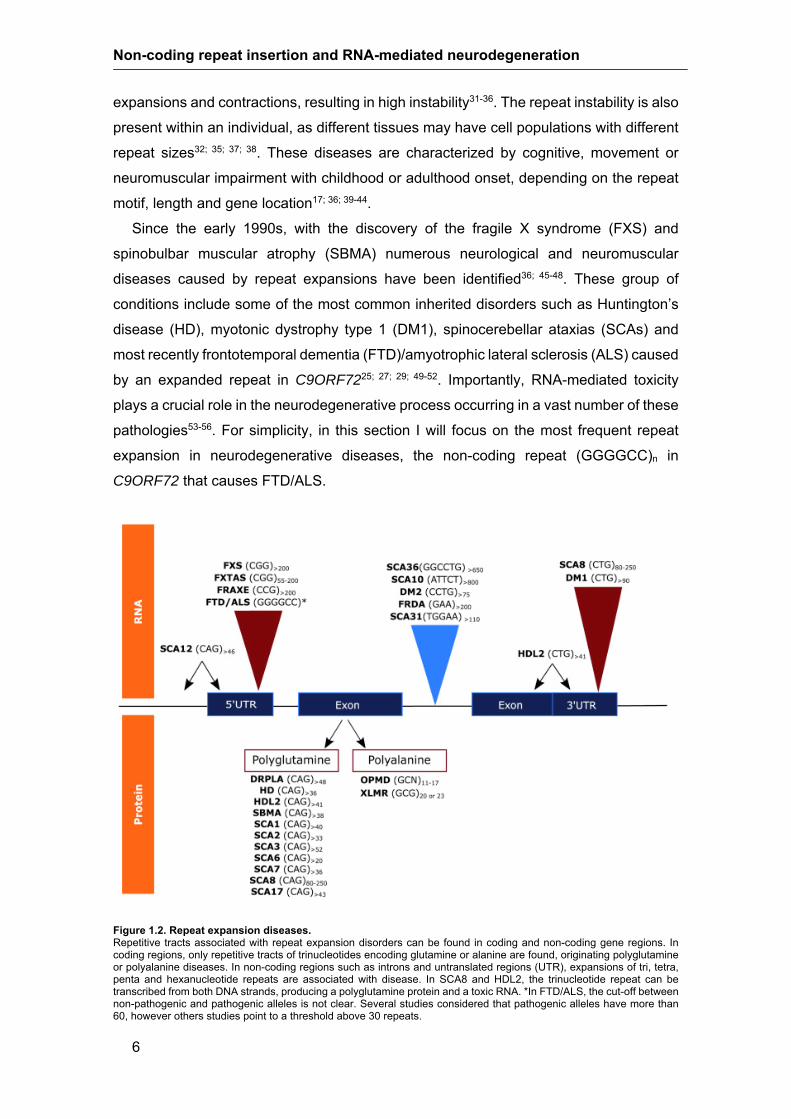
In these diseases, the repetitive tracts in DNA and RNA are trinucleotide,
tetranucleotide, pentanucleotide or hexanucleotide repeats that expand above a given
repeat size25-30 (Figure 1.2). The repeat expansions are a class of mutations named
dynamic mutations because the repeat size changes through generations originating
Non-coding repeat insertion and RNA-mediated neurodegeneration
6
expansions and contractions, resulting in high instability31-36. The repeat instability is also
present within an individual, as different tissues may have cell populations with different
repeat sizes32; 35; 37; 38. These diseases are characterized by cognitive, movement or
neuromuscular impairment with childhood or adulthood onset, depending on the repeat
motif, length and gene location17; 36; 39-44.
Since the early 1990s, with the discovery of the fragile X syndrome (FXS) and
spinobulbar muscular atrophy (SBMA) numerous neurological and neuromuscular
diseases caused by repeat expansions have been identified36; 45-48. These group of
conditions include some of the most common inherited disorders such as Huntington’s
disease (HD), myotonic dystrophy type 1 (DM1), spinocerebellar ataxias (SCAs) and
most recently frontotemporal dementia (FTD)/amyotrophic lateral sclerosis (ALS) caused
by an expanded repeat in C9ORF7225; 27; 29; 49-52. Importantly, RNA-mediated toxicity
plays a crucial role in the neurodegenerative process occurring in a vast number of these
pathologies53-56. For simplicity, in this section I will focus on the most frequent repeat
expansion in neurodegenerative diseases, the non-coding repeat (GGGGCC)n in
C9ORF72 that causes FTD/ALS.
Figure 1.2. Repeat expansion diseases. Repetitive tracts associated with repeat expansion disorders can be found in coding and non-coding gene regions. In coding regions, only repetitive tracts of trinucleotides encoding glutamine or alanine are found, originating polyglutamine or polyalanine diseases. In non-coding regions such as introns and untranslated regions (UTR), expansions of tri, tetra, penta and hexanucleotide repeats are associated with disease. In SCA8 and HDL2, the trinucleotide repeat can be transcribed from both DNA strands, producing a polyglutamine protein and a toxic RNA. *In FTD/ALS, the cut-off between non-pathogenic and pathogenic alleles is not clear. Several studies considered that pathogenic alleles have more than 60, however others studies point to a threshold above 30 repeats.
Chapter 1| General introduction
7
1.2.2. Neurodegeneration by repeat expansion
Repeat diseases can result from expansion of repetitive sequences in exons, 5’-
UTRs, 3’-UTRs or intronic sequences of protein-encoding genes originating coding or
non-coding repeat expansions. Depending on repeat location in the gene and nucleotide
composition, three distinct neuropathological mechanisms can be associated with these
pathologies: (1) polyglutamine or polyalanin gain-of-function, (2) protein loss-of-function
and (3) RNA gain-of-function (Figure 1.3). In coding gene regions only trinucleotide
expanded repeats have been identified as causing disease. Trinucleotide repeat
expansions originate homopolymeric stretches of one amino acid, like polyglutamine
tracts in the CAG repeat disorders HD and Machado-Joseph disease/SCA336. These
homopolymeric stretches, form insoluble ubiquitin-positive aggregates and disturb
cellular homeostasis57. In non-coding repeat conditions two neuropathological
mechanisms can be identified. One is exemplified by FXS and Friedreich ataxia (FRDA),
in which epigenetic alterations lead to transcriptional repression and consequent gene
loss-of-function36. On the other hand, when the repeat is transcribed a toxic RNA is
produced causing a toxic gain-of-function as in SCA36 and C9ORF72 FTD/ALS. This
RNA-mediated toxicity is a complex mechanism in which several nonexclusive
pathogenic pathways have been identified, including (1) RNA foci formation by the
repeat-containing transcripts with perturbed activity of RNA-binding proteins that
regulates critical aspects of RNA metabolism in different cell types and tissues, notably
in neurons (2) alternative splicing misregulation originating imbalance in the expression
ratio of protein isoforms in the brain and (3) generation of neurotoxic peptides resulting
from repeat-associated non-ATG-initiated (RAN) translation56; 58-60.
RNA foci are identified in all non-coding repeat disorders when transcription of the
expanded repeat occurs. These structures correspond to RNA/protein aggregates in the
cell nucleus due to an excessive recruitment of proteins such as RNA-binding proteins
(RNABP) by the expanded RNA that forms secondary structures and hairpin loops.
Splicing factors are the most commonly recruited RNABPs in nuclear RNA foci. Thus,
due to their lower cellular availability, alternative splicing of other mRNAs is impaired.
Splicing misregulation has been deeply studied in DM1, where a few hundred alternative
splicing events are misregulated50; 61. In addition to the mechanisms described, it has
been shown that some of these expanded repeats have bidirectional transcription,
meaning that both sense and antisense RNAs can contribute to the neuropathology. In
SCA8, Huntington’s disease-like 2 (HDL2) and HD, both polyglutamine and RNA gain-
of-function are associated with the pathology and in FTD/ALS both sense and antisense
transcripts contribute to RNA gain-of-function23; 58; 62-65. A recent mechanism associated

Non-coding repeat insertion and RNA-mediated neurodegeneration
8
with neuropathology in repeat expansion diseases is RAN translation. This process has
been associated with tri, tetra, penta and hexanucleotide expansion disorders59; 66. In
RAN translation, the RNA repeat is translated in all reading frames across the expanded
repeat in the absence of the AUG start codon. This likely occurs due to the formation of
secondary structures in repetitive RNA able to recruit ribosomal subunits. The RAN
proteins resulting from translation of expanded repeats are toxic to the cell58; 62; 66.
Notably, in several of these pathologies there is no simple disease mechanism and key
evidence of gain and loss-of-function have been observed62; 67. Two hexanucleotide
repeat expansions have accelerated the pace of understanding of the
neurodegeneration mechanisms driven by expanded repeats and particularly C9ORF72
repeat expansion25-27; 29.
Figure 1.3. Pathogenic mechanisms associated with repeat expansion disorders.

Chapter 1| General introduction
9
Three main disease mechanisms are associated with repeat expansions. (1) In coding regions, repeat expansions originate homopolymeric stretches of one amino acid, such as glutamine (Q), leading to protein gain-of-function. (2) The protein with the polyQ stretch can be cleaved and the polyQ tract (3) aggregates in the cytoplasm or is (4) imported to the nucleus. Cytoplasmic and nuclear aggregates are ubiquitin (U) positive and toxic to the neuronal cell. (5) On the other hand, the expanded CAG RNA has an increased interaction with MBLN1, resulting in its nuclear retention and in decreased protein from translation. In non-coding regions, repeat expansions lead to two exclusive pathogenic mechanisms; (6) gene loss-of-function due to hypermethylation and consequent gene silencing as in FRDA and FXS or (7) RNA gain-of-function, which includes several parallel mechanisms. (8) For the GGGGCC repeat expansion in C9ORF72, the hexanucleotide forms G-quadruplex structures in DNA and DNA:RNA hybrids (R-loop). (9) The G-quadruplexes are also detected in the repetitive expanded RNA. (10) In all non-coding RNA-mediated disorders, the expanded RNA forms RNA foci due to recruitment of RNABP (green squares and purple circles). (11) The consequent sequestration of RNABP to RNA foci leads to alternative splicing misregulation of other neuronal mRNAs. (12) In expanded C9ORF72, the repetitive RNA forms secondary structures able to recruit nucleolin (NCL), leading to nucleolar stress, that compromises rRNA biogenesis. (13) The C9ORF72 hexanucleotide RNA can also sequester the RanGAP1 and interact with other proteins of the nuclear pore complex, disrupting nucleocytoplasmic transport and causing (14) accumulation of other mRNAs in the nucleus. (15) Repeat-containing transcripts can escape from the nucleus to the cytoplasm and be translated by RAN, leading to the synthesis of repetitive peptides, such as the dipeptide species of poly-(Pro-Arg) (PR) and poly-(Gly-Arg) (GR) from the C9ORF72 repeat expansion. These RAN peptides are able to (16) aggregate in the cytoplasm, (17) cause nucleolar stress, (18) compromise nucleocytoplasmic transport and /or (19) impair mRNA splicing of other neuronal mRNAs. (20) For coding repeat expansion diseases such as SCA3 and HD, in addition to protein gain-of-function, RNA-mediated toxicity has also been detected.
1.2.3. Hexanucleotide repeat expansion in C9ORF72
In families with a mixed phenotype of autosomal dominant FTD and ALS, genetically
linked to chromosome 9p21, an expanded GGGGCC hexanucleotide repeat has been
identified in the C9ORF72 gene25; 29. FTD is a progressive form of dementia
characterized by changes in personality, behaviour, cognitive impairment and atrophy of
the frontal and temporal lobes of the brain. In most of the cases, the disease starts before
the age of 65 years. ALS is characterized by onset around the 60s with rapid
degeneration of motor neurons, resulting in progressive paralysis and death from
respiratory failure. Repeat expansions in C9ORF72 account for approximately 12-25%
of familial and 6-7% of sporadic patients with FTD and around 10-50% of familial and 5-
7% of sporadic patients with ALS68-72. In patients, hexanucleotide repeat sizes in
expanded alleles range from ~30 to ~4,000, whereas in healthy individuals repeat
number is usually from 2 to <~30 repeats71; 73-77. Some elderly carriers of intermediate
repeats from ~30 to ~400 do not show disease symptoms, indicating that this range is of
pathogenic reduced penetrance73; 77. Of note is the jump in size from 70 to ~1,750 repeats
during transmission from an unaffected 89-year-old father to four of his children, showing
that small expansions have tendency to expand78. Remarkably, there is an extensive
intra-individual variation of repeat number between non-neuronal and neuronal tissues
that can reach 1,000 repeats more in some neuronal regions78; 79. Thus, intermediate
alleles of reduced penetrance, extreme size variability between peripheral and neuronal
tissues and lack of adequate methodologies can complicate molecular testing for this
disease80; 81. Interestingly, the length of non-pathogenic alleles in C9ORF72 mutation
carriers is not associated with disease phenotype or age at onset82. Regarding the
disease origin, the risk haplotype is shared by all patients with the repeat expansion,

Non-coding repeat insertion and RNA-mediated neurodegeneration
10
which might have arisen in an ancestral population and later have been spread
worldwide71; 83; 84, resembling other repeat expansion diseases52; 85-89. On the other hand,
some data support the possibility of multiple mutation events in the risk haplotype for the
worldwide distribution of the mutation70.
The C9ORF72 gene has shown, by bioinformatic analysis, high homology with the
differentially expressed in normal and neoplastic cells (DENN) domain present in
guanine exchange factors (GEFs) that activate RAB GTPases, indicating a role in
membrane trafficking90. Further studies, by depletion of C9ORF72 protein, have shown
inhibition of transport from plasma membrane to Golgi apparatus, of internalization of
TrKB receptors and altered ratio of an autophagosome marker, indicating that C9ORF72
regulates endocytosis and autophagy91. During mouse development, C9ORF72 protein
is located in synaptic-rich fractions, which supports its involvement at the synapse. In
addition, the differential expression of isoforms in the nucleus and cytoplasm at different
developmental stages suggests distinct roles for C9ORF7292. The first studies could not
establish if the hexanucleotide repeat expansion interferes with normal expression of the
protein C9ORF72 probably because of lack of antibody specificity25; 29. Further
investigation in several brain regions with a newly generated antibody identified reduction
of C9ORF72 protein in frontal cortex, but not in cerebellar cortex93. However, abnormal
RNA expression is probably an important player in the course of the disease. The repeat
is located between two non-coding first exons, and depending on the transcript, the
expanded repeat is either located in the promotor region or in intron 1 and in both cases
causes decrease in mRNA levels in lymphoblastoid cells and frontal cortex25; 70; 93. In
patients with the C9ORF72 repeat expansion, induced pluripotent stem cells (iPSCs)
differentiated in neurons (iPSN) also showed mRNA reduction of all variants94,
confirming previous observations.
The finding of decreased RNA expression in patients with FTD/ALS prompted the
investigation of CpG methylation in C9ORF72. The results showed hypermethylation of
the CpG islands 5’ of the GGGGCC repeat in a proportion of expansion carriers with ALS
and FTD95; 96. Carriers of hypermethylated alleles have reduced RNA expression,
whereas the carrier of an unmethylated allele with 43 repeats showed an expression
level similar to noncarriers. Remarkably, no methylation was detected for normal or
intermediate alleles up to 43 repeats, which raised the question of the threshold of 30
repeats for pathological alleles95; 96. As methylation of CpGs is known to repress unstable
DNA elements in the genome and silence inappropriate gene expression, it was
suggested that this mechanism could be a protective contra-regulatory response to
repeat expansion. To investigate this hypothesis DNA methylation and RNA expression
levels from repeat expansion carriers have been analyzed, showing that

Chapter 1| General introduction
11
hypermethylation of the promoter is associated with transcriptional silencing and reduced
accumulation of C9ORF72 intronic RNA. Moreover, demethylation of mutant C9ORF72
resulted in increased vulnerability of repeat expansion cells to oxidative and autophagic
stress97. Therefore, hypermethylation of the C9ORF72 promoter seems to protect
against repeat expansion mediated toxicity by transcriptional silencing; it causes
decrease in RNA foci formation and in accumulation of RAN pathological aggregates97.
Furthermore, the GGGGCC repeat is itself methylated in carriers of expanded alleles
with >90 repeats, whereas alleles with <43 repeats have shown no methylation98. This
methylation occurs in both blood and brain and is stable over time being linked to repeat
size98. Carriers of large expansions present GGGGCC methylation irrespective of
whether they are hypermethylated at the promoter98. This suggests that in some cases
methylation of the GGGGCC repeat can spread to the promoter region. Another
mechanism involved in reducing C9ORF72 mRNA expression in expanded repeat
carriers is histone H3 and H4 trimethylation of lysine residues that bind strongly to
expanded repeats in brain tissue, but not to non-expanded repeats99.
There is evidence for both loss-of-function and gain-of-function mechanisms in
C9ORF72 FTD/ALS. Reduced mRNA expression supports a role for loss-of-function in
the disease, but it is more likely to modify disease phenotype rather than be the main
origin of the pathology. Moreover, C9ORF72 loss-of-function by conditional knockout in
mice does not induce motor neuron degeneration, defects in motor function or altered
survival100. The data obtained until the present point to an RNA gain of a toxic function,
through RNA foci formation with RNABP sequestration and toxic RAN translation, as the
primary mechanism, confirmed by the behavioral and neuropathological findings in a
mouse model expressing 66 hexanucleotide repeats15; 62; 70; 101-105.
1.2.3.1. RNA gain-of-function in C9ORF72 FTD/ALS
Sequestration of RNABP by sense and antisense repeat-containing transcripts is a
hallmark of RNA-mediated diseases. In C9ORF72 pathology, intranuclear RNA foci
formed by transcripts in the sense orientation have been detected in frontal cortex and
spinal cord tissue from affected subjects25. Antisense transcripts resulting from
bidirectional transcription of the expanded repeat are also prone to RNA foci formation
in frontal cortex, spinal cord and cerebellum of patients106. In the brain, RNA foci are
significantly enriched with proteins containing RNA recognition motifs and involved in
splicing, mRNA nuclear export and translation such as SRSF2, heterogeneous nuclear
ribonucleoprotein (hnRNP) H1/F, hnRNP A1 and Aly/REF export factor (ALYREF)75; 76;
107. In a Drosophila model, the RNABP Pur α binds to RNA C9ORF72 expanded repeats

Non-coding repeat insertion and RNA-mediated neurodegeneration
12
causing neurodegeneration108. Remarkably, the expression of hexanucleotide repeats
as small as 38 and 72 form RNA foci that cause cell death in neuronal cell lines and in
zebrafish76. Furthermore, adenosine deaminase RNA-specific B2 (ADARB2) colocalizes
and interacts with nuclear hexanucleotide repeat RNA foci in iPSN94. Splicing factors are
the proteins most found in RNA foci and sequestration of these factors causes
downstream changes in splicing of target mRNAs, a fraction of which are likely
responsible for neurodegeneration. Moreover, alternative splicing misregulation in the
cerebellum and frontal cortex of patients with the C9ORF72 repeat expansion have been
reported, namely for (Ataxin 2) ATXN2 and microtubule associated protein tau (MAPT)
transcripts originating imbalance of isoforms with altered tissue specificity or altered ratio
of tau proteins containing three or four microtubule-binding domains, respectively104.
Proteins involved in nuclear mRNA export may be involved in export of repeat-containing
RNA from the nucleus for protein translation. Their sequestration in RNA foci may
impede export of their other targets and affect downstream cellular functions. Notably,
transcription studies have revealed several genes aberrantly expressed in CNS tissues
and iPSNs from C9ORF72 ALS patients94; 102. The sequestration of multiple RNABP
from their normal functions suggests the implication of several downstream pathways
that can together, usually in aging carriers, lead to cell death.
During transcription, the newly generated transcripts leave their site of creation and
follow their downstream processes unless there is invasion of a DNA duplex by an
emerging transcript forming an R-loop structure with RNA:DNA hybrids. This process is
favoured by guanine clusters and R-loops are further stabilized by guanine (G)-
quadruplexes. The GGGGCC hexanucleotide repeat expansion is able to form DNA and
RNA G-quadruplex secondary structures and RNA:DNA hybrids. The RNA repeat folds
into a very stable parallel intramolecular structure109. The DNA repeat expansion forms
both antiparallel and parallel stranded G-quadruplexes, but the antiparallel G-quadruplex
structure seems to be the most dominant and stable conformation at physiologically
relevant conditions110; 111. The in vitro transcription assay of hexanucleotide repeats of
varying lengths produced increased amounts of truncated and decreased levels of full-
length transcripts for longer repeats. In contrast, a recent study established that in a
Drosophila model expressing 160 hexanucleotide repeats, flanked by human intronic
and exonic sequences, the repeat is transcribed and spliced in vivo112. Moreover, the
RNA:DNA hybrids and R-loops bind to ribonucleoproteins including the nucleolar protein
nucleolin originating nucleolar stress109; 110. However, the toxicity of RNA foci is not
completely settled in the C9ORF72 repeat disease and a recent study favors dipeptide-
repeat proteins in the neurotoxicity process112.

Chapter 1| General introduction
13
1.2.3.2. RAN and nucleolar stress cytotoxicity in expanded C9ORF72
Like in other neurodegenerative diseases, characteristic intracellular inclusions of
proteins are typical of C9ORF72 pathology. The repeat-containing transcripts can
escape the nucleus and be bound by ribosomal complexes being translated by RAN
translation into aggregating dipeptide-repeat proteins of poly-(Gly-Ala) and to a lesser
amount poly-(Gly-Pro) and poly-(Gly-Arg) that form ubiquitin and p62-positive
cytoplasmic inclusions106; 113-116. RAN translation from antisense transcripts resulting
from bidirectional transcription have also been observed in affected brains66; 106. A
component that has recently been identified in these dipeptide-repeat aggregates is
Drosha protein, a player in miRNA biogenesis117. The Drosha protein forms neuronal
cytoplasmic inclusions in the hippocampus, frontal cortex and cerebellum of patients with
the C9ORF72 repeat expansion. These cytoplasmic Drosha inclusions colocalize with
p62 and ubiquilin-2, two important proteins involved in protein degradation117. The
mislocalization of the Drosha protein to neuronal cytoplasmic inclusions in affected brain
tissues suggests a role for disturbance of RNA/miRNA processing in this pathology.
The mechanism of cytotoxicity by C9ORF72 repeat expansion is now more elucidated
with the observation that two of the RAN translation products behave as cytotoxins in
vitro and in vivo 66; 118; 119. Poly-dipeptides like poly-(Gly-Arg) from sense and poly-(Pro-
Arg) from antisense RAN translation mimic RNA splicing factors containing serine-
arginine repeats and bind hydrogel droplets formed from polymeric fibers of hnRNPA2
but independent of reverse binding upon phosphorylation by CDC2-like kinase 1 and 2
(CCLK1/2)118. Synthetic poly-(Gly-Arg) and poly-(Pro-Arg) peptides when applied to
U2OS cells were able to enter the cells, migrate to the nucleus, bind to nucleoli and
cause cell death. Also in a Drosophila model, arginine-rich dipeptide-repeat proteins
contribute to C9ORF72-mediated neurodegeneration119. Further analysis showed that
exposure of cells to poly-(Pro-Arg) peptide originate global alterations in pre-mRNA
splicing as demonstrated by splicing alterations in mRNAs of the Ran guanosine
triphosphatase (Ran GTPase), pentraxin-related protein PTX3, nascent polypeptide-
associated complex subunit alpha (NACA) and growth arrest and DNA damage-inducible
GADD45A. Moreover, the central task of nucleoli to synthesize mature rRNA was shown
to be impaired with changes in the cellular levels of rRNA precursors with evidence of
nucleolar dysfunction118. Accumulating data indicate that nucleolar stress and impaired
stress granule formation are induced by arginine-rich RAN proteins120. Stress granules
are originated from self-assembly of non-translating mRNAs with various proteins that
regulate mRNA translation, localization and turnover into cytoplasmic structures. They
have important functions either in spatio-temporal control of mRNAs that drive

Non-coding repeat insertion and RNA-mediated neurodegeneration
14
developmental processes or regulate synaptic plasticity and memory in neurons. These
granules assemble, disassemble and travel through cells. Aberrant granule assembly,
namely stress granules formed during cellular stress, has lately been shown to be an
important mechanism in C9ORF72 pathology53; 66; 118; 120.
1.2.3.3. C9ORF72 repeat expansions disrupt nucleocytoplasmic transport
Three recent studies have revealed a novel mechanism of neurodegeneration
originated by dysfunction of the nucleocytoplasmic transport through the nuclear pore. A
candidate genetic screen of disease modifiers in Drosophila expressing 30
hexanucleotide repeats identified an orthologue of human Ran GTPase activating
protein (RanGAP1), regulator of nucleocytoplasmic transport, as a potent suppressor of
neurodegeneration24. In addition, another screen in transgenic flies expressing
transcripts with 58 repeats, without a translation start site, showed RAN translation of
dipeptide repeat proteins and neurodegeneration. This system allowed the identification
of 18 genetic modifiers that encode components of the nuclear pore complex, as well as
the machinery that regulates the export of nuclear RNA and the import of nuclear
proteins16. In agreement with these results, Drosophila salivary gland cells expressing
58 repeats showed nuclear envelope abnormalities and accumulation of nuclear RNA.
In patient motor cortices, RanGAP1 binds to hexanucleotide repeat RNA and exhibits
abnormal nuclear localization and aberrant nuclear aggregates24. In iPSN from
C9ORF72 patients RanGAP1 also co-localizes with the hexanucleotide repeat RNA and
forms aggregates. In the Drosophila salivary gland, the expression of a GFP protein,
tagged with a classical nuclear localization signal (NLS) and nuclear external signal
(NES), showed a reduced nuclear/cytoplasmic ratio in cells expressing the expanded
hexanucleotide repeat, indicating impairment in nucleocytoplasmic transport. Further
studies demonstrated decrease in nuclear import of proteins24 and disturbance in nuclear
export of RNAs16, in hexanucleotide repeat expansion-expressing flies in vivo. Potentially
affected by these defects is TDP-43, a predominantly nuclear protein depleted from the
nucleus of neurons in ALS, being a hallmark of the disease24.
The third work on modifiers of hexanucleotide repeat C9ORF72 toxicity identified
potent suppressors and enhancers of dipeptide repeat toxicity in Saccharomyces
cerevisiae121. This study focused specifically on dipepetide-repeat toxicity and not RNA-
related toxicity using codon-optimized constructs to express each dipeptide repeat
independently without the hexanucleotide repetitive sequence. Many of the modifiers
identified in yeast are genes with a function in nucleocytoplasmic transport, but genes
involved in other processes and cellular localizations such as in ribosome biogenesis

Chapter 1| General introduction
15
and function, ubiquitination and proteasome, mitochondria, transcription, RNABP and
serine/threonine protein kinases were also reported.
Surprisingly, these three studies of modifiers of expanded RNA repeat toxicity,
dipeptide repeat toxicity or both, in model organisms and neurons derived from patients,
identified a fundamental pathway for FTD caused by compromise of nucleocytoplasmic
transport through the nuclear pore. Less understood is the role of C9ORF72 loss-of-
function in disruption of this transport as one of its isoforms localizes to the nuclear
envelope and interacts with regulators of the nucleocytoplasmic transport122. Notably, the
disrupted nucleocytoplasmic transport is potentially amenable to therapy16; 24; 121.
1.2.4. Therapy for RNA gain-of-function
In diseases like C9ORF72 FTD/ALS caused by gain of a toxic function at the RNA
level, promising therapies should target the expanded RNA. The FTD/ALS caused by
C9ORF72 repeat expansion has shown to be a good candidate for antisense
oligonucleotide (ASO) treatment. The RNA gain-of-function causing RNA toxicity and
consequent RAN translation with production of toxic proteins can be targeted without a
better understanding of the contribution of each of these mechanisms in disease
pathology. This has been confirmed by the employment of ASOs targeting the sense
transcript that showed reduction of sense RNA foci and rescue of the toxic phenotype in
C9ORF72 patient-derived cells94; 123; 124. The ASOs used in these studies mediated
RNase H-dependent degradation of the repeat-containing toxic RNA transcripts from the
sense strand. However, ASOs targeting the antisense transcripts may also be needed
as ASOs for the sense strand may not correct all the transcriptional changes as shown
in patient cells after treatment123. Notably, ASOs targeting the C9ORF72 RNA are able
to rescue the disrupted nuclear/cytoplasmic Ran GTPase ratio observed in iPSN from
ALS patients with the repeat expansion24. These ASO molecules can be modified for
stability in biological conditions and after the requirements for safety and efficiency have
been fulfilled, they hold promise for brain delivery and treatment of these
neurodegenerative diseases125; 126.
1.3. Microsatellite repeats located in Alu elements
In the genome of primate species, approximately 22% of microsatellite repeats are
located in poly-As or in the vicinity of Alu elements127. Interestingly, poly-A repeats with
less than 16 units are more mutable when located in Alu elements128. Thus, poly-A

Non-coding repeat insertion and RNA-mediated neurodegeneration
16
stretches of Alu elements can be the basis for nucleotide substitution and microsatellite
birth either by errors introduced during Alu reverse transcription, or after insertion, by the
accumulation of mutations in Alu poly-A stretches129; 130. The second mechanism is the
most probable for explaining mutations in the Alu 3’-tail due to its high relevance for
retrotransposition. After microsatellite birth, mutations can progressively cause the
expansion or contraction of the repeat. With Alu poly-As involved in microsatellite birth
and mutability, it is not surprising that some pathogenic repeats have been found in these
regions, as observed in DM2, FRDA, SCA10 and SCA31 (Table 1.1)131-134.
Disease-associated repeats located within poly-A stretches of Alu elements have
different evolutionary histories, resulting in different motif complexities. The simpler GAA
repeat, expanded in FRDA, is to our knowledge the oldest of these repeats, originated
before New World monkeys radiation; the ATTCT repeat in the SCA10 locus was
originated after or before Old World monkeys radiation; and the tetranucleotide repeat
associated with DM2 is the youngest, originated before the divergence of great apes131;
133; 135. Regarding SCA31, it is known that the repeat insertion occurred in humans, but
the origin of the flanking repeats still needs to be investigated.
Table 1.1. Most common non-coding pathogenic repeats
Disease Causal repeat
Gene
Non-pathogenic small size
alleles
Pathogenic alleles
Location in Alu element
DM1-Myotonic dystrophy type 1 CTG DMPK 5-34 >90 *
DM2-Myotonic dystrophy type 2 CCTG ZNF9 11-26 >75 AluSx
(3'-poly-A)
FXS- Fragile X syndrome CGG FMR1 5-44 >200 *
FRAXE-Mental retardation, X-linked, FRAXE type
CCG FMR2 6-25 >200 *
FRDA-Friedreich's ataxia GAA FXN 5-33 >200 AluSx
(midle-poly-A)
FTLD/ALS-Frontotemporal dementia/ Amyotrophic lateral sclerosis
GGGGCC C9ORF72 <25 >60a *
FXTAS-Fragile X Tremor/Ataxia Syndrome
CGG FMR1 5-44 55-200b *
HDL2-Hungtinton disease like 2 CAG JPH3 6-28 >41 *
SCA8-Spinocerebellar ataxia type 8 CTG/CAG ATXN8OS 15-50c 80-250c *
SCA10- Spinocerebellar ataxia type 10 ATTCT ATXN10 10-32 >800 AluSz
(3'-poly-A) SCA12-Spinocerebellar ataxia type 12 CAG PPP2R2B 4-32 >46 *
SCA31-Spinocerebellar ataxia type 31 TGGAA BEAN (TGGAA)n insertion
AluSx (3′-poly-A)
SCA36-Spinocerebellar ataxia type 36 GGCCTG NOP56 3-16 >650 *
*Not applicable; ain some studies >30 repeats; bat risk of developing the disease; ccombined (CTA)n(CTG)n repeats.

Chapter 1| General introduction
17
1.4. Microsatellite repeat instability
In non-pathogenic chromosomes, repeat size variation of the disease-associated
microsatellite occurs typically through a stepwise mechanism, by gain or loss of one
repeat unit. After expanding above a given threshold, however, these microsatellites
become unstable during intergenerational transmissions and in somatic tissues, a
feature that confers to pathogenic repeat expansions the designation of dynamic
mutations. The degree of variation in size of unstable alleles upon intergenerational
transmission, as well as in somatic tissues of the same individual, depends on the locus
and its location in the genome. Repeats located in non-coding regions are usually more
unstable than repeats located in coding regions. For instances, in SCA7 caused by an
exonic (CAG)n expansion, the repeat may reach 460 units, but alleles as large as 11,000
repeats can be detected in non-coding regions, such as in DM247; 136. Independent of
their location, stable non-pathogenic alleles have usually fewer than 40 repeat units
(Table 1.1).
The repeat size in polyglutamine diseases and some non-coding repeat expansions,
like SCA10 and DM1, correlates negatively with the age of onset and positively with
severity of disease symptoms, evidencing the importance of knowing the degree of
repeat size instability for clinical purposes137-143.
1.4.1. Genetic factors associated with repeat length instability
The mechanisms leading to repeat length instability in pathogenic repeats are not fully
understood, however, it is known that mitotic and meiotic repeat instability depend on
several genetic factors, such as (1) initial repeat length, (2) presence of repeat
interruptions and (3) haplotype background. Other factors like the type of tissue,
developmental stage and proliferative stage of the cell, also contribute to instability.
The initial repeat size is a crucial factor in repeat length variation because larger
repeats usually trigger larger changes in size144. However, the presence of repeat
interruptions in the repetitive tract, that occurs in some repetitive loci, contributes to
stabilize the repeat. The presence of interruptions in a repetitive tract destabilizes non-
B-DNA conformations that are associated with repeat instability145. These interruptions
correspond to nucleotide motifs usually with the same size of the repeat unit and their
loss leads to increased repeat instability. This phenomenon was deeply assessed for the
FMR1 CGG repeat, where the largest repeat lengths have no interruptions. In FMR1,
non-pathogenic stable alleles are usually interrupted by two AGGs, one every 9 or 10

Non-coding repeat insertion and RNA-mediated neurodegeneration
18
CGGs, whereas unstable alleles in the premutation range (55-200 CGGs) have no AGG
interruptions (in most of them) or 1 or 2 interruptions; FMR1 full mutations (with more
than 200 CGGs) are generally pure146; 147. This loss of AGG interruptions is polarized,
occurring from 3’ to 5’ of the CGG tract, and is associated with the probability of
expansion; large pure CGG repeats at the 3’-end of premutation alleles increase the risk
of full mutation expansion146; 148. Also, in SCA1, the loss of interruptions in the ATXN1
gene increases repeat instability; alleles in the non-pathogenic size range have from 1
up to 3 CAT interruptions, while expanded alleles are usually composed of pure CAG
tracts149; 150. Similarly to FMR1, the acquisition of instability in ATXN1 is influenced by the
loss of interruptions in the repetitive stretch, though some alleles beyond the normal
range have been detected containing CAT interruptions 149-151. In other diseases,
interruptions are more likely to be present in expanded alleles, as in DM1, where normal
DMPK alleles are pure CTGs and 3-8% of expanded alleles contain CCG, CTC, CGG or
CAG interruptions152-156. Interrupted expanded alleles in DMPK showed a tendency to
contraction in maternal transmissions (instead of the expected expansion bias of pure
alleles, which results in allele sizes into the range of congenital DM1); this suggests that
the presence of interruptions stabilizes the DM1 repeat at the germline level and turns it
prone to contraction152-154; 156. Trinucleotide repeats are not the only interrupted repeats;
the ATXN10 pentanucleotide repeat associated with SCA10 may also be interrupted. In
ATXN10, small size non-pathogenic alleles are usually pure, whereas large non-
pathogenic alleles (>17 ATTCT units) may be, very rarely, interrupted; however,
expanded alleles present several pentanucleotide or heptanucleotide ATTGT, TTTCT,
ATGCT, ATCCT, ATTCTAT and ATATTCT interruptions. These interruptions only
stabilize the repeat tract locally, evidenced by low repeat size variability in the allele
positions close to interruptions; however, the presence of interruptions do not influence
the overall repeat instability157-159.
The genomic context flanking the repeat is another important factor for repeat stability,
in fact, depending on the flanking sequence, repetitive regions have shown different
levels of instability160. This is supported by mouse models of the polyglutamine disease
SCA7, showing that the full-length ATXN7 cDNA with an expanded repeat alone had low
repeat instability in germline and somatic cells, contrarily to the fragment containing the
expanded repeat and the flanking genomic region (both flanking introns and the
downstream exon)161. These different levels of repeat instability indicate the presence
of cis elements affecting instability. In fact, haplotype studies in DM1, MJD/SCA3 and
FXS patients from different populations have shown some polymorphic (SNP and/or
STR) alleles linked to expanded alleles suggesting either evidence of independent origin
mutations or the presence of cis elements that may predispose the region to repeat

Chapter 1| General introduction
19
instability, with haplotypes of expanded alleles shared by different independent-origin
mutations86; 162; 163.
1.4.2. Germline repeat length instability
In dynamic mutations, the change in repeat length upon parent-offspring transmission
is often associated with the gender of the transmitting parent. In diseases like FXS and
SCA8, repeat expansions usually occur after maternal transmission, while in
polyglutamine diseases such as SCA1, 2, 3/MJD, 7 and HD, as well as SCA10 and
DRPLA, alleles are more prone to expand after paternal transmission32; 144; 150; 164-169. In
DM1, the sex-bias for instability depends on allele size of the transmitting-parent. Small
expansions (less than 100 repeats) are more unstable upon male transmission, but
congenital DM1 (resulting in expansions of more than 1000 repeats) is almost exclusive
of maternal transmission170-172. When analysing sperm cells from DM1 male carriers,
expansions in sperm are indeed detected, but with a size apparently lower than 1000
CTGs173.To explain this differential inheritance of large alleles it has been suggested that
selection might occur in sperm cells with very large expansions170. Interestingly,
although unstable repeats have a tendency to increase in size, in some diseases,
contractions are detected after paternal transmission as is the case of trinucleotide
repeats in FXS, FRAXE, FRDA and SCA8. In these diseases, repeat contraction events
were detected not only when comparing blood cells of parent-offspring pairs, but also by
analysing sperm of the affected parents33; 35; 164; 174-177. In fact, this bias for repeat
expansion after gender-specific transmission indicates the involvement of molecular
mechanisms specific of oocyte and sperm development. To shed light on the timing and
pathways of gametogenesis involved in this process, some studies were performed using
knock-in animal models for unstable repeats. Regarding male mice germline, in FXS,
expansions are detected in primary spermatocytes, with no additional increase in size in
later stages, whereas in FXS female mice germline, expansions occur in non-dividing
post-natal oocytes178. These results show why large expansions are only maternally
transmitted.
Studies of instability on sperm cells from individuals with dynamic mutations have
shown that repeat sizes are very heterogeneous and different in length from blood cells.
In HD, DM1, SCA1, SCA8 and SCA10, the expanded repeat size is more variable in
sperm than in blood cells, which is a factor contributing to the instability observed upon
transmission32; 35; 179-181. Remarkably, in sperm cells from SCA1 subjects there is a

Non-coding repeat insertion and RNA-mediated neurodegeneration
20
tendency for repeat length increase, which correlates with the paternal tendency for
expansion observed in pedigrees179.
The study of gametes of individuals with repeat expansions is challenging due to the
difficulty in obtaining biological material. However, some studies using mouse models
suggest that the instability of repeats at the germinal level may increase with the age of
the individual as is the case of mouse models for DM1 and DRPLA182; 183. In DRPLA, this
age dependent instability is corroborated when analysing the unstable allele
transmission in pedigrees of affected families183.
In general, although repeat length instability is highly variable among repeat diseases,
there is a bias towards repeat size expansion over successive generations35; 52; 144; 148; 150;
184; 185. This tendency is the molecular basis for decrease in disease age-of-onset and
increase of disease severity over generations, a phenomenon called anticipation186; 187.
1.4.3. Somatic repeat length instability
In somatic tissues from the same affected individual, cell populations may have
different repeat sizes, resulting in somatic instability or mosaicism. Similarly to germline
instability, at the somatic level repeat length instability is highly variable, depending on
the locus, tissue and developmental stage.
In individuals and animal models carrying unstable alleles, several studies have
reported repeat expansions and contractions in both proliferative and non-proliferative
cells and at different life stages188; 189. In some repeat diseases, affected individuals
present larger repeat expansions in relevant tissues for disease progression; this is the
case of FRDA, a disease with a phenotype of progressive ataxia, cardiomyopathy and
diabetes190, in which larger expansions are observed in the heart, pancreas and
cerebellum. In other diseases, like SCA1, 2 and 3/MJD, the cerebellum, the most
affected brain region, is the human brain region where lower repeat sizes are detected179;
185; 191.
Somatic repeat length instability increases with aging. Age-dependent expansion bias
was detected in human tissues of individuals with FRDA, when comparing tissues from
an 18 week fetus homozygous for an allele with 620 GAAs. Both expanded alleles
inherited by the fetus have shown somatic instability, according to the repeat sizes
detected in the blood of the parents. However, in the fetus, the observed repeat instability
in different brain regions, pancreas, heart and blood was much lower than in the
corresponding tissue of the affected adult189. Evaluation of age-dependent instability in
different early developmental stages has also been performed in congenital DM1, from

Chapter 1| General introduction
21
10 weeks fetus up to neonatal periods. Somatic instability was not detected up to 13
weeks of gestation, but started to appear from 16 week onward192. This age-dependent
instability was also detected in human peripheral blood cells of DM1 affected individuals,
carrying similar size expanded alleles, but with ages ranging from 0.5-62 years old. In
earlier stages of development, the repeat is more stable than in aging, thus the rate of
instability is progressive through aging. When evaluating the repeat size of the same
individual in peripheral blood collections separated by two years there is a clear tendency
for repeat size increase with aging193.This age-dependent expansion bias is also
detected in post-mitotic cells, in brain regions of HD and SCA1 mouse models at different
time points194; 195.
The high somatic variability, regarding repeat size in different cells and tissues, found
in a same individual, turns the repeat length assessed in peripheral blood (the biological
material used for molecular diagnosis) not always corresponding to the repeat expansion
size in the affected tissue, a phenomenon that was observed in SCA1, SCA2, and FRDA 179; 185; 189; 190. This difference can justify, at least partially, why repeat expansion length in
peripheral blood explains only approximately half of the variation in age of onset. In fact,
in HD, the somatic instability and size of expansions in the cortex, an affected brain
region, shows higher inverse correlation with age of onset in a cohort of affected
individuals, suggesting that greater repeat length instability in the affected brain region
accelerates disease progression196.
1.4.4. Cellular mechanisms implicated in repeat length instability
The repeat length instability occurring at different cell stages is not due to a single
molecular mechanism, but results from the action of several cellular pathways. Repeat
length and homogeneity is correlated with the formation of non-canonical DNA structures
(hairpins, triplexes, G-quadruplexes and slipped strands) during DNA replication,
transcription and repair. These structures are known to have mutagenic propensity.
Repeats are also associated with DNA damage and chromosomal fragility (breaks) that
may be repaired by several cellular mechanisms. Their use depends on the phase in cell
cycle and type of lesion, and are often implicated is repeat size variability197. The
mechanisms and proteins involved in expanded repeat instability may be different from
small scale repeat size variability as shown in Saccharomyces cerevisiae198.

Non-coding repeat insertion and RNA-mediated neurodegeneration
22
1.4.4.1. DNA Replication
Cell proliferation is one factor associated with repeat instability. In dividing cells, DNA
replication and post-replication repair are major contributors for repeat expansion.
Several non-exclusive mechanisms have been proposed to explain repeat length
instability in proliferative cells. Most of them are based on the difficulty that the DNA
polymerase complex finds when progresses through repetitive regions and their non-
canonical structures, which may result in replication fork stalling and collapse.
In replication, as shown in yeast, repeat instability is more associated with the lagging
DNA strand and processing of Okazaki fragments. The 5’-single stranded tail or flap
structure, resulted from strand displacement during Okazaki fragment maturation, may
fold in a secondary structure, which may then inhibit flap endonuclease 1 (FEN1) activity.
This way, the 5’-single stranded tail may be ligated at 3’end of the preceding Okazaki
fragment and be part of the newly synthetized strand, creating an expansion199.
A factor that influences the probability of a repeat to expand is the position of the
replication initiation200. Three models, named the origin switch, origin shift and fork-shift,
suggested that the distance of the replication origin to the repeat and the sequence
orientation of repeat replication influenced CTG repeat instability due to the formation of
secondary structures during replication (Figure 1.4)201.
Figure 1.4. Positioning and orientation of repeat replication, adapted from Mirkin, 2007202. (A) Origin switch model: the change in replication direction in an allele with a repeat expansion compared to small size alleles leads to a change in the repeat sequence used as template, putting the sequence prone to form a non-canonical structure in the lagging strand. (B) Origin shift model: the shifting in the location of the replication origin may change the positioning of the repetitive sequence in the Okazaki initiation zone (OIZ). (C) Fork-shift model: a change in the mode of replication fork progression due, for example, to an epigenetic change that may alter the repeat position in OIZ. Orange- replication origin, blue- DNA sequence that change the distance between the replication origin and the repeat, blue brackets- OIZ, blue circle- epigenetic change, red- repetitive sequence prone to form a non-canonical structure and green- complementary sequence.

Chapter 1| General introduction
23
One of the first models proposed to explain repeat instability was the replication
slippage. This is also the model accepted for the generation of non-pathological
microsatellite polymorphisms. According to this model, during new DNA strand
synthesis, when the replication complex finds a tandem repeat, it can stop the replication
and release the template DNA strand. Then, due to complementarity, the new DNA
strand may anneal with repeats upstream or downstream from the template strand and
when the polymerase complex restarts the DNA polymerization, this misalignment will
result in increase or decrease of the number of repeats in the daughter DNA strand
(Figure 1.5)203; 204.
Figure 1.5. Replication slippage model, adapted from Richard et al., 2008205. (A) Replication of a tandem repeat sequence. (B) During DNA synthesis, the replication machinery may pause on the lagging strand, due to secondary structures or other kinds of lesions and release the template DNA. (C) Partial unwinding of the lagging strand may lead to replication slippage. (D) The replication restart gives rise to an expansion or a contraction of the repeat tract, depending on what strand (newly synthesized strand or template) slippage occurred. (E) Alternatively, partial unwinding of the lagging strand may lead to lesion bypass by homologous recombination with the sister chromatid, also leading to contractions or expansions of the repeat tract. Template strands are drawn in red, and the newly synthesized strands are drawn in blue.
Another model suggested to explain repeat expansion during DNA synthesis was the
template switching. According to this model, while the DNA from the repetitive sequence

Non-coding repeat insertion and RNA-mediated neurodegeneration
24
is synthetized, either on the leading or lagging strand, the DNA polymerase may switch
from the template to the nascent DNA and polymerize DNA, using the last as template.
When the DNA polymerase returns to the corresponding template, the fork progresses,
resulting in an increase in DNA repeat size, that forms a loop in the nascent strand and
may become an expansion in the next replication round (Figure 1.6) 206; 207. Mutations in
replicative DNA polymerase that destabilize the fork, favour the template switch model
in increasing the repeat expansion rate208. Template switching is more expected in
lagging strand having been shown in the Saccharomyces cerevisiae model of FRDA, a
link between scale of expansion and Okazaki fragment size208. In fact, the repeat
expansion is more likely when the repeat length exceeds the size of an Okazaki
fragment, or in other words, when both leading and lagging strand are being replicated
through the repeat208.
Figure 1.6. Template switching model, adapted from Shah et al., 2012208. (A) Progression of the replication fork through a long tandem repeat. (B) During repeat replication, a switch from template DNA to nascent strand may occur. (C) Then, the leading strand is replicated from the repetitive Okazaki fragment. (D) The leading strand switch to its original template and fork restart. (E) Replication products containing repetitive loop-outs and gaps. Complementary strands of the repeat are shown in green and red; 3′-ends of growing DNA chains are shown by arrows.
Another factor that influences repeat instability is the presence of nucleosomes in the
repetitive region209. When the lagging strand invades the complementary strand, if
nucleosomes are positioned in that chromatid they limit the region for strand invasion,
whereas if no nucleosomes are present the new DNA strand has a larger region to
invade, increasing the probability of repeat expansion209.
Although several mechanisms have been implicated in DNA replication mediated
instability, they only explain repeat size variation in proliferative cells. The contribution of

Chapter 1| General introduction
25
these mechanisms for repeat instability in the central nervous system, should be
negligible, where other mechanisms independent of DNA replication are involved.
1.4.4.2. Recombination
The simplest mechanism connecting recombination with repeat size instability is the
meiotic homologous recombination. In this mechanism, the unequal cross-over among
the repeats in homologous chromosomes results in repeat expansion and contraction.
This mechanism was proposed for the evolution of FMR1 CGG repeats and in the
expansion of CGN repeats in HOXD13210; 211.
Recombination is an important mechanism to repair DNA damage like double-strand
DNA breaks (DSB), gaps, and nicks that result from DNA replication and repair. DSB are
generated during DNA replication, as a result of replication complex stalling and reverse.
DNA DSB resulting from both repeat replication and transcription promote break induced
replication (BIR), a type of DNA damage repair that includes the invasion of sister
chromatids (homologous recombination) and the misalignment of the repetitive
sequence with consequent repeat contraction or expansion, depending on how more
downstream or upstream the strand invasion occurs (Figure 1.7)212.

Non-coding repeat insertion and RNA-mediated neurodegeneration
26
Figure 1.7. Replication fork stalling followed by break induced replication adapted from Kononenko et al., 2018212. Repetitive regions in the genome may lead to replication fork stalling and occasionally to fork reversal. Resolution of the holyday junction (isomerization) results in DSB. Repair of DNA damage via BIR may lead to a repeat contraction or expansion. Repeats are represented in brown and blue.
1.4.4.3. Transcription
Repeat instability is also a feature of cells with low or without proliferative capability
such as oocytes (arrested during the first meiotic division from late fetal stages up to
puberty) and neurons (post-mitotic cells). This indicates that repeat size instability in cell
types involved in disease inheritance and pathology must be mediated by other cellular
mechanisms independent of DNA replication.
Gene expression levels are in fact correlated with repeat size instability209. Induction
of gene transcription, more than transcription through the repeat, increases the rate of
expansion209. This instability is further aggravated if the repeat has bidirectional
transcription213.
Three stranded structures formed during transcription or R-loops- a stable DNA:RNA
hybrid with the template DNA strand; the nascent RNA; and a loop of single stranded
non-template DNA, also have an impact in repeat instability214. Although these structures
play important roles in transcription initiation and termination, they may also be formed
in repetitive regions as in expanded CAG, CGG, GGGGCC and GAA repeats110; 214; 215.

Chapter 1| General introduction
27
These structures interfere with RNA polymerase progression and are potentially
mutagenic and prone to DSB. During transcription, repetitive tracts and R-loops
contribute to instability in two ways, as detected in vitro in E. coli and human cells: the
strength of DNA pairing with the RNA and the rate of re-pairing of the separated DNA
strands216. The DNA:RNA hybrids lead to replication stall with consequent DSB formation
that are repaired by BIR, promoting repeat expansion. On the other hand, the R-loop
structure leads to a loose non-template single strand DNA that is prone to form hairpins
and consequently repeat size variation when the RNA is removed and the template and
non-template DNA hybridize again216.
1.4.4.4. DNA Repair
DNA damage occurs naturally in living cells, not only during replication or
transcription, but also (among others) due to cell exposition to products of endogenous
metabolic processes like reactive oxygen species217. This damage is repaired by suitable
cellular mechanism being the errors in these pathways potentially mutagenic for the cell.
Genome-wide association studies in individuals affected with diseases caused by
CAG, GAA and CGG repeat expansions, have allowed to identify variants close to genes
of DNA damage response and mismatch repair pathways with significant association
with age of onset and/or repeat instabillity218-221. A gene commonly found in these studies
is FANCD2/FANCI-associated nuclease 1 (FAN1) that encodes an interactor of the
mismatch repair protein, MLH1, previously associated with repeat instability and
implicated in recovery of replication forks222.
Mismatch repair enzymes have a central role in somatic instability. They were
identified driving repeat expansion by repairing slipped DNA strands or endogenous
damage. In CAG and CTG repeats, many proteins have been associated with somatic
instability like the enzymes MSH2, MSH3 and MSH6 that recognize DNA mismatch, the
effectors MLH1 and MLH3 and the downstream PMS2222-224.
Although many research studies have focused on investigating the mechanisms
leading to trinucleotide repeat instability, they are not fully understood. More studies in
other repeats beside trinucleotides are needed to identify shared proteins involved in
instability of tetra, penta and hexanucleotide repeats causing disease.
1.5. Investigating non-coding pathogenic repeats
The identification of pathogenic repeats is challenging since they are not detected
with high-throughput genotyping technologies and the molecular mechanisms triggered
Non-coding repeat insertion and RNA-mediated neurodegeneration
28
by them, are not fully elucidated. However, their study is highly valuable being applied in
molecular diagnosis, genetic counselling and development of suitable therapeutic
strategies to stop or delay the progression of these diseases that have no cure.
This PhD project aims to investigate the role in neurodegeneration of a non-coding
repeat region in DAB1 genetically linked to a new type of spinocerebellar ataxia. To
achieve this goal, we addressed the main questions below:
Is the non-coding repeat sequence in DAB1 alleles of affected individuals
different from unaffected subjects?
Is the non-coding repeat sequence in DAB1 toxic?
What is the molecular mechanism mediated by this non-coding repeat
sequence in DAB1?
How can the pathological non-coding repeat be molecularly diagnosed?
What is the mutational mechanism in the origin of the pathological non-coding
repeat in DAB1?

Chapter 2. Non-coding ATTTC repeat insertion
causes neurodegeneration by RNA toxicity
Adapted from A Pentanucleotide ATTTC Repeat Insertion in the Non-Coding
Region of DAB1, Mapping to SCA37, Causes Spinocerebellar Ataxia,
American Journal of Human Genetics: 101: 87-103, 2017


Chapter 2| A non-coding ATTTC repeat insertion causes neurodegeneration by RNA toxicity
31
A Pentanucleotide ATTTC Repeat Insertion in the Non-Coding Region of
DAB1, Mapping to SCA37, Causes Spinocerebellar Ataxia
Ana I. Seixas,1,2,11,12 Joana R. Loureiro,2,3,4,12, Cristina Costa,5 Andrés Ordóñez-Ugalde,2,3,6 Hugo
Marcelino,2,7 Cláudia L. Oliveira,2,3 José L. Loureiro,1,2,8 Ashutosh Dhingra,9 Eva Brandão,8 Vitor T. Cruz,8
Angela Timóteo,5 Beatriz Quintáns,6 Guy A. Rouleau,10 Patrizia Rizzu,9 Ángel Carracedo,6 José Bessa,2,7
Peter Heutink,9 Jorge Sequeiros,1,2,4 Maria J. Sobrido,6 Paula Coutinho,1,2 Isabel Silveira,2,3
1UnIGENe, i3S-Instituto de Investigação e Inovação em Saúde, Universidade do Porto, 4200-135 Porto, Portugal;
2IBMC- Institute for Molecular and Cell Biology, Universidade do Porto, 4200-135 Porto, Portugal; 3Genetics of Cognitive
Dysfunction Laboratory, i3S-Instituto de Investigação e Inovação em Saúde, Universidade do Porto, 4200-135 Porto,
Portugal; 4ICBAS, Universidade do Porto, 4050-313 Porto, Portugal; 5Department of Neurology, Hospital Pr. Dr.
Fernando Fonseca, E.P.E, 2720-276 Amadora, Portugal; 6Instituto de Investigación Sanitaria and Fundación Pública
Galega de Medicina Xenómica, Centro para Investigación Biomédica en Red de Enfermedades Raras (CIBERER),
15706 Santiago de Compostela, Spain; 7Vertebrate Development and Regeneration Laboratory, i3S-Instituto de
Investigação e Inovação em Saúde, Universidade do Porto, 4200-135 Porto, Portugal; 8Department of Neurology,
Hospital São Sebastião, 4520-211 Feira, Portugal; 9German Center for Neurodegenerative Diseases, 72076 Tübingen,
Germany; 10Montreal Neurological Institute, and department of Neurology and Neurosurgery McGill University, Montréal,
QC H3A 2B4, Canada; 11Present Affiliation: Glial Cell Biology Laboratory, i3S-Instituto de Investigação e Inovação em
Saúde and IBMC – Institute for Molecular and Cell Biology, Universidade do Porto, 4200-135 Porto, Portugal; 12These
authors contributed equally for this work
Advances in human genetics in recent years have largely been driven by next
generation sequencing (NGS); however, the discovery of disease-related gene
mutations has been biased towards the exome because the large and very repetitive
regions that characterize the non-coding genome remain difficult to reach by that
technology. For autosomal-dominant spinocerebellar ataxias (SCAs) 28 genes have
been identified, but only five originate from non-coding mutations. Over half of the SCA
families, however, remain without a genetic diagnosis. We used genome-wide linkage
analysis, NGS and repeat analysis to identify an (ATTTC)n insertion in a polymorphic
ATTTT repeat in DAB1, in chromosomal region 1p32.2 as the cause of autosomal-
dominant SCA; this region has been previously linked to SCA37. The non-pathogenic
and pathogenic alleles have the configurations of [(ATTTT)7-400] and [(ATTTT)60-
79(ATTTC)31-75(ATTTT)58-90], respectively. (ATTTC)n insertions are present on a distinct
haplotype and show an inverse correlation between size and age of onset. In the DAB1-
oriented strand, the (ATTTC)n is located in 5’-UTR introns of cerebellar-specific
transcripts arising mostly during human fetal brain development from the usage of
alternative promoters, but it is maintained in adult cerebellum. Overexpression of the
transfected (ATTTC)58 insertion, but not the (ATTTT)n, leads to abnormal nuclear RNA
accumulation. Zebrafish embryos injected with the (AUUUC)58 insertion, but not the

Non-coding repeat insertion and RNA-mediated neurodegeneration
32
(AUUUU)n, showed lethal developmental malformations. Together these results
establish an unstable repeat insertion in DAB1 as a cause of cerebellar degeneration;
on the basis of the genetic and phenotypic evidence, we propose this mutation as the
molecular basis for SCA37.
2.1. Introduction
The spinocerebellar ataxias (SCAs) are a group of clinically and genetically very
heterogeneous diseases, usually characterized by adult onset of progressive gait, limb
and speech ataxia caused by loss of cerebellar neurons1,2. The estimated prevalence of
autosomal-dominant SCAs varies considerably in different world regions, from 1.6-
5.6/100,000 inhabitants1; 3; Machado-Joseph disease (MJD)/SCA3 [MIM: 109150] is the
most frequent in Portugal and worldwide, but a definite genetic diagnosis is still lacking
for more than half of the families affected by these neurodegenerative diseases1,4.
Currently 28 genes are known to harbor mutations causing SCA pathology. Seven of
these are CAG repeat expansions that encode aberrant polyglutamine tracts in the
protein products, rendering them toxic. Point mutations, deletions or duplications in
genes functioning in a wide range of neuronal processes explain 16 additional SCA
types5-7. Unstable repeat expansions in non-coding regions of protein-encoding genes
have been reported only in five SCAs8.
The disease mechanism(s) associated with non-coding repeat expansion remained
obscure until the concept emerged that RNA transcripts harboring expanded repeats
were toxic in myotonic dystrophy type 1 (DM1 [MIM: 160900])9,10. RNA-associated
pathogenesis was subsequently identified in other neurodegenerative diseases also
caused by non-coding expanded repeats, such as DM2 [MIM: 602668], fragile X
tremor/ataxia syndrome (FXTAS [MIM: 300623]), HD-like disease type 2 (HDL2 [MIM:
606438]), frontotemporal dementia and/or amyotrophic lateral sclerosis (FTDALS [MIM:
105550]) and in several SCAs caused by non-coding repeat mutations8,10,11.
In SCAs caused by non-coding mutations, such as SCA8 (MIM: 608768), SCA10
(MIM: 603516), SCA36 (MIM: 614153) and SCA31 (MIM: 117210), the untranslated RNA
repeat accumulates in affected brain tissue and correlates with the pathogenesis1,10,12-14.
SCA8 is associated with bidirectional transcription of a CTG/CAG repeat located in the
UTR of ATXN8OS (MIM: 603680) and in a short ORF in the overlapping ATXN8 (MIM:
613289) gene15. In SCA10, 31 and 36, very large intronic penta or hexanucleotide
repeats are found in ATXN10 (MIM: 611550), TK2 (MIM: 188250) and BEAN1 (MIM:

Chapter 2| A non-coding ATTTC repeat insertion causes neurodegeneration by RNA toxicity
33
612051) genes, and NOP56 (MIM: 614154) respectively12-14,16. Studies on RNA-
mediated pathogenesis point towards a complex combination of different effects
including aberrant transcript expression and processing and the formation of RNA
aggregates in affected cells and tissues, which are also detected in cell and animal
models that overexpress the repeat8,11,17-19.
Here we describe a SCA in three large Portuguese families identified during a
population-based survey of hereditary ataxias in Portugal3.The affected individuals from
these families share a pure cerebellar ataxia phenotype and, distinctively, onset by
dysarthria in late adolescence to adulthood. In this study, we mapped this SCA by
genome-wide linkage analysis of the families and identified a pathological (ATTTC)n
repeat insertion in the non-coding region of the DAB1 (DAB1, reelin adapter protein
[MIM: 603448]), in chromosomal region 1p32.2, which overlaps the SCA37 linkage
region (MIM: 615945)20. Given the significant death caused by injection of the RNA
repeat insertion in zebrafish, we suggest that RNA-mediated toxicity could be one
pathogenic mechanism implicated in this SCA.
2.2. Subjects and Methods
2.2.1. Subjects
Families were ascertained during a nationwide, population-based, systematic survey
of hereditary ataxias and spastic paraplegias in Portugal, performed from 1994 to 20043,
and with affected individuals referred for diagnostic purposes to the authorized Center
for Predictive and Preventive Genetics (CGPP), at IBMC; they were tested for mutations
in genes associated with SCA1, DRPLA, SCA2, MJD/SCA3, SCA6, SCA7, SCA8,
SCA10, SCA12, SCA14 and SCA17. This study used the de-identified, previously
collected DNA samples from families that were stored at the CGPP biobank, as well as
anonymized samples from Fundación Pública Galega de Medicina Xenómica, Santiago
de Compostela, Spain and from the Montreal Neurological Institute, McGill University,
Montréal, Canada. For the study of relevant polymorphisms in the Portuguese
population, anonymized DNA samples were obtained from both dried-blood spots from
the public health laboratory newborn screening program and internal lab control samples.
For skin biopsies, informed written consent was given to participate in this study, which
was approved by the health ethics board of the Hospital Prof. Doutor Fernando Fonseca.

Non-coding repeat insertion and RNA-mediated neurodegeneration
34
2.2.2. Genotyping and Linkage Analysis
DNA samples from 65 individuals from three Portuguese families were hybridized to
Affymetrix GeneChip Human Mapping 10K array Xba 142 2.0, according to the
manufacturer’s recommendations (Affymetrix). Each individual genotype was obtained
with the GeneChip® DNA Analysis Software 3.0. After batch import of pedigree and
sample information, data were checked for Mendelian errors. The file obtained was
imported into ALOHOMORA21 and analyzed with PEDSTATS22 for a check of the
consistency of the pedigree and identification of uninformative and/or mistyped SNPs for
removal from the data set. SNPs were reformatted as individual chromosomes for
linkage in Merlin23. Multipoint linkage analysis was performed with a parametric model
assuming an autosomal-dominant mode of inheritance, a disease-allele frequency of
0.0001, and a 95% penetrance.
Short tandem repeat (STR) markers from chr1:51,245,557-59,958,568 (UCSC
Genome Browser built hg19) were analyzed in families, by PCR. Allele sizes were
assessed in ABI3730xl DNA-analyzer using GeneMapper software v4.0 (Applied
Biosystems).
2.2.3. Mutation Screening
Mutation screening was performed in affected individuals and unaffected relatives by
Sanger sequencing of exon and exon-intron boundaries and by NGS. NGS was carried
out in six affected individuals (family M: II.9, III.1; G: II.1, III.6; R: II.7, III.5) and four
unaffected relatives (family M: II.10; G: II.6, III.5; R: II.3) at the sequencing facility of the
Fundación Pública Galega de Medicina Xenomica. Enrichment was performed using the
SureSelect Target Enrichment (Agilent Technologies), targeting 3.4 Mb of genomic
sequence, on chr1:55,800,000-59,200,000 (hg19). Each genomic library was sequenced
using a SOLiD 5500xl (Life Technologies) as paired-end reads (75 + 35 bp). Reads were
aligned to the reference genome (hg19) using SOLiD BioScope software version 1.3.0
followed by variant calling with GATK version 2.124. In the 10 individuals analyzed by
NGS, the number of reads matching the target region ranged from 2,366,593 to
3,444,594; 77.7% to 84.12% of the target region was covered at least 20X, whereas
92.2% to 93.4% was covered at least 5X; the average depth of coverage ranged from
35.11 to 51.29. Variant calling required at least 6X of minimum depth coverage. The next
variant filtering steps required that the variant was (1) heterozygous, and (2) absent (or

Chapter 2| A non-coding ATTTC repeat insertion causes neurodegeneration by RNA toxicity
35
had a frequency below 0.01) from public databases (dbSNP135 and 1000 Genomes
Project, February 2012).
Repeat expansion screening was performed by standard PCR analysis in a family
branch from each of the M, G and R pedigrees. Approximate allele size was assessed
in 2% agarose gel. Non-polymorphic repeats were confirmed by PCR amplification using
a 6-FAM labeled reverse primer and detected on an ABI3730xl DNA-analyzer.
2.2.4. Southern Blot Analysis
Genomic DNA (10 µg) was digested with XbaI (Fermentas Life Sciences). Digestion
products were separated by electrophoresis on a 0.8% agarose gel, denatured with 0.4M
NaOH, and transferred in 10x SSC onto a Hybond-N+ nylon membrane (GE Healthcare)
using a vacuum blotter (Bio-Rad). DNA was immobilized onto the membrane by UV
cross linking. The hybridization probe was synthesized from genomic DNA by PCR using
the HotStarTaq Master Mix Kit (QIAGEN) and primers SB24 (Table S2.1 and Figure
2.2D). The probe was labeled with [α-32P]-dCTP with a specific activity of 3000Ci/mmol
using the Prime It II Random Primer Labeling Kit (Agilent Technologies). Hybridization
was carried out overnight, at 65ºC, and the signal detected by X-ray film exposure or by
phosphor storage screen and digital image acquisition on a Typhoon imaging system
(GE Healthcare).
2.2.5. PCR Amplification and Sequencing of Pentanucleotide Repeat Alleles
Short pentanucleotide repeats were analyzed by standard PCR using primers 24F
and 24R (Table S2.1 and Figure 2.2D) with HotStarTaq Master Mix Kit (QIAGEN). PCR
products were purified and repeat size was assessed by Sanger sequencing. Large
pentanucleotide repeat alleles were analyzed by long-PCR, using 200 ng of DNA, 0.3
μM of primers ALU24F and ALU24R (Table S2.1); 200 μM of dNTPs; 1.4 mM MgCl2; 60
mM Tris-SO4, pH 9.1; 18 mM (NH4)2SO4, 2mM MgSO4; 1 μL of Elongase enzyme mix
(Invitrogen); in 50 μL. After 3 min at 94ºC, DNA samples underwent ten cycles of
amplification (94ºC for 30s, 62ºC for 7 min) followed by an additional 30 cycles (94ºC for
30s, 62ºC for 7 min with 20s increments per cycle). PCR products were separated by
electrophoresis in 1% agarose gels; DNA was extracted from the gel, and the number of
repeat units and insertion composition was determined by Sanger sequencing using
internal primers 24F and 24R4 (Table S2.1).

Non-coding repeat insertion and RNA-mediated neurodegeneration
36
2.2.6. Repeat-primed PCR for (ATTTC)n Insertion Alleles
The repeat insertion sequence was amplified by RP-PCR with primers 24R, FLAG
and RP-TTCAT (Table S2.1). Reverse primer 24R was a 6-FAM-labeled locus-specific
primer, primer RP-TTCAT was a repeat insertion-specific primer with a DNA tail
sequence at the 5’ end, whereas reverse primer FLAG contained the same 5’ tail
sequence as primer RP-TTCAT. PCR was performed with 100 ng of genomic DNA, 0.4
μM of primer 24F and primer FLAG, and 0.04 μM of primer RP-TTCAT using HotStarTaq
Master Mix (QIAGEN). Initial RP-PCR step was at 95ºC for 15min, followed by 35 cycles
(95 ºC for 45s, 58ºC for 30s and 72 ºC for 2 min, with 20s increment per cycle). PCR
products of RP-PCR were detected on an ABI3730xl DNA-analyzer.
2.2.7. Pentanucleotide Repeat Cloning
For cloning, PCR amplification of normal alleles with 7 to 139 ATTTTs as well as the
pathological (ATTTT)57(ATTTC)58(ATTTT)73 abbreviated Ins(ATTTC)58 insertion allele
was carried out using primers flanking the Alu sequence, containing EcoRI and NotI
restriction sites (Table S2.1). PCR products were separated by agarose gel
electrophoresis, DNA extracted with QIAquick Gel extraction kit (QIAGEN), ligated into
pCDH-CMV-MCS-EF1-GFP-T2A-Puro (System Biosciences) and transformed into
DH5α E.Coli strain. Plasmid DNA was isolated with the QIAGEN Plasmid Midi kit and
the insert sequence was confirmed by Sanger sequencing.
2.2.8. Cell Culture
Primary cultures of fibroblasts, established from skin biopsies, and HEK293T cells
were cultured in DMEM with 10% fetal bovine serum at 37ºC with 5% CO2.
2.2.9. Analysis of Gene Expression
For RT-PCR, RNA first-strand cDNA was synthesized, using SuperScript III First-
Strand Synthesis SuperMix (Invitrogen), from total human cerebellum RNA (Clontech).

Chapter 2| A non-coding ATTTC repeat insertion causes neurodegeneration by RNA toxicity
37
2.2.10. Cell Transfection and RNA Fluorescent In situ Hybridization (FISH)
Analysis
For RNA in situ hybridization, a digoxigenin 3’- and 5’- labeled LNA probe (Exiqon)
was synthesized containing the sequence (TGAAA)5TGA predicted to hybridize to
(AUUUC)n. HEK293T cells were transfected with the plasmids N(ATTTT)7, N(ATTTT)139,
or Ins(ATTTC)58 using jetPRIME transfection reagent (Polyplus-transfection). After 48h
post-transfection, cells were fixed in 4% PFA. Permeabilization was performed in 0.2%
Triton in PBS for 10 min. Following this, the coverslips were incubated in prehybridization
solution (50% formamide, 10% dextran sulphate, 2X SSC, 50 mM sodium phosphate
buffer, 10 mM ribonucleosyde vanadyl complex and 100 g/ml yeast tRNA) for 20 min,
at 60ºC. The LNA probe was used at working solution of 40 nM after dilution in
prehybridization buffer, incubated for 2h, at 60 ºC. This was followed by several washes
(5 min, 2X SSC, 0.1% Tween20) at room temperature and (10 min, 0.2X SSC), at 60ºC.
Coverslips were blocked with 5% BSA in PBST, for 30 min, and incubated overnight with
anti-digoxigenin-rhodamine antibody (Roche Diagnostics). They were counterstained
with DAPI. Fluorescence in situ hybridization (FISH) signals were scored using a Leica
Microsystems SP5 II confocal microscope, with a 63x glycerol objective. For DNase or
RNase treatment, before prehybridization, coverslips were incubated for 1h at 37ºC with
200 U/mL of DNaseI or 100 µg/mL of RNase A (ThermoFisher). Images were analyzed
post-acquisition using Fiji25.
2.2.11. Protein Analysis
Total protein extracts were prepared from cultured cells or frozen human cerebellum
in RIPA solution, containing a cocktail of protease and phosphatase inhibitors. For cells,
direct lysis was performed on ice by scrapping. Tissue was homogenised on ice, followed
by sonication, a 30 min incubation on ice, and a final centrifugation. Protein lysates were
resolved by standard PAGE and transferred onto nitrocellulose membrane, using a rapid
transfer system (Bio-Rad). Membranes were incubated overnight with a rabbit polyclonal
anti-DAB1 antibody (sc-13981, Santa Cruz Biotechnology, at 1:500 dilution), at 4ºC,
followed by a 1h incubation with an HRP-conjugated antibody and signal detection using
an ECL HRP substrate (SuperSignal West Pico). Signal acquisition was performed
digitally using a ChemiDoc XRS+ (Bio-Rad).

Non-coding repeat insertion and RNA-mediated neurodegeneration
38
2.2.12. In Vitro Synthesis of RNA
Two complementary oligonucleotides were used to clone a T7 promoter in the pCDH-
CMV-MCS-EF1-GFP-T2A-Puro vector harboring the normal N(ATTTT)7, N(ATTTT)139
and the pathological Ins(ATTTC)58 alleles. Oligonucleotides have overhanging
sequences compatible with the XbaI and EcoRI, restriction sites used for cloning (Table
S2.1). A mix of T7A and T7B oligonucleotides were denatured at 95ºC during 5 min,
annealed at room temperature and used in the pCDH-CMV-MCS-EF1-GFP-T2A-Puro
ligation. After cloning, T7-pCDH-CMV-MCS-EF1-GFP-T2A-Puro vectors were linearized
with NotI, purified by phenol/chloroform and RNA was transcribed in vitro using T7 RNA
polymerase. Cas9 mRNA was used as a control RNA for the injections and was
transcribed in vitro as previously described26.
2.2.13. Zebrafish RNA Microinjection
All zebrafish (Danio rerio) experiments complied with standard animal care guidelines
and national legislation for animal experimentation. Zebrafish animals were crossed to
generate embryos for RNA injections; 5nL of the in vitro synthesized RNA for the normal
N(ATTTT)7, N(ATTTT)139 and the pathological Ins(ATTTC)58 alleles and a control RNA
were microinjected in the yolk of 1 to 2 cell stage zebrafish embryos27 at a concentration
of (100ng/µl). Each RNA was microinjected 3 times, in at least 200 embryos per injection.
Embryos were raised at 28ºC in E3 medium, and observed at 6 and 24 hours post
fertilization (hpf). Embryos were anaesthetized by adding tricaine (ethyl 3-
aminobenzoate; Sigma) to the E3 medium and phenotypes were documented at 24hpf
using a stereomicroscope Leica M205FA (Leica Microsystems) with an imaging
acquisition system Orca Flash 4.0LT (Hamamatsu Photonics). For lethality rate and
normal developmental analysis the statistical significance was assessed using the χ2
test.
2.3. Results
2.3.1. Mapping a Spinocerebellar Ataxia to Chromosome 1p32.2
We studied three large families from southern Portugal affected by an unknown
genetic type of autosomal-dominant spinocerebellar ataxia with adult onset and

Chapter 2| A non-coding ATTTC repeat insertion causes neurodegeneration by RNA toxicity
39
characterized by dysarthria as the first symptom in most affected individuals (Figure 2.1).
All affected subjects had a similar clinical presentation, consisting of progressive pure
cerebellar ataxia, starting in their late teens to early sixties (Table 2.1). Brain MRI showed
cerebellar atrophy in all affected individuals for whom it was available (Figure 2.2A and
2.2B). Analysis of genes previously associated with SCA failed to detect mutations (data
not shown), and therefore we performed a whole-genome linkage analysis on 65
individuals, using the Affymetrix GeneChip Human Mapping 10K array Xba 142 2.0.
Linkage analysis with Merlin showed maximum multipoint LOD scores of 5.1, 4.4 and
2.2 on chromosome 1p32 for families M, R and G, respectively (Figure 2.2C). Haplotype
analysis in these families established a candidate region of approximately 8 Mb
containing the disease gene mutation. Fine-mapping with 14 STRs spanning this critical
region positioned the disease gene mutation between markers D1S200 and D1S2869,
comprising approximately 2.8 Mb on chr1:56,010,121-58,760,479 (hg19) (Figure 2.1).
This candidate region contained the genes PPAP2B, PRKAA2, C1orf168, C8A, C8B and
DAB1 (Figure 2.2D) and overlapped the SCA37 locus mapped in a Spanish family with
pure SCA, starting in most affected individuals by falls, dysarthria or clumsiness followed
by complete cerebellar syndrome20.

Non-coding repeat insertion and RNA-mediated neurodegeneration
40
Figure 2.1. Pedigrees of SCA Families and STR Haplotypes. Pedigree structures of the three families used for genome-wide linkage analysis with the corresponding individual genotypes and pentanucleotide repeat configuration. Below are the disease haplotypes from selected affected individuals, identified by pedigree number, for 14 markers from locus D1S427 (51,245,557 bp) to D1S1577 (59,958,568). The disease haplotypes found in families established an initial candidate region between markers D1S200 and D1S2869 spanning 2.8 Mb on chromosome 1p32.2 (hg19), shown in a box. Symbols in pedigrees were modified for privacy protection; nd-not determined; +-individuals for whom DNA was available

Chapter 2| A non-coding ATTTC repeat insertion causes neurodegeneration by RNA toxicity
41
Table 2.1. Clinical and Genetic Features of Selected Affected Individuals
Family Affected Individual
Gender Onset Age (y)
Disease Progression (y)
Onset Symptom
Ataxia (ATTTC)na
M
M1 F 30 34 DA +++ 60 M2 M 30 40 DA NA 67 M3 F 18 24 DA NA 75 M4 F 32 3 DA NA 74 M5 F 30 27 DA ++ 53 M6 F 34 6 DA ++ 62 M7 F 40 6 DA ++ 60 M8 M 27 30 GI +++ 73 M9 F 23 38 DA, GI ++ 54 M10 F 46 6 DA, GI + 60 M11 M 30 8 DA, GI + 61 M12 F 26 15 DA, GI ++ 60 M13 F 29 2 DA + 65
G
G2 F 58 10 DA, GI ++ 43 G6 F 55 20 GI ++ 31 G8 F 40 14 DA ++ 51 G9 M 45 13 DA ++ 53 G11 F 35 2 DA + 50
R
R1 F 30 47 DA ++ 56 R2 F 40 29 DA ++ 52 R3 F 31 47 DA ++ 57 R4 M 55 14 DA ++ 59 R5 F 28 12 DA + 71 R6 F 30 5 DA + 71 R7 M 32 5 DA + 69 R8 F 23 54 DA +++ 55
MS
MS1 M 40 38 DA, GI +++ 46 MS2 M 47 31 GI +++ 52 MS3 M 57 14 DA, GI +++ 46 MS4 F 31 6 DA ++ 61
aThe ATTTC repeat insertion is always flanked on both sides by (ATTTT)>58; DA-dysarthria; GI-gait imbalance; NA-not available.
2.3.2. A Rare Haplotype on Chromosome 1p32.2 Shared by Affected
Individuals
We carried out mutation screening of candidate genes, first by Sanger sequencing of
known exons and corresponding exon-intron boundaries, followed by NGS of the entire
candidate region in affected individuals and unaffected relatives. In affected subjects, we
detected 20 heterozygous intergenic and intronic variants not found in healthy relatives
and absent from the 1000 Genomes Project and dbSNP databases or having a
frequency below 1% (Table S2.3). None of the identified variants was located in exonic
or consensus splicing regions. We then carried out genotype analysis of variants with

Non-coding repeat insertion and RNA-mediated neurodegeneration
42
frequency lower than 0.1% in 150 autosomal-dominant SCA families of unknown genetic
type, 34 of them from southern Portugal, and in the general Portuguese population. We
found three additional pedigrees sharing a core disease haplotype with the three linked
families and confirmed that these variants were very rare in the control population. All
the six families with the disease haplotype were from southern Portugal. We identified
four different haplotypes around the core haplotype in families with this SCA (Table 2.2).
Haplotype analysis in the additional affected individuals narrowed the candidate region
down to 1.8 Mb, from rs537634498 to marker D1S2869. We then performed multiplex
ligation-dependent probe amplification (MLPA) analysis on genes from this region that
are expressed in brain tissue, namely PPAP2B, PRKAA2 and DAB1, but no copy number
variations were detected (data not shown).
Figure 2.2. Chromosome Mapping and Mutation Screening Brain MRI of the affected individual M8 (Table 2.1), showing (A) atrophy of the cerebellar cortex on sagittal T1 and (B) of the middle cerebellar peduncles on axial T2-weighted image. (C) Linkage analysis using genotypes from 18 individuals from family M showing a maximum multipoint LOD score of 5.1 obtained on chromosome 1p32. (D) Schematic physical map of the SCA candidate region; also depicted are the intronic repeat region with the XbaI restriction sites, the location of the Southern blot probe and primers used for standard PCR; the (ATTTT)n position in hg19 is shown. (E) Standard PCR analysis of the pentanucleotide repeat ATTTT/AAAAT in the intronic 5’-UTR of DAB1, showing lack of PCR amplification of large repeat tracts that are not amplified by standard PCR. (F) Corresponding Southern blot analysis for the same family branch shown in panel E using a probe hybridizing near the pentanucleotide repeat ATTTT/AAAAT in DAB1, showing a fragment of approximately 5.1 kb
corresponding to ~200 pentanucleotide repeats that is transmitted from the affected mother to affected offspring. Large non-pathogenic (ATTTT)n alleles are unstable and the estimated allele size of 120 on Southern blot was shown by sequencing analysis to vary from 127-139 units. Individual ID number is according to individual ID number in each pedigree of Figure 2.1.

Chapter 2| A non-coding ATTTC repeat insertion causes neurodegeneration by RNA toxicity
43
2.3.3. An (ATTTC)n Insertion in DAB1 Segregates with SCA
Next, we screened, by standard PCR, 278 repetitive sequences within the candidate
region containing tri, tetra, penta, hexa, heptanucleotide and other complex repeats
(Table S2.2 and Figure 2.2E). PCR analysis of a pentanucleotide repeat ATTTT/AAAAT
in the 5’-UTR intron 3 of DAB1, reelin adaptor protein gene (DAB1; GenBank:
NM_021080) showed no apparent transmission of this pentanucleotide repeat (Figure
2.2E and Figure S2.1A). We consistently observed a missing PCR product in all affected
parent to affected offspring transmissions analyzed (Figure S2.1A) and hypothesized
that this resulted most likely from the presence of large alleles that were not amplifiable
under standard PCR conditions. To investigate this further, we performed Southern blot
analysis and identified an approximately 5.1 kb band that was transmitted only from
affected individuals to their affected offspring (Figure 2.2F and Figure S2.1B). The
Table 2.2. Refinement of the candidate region
Gene/ Intergenic
Marker Position on Chr1 (hg19)
Haplotypes
A B C D
rs565332393 56,090,535 C C T T rs762335464 56,223,397 C C T T rs142969184 56,251,658 A A C C rs761751006 56,305,070 G G A A ss2137493855a 56,453,113 G G A A D1S2742 56,693,429 3 3 4 2 rs777060331 56,517,710 C C T T ss2137493856a 56,545,567 G G A A rs138928773 56,753,932 C C T T
rs528859858 56,810,200 A A A G PPAP2B rs537634498 56,964,113 CCCAGC CCCAGC CCCAGC delCCCAGC
PRKAA2 D1S2690 57,155,539 2 2 2 2 C1orf168 rs555296478 57,250,815 delT delT delT delT C8A rs572272180 57,367,559 delTTG delTTG delTTG delTTG rs115293800 57,438,757 C C C C rs866411539 57,444,772 T T T T
DAB1
rs145962085 57,481,145 C C C C ss2137493861a 57,491,966 G G G G D1S2867 57,608,090 2 2 2 2 D1S2665 57,693,122 5 5 5 5 (ATTTC)n 57,832,716 (ATTTC)n (ATTTC)n (ATTTC)n (ATTTC)n D1S2890 57,873,400 1 1 1 1 ss2137493857a 57,551,605 A A A A rs192485043 57,926,567 A A A A rs145097803 58,201,704 A A A A ss2137493862a 58,215,160 G G G G D1S476 58,238,915 4 4 4 4
D1S2869 58,760,479 5 1 * * *Not determined; asubmitted to NCBI, ss#.

Non-coding repeat insertion and RNA-mediated neurodegeneration
44
amplification of these large alleles by long-PCR of the repetitive region, followed by direct
sequencing, enabled us to find a heterozygous (ATTTC)n insertion within the simple
ATTTT/AAAAT repeat, at nucleotide 57,832,716 on chromosome 1 (hg19). We identified
complex pentanucleotide repeats with the structure [(ATTTT)60-79(ATTTC)31-75(ATTTT)58-
90] in 3’-fully sequenced alleles (Figure 2.1 and Figure 2.3A-C and Table S2.4). We
confirmed the presence of this (ATTTC)n insertion with its flanking (ATTTT)n, by Sanger
sequencing analysis, in all of the 35 affected individuals from the initial three large
kindreds and in the six available affected subjects from the three additional pedigrees
with the shared core haplotype (Figure 2.1 and Table S2.4). The heterozygous (ATTTC)n
insertion, ranging from 31-75 repeats (Figure 2.4A), is always flanked by (ATTTT)n tracts
larger than 58 repeats and can be detected by repeat-primed PCR (Figure 2.3D). This
(ATTTC)n insertion segregates with the disease in all families and was not detected by
sequencing analysis of 520 chromosomes from the normal Portuguese population. In
every disease allele the insertion site is identical and placed in the middle of the normal
ATTTT repeat, thus maintaining the pentanucleotide repeat structure. This genetic
evidence establishes the (ATTTC)n insertion in DAB1 as causative for this type of
autosomal-dominant SCA.
2.3.4. Normal Polymorphic ATTTT Alleles Are Rarely Very Large
The ATTTT/AAAAT repeat localizes to the polymorphic middle A-rich region of an
AluJb sequence (Figure 2.3A). After sequencing analysis in 260 subjects from the normal
population, we found normal alleles of 7-400 ATTTT/AAAAT repeat tracts that never
contained the pathological ATTTC repeat insertion (Figure 2.4B). We also performed
sequencing analysis of a cohort of 452 subjects with neurodegenerative diseases of
European and North American origin, including 101 unrelated subjects presenting ataxia,
tremor or cognitive decline with onset after the age of 50 years and no family history; we
did not find the (ATTTC)n insertion in these subjects and the ATTTT/AAAAT repeat allele
distribution was similar to that of the normal population (Figure 2.4C). Overall, the
distribution of normal ATTTT/AAAAT repeats comprised mostly alleles shorter than 30
and a rare group of larger alleles, ranging from 30-400 repeats (with a frequency of 7.3%
in the normal population and of 6.7% in subjects with neurodegenerative diseases).

Chapter 2| A non-coding ATTTC repeat insertion causes neurodegeneration by RNA toxicity
45
Figure 2.3. Identification of an Unstable ATTTC Repeat Insertion in DAB1. (A) Schematic representation of the ATTTT/AAAAT simple repeat present in the polymorphic middle A-rich region of an AluJb sequence flanked by the left (LM) and right (RM) monomers, in an intron of the DAB1 5’-UTR; also depicted is the structure of normal alleles (NA) with pure ATTTT/AAAAT repeat and mutant alleles (MA) with the (ATTTC)n insertion. (B) Sequencing analysis of the DAB1 repeat showed the (ATTTC)n insertion in the middle of the ATTTT simple repeat in affected individuals. (C) Long-PCR across the Alu sequence was used to assay alleles up to 2.7 kb. (D) In this family branch shown for long-PCR, the ATTTC repeat insertion is readily detected by RP-PCR analysis in affected individuals (mother I.2 and offspring II.1 and II.2) but not in the non-affected I.1 and II.3.
2.3.5. Length of the Unstable (ATTTC)n Insertion Correlates Inversely with
Age of Onset
We observed a significant inverse correlation between ATTTC insertion size and age
of onset (Figure 2.5A) in which affected individuals with larger insertion sizes present
earlier onset (r = -0.68, p < 0.001, n = 33). Thus, the ATTTC insertion size explains
approximately 50% of variation in the age of disease onset (R2 = 0.46). Analysis of the

Non-coding repeat insertion and RNA-mediated neurodegeneration
46
ATTTC repeat insertion in families showed instability upon transmission in 81% (13/16)
of parent-offspring transmissions (Figure 2.5B). The repeat insertion increased in length
in all paternal transmissions, by 2-12 ATTTCs; but only in 67% (6/9) of the maternal
transmissions, with an increase of 2-7 ATTTC repeats. In Figure 2.5C is shown the
difference in (ATTTC)n insertion size inherited by pairs of siblings, from paternal and
maternal transmissions. In sibships, paternal transmissions led to inherited repeat
insertions varying by 0-19 units, whereas the difference was of 1-6 ATTTCs upon
maternal transmissions. These results suggest that the (ATTTC)n insertion is highly
unstable, but more so when the father is the transmitting parent.
Figure 2.4. Distribution of Pathological and Normal alleles. (A) The absolute frequency of pathological (ATTTC)n allele sizes in SCA subjects (n=41). (B) Distribution of (ATTTT)n allele sizes in chromosomes from the normal population (n=520) and (C) in chromosomes from affected individuals with other neurodegenerative diseases (n=904).

Chapter 2| A non-coding ATTTC repeat insertion causes neurodegeneration by RNA toxicity
47
2.3.6. Cerebellar Expression of DAB1 Transcripts with the (ATTTC)n
Insertion Intron
In the DAB1-oriented strand, the ATTTC repeat insertion is located in a 5’-UTR intron
of the gene. The DAB1 gene encompasses a region of 1.25 Mb of genomic DNA. The
5’-UTR is composed of very large introns and spreads over 961 kb28. At the expression
level, the gene is very complex presenting several alternative first exons, which can
result in transcripts with variable 5’-UTRs. The largest known coding transcript
(ENSEMBL: ENST00000371236) comprises 15 exons, encodes a 5298-bp mRNA and
a 555-aminoacid protein. There are two validated DAB1 transcripts with exons flanking
the repeat insertion site, both encoding a known 555-aminoacid protein, represented in
Figure 2.6A as V1 and V2. Additionally, two other transcripts found in the UCSC and/or
ENSEMBL databases (hg19) position the (ATTTC)n insertion region in 5’-UTR introns.
These are variants V3, encoding a predicted 213-aminoacid protein and variant V4
encoding a known protein of 555 residues (Figure 2.6A). We confirmed the expression
of transcripts encompassing the intronic repeat insertion region by full-length RT-PCR of
human cerebellum total RNA (Figure S2.2). We also analyzed the data obtained by cap
analysis of gene expression (CAGE) from the FANTOM5 promoterome project29 and
compared the expression of these transcripts in multiple human CNS regions, skin
fibroblasts and lymphoblastoid cell lines (Figure 2.6B). In human adults, the transcript
variants DAB1 V1+V3 were highly expressed in cerebellum and modestly expressed in
the hippocampus, the contrary being true for variant V2. The transcript variants V1+V3
and V4 showed low levels of expression in CNS regions except for the cerebellum and
pineal gland, where they showed high levels. The expression of DAB1 was mainly brain-
specific and lymphoblastoid cells and fibroblasts showed very low levels of each
transcript. In human fetal brain tissue, all transcript variants showed much higher
expression as compared to that of adult brains (Figure 2.6C). During development of the
mouse cerebellum, Dab1 gene expression is tightly regulated from embryonic day 11 to
postnatal day 9, as shown by analysis of the data from FANTOM5 (Figure 2.6D)30, which
is in line with its known essential function in the formation of this brain structure. The
Dab1 protein is a reelin signal transducer responsible for the accurate neuronal
positioning of cerebellar, hippocampal and cortical neurons during development31. Dab1
function relies on an N-terminal protein interaction/phosphotyrosine binding (PID/PTB)
domain involved in reelin receptor binding. Importantly, all four transcript variants
spanning the repeat insertion region, including those abundantly expressed in adult
cerebellum, encode the functional PID/PTB domain (Figure 2.6A). Concomitant with the
transcriptional data analysis, we found that DAB1 protein is present in human adult and
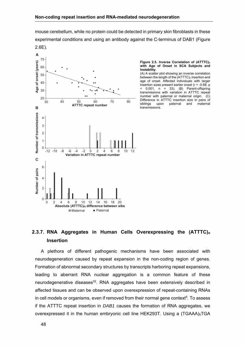
Non-coding repeat insertion and RNA-mediated neurodegeneration
48
mouse cerebellum, while no protein could be detected in primary skin fibroblasts in these
experimental conditions and using an antibody against the C-terminus of DAB1 (Figure
2.6E).
Figure 2.5. Inverse Correlation of (ATTTC)n with Age of Onset in SCA Subjects and Instability. (A) A scatter plot showing an inverse correlation between the length of the (ATTTC)n insertion and age of onset. Affected individuals with larger insertion sizes present earlier onset (r = -0.68, p < 0.001, n = 33). (B) Parent-offspring transmissions with variation in ATTTC repeat number with paternal or maternal origin. (C) Difference in ATTTC insertion size in pairs of siblings upon paternal and maternal transmissions.
2.3.7. RNA Aggregates in Human Cells Overexpressing the (ATTTC)n
Insertion
A plethora of different pathogenic mechanisms have been associated with
neurodegeneration caused by repeat expansion in the non-coding region of genes.
Formation of abnormal secondary structures by transcripts harboring repeat expansions,
leading to aberrant RNA nuclear aggregation is a common feature of these
neurodegenerative diseases32. RNA aggregates have been extensively described in
affected tissues and can be observed upon overexpression of repeat-containing RNAs
in cell models or organisms, even if removed from their normal gene context9. To assess
if the ATTTC repeat insertion in DAB1 causes the formation of RNA aggregates, we
overexpressed it in the human embryonic cell line HEK293T. Using a (TGAAA)5TGA

Chapter 2| A non-coding ATTTC repeat insertion causes neurodegeneration by RNA toxicity
49
probe predicted to hybridize to (AUUUC)n, we observed widespread formation of RNA
aggregates detected by FISH 48h post-transfection of a plasmid containing the repeat
insertion with its flanking ATTTTs and AluJb monomers, Ins(ATTTC)58, while none were
detected in cells transfected with the corresponding normal N(ATTTT)7 or N(ATTTT)139
sequences (Figure 2.7A). These RNA aggregates had generally a nuclear localization
(Figure 2.7B). Enzymatic treatment of the cells with RNase, but not with DNase, led to
the disappearance of the FISH signal, suggesting that the labelled probe was hybridizing
specifically to RNA (Figure 2.7C).
2.3.8. (AUUUC)n-Containing RNA Impairs Early Embryonic Development
To investigate if the (ATTTC)n repeat insertion is deleterious in vivo, we injected 1 to
2 cell stage zebrafish embryos with RNA containing the pathological DAB1 repeat
insertion with its flanking AUUUUs and AluJb monomers and assessed animal viability,
morphology and survival. We injected RNA obtained from the normal alleles N(ATTTT)7
and N(ATTTT)139 and from the pathological Ins(ATTTC)58. At 24 hpf, the lethality rate
detected in the Ins(AUUUC)58 injected embryos was significantly increased when
compared with the other conditions (Figure 2.8A; % of lethality: Control RNA-21.51%;
N(AUUUU)7-18.51%; N(AUUUU)139-14.40%; Ins(AUUUC)58-58.79%; average of 3
replicas with at least 200 embryos/replica; χ2 test p≤0.001). This difference was even
higher when discriminating phenotypes in the surviving embryos, showing that the
number of embryos that developed normally in the Ins(AUUUC)58 injection was
significantly lower when compared with the other conditions (Figure 2.8A; % of normal
embryos: Control RNA-77.52%; N(AUUUU)7-79.79%; N(AUUUU)139-85.37%;
Ins(AUUUC)58-7.76%; χ2 test p<0.0001). Figure 2.8B shows the distribution of
phenotypic classes observed in Ins(AUUUC)58 injected embryos. These results further
suggest that the DAB1 repeat insertion identified in this SCA is potentially deleterious in
vivo.

Non-coding repeat insertion and RNA-mediated neurodegeneration
50
Figure 2.6. Expression of DAB1 Transcripts Spanning the Region Containing the Repeat Insertion and DAB1 protein. (A) Schematics of DAB1 genomic position on chromosome 1, showing transcripts identified by CAGE (FAMTOM5) that result from alternative promoter usage. These transcripts are also annotated in ENSEMBL or UCSC databases (hg19) and all have the repeat insertion region in the 5’-UTR. Coding and non-coding exons in transcripts are represented by closed and open boxes, respectively; the location of the (ATTTC)n insertion region in transcript variants is represented by a diamond and indicated with an arrow; ATG start codon and TAG stop codon locations are shown; PID = phosphotyrosine interaction domain. (B) Mean expression levels of DAB1 transcripts in different CNS regions, skin fibroblasts and B lymphoblastoid cells from human adult analysed from CAGE data29. (C) Mean expression levels of DAB1 transcript variants in CNS regions of 20 to 29 weeks-old human fetus. (D) Mean developmental expression levels of Dab1 transcripts in mouse cerebellum from embryonic day 11 (E11) to postnatal day 9 (P9) analysed from CAGE data30; samples available for expression analysis of each CNS region or cell type ranged from 1 to 3 in human adult and from 1 to 2 in human fetus, while in mouse cerebellum 3 samples were available for all stages; represented is the mean ± SD. E) Western blot analysis of DAB1 protein expression in adult human and mouse cerebellum, and in primary skin fibroblasts. DAB1 protein from HEK293T cells overexpressing human DAB1 cDNA plasmid (pCMV6-hDAB1, OriGene Technologies) was used as control. Boxes indicate cropped areas from digital images of the same membrane obtained with different exposure times (30 seconds for the first lane and five minutes for the other three lanes).

Chapter 2| A non-coding ATTTC repeat insertion causes neurodegeneration by RNA toxicity
51
Figure 2.7. Formation of (AUUUC)n RNA Aggregates in a Human Cell Line. (A) Two normal Alu fragments with a simple ATTTT of different size and the pathogenic ATTTC repeat insert (with the configuration (ATTTT)57(ATTTC)58(ATTTT)73) were directly amplified from genomic DNA and cloned into the pCDH-CMV-EF1-GFP-Puro vector. (B) Transient overexpression of the pathogenic ATTTC repeat insertion, but not the normal ATTTT repeat of 7 or 139 units, in HEK293T cells leads to widespread formation of nuclear RNA aggregates visible after FISH staining with a probe (TGAAA)5TGA predicted to hybridize to (AUUUC)n. GFP expression was used as a marker for transfection. Represented are single-plane confocal images. (C) Aggregates, variable in size and number in individual cells, were sensitive to enzymatic treatment with RNase but not DNase. Representative images are projections of z-stacked images collected at 0.21 m intervals with a confocal microscope. (B, C) Unlabelled scale bar is 5 m.

Non-coding repeat insertion and RNA-mediated neurodegeneration
52
2.4. Discussion
We report the identification of an autosomal-dominant SCA, which is characterized by
pure cerebellar ataxia, with dysarthria as the first clinical manifestation, caused by an
insertion of an unstable ATTTC repeat in the non-coding region of the
neurodevelopmental DAB1, reelin adaptor protein gene, which locates to the mapped
SCA37 locus. The mutation consists in an insertion of an ATTTC repeat in the middle of
an (ATTTT)n tract. The (ATTTC)n insertion (with variable size, ranging from 31 to 75 pure
ATTTC units) was found in affected individuals and absent in 520 control chromosomes.
It completely segregates with the disease in the six families studied. The DAB1 mutation
leads to an aberrant behavior of RNAs containing the AUUUC repeat insertion, rendering
them prone to aggregation in a human cell line. In vivo assessment of the pathogenicity
of this DAB1 insertion suggests that this abnormal RNA is toxic to zebrafish embryos,
causing developmental defects and increased lethality.
The systematic epidemiological survey of hereditary ataxias performed in
Portugal has allowed the genetic characterization of a large number of families3. All
affected subjects with this genetic type of SCA are originally from southern Portugal. It
is interesting to notice that while in the archipelago of Azores and in northern and center
mainland Portugal Machado-Joseph disease (MJD)/SCA3 is the most frequent, it is
rarely found in the south3. In the southern provinces of the country the SCA described
here is the most common, representing 14% of autosomal-dominant forms.
It is remarkable that an (ATTTC)n of 31 to 75 units is disease-causing, whereas
an (ATTTT)n of much larger size that can reach 400 repeats is not pathogenic. The
(ATTTT)n length in healthy individuals ranged from 7-400 pentanucleotide units, whereas
the ATTTT repeat tracts that flank the (ATTTC)n insertion in affected individuals from this
study are larger than 58 units. The total pentanucleotide repeat size for the complex
(ATTTT)n(ATTTC)n(ATTTT)n assessed in this cohort varies from approximately 190 to
220 units. For these sizes, the RP-PCR can efficiently detect the repeat insertion in the
middle of this complex repeat, but larger flanking (ATTTT)n sizes would not allow RP-
PCR insertion detection; sequencing is required for confirmation of its presence.
We observed significant intergenerational instability of the (ATTTC)n insertion, a
feature common to most repeat expansion disseases8,33. This repeat insertion is more
unstable when the father is the transmitting parent, with larger increase in size when
compared to maternal transmissions. This suggests that meiotic instability may play a
relevant role in this dynamic feature, but we cannot exclude the possibility of mitotic
instability during development and aging causing somatic mosaicism in the brain regions
affected.

Chapter 2| A non-coding ATTTC repeat insertion causes neurodegeneration by RNA toxicity
53
Notably, in this cohort there is an inverse correlation between age of disease onset
and the size of the (ATTTC)n insertion, with larger repeats resulting in earlier clinical
manifestation, a feature of many repeat expansion disorders. In fact, the repeat insertion
size accounts for approximately 50% of the variability in age of onset (from late
adolescence to the early 60s). Interestingly, recent evidence from a mouse model of
SCA1 points to the existence of neuronal dysfunction, from development to adulthood,
preceding the appearance of the neurological symptoms and neuronal cell death34. Here
we identified a mutation in a neurodevelopmental gene causing SCA, raising the
possibility that neuronal dysfunction starting early in brain development may contribute
to disease pathogenesis.
The expression of the DAB1, reelin adaptor protein gene is tightly regulated during
development, and across various brain regions. In the human CNS, expression analysis
showed alternative promoter usage for DAB1 and region-specific expression of different
transcripts. The (ATTTC)n insertion region reported here is included in all four transcripts
identified in human brain. Three of them encode the same protein of 555 residues (V1,
V2 and V4), but differ in their 5’-UTRs. The promoter of transcript V2 is the most widely
used in the CNS, while the promoters for V1+V3 and V4 are less used in general, except
in the cerebellum. Altogether, this suggests that transcriptional regulation likely plays an
important role in tuning DAB1 function, especially in cerebellum, the brain structure most
affected in this SCA. Future studies will address to what extent the ATTTC repeat
insertion in the non-coding DAB1 may alter its expression and normal function.
Given that injection of one-cell-stage zebrafish embryos with AUUUC repeat RNA led
to lethal developmental malformations causing significant animal death, we hypothesize
that an RNA-mediated mechanism may contribute to the pathogenesis of this SCA. This
RNA-mediated lethality seen in zebrafish embryos might be initiated by different, not
mutually exclusive, mechanisms. Considering also that overexpression of the ATTTC
repeat insertion leads to the formation of nuclear RNA aggregates in human cell lines,
and in light of what we learnt from other non-coding repeat diseases, these aberrant
RNAs can be pathogenic by (1) sequestering critical RNA-binding proteins and
preventing them from performing their normal function32; (2) initiating non-AUG
dependent translation and producing toxic peptides19,35; and (3) disrupting RNA
metabolism processes such as splicing or nuclear export8. A possible pathological
contribution of bidirectional transcription across the repeat insertion region, described in
several repeat diseases8,15, remains to be investigated.

Non-coding repeat insertion and RNA-mediated neurodegeneration
54
Figure 2.8. In vivo Deleterious Effects of the (ATTTC)n Insertion. (A) Percentage of embryos that presented lethality (Dead), developmental defects (Defects) or a wild type phenotype (Normal) at 24 hpf in RNA injection of control Cas9RNA, N(AUUUU)7, N(AUUUU)139 and Ins(AUUUC)58 (average of 3 replicas with at least 200 embryos/replica; *** p≤0.001; χ2 test for the lethality rate). (B) Distribution of phenotypic classes (a to d) observed in Ins(AUUUC)58
injected embryos at 24hpf and representative images of the observed phenotypic classes comprising wild type (a), severe defects in the tail and head (b), mild defect in the tail (c) and severe defects in the anterior/posterior axis (d).
This form of cerebellar-specific degeneration is caused by a repeat insertion in the
DAB1 gene encoding an adaptor protein of the reelin signaling pathway, which controls
neuronal migration and maturation of synaptic connections in the brain during
development36. Homozygous mutations in the mouse Dab1 gene cause the scrambler
and yotari phenotypes characterized by aberrant splicing forms of Dab1 transcripts and
little or no Dab1 protein causing widespread misplacement of neurons in the cerebellum,
hippocampus and cortex, and the associated ataxia31,36,37. In young-adult mouse
hippocampus, the reelin/DAB1 pathway regulates the maturation of dendritic spines,
synaptogenesis and glial ensheathment of newborn granule cells38. In humans, a
homozygous deletion of the VLDLR [MIM: 192977], that encodes a receptor for reelin,

Chapter 2| A non-coding ATTTC repeat insertion causes neurodegeneration by RNA toxicity
55
originates an autosomal-recessive nonprogressive cerebellar ataxia and mental
impairment [MIM: 224050]39. The presence of cerebellar symptoms is a common feature
when the reelin/DAB1 pathway is impaired; however, the cerebellar atrophy exhibited by
the affected individuals in this study is very mild when compared to the recessive
phenotypes referred above.
It is interesting that the (ATTTC)31-75 insertion in DAB1 intronic 5’UTR is structurally
similar to the (ATTCT)800-4,500 expansion in intron 9 of the ATXN10 gene that causes
SCA1016. The size ranges of the pathological pentanucleotide repeats for fully penetrant
alleles are far apart in these two diseases. One can speculate that these different size
requirements for pathology is related with the specific gene-context and localization in
genomic elements40. Nevertheless, based on the bias towards increased (ATTTC)n size
upon transmission larger alleles may be expected in affected individuals from other
families with this repeat insertion. Notably, alleles of 280 to 850 units may show reduced
penetrance in SCA10 and alleles of 33 to 280 repeats have not been observed41-43.
The DAB1 lies within the mapped SCA37 region on chromosome 1p32, in a family
originating from Spain20. The occurrence of multiple SCA gene mutations clustered in
small genomic regions is rare and only seen in PDYN [MIM: 131340] (SCA23 [MIM:
610245]), NOP56 (SCA36) and TGM6 [MIM: 613900] (SCA35 [MIM: 613908]), which are
contained in a 750 kb region on human chromosome 20p12,44,45. The DAB1 gene
encompasses 1.25 Mb within the SCA37 locus and there is a possibility that the same
ATTTC repeat insertion described here is the mutation in the SCA37 family, especially
since this kindred is from Spain, a country that borders Portugal. Thus, the possibility
exists that the high incidence of DAB1 repeat insertions found in Portuguese SCA
subjects may extend to Spain and likely other populations of European ancestry.
The highly polymorphic (ATTTT)n where the ATTTC insertion occurred is located in
the midle A-rich stretch of an AluJb element. Other repeat diseases, like Friedrich ataxia
[MIM: 229300], DM2, SCA10 and SCA31 originate from expansion or insertion of repeats
in poly-A tracts in the middle or in the 3’ end of Alu elements14,46-48. These A-rich tracts
can be the basis for nucleotide substitution leading to microsatellite birth either by errors
introduced during Alu reverse transcription or by the accumulation of mutations in Alu
poly-A stretches after insertion40.
This is the second neurodegenerative disease (in addition to SCA31), caused by the
insertion in a polymorphic repeat in an A-rich region of an Alu element14. In both cases,
the insertion of the pathogenic repeat is flanked by ATTTT/AAAAT tracts, creating a large
unstable repeat that shares similarities with expanded repeats. These unstable repeat
insertions may thus constitute a new class of dynamic mutations, difficult to identify with
the common strategies currently in use. Furthermore, its occurrence in two genetic types

Non-coding repeat insertion and RNA-mediated neurodegeneration
56
of SCA, suggests that they could be implicated in other brain diseases of unknown
molecular etiology. The discovery of a pathogenic (ATTTC)n insertion in DAB1 could
contribute towards a better understanding of neurodegenerative diseases.
2.5. Acknowledgments
We thank the families who participated in this study. We are very grateful to Gonçalo
Abecasis, Miguel Costa, Tito Vieira, André Torres for their help with Merlin analysis;
Beatriz Sobrino, Jorge Amigo, Pilar Cacheiro for support with NGS sequencing,
performed at the Santiago de Compostela node of the Spanish National Genotyping
Center (CEGEN-ISCIII), or NGS data analysis; Nuno Santarém and Anabela Cordeiro-
da-Silva for assistance with cloning; António Amorim, Laura Vilarinho and Paula Jorge
for the anonymized DNA samples from the Portuguese population and Paula Magalhães
from the IBMC Cell Culture and Genotyping Core for assistance with DNA extraction.
This work was financed by FEDER - Fundo Europeu de Desenvolvimento Regional funds
through the COMPETE 2020 - Operational Programme for Competitiveness and
Internationalisation (POCI), Portugal 2020, and by Portuguese funds through FCT -
Fundação para a Ciência e a Tecnologia/ Ministério da Ciência, Tecnologia e Inovação
in the framework of the project "Institute for Research and Innovation in Health Sciences"
(POCI-01-0145-FEDER-007274); by FCT Grant PTDC/SAU-GMG/098305/2008 to I.S.
A.I.S. was the recipient of scholarships from FCT (SFRH/BD/30702/2006) and from FCT
Grant PTDC/GMG-SAU/098305/2008. J.R.L. was supported by scholarships from FCT
Grant PTDC/GMG-SAU/098305/2008, PEst-C/SAU/LA0002/2013 and EMBO
(ASTF494-2015). A.O.U. was the recipient of a scholarship from FCT Grant
PIC/IC/82897/2007. C.L.O was supported by a scholarship from PEst-
C/SAU/LA0002/2013. This work was also financed by Norte-01-0145-FEDER-000008 -
Porto Neurosciences and Neurologic Disease Research Initiative at I3S, supported by
Norte Portugal Regional Operational Programme (NORTE 2020), under the PORTUGAL
2020 Partnership Agreement, through the European Regional Development Fund
(FEDER), Portugal. Fondo de Investigación Sanitaria-Instituto de Salud Carlos III grant
PI12/00742, Spain.

Chapter 2| A non-coding ATTTC repeat insertion causes neurodegeneration by RNA toxicity
57
2.6. Web Resources
The URLs for data presented herein are as follow:
1000 Genomes, http://www.1000genomes.org/
dbSNP, http://www.ncbi.nlm.nih.gov/SNP/
Ensemble Genome Browser, http://www.ensembl.org/index.html
Genome Analysis Toolkit (GATK), http://www.broadinstitute.org/genome-analysis-
toolkit
Online Mendelian Inheritance in Man (OMIM), http://www.omim.org
UCSC Genome Browser, http://genome.ucsc.edu/
GenBank, http://www.ncbi.nlm.nih.gov/genbank/
FANTOM, http://fantom.gsc.riken.jp/Zenbu/
2.7. References
1. Sequeiros, J., Martins, S., and Silveira, I. (2012). Epidemiology and population
genetics of degenerative ataxias. In Handb Clin Neurol. (Netherlands), pp 227-251.
2. Durr, A. (2010). Autosomal dominant cerebellar ataxias: polyglutamine expansions
and beyond. Lancet Neurol 9, 885-894.
3. Coutinho, P., Ruano, L., Loureiro, J., Cruz, V., Barros, J., Tuna, A., Barbot, C.,
Guimaraes, J., Alonso, I., Silveira, I., et al. (2013). Hereditary Ataxia and Spastic
Paraplegia in Portugal A Population-Based Prevalence Study. JAMA Neurol 70,
746-755.
4. Silveira, I., Miranda, C., Guimaraes, L., Moreira, M., Alonso, I., Mendonca, P., Ferro,
A., Pinto-Basto, J., Coelho, J., Ferreirinha, F., et al. (2002). Trinucleotide repeats
in 202 families with ataxia - A small expanded (CAG)(n) allele at the SCA17 locus.
Arch Neurol 59, 623-629.
5. Carlson, K.M., Andresen, J.M., and Orr, H.T. (2009). Emerging pathogenic pathways
in the spinocerebellar ataxias. Curr Opin Genet Dev 19, 247-253.
6. Coutelier, M., Blesneac, I., Monteil, A., Monin, M.L., Ando, K., Mundwiller, E., Brusco,
A., Le Ber, I., Anheim, M., Castrioto, A., et al. (2015). A Recurrent Mutation in

Non-coding repeat insertion and RNA-mediated neurodegeneration
58
CACNA1G Alters Cav3.1 T-Type Calcium-Channel Conduction and Causes
Autosomal-Dominant Cerebellar Ataxia. Am J Hum Genet 97, 726-737.
7. Di Gregorio, E., Borroni, B., Giorgio, E., Lacerenza, D., Ferrero, M., Lo Buono, N.,
Ragusa, N., Mancini, C., Gaussen, M., Calcia, A., et al. (2014). ELOVL5 mutations
cause spinocerebellar ataxia 38. Am J Hum Genet 95, 209-217.
8. Loureiro, J.R., Oliveira, C.L., and Silveira, I. (2016). Unstable repeat expansions in
neurodegenerative diseases: nucleocytoplasmic transport emerges on the scene.
Neurobiol Aging 39, 174-183.
9. Mankodi, A., Logigian, E., Callahan, L., McClain, C., White, R., Henderson, D., Krym,
M., and Thornton, C.A. (2000). Myotonic dystrophy in transgenic mice expressing
an expanded CUG repeat. Science 289, 1769-1773.
10. La Spada, A.R., and Taylor, J.P. (2010). Repeat expansion disease: progress and
puzzles in disease pathogenesis. Nat Rev Genet 11, 247-258.
11. Wojciechowska, M., and Krzyzosiak, W.J. (2011). Cellular toxicity of expanded RNA
repeats: focus on RNA foci. Hum Mol Genet 20, 3811-3821.
12. Kobayashi, H., Abe, K., Matsuura, T., Ikeda, Y., Hitomi, T., Akechi, Y., Habu, T., Liu,
W., Okuda, H., and Koizumi, A. (2011). Expansion of intronic GGCCTG
hexanucleotide repeat in NOP56 causes SCA36, a type of spinocerebellar ataxia
accompanied by motor neuron involvement. Am J Hum Genet 89, 121-130.
13. Garcia-Murias, M., Quintans, B., Arias, M., Seixas, A.I., Cacheiro, P., Tarrio, R.,
Pardo, J., Millan, M.J., Arias-Rivas, S., Blanco-Arias, P., et al. (2012). 'Costa da
Morte' ataxia is spinocerebellar ataxia 36: clinical and genetic characterization.
Brain 135, 1423-1435.
14. Sato, N., Amino, T., Kobayashi, K., Asakawa, S., Ishiguro, T., Tsunemi, T.,
Takahashi, M., Matsuura, T., Flanigan, K.M., Iwasaki, S., et al. (2009).
Spinocerebellar ataxia type 31 is associated with "inserted" penta-nucleotide
repeats containing (TGGAA)n. Am J Hum Genet 85, 544-557.
15. Moseley, M.L., Zu, T., Ikeda, Y., Gao, W., Mosemiller, A.K., Daughters, R.S., Chen,
G., Weatherspoon, M.R., Clark, H.B., Ebner, T.J., et al. (2006). Bidirectional
expression of CUG and CAG expansion transcripts and intranuclear polyglutamine
inclusions in spinocerebellar ataxia type 8. Nat Genet 38, 758-769.
16. Matsuura, T., Yamagata, T., Burgess, D.L., Rasmussen, A., Grewal, R.P., Watase,
K., Khajavi, M., McCall, A.E., Davis, C.F., Zu, L., et al. (2000). Large expansion of

Chapter 2| A non-coding ATTTC repeat insertion causes neurodegeneration by RNA toxicity
59
the ATTCT pentanucleotide repeat in spinocerebellar ataxia type 10. Nat Genet
26, 191-194.
17. Cleary, J.D., and Ranum, L.P. (2017). New developments in RAN translation: insights
from multiple diseases. Curr Opin Genet Dev 44, 125-134.
18. Pearson, C.E. (2011). Repeat associated non-ATG translation initiation: one DNA,
two transcripts, seven reading frames, potentially nine toxic entities! PLoS Genet
7, e1002018.
19. Ishiguro, T., Sato, N., Ueyama, M., Fujikake, N., Sellier, C., Kanegami, A., Tokuda,
E., Zamiri, B., Gall-Duncan, T., Mirceta, M., et al. (2017). Regulatory Role of RNA
Chaperone TDP-43 for RNA Misfolding and Repeat-Associated Translation in
SCA31. Neuron 94, 108-124 e107.
20. Serrano-Munuera, C., Corral-Juan, M., Stevanin, G., San Nicolas, H., Roig, C.,
Corral, J., Campos, B., de Jorge, L., Morcillo-Suarez, C., Navarro, A., et al. (2013).
New subtype of spinocerebellar ataxia with altered vertical eye movements
mapping to chromosome 1p32. JAMA Neurol 70, 764-771.
21. Rüschendorf, F., and Nürnberg, P. (2005). ALOHOMORA: a tool for linkage analysis
using 10K SNP array data. Bioinformatics 21, 2123-2125.
22. Wigginton, J.E., and Abecasis, G.R. (2005). PEDSTATS: descriptive statistics,
graphics and quality assessment for gene mapping data. Bioinformatics 21, 3445-
3447.
23. Abecasis, G.R., Cherny, S.S., Cookson, W.O., and Cardon, L.R. (2002). Merlin--
rapid analysis of dense genetic maps using sparse gene flow trees. Nat Genet 30,
97-101.
24. McKenna, A., Hanna, M., Banks, E., Sivachenko, A., Cibulskis, K., Kernytsky, A.,
Garimella, K., Altshuler, D., Gabriel, S., Daly, M., et al. (2010). The Genome
Analysis Toolkit: a MapReduce framework for analyzing next-generation DNA
sequencing data. Genome Res 20, 1297-1303.
25. Schindelin, J., Arganda-Carreras, I., Frise, E., Kaynig, V., Longair, M., Pietzsch, T.,
Preibisch, S., Rueden, C., Saalfeld, S., Schmid, B., et al. (2012). Fiji: an open-
source platform for biological-image analysis. Nat Methods 9, 676-682.
26. Hwang, W.Y., Fu, Y., Reyon, D., Maeder, M.L., Tsai, S.Q., Sander, J.D., Peterson,
R.T., Yeh, J.R., and Joung, J.K. (2013). Efficient genome editing in zebrafish using
a CRISPR-Cas system. Nat Biotechnol 31, 227-229.

Non-coding repeat insertion and RNA-mediated neurodegeneration
60
27. Mitsuhashi, H., Mitsuhashi, S., Lynn-Jones, T., Kawahara, G., and Kunkel, L.M.
(2013). Expression of DUX4 in zebrafish development recapitulates
facioscapulohumeral muscular dystrophy. Hum Mol Genet 22, 568-577.
28. Bar, I., Tissir, F., Lambert de Rouvroit, C., De Backer, O., and Goffinet, A.M. (2003).
The gene encoding disabled-1 (DAB1), the intracellular adaptor of the Reelin
pathway, reveals unusual complexity in human and mouse. J Biol Chem 278, 5802-
5812.
29. Forrest, A.R., Kawaji, H., Rehli, M., Baillie, J.K., de Hoon, M.J., Haberle, V.,
Lassmann, T., Kulakovskiy, I.V., Lizio, M., Itoh, M., et al. (2014). A promoter-level
mammalian expression atlas. Nature 507, 462-470.
30. Arner, E., Daub, C.O., Vitting-Seerup, K., Andersson, R., Lilje, B., Drablos, F.,
Lennartsson, A., Ronnerblad, M., Hrydziuszko, O., Vitezic, M., et al. (2015).
Transcribed enhancers lead waves of coordinated transcription in transitioning
mammalian cells. Science 347, 1010-1014.
31. Howell, B.W., Gertler, F.B., and Cooper, J.A. (1997). Mouse disabled (mDab1): a Src
binding protein implicated in neuronal development. EMBO J 16, 121-132.
32. Todd, P.K., and Paulson, H.L. (2010). RNA-mediated neurodegeneration in repeat
expansion disorders. Ann Neurol 67, 291-300.
33. Silveira, I., Alonso, I., Guimarães, L., Mendonça, P., Santos, C., Maciel, P., Fidalgo
De Matos, J.M., Costa, M., Barbot, C., Tuna, A., et al. (2000). High germinal
instability of the (CTG)n at the SCA8 locus of both expanded and normal alleles.
Am J Hum Genet 66, 830-840.
34. Hatanaka, Y., Watase, K., Wada, K., and Nagai, Y. (2015). Abnormalities in synaptic
dynamics during development in a mouse model of spinocerebellar ataxia type 1.
Sci Rep 5, 16102.
35. Zu, T., Gibbens, B., Doty, N.S., Gomes-Pereira, M., Huguet, A., Stone, M.D.,
Margolis, J., Peterson, M., Markowski, T.W., Ingram, M.A., et al. (2011). Non-ATG-
initiated translation directed by microsatellite expansions. Proc Natl Acad Sci USA
108, 260-265.
36. Sheldon, M., Rice, D.S., D'Arcangelo, G., Yoneshima, H., Nakajima, K., Mikoshiba,
K., Howell, B.W., Cooper, J.A., Goldowitz, D., and Curran, T. (1997). Scrambler
and yotari disrupt the disabled gene and produce a reeler-like phenotype in mice.
Nature 389, 730-733.

Chapter 2| A non-coding ATTTC repeat insertion causes neurodegeneration by RNA toxicity
61
37. Goldowitz, D., Cushing, R.C., Laywell, E., D'Arcangelo, G., Sheldon, M., Sweet, H.O.,
Davisson, M., Steindler, D., and Curran, T. (1997). Cerebellar disorganization
characteristic of reeler in scrambler mutant mice despite presence of reelin. J
Neurosci 17, 8767-8777.
38. Bosch, C., Masachs, N., Exposito-Alonso, D., Martinez, A., Teixeira, C.M., Fernaud,
I., Pujadas, L., Ulloa, F., Comella, J.X., DeFelipe, J., et al. (2016). Reelin Regulates
the Maturation of Dendritic Spines, Synaptogenesis and Glial Ensheathment of
Newborn Granule Cells. Cereb Corte 26, 4282-4298.
39. Boycott, K.M., Flavelle, S., Bureau, A., Glass, H.C., Fujiwara, T.M., Wirrell, E., Davey,
K., Chudley, A.E., Scott, J.N., McLeod, D.R., et al. (2005). Homozygous deletion
of the very low density lipoprotein receptor gene causes autosomal recessive
cerebellar hypoplasia with cerebral gyral simplification. Am J Hum Genet 77, 477-
483.
40. Kelkar, Y.D., Eckert, K.A., Chiaromonte, F., and Makova, K.D. (2011). A matter of
life or death: how microsatellites emerge in and vanish from the human genome.
Genome Res 21, 2038-2048.
41. Alonso, I., Jardim, L., Artigalas, O., Saraiva-Pereira, M., Matsuura, T., Ashizawa, T.,
Sequeiros, J., and Silveira, I. (2006). Reduced penetrance of intermediate size
alleles in spinocerebellar ataxia type 10. Neurology 66, 1602-1604.
42. Seixas, A., Maurer, M., Lin, M., Callahan, C., Ahuja, A., Matsuura, T., Ross, C.,
Hisama, F., Silveira, I., and Margolis, R. (2005). FXTAS, SCA10, and SCA17 in
American patients with movement disorders. Am J Med Genet A 136, 87-89.
43. Matsuura, T., Fang, P., Pearson, C.E., Jayakar, P., Ashizawa, T., Roa, B.B., and
Nelson, D.L. (2006). Interruptions in the expanded ATTCT repeat of
spinocerebellar ataxia type 10: repeat purity as a disease modifier? Am J Hum
Genet 78, 125-129.
44. Bakalkin, G., Watanabe, H., Jezierska, J., Depoorter, C., Verschuuren-Bemelmans,
C., Bazov, I., Artemenko, K.A., Yakovleva, T., Dooijes, D., Van de Warrenburg,
B.P., et al. (2010). Prodynorphin mutations cause the neurodegenerative disorder
spinocerebellar ataxia type 23. Am J Hum Genet 87, 593-603.
45. Wang, J.L., Yang, X., Xia, K., Hu, Z.M., Weng, L., Jin, X., Jiang, H., Zhang, P., Shen,
L., Guo, J.F., et al. (2010). TGM6 identified as a novel causative gene of
spinocerebellar ataxias using exome sequencing. Brain 133, 3510-3518.

Non-coding repeat insertion and RNA-mediated neurodegeneration
62
46. Clark, R.M., Dalgliesh, G.L., Endres, D., Gomez, M., Taylor, J., and Bidichandani,
S.I. (2004). Expansion of GAA triplet repeats in the human genome: unique origin
of the FRDA mutation at the center of an Alu. Genomics 83, 373-383.
47. Kurosaki, T., Matsuura, T., Ohno, K., and Ueda, S. (2009). Alu-mediated acquisition
of unstable ATTCT pentanucleotide repeats in the human ATXN10 gene. Mol Biol
Evol 26, 2573-2579.
48. Kurosaki, T., Ueda, S., Ishida, T., Abe, K., Ohno, K., and Matsuura, T. (2012). The
unstable CCTG repeat responsible for myotonic dystrophy type 2 originates from
an AluSx element insertion into an early primate genome. PLoS One 7, e38379.

Chapter 2| A non-coding ATTTC repeat insertion causes neurodegeneration by RNA toxicity
63
2.8. Supplemental data
Fig S2.1. PCR and Southern blot for (ATTTT)n in three large families. Related to Figure 2.2. (A) Standard PCR amplification shows no apparent (ATTTT)n transmission from all affected parent to affected offspring, (ATTTT)n size was assessed by sanger sequencing and is indicated below each lane in the agarose gel image. (B) Southern blot analysis with a probe hybridizing near the pentanucleotide repeat ATTTT/AAAAT in DAB1 showing a fragment of approximately 5.1 kb corresponding to ~200 pentanucleotide repeats that is transmitted from the affected parent to affected offspring. Individual ID number is according to individual ID number in each pedigree of Figure 2.1.

Non-coding repeat insertion and RNA-mediated neurodegeneration
64
Figure S2.2. Cerebellar Expression of DAB1 transcripts with the (ATTTC)n Insertion Intron. Related to Figure 2.6. Bellow is the RT-PCR analysis of DAB1 transcripts from human total cerebellum RNA (Clontech). PCR products correspond to amplification with specific primers, represented with arrows, in the schematic representation above of DAB1 transcripts, for the first and last exons of variant 1 (V1), V2, V3 and V4.

Chapter 2| A non-coding ATTTC repeat insertion causes neurodegeneration by RNA toxicity
65
Table S2.1. Primers used for PCR, Sequencing, Long range PCR, RP-PCR, Southern Blot, rt-PCR and T7 promoter cloning
Primer ID Primer sequence (5'-3')
(ATTTT)n screening
24F GAAGTGGTCCTCCCAAGTCA
24R ACACTTTGGGAGGCAGAGG
Sequencing primers
24F GAAGTGGTCCTCCCAAGTCA
24R4 GAGACCAGCCTGGGCAAC
Long PCR
ALU24F ATTTGCCCTTTGCTGATTGA
ALU24R TGAAACTGAGGCTCAAAATGA
RP-PCR
24R [6FAM]ACACTTTGGGAGGCAGAGG
RP-TTCAT TACGCATCCCAGTTTGAGACGTTCATTTCATTTCATTTCATTTCAT
FLAG TACGCATCCCAGTTTGAGACG
AluJb (ATTTT)n and AluJb (ATTTC)n allele Cloning
ALUEcoRIF GTCAGTGAATTCAATTAATTTGCCCTTTGCTGATT
ALUNotIR GTCAGTGCGGCCGCTGAAACTGAGGCTCAAAATGA
Southern Blot probe
SB24F CAGGAGGGAGGTGCTTCTG
SB24R TCACACAAAATGGTACTCTGAAAAA
rt-PCR full length DAB1 transcript variants
V1F GAGGAGGATGCTCTGGGCTA
V1R TCCCAGACCTGCGCTATCTA
V2F CCCAGGGAACAAAAGCGGA
V2R TTGATCTGCGCGTACAGAGG
V3F AGGAGGATGCTCTGGGCTAGG
V3R CCAGGTTTTGCAGTCAGTGGA
V4F GTTCGGCTCATAAAGCAGGC
V4R CACTGGGCTCACCAAATGGA
T7 promoter cloning in pCDH-CMV-MCS-EF1-GFP-T2A-Puro
T7A CTAGGGATCCTAATACGACTCACTATAGGG
T7B AATTCCCTATAGTGAGTCGTATTAGGATCC

Non-coding repeat insertion and RNA-mediated neurodegeneration
66
Table S2.2. Repeat Sequences Studied in SCA Families Linked to Chr 1
Gene Copies Sequence hg19
(strand plus) Location Position Chr1 hg19 Primer F Primer R Polymorphic
AK127270 2.1 ATTTCTCTGATGGTCA intron 1 56,062,233-56,062,266 TGCTTTCCAAAATGGCTGTA GAGCAGAAGACCTGAATAGA
TGTTT NO
AK127270 2.2 AAAGGAAAGGTACATTTAT intron 1 56,068,484-56,068,524 GCACCAGAATATGCCACCTT GAGCTGTCCCAAGGAGTAG
C NO
AK127270 1.9 GTGTGTGTATGTGC intron 2 56,093,994-56,094,020 ATTTGCCAGTGGCCAGTTAG GAATTTTTCAGTGCCATTGT
GA NO
AK127270 2 TAATTAGATATTAA intron 2 56,094,897-56,094,924 TGCTTCTCCTCTACTGTATTCTT
CTC AAATGGTTCTCCCAATGGTA
A NO
AK127270 2 TAAAAAGGAAGAAAGAGAAAA intron 2 56,124,544-56,124,584 CCCAGGCTAAAGCTTCCTTC TTCCCTCTGTAGCCTTTCTCA NO
AK127270 2.1 CAGTTTCATTCTTCTA intron 2 56,127,033-56,127,066 TTCTTTGCCTAACCCAATGTC TGGTTCTAGGAAAAGTGGCA
AG NO
AK127270 4.9 ACATATATAC intron 2 56,165,413-56,165,461 GGTTCTCCAGAGATACAGAGTC
AA CTTACCAGCCCCCATAACTG NO
AK127270 2.1 TATATAATGTATAATTTACATAA intron 2 56,168,155-56,168,202 CAGACTCAGGCCAATATCAGA CATGCATCCCAGTGTCTCC NO
AK127270 2.9 AGAGAAGAAGAGGGAAGGA intron 2 56,169,302-56,169,360 AGGGGATAAGGCCTGAGAAG AGGAGTTCAAAGCCCCAAAT NO
AK127270 2 CTCTGGGCCAACAGAGAA intron 2 56,174,094-56,174,129 TGTGAGGTGAGGGATGACAG ATGGAGTGGATGATGCCTTC NO
AK127270 3.1 ATAGTAGACATAAACAAGTAA intron 2 56,175,575-56,175,639 TCTGGGCATTGAGGTTACAA GGAAATCATCTCTCCCACTC
C NO
AK127270 1.9 TTTTTTTTCTTTTTC intron 2 56,191,420-56,191,448 TCCGCATCTACTATCAGCACA CAGCTATGTGGGAGGCTGA
G YES
AK127270 2.4 AAAACAAATAGTAAATCAA intron 2 56,196,781-56,196,825 AGCTGGCAGAACCTTCACAT GAATCATCACCCCTGCATCT NO
C1Orf 168 1.9 CATCTTTCTTCCCATA intron 2 57,256,201-57,256,231 TTGGGCTCAGAGTTTTTACCA TGATGAAGGGATGAGGAAGA
A NO
C1Orf 168 2.5 AAACAAACAAACAAAAACAA intron 1 57,258,495-57,258,543 GGAAGAGGTGGAGCATCAAG CTAACATTGCCCAGAAGCAA NO
C1Orf 168 9.5 AATA intron 1 57,270,403-57,270,440 CGAGATGGCACCACTACACT AGTGGTGGGAAATGAAGCTG YES
C1Orf 168 1.9 GTCTTTATCAGCAT intron 1 57,271,601-57,271,627 CCCAGCCATATGGAACTGTAA TGTGTCTGCCTCATGCATTT NO

Chapter 2| A non-coding ATTTC repeat insertion causes neurodegeneration by RNA toxicity
67
Gene Copies Sequence hg19
(strand plus) Location Position Chr1 hg19 Primer F Primer R Polymorphic
C1Orf 168 2.1 AGAGAAAGAAGGAGGAGGAAG
GGGAGAAGGA intron 1 57,275,692-57,275,758 ACATTAACCTTGGCCTGGTG
TCTGCCACAGTCATTTCATCA
NO
C1Orf 168 7 CAAA intron 1 57,277,429-57,277,456 CCCAATCCAATGCTATTTCG CCACCTTCCACACCCTTTTA NO
C8A 16.2 TCCC intron 1 57,329,495-57,329,561 TGCCTATATCCCAGAAATTGGT GTTCCCAGCACCTATCATGG YES
C8A 6.1 TCCTTTCTTCCT intron 1 57,329,549-57,329,623 TGCCTATATCCCAGAAATTGGT GTTCCCAGCACCTATCATGG YES
C8A 2.5 AGGTAGGTGATGGGC intron 2 57,341,370-57,341,406 AAAAGCATGGGGATCAGAGA CCAGTCTGCCCATGTAATCC NO
C8A 8.5 GAAT intron 9 57,373,333-57,373,366 TGTGCCCACATTGAGTCCTA CCATCCTCTGGAGTGGAGAA NO
C8A 2.1 TGGCACAGTCCTACTGCTGACT
TGCTC intron 9 57,376,526-57,376,581 CCACAGGCCAAAGTCTGTTT GGAAGTGATTTGCCAAGGTC NO
PPAP2B 3 GCCTTAG intron 4 56,977,441-56,977,461 GCTAGAGAGCCCTCGTTCCT CTACGAGCCCAAAACCTCTG NO
PPAP2B 3.3 AAATAAATAAGT intron 4 56,978,288-56,978,327 CACCACTGCAGTTCGGTCT GCAGTGTGCTAAGGCCATTT NO
PPAP2B 2 GTGGCTCATGCCTGTAATCCCAACACTTTGGGAGGCTGAGGTGG
GTG intron 4 56,986,265-56,986,359 TTTGCAAATCAGCCAAACCT
TTTAATAGAGACAGGGTTTCACCA
NO
PPAP2B 2.3 TCCTCTCTCCCTCTCTCTTCCTT intron 4 56,987,383-56,987,435 CAGGTATAAGCCACCATCCAG GGGATGATGGGTGAGAGGT
AT YES
PPAP2B 1.9 CCAACTAGGCAACTAGT intron 1 57,006,472-57,006,504 AGGGTGATGCCAGTCAAATC TGGAACTAAAGGTTGTTGGA
GAA NO
PPAP2B 2.1 TATCCTTCAAGACTTAAA intron 1 57,010,726-57,010,762 CAACTCCATGAAGTAATGTCTTT
AACA CTGCCTGACTCCAAAGATCA NO
PPAP2B 2 TGTCATTTGAATGCCA intron 1 57,014,508-57,014,539 GATGCCCTCAGCACATGAC TGGGAAGAAAACCTTTTCAG
TT NO
PPAP2B NA TTCGG intron 1 57,016,953-57,017,000 GGAAGGGGAGGGACTTGTAT ACCATTTCACAGCCCATGTT YES
PPAP2B 2.8 AAAAAAAAACAAAAAAAAA intron 1 57,021,107-57,021,158 GGAGAAGGAAGGCCAAACAC TCCATTCAGGGACACAGTGA NO
PPAP2B 15.1 GAGAGAGAAAGA intron 1 57,022,591-57,022,771 TTGGCCAGGATTGTCTTGAT GGCTGAGGTGGATAGATTGC YES
PPAP2B 2 TTCAGGTGCTTTCAATTATAAAA
TTG intron 1 57,025,945-57,025,996 CCTGGTGCATTAAAGGTGCT
CCAGCATCAGCTATGTTTGGT
NO
PPAP2B 2.2 ACATGCATGCATGCACAC intron 1 57,042,467-57,042,503 GGGGATAGTGGGGAAATGAA GCAGTCCCAAGAGATGGTGT NO
PPAP2B NA CGCGG intron 1 57,044,450-57,044,478 CCTCTTCTGCCTCTTCATGG CCCAGACCCACTCAAGTACA
G NO

Non-coding repeat insertion and RNA-mediated neurodegeneration
68
Gene Copies Sequence hg19
(strand plus) Location Position Chr1 hg19 Primer F Primer R Polymorphic
PPAP2B 14 CCT 5'UTR 57,045,139-57,045,180 GCCAGCCCCAACTCTAACTT CCCCCACGTCTTCTCTTTTT NO
PPAP2B 5 CCTCCTGCT 5'UTR 57,045,139-57,045,183 GCCAGCCCCAACTCTAACTT CCCCCACGTCTTCTCTTTTT NO
PRKAA2 9 GATA intron 1 57,115,206-57,115,241 CAGCATAGGGAGACCCTGTC CAGCCTGGTCAACATAGTCC NO
PRKAA2 2.1 AGAGTCTACTGAATGCTAATACA
TTCTCCAACAGTGTTAA intron 1 57,124,141-57,124,223 CGCTTTGGGATAAGTGTTGC
TGAGACCCAGACTCAAAACAAA
NO
PRKAA2 2.3 GAAAGTGCCTTCTTAAGAT intron 1 57,127,854-57,127,896 CATTTGTTAGGCAAGTGTGTCTT TCCCAAATATAATCTGATGTT
TCC NO
PRKAA2 2.4 TGTTTTTTGTTTGTTTGT intron 1 57,128,741-57,128,784 TGTTGCCATGAACTCTGCTC CTACTCAGGGGGCTGAAACA NO
PRKAA2 2.9 TGTTTCTAAATTAGTACTCAATTT
A intron 2 57,140,386-57,140,459
TTTGCATACTCTTGGTTAGACTGAA
TGTCAAAGCCCTGAATGACA NO
PRKAA2 2.1 TTTTTAAAATATAA intron 7 57,165,257-57,165,285 CAAACAATTATGGCCATTTGACT
A ATAGGAGGAAGGGGAAATG
G NO
PRKAA2 2.1 TTTGGTTAATTTCCAGGGTTCTG
AAAAAATTGATTTTCACAGTT intron 7 57,166,693-57,166,784 AAAGCCACAAATCTCACCTTTC
TCAATGACAAATAGAAAAGTTAAGCA
NO
C8B 2.9 TTTTTTCTTTTC intron 10 57,402,250-57,402,284 AGAAAAATTGGCTGGGTGTG AGACTTTCAATCACTGTCCC
TTT NO
C8B 2.4 AATTTAATTGATTA intron 10 57,405,884-57,405,916 TGTTGGTTGAATGCATAATGTTT AGTGATCCTCCCACTTTTGC NO
C8B 82.8 AGAT intron 9 57,406,959-57,407,278 TGCTAGTTTCTGAGTAAGTTTAT
GGA GGCTCCTAAAATTGCTATCT
GG YES
C8B 7.4 AAAAT intron 9 57,408,582-57,408,619 TGAGGGTACCGAGGCTTGTA TCAGGCAATCCTCTCACCTC NO
C8B 5.9 AAAAAAT intron 9 57,408,748-57,408,786 GCTGGGTTTTAGAGGGTGTG GCCCAAGAGTTTGAGACCAG NO
C8B 3.5 CAGGAAGAGATC intron 8 57,411,201-57,411,241 TTCAGCTGGTGAGATTTGCT CCTAGCTCCCTAGGCCATTC NO
C8B 8.8 AAAAC intron 7 57,413,605-57,413,645 GGAGGTGGAGGTTGAAGTGA GGAGCAGCAAACATGAAAGC YES
C8B (-) CCTT intron 4 57,421,539-57,421,878 TCATTTTTGCTGGAGATGGA TCCATGATATACCCGGTAAA
AA NO
C8B-DAB 2.9 ATTAAATAAATTAGATTATAATTA
ATTATT intergenic 57,432,431-57,432,516 TCTGGGCCTAAGTTCCTTCA AATGTGCCAGGCAATGTTC NO
C8B-DAB 7.2 CAAAA intergenic 57,432,984-57,433,018 ACGAGGTCAGGAAATCGAGA GGAGTCAGGGATGGTTTTCA NO

Chapter 2| A non-coding ATTTC repeat insertion causes neurodegeneration by RNA toxicity
69
Gene Copies Sequence hg19
(strand plus) Location Position Chr1 hg19 Primer F Primer R Polymorphic
C8B-DAB 4.7 CATATATATATATAGACATATATA
TATAGA intergenic 57,448,691-57,448,834 CAGGGGTCTTGTGAGAAAATG TCCCACCTTCTCCTTACAGC YES
C8B-DAB 1.9 TTCAGAATCTGAATTC intergenic 57,462,253-57,462,283 AGTGGGGAGGTGGGAGAGT AGCAGCTAGTGAACGGGAA
G NO
DAB1 16 TGA intron 16 57,474,084-57,474,131 GGAGCGTGCATTAATTGGAT TGTGAAGCTCTTAGCACTGT
GT NO
DAB1 2.2 TGAGGTAGCCTCAGAATCCATT
TA intron 16 57,475,209-57,475,260
TGTCTAACTTACGTGTGGGCTAA
AAACACACAGGACTGCAAGG NO
DAB1 2.8 AGCTCAGGCAAGGAGAAATGGT
GCAAACATCAGCAATAC intron 16 57,476,158-57,476,265 GACATGGTGCAGACACTGGT TCTGCTTTCCCTTTTGGTTG NO
DAB1 NA CTG intron 13 57,485,186-57,485,201 CGGCATAAACAGATGCTCCT GCTGTGCACACTTTTCTGGA NO
DAB1 3 CTCTG intron 11 57,500,591-57,500,606 GAAACAGGTGGTTGGTGGTT GAGGAGAGCTTTTGGCAATG NO
DAB1 2.1 TTTAATAAATGG intron 11 57,503,518-57,503,542 CTTTCCCATTTCTTGGCAGA TATTGGACAGGGCTGGTCTC NO
DAB1 2.8 CACACACACACACATACA intron 11 57,508,501-57,508,550 TGGCATTCCATATGTTGTTTTC CCAAATGGCAGGGTAGATGT NO
DAB1 NA CAG intron 11 57,512,335-57,512,350 AAGATGCAGGTTCCCTGATG TGCCAGCATCAATGGATATT
T NO
DAB1 2.1 CATTTAGTCTTTACA intron 11 57,521,655-57,521,686 GTGAATCCTGCCCTCTAGGA TCCACTCTCCATGTCCTCAT
C NO
DAB1 2.6 AAAAAAAAAAAAAG intron 11 57,522,181-57,522,217 GGTAAGCTGAGGCAGGAGGA GCCCACTTCTGCTGAACTTC
T NO
DAB1 8.6 TTTTA intron 11 57,525,799-57,525,841 TCAGGCAGCCCAGATATTTT CTGGGTGACAGAGCCAAACT YES
DAB1 2.1 TGGTTTTTGTTTCTTT intron 6 57,557,518-57,557,550 TGAAACAGTTCCTGGCACCT ATCCCATGTGCCAAAGAGAG NO
DAB1 13.8 TTTA intron 6 57,563,413-57,563,467 TCTTCCATACTTGGAGATCACAG
A CCCCGTCCCTATTAAAAATA
CA NO
DAB1 8 TCTCTG intron 6 57,567,864-57,567,911 ATTTCAATGCAGTGCGGTCT GTGGCAATTGTTTGTGATGG NO
DAB1 NA CAG intron 6 57,568,910-57,568,922 CTGGGAGATCTGGGCCTAA GGCTGGTGAAGAAAAACCAA NO
DAB1 NA GCAG intron 6 57,584,195-57,584,207 CAAGGCAGCTGATGAAGGAT CACCTCCCTCAGCTTAAAAC
A NO
DAB1 17.3 AAGAAGAGG Intron 6 57,586,779-57,586,931 CTTGTGTGAAAGCCATTGGA AAAGGAAGGGGCAAAGAGA
T YES
DAB1 2.3 AAAAAGAAAAAAG intron 6 57,587,228-57,587,256 GGGAAGGGATTCCAAATGTC TCATAATGAAAACCACATTCA
GTTC NO

Non-coding repeat insertion and RNA-mediated neurodegeneration
70
Gene Copies Sequence hg19
(strand plus) Location Position Chr1 hg19 Primer F Primer R Polymorphic
DAB1 10.5 AAAT intron 6 57,593,826-57,593,867 GTTGCAGCAAACAGAGATCG GTGGCTGATTTTTGGGATGA YES
DAB1 8.3 ATGTAT intron 5 57,609,577-57,609,626 TTTGAAAATATTTCATTGACAAA
GTC AAAATTTATACAGTTTCCTGT
CTTGG NO
DAB1 3.1 TAATTATTTATAAC intron 4 57,618,389-57,618,432 TGGTTTCAAAAGTGTCTCCAAG GCACTTACCAACTCAGTTCT
GTTC NO
DAB1 2.1 ATCCTCAATAACTAATC intron 4 57,635,328-57,635,362 CATGCTTTCATCAGGCCTCT AGGGAAGCCAGATACCACCT NO
DAB1 6 TTTTG intron 4 57,647,549-57,647,577 GCTAAGCCCCTTCCTAGCTG AGGCTGAGGCAGGAGTATC
A NO
DAB1 1.9 AATTAACTAAAAT intron 4 57,648,574-57,648,598 TGGGAAAGAATGCAGAAATG GAGGCTGGTGTGACTGAACA NO
DAB1 1.9 AGAGAGATCCCCCACAAAA intron 4 57,653,519-57,653,554 CAACCATTCAGCTAATCCCAGT ATTGCTGCATCCAAAGAGGA NO
DAB1 NA CCA intron 4 57,656,335-57,656,347 AGTGCATGGAGTTGCCTCTT CCTTTCCCCAGTATGCAAAA NO
DAB1 NA CATG intron 4 57,667,553-57,667,579 AGCCTAGCACAGAGCCTGAA CTTTCCTGTTCCTCCACCTG NO
DAB2 NA CTG intron 4 57,668,562-57,668,571 TCTGTTCCCACTGTTTGCTG ACCAAGGCCACCATATCTGA NO
DAB1 2.2 TTCTTTTCTTCTTTCT intron 4 57,672,598-57,672,632 AGACTGCGCCATTACACTCC CCCTGATTTAGCCCCTTTTT YES
DAB1 3.2 AAAATACATGACAG intron 4 57,687,939-57,687,985 CCTTTCCTTCCTTCCTTGCT CAACAGCCATCTCCCTGACT NO
DAB1 1.9 AACTACTTACATCAACATGAATG
G intron 4 57,694,570-57,694,615
TCGTAAACAGAAGACTCGGTAGA
TTTGGAACTTTATAAAAATGGATTCAC
NO
DAB1 1.9 ACATCACTCTGAA intron 4 57,702,398-57,702,422 GCAGAGGAAGCCAACAACTG CCTCCGCATTCTCTCGTAAG NO
DAB1 1.9 ATGCACGTGTGTGTGT intron 4 57,709,144-57,709,174 TGCCCCTCAACTCTCTGTTT AGTGGGTTGGATGCAAGAAC NO
DAB1 NA CTGAA intron 4 57,728,002-57,728,017 ATTAACCATGGCACCCAATG ACTTCCCCAAGGTCACACAG NO
DAB1 10 GCCT intron 4 57,739,190-57,739,229 GCTAGGGGAGCCATCAGAC CTCCAATAAGACCTGGCTGA
A YES
DAB1 15.5 CCTT intron 4 57,739,227-57,739,288 CACAATGCATGTTTGACTCG CCTGGCTGAAGTTGCCT YES
DAB1 2.9 AAGATAGAGAGAAA intron 4 57,740,793-57,740,833 TTGCAAGCAGTTCTGATCCTT TGACAATTTTTAGGCCAGGT
C NO
DAB1 1.9 AAAAATCATTATATGAGAATATA
ACATTAACTGAT intron 4 57,752,348-57,752,414 TTATTCAGCATTTCATTCCTACC
GGTGGGTTTGATTCGTTAGGT
NO
DAB1 8.8 TTTA intron 4 57,753,451-57,753,485 CATCATCAAAACTGAAGAACAA
GAA GAGGCAGAGGTTACGGTGA
G YES

Chapter 2| A non-coding ATTTC repeat insertion causes neurodegeneration by RNA toxicity
71
Gene Copies Sequence hg19
(strand plus) Location Position Chr1 hg19 Primer F Primer R Polymorphic
DAB1 2.9 AAAAAAGAAAAA intron 3 57,771,641-57,771,674 GAGATCGAGCCACTGCACTC GAAGTAACAGCATGGGGAAT
G NO
DAB1 8 TCTAGGCCACACCCCTTTCCTG
TCTGTTGAA intron 3 57,773,216-57,773,474 GGTGAGTTGCCCACAATCTT
GGGAAAGAAGGTGGTCTAGGA
YES
DAB1 1.9 TAACATAACTTACCAAAACTCAG
G intron 3 57,775,184-57,775,229 GTTGTTGGGGTTTGTTTGCT
CTTCTGCCCACACTTTAGAGG
NO
DAB1 3 GATAGACAGACA intron 3 57,776,689-57,776,724 CGGCTACTTGGGAGACTGAG ATCTGAGTTCTGCCGCTTGT NO
DAB1 NA GCA intron 3 57,782,158-57,782,167 AGGCCACCTGAGAGCTTGT GACAGTCAGGAGCCATCCAC NO
DAB1 2.1 TGCGCATTAGAGAGGAGAATGA intron 3 57,790,224-57,790,269 TGGGTCTTGGAGTCCATAAAA GCTGAGAAAGAAAGCGGAT
G NO
DAB1 12.2 TTCCC intron 3 57,803,519-57,803,580 TACCACTGCATTCCATCCTG AAAGTTCCAGGACACCAAGG YES
DAB1 NA TTC intron 3 57,803,648-57,803,668 GAGGCTGCAGTGAATTGTGA CTCCTTCCTTGCTTCCTCCT NO
DAB1 NA GCCG intron 3 57,809,406-57,809,418 CTTTTGGGGAGGTGTGGAT CGAGAAATTGAGCACAGCAG NO
DAB1 1.9 CAAACAAGAGAGAAT intron 3 57,818,580-57,818,608 GGAGGGTGTTTTTGGATGAG AGCAGCCTTGAAACTCAGGA NO
DAB1 NA CTTC intron 3 57,820,901-57,820,974 CCTTTCCTTCCTGGCATACA TGACTTTTTCAACCTGGGAA
T NO
DAB1 3.1 TCGTGGTATT intron 3 57,821,327-57,821,357 GGGCATGAGTGTGAACAATG GGCCTGCCTACATTTTCTTG NO
DAB1 7.9 TATATATAAAGAATATATATAAAT
ATATATTTTT intron 3 57,830,748-57,831,026 TTCATAAATCATCCACATACGC
TTTCCCAGTGTAACCCATTTTT
YES
DAB1 16.6 AAAAT intron 3 57,832,716-57,832,797 GAAGTGGTCCTCCCAAGTCA ACACTTTGGGAGGCAGAGG YES
DAB1 2.6 AATGATGATAATGAT intron 3 57,846,215-57,846,253 TCCTTAAAGTGGCCAGCAGT CTAGAGGCCAATGCCATTTC NO
DAB1 NA CAG intron 3 57,850,776-57,850,787 TGCACTGATTGGCAGTGTTT TACTCCCCCATGCTATGACA NO
DAB1 NA CAA intron 3 57,858,044-57,858,069 AAGGCAGGAGAATTGCTTGA CCAATATGTTTTGAATGCAG
GA NO
DAB1 2.3 GGGGGGTGGCGGGGG intron 3 57,887,565-57,887,598 ATCCCCACCCCTTGAATTAG TTGTCACCAAGCCTTGTCAG YES
DAB1 NA GGGGGGTGGCGGGGG intron 3 57,887,651+57,887,679 ATCCCCACCCCTTGAATTAG TTGTCACCAAGCCTTGTCAG NO
DAB1 2.1 GGGCTCGCCCCC intron 3 57,888,036-57,888,060 ACGCCTGTGTTTAGCTCCAT CAGGAAGGAGGAAGAAACG
A NO
DAB1 2 CCCGCCGCCCCCCGCGCCCGC
CCGC intron 3 57,889,796-57,889,844 GCTGGGATGGAGAGAGAGTG GAGAAGGCGAAAAACTGCTG NO

Non-coding repeat insertion and RNA-mediated neurodegeneration
72
Gene Copies Sequence hg19
(strand plus) Location Position Chr1 hg19 Primer F Primer R Polymorphic
DAB1 6.5 CGAG intron 3 57,890,006-57,890,031 GCTGGGATGGAGAGAGAGTG CTCCGAGAGCTTAGCCGAGT NO
DAB1 NA TTG intron 3 57,894,570-57,894,590 CACCAACAGCGTACAAAGGTT CCAGGACTTTGAGGCTGAAC NO
DAB1 3.2 TTTTTCTTTTTT intron 3 57,900,700-57,900,738 AAGTGGCGAGTGAATGTGAA TTGAACCTGGGAGACAGAG
G NO
DAB1 2.6 GGAAGGAAGAAAGAAAGGAGA
GAG intron 3 57,906,197-57,906,258 CCTTTCGAAGAGATGGGAAC TTGCCTTTAGCATCCTTTGG NO
DAB1 31.8 TTTC intron 3 57,906,918-57,907,041 AAGTGAGAACCCTGCCTCAA CATCCATGTGACTGCAAAGG YES
DAB1 2.1 CAAAGGACCATGAGAAA intron 3 57,908,978-57,909,013 CTGGTTGTGTGCCAGTTCAC ATGTTCAGGGAGCAAGGAGA NO
DAB1 2.8 AGGAAGGAAAAAAGAAAGAAAG
GA intron 3 57,918,572-57,918,636
GCTCCTAAATTTTACCAGTTTACTTC
CCAAAGTAGGCACTCAGTAGATG
NO
DAB1 NA TTCA intron 3 57,929,433-57,929,456 TGCCTGACACCAAATAGTTTC TTTTCTGCAGCACTTAGCTC
AT NO
DAB1 7.5 AATG intron 3 57,931,105-57,931,135 CCTTTGCCTTCTGTGGTAGC GTTTGCACAAGCAAGTTCCA NO
DAB1 NA TGAA intron 3 57,931,480-57,931,496 TGCTTGTTTCAACCTGGTCTT TAGCACACCAGGAGGAGCTT NO
DAB1 2.1 AAAATACAAAAATTAGCC intron 3 57,940,771-57,940,806 TGTTTGGGACAGAGTCTTGC GAGACCAGTCCGTCCAACAT NO
DAB1 8.5 TGAA intron 3 57,941,544-57,941,577 AGTGGGGGAACTGTGTCTTG GATAACAGGGGTGAGCCACT NO
DAB1 2.7 AAAAAAAAAAAAC intron 3 57,941,901-57,941,935 GGAGAATCGCTTGAATCTGG TTTTCCCCATTTTCCATTCA NO
DAB1 6.8 AAAATA intron 3 57,954,349-57,954,389 GATCGCATCATTGCACTCC CAGTGAGTGGCAGGTGAGT
C NO
DAB1 NA CTAG intron 3 57,966,381-57,966,393 ACAAAGGCTCTTGCTTTCCA CCTCCAGCTCCTCACTCAAG NO
DAB1 NA AGAA intron 3 57,967,983-57,967,999 GCCCTGCTTCAACTCTTTTG GAGGCGGAGCTTGTAGTGA
G NO
DAB1 5.7 ACATTTATATATGACATACATAT
ATAAAAT intron 3 57,974,578-57,974,753 AATTTGTCCACAACCACTCAG CCTTGGCTAGACTGCCAGAT NO
DAB1 8.4 AAAGA intron 3 57,980,777-57,980,818 GGCTTGATTTTTAACCCAGGA GGAAGAACGGAAACGAAGG
T NO
DAB1 2
TTCTCCAGAACCAAGAATACAGTCGCTGTTCAAACCTCAGAGCAACACATAATATTTGAAGCAAGGTA
TTTATTTCTATCAGCTGA
intron 3 57,981,060-57,981,228 CATCATGGCTGTCCTCTTCA CCAGTTCTTTTCACCTTCTG
G NO
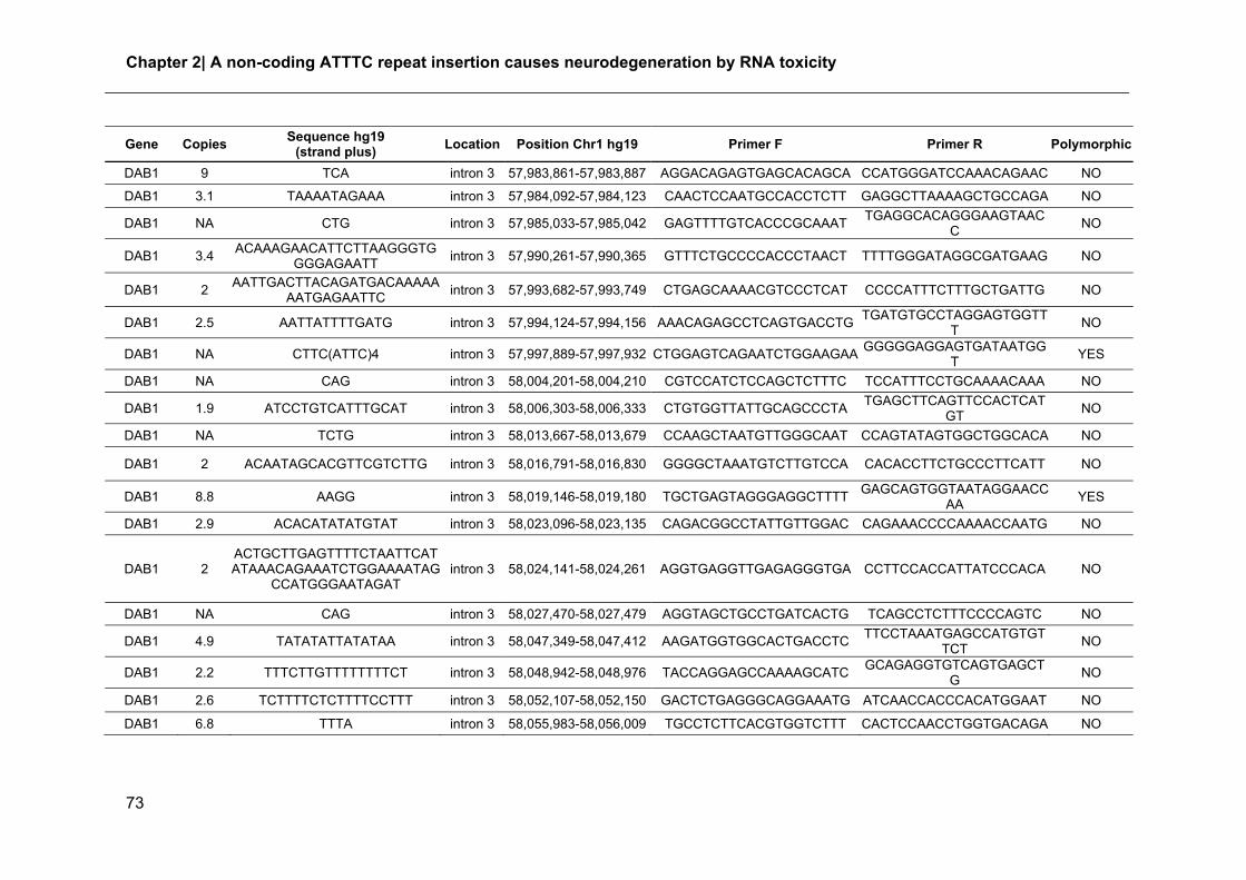
Chapter 2| A non-coding ATTTC repeat insertion causes neurodegeneration by RNA toxicity
73
Gene Copies Sequence hg19
(strand plus) Location Position Chr1 hg19 Primer F Primer R Polymorphic
DAB1 9 TCA intron 3 57,983,861-57,983,887 AGGACAGAGTGAGCACAGCA CCATGGGATCCAAACAGAAC NO
DAB1 3.1 TAAAATAGAAA intron 3 57,984,092-57,984,123 CAACTCCAATGCCACCTCTT GAGGCTTAAAAGCTGCCAGA NO
DAB1 NA CTG intron 3 57,985,033-57,985,042 GAGTTTTGTCACCCGCAAAT TGAGGCACAGGGAAGTAAC
C NO
DAB1 3.4 ACAAAGAACATTCTTAAGGGTG
GGGAGAATT intron 3 57,990,261-57,990,365 GTTTCTGCCCCACCCTAACT TTTTGGGATAGGCGATGAAG NO
DAB1 2 AATTGACTTACAGATGACAAAAA
AATGAGAATTC intron 3 57,993,682-57,993,749 CTGAGCAAAACGTCCCTCAT CCCCATTTCTTTGCTGATTG NO
DAB1 2.5 AATTATTTTGATG intron 3 57,994,124-57,994,156 AAACAGAGCCTCAGTGACCTG TGATGTGCCTAGGAGTGGTT
T NO
DAB1 NA CTTC(ATTC)4 intron 3 57,997,889-57,997,932 CTGGAGTCAGAATCTGGAAGAA GGGGGAGGAGTGATAATGG
T YES
DAB1 NA CAG intron 3 58,004,201-58,004,210 CGTCCATCTCCAGCTCTTTC TCCATTTCCTGCAAAACAAA NO
DAB1 1.9 ATCCTGTCATTTGCAT intron 3 58,006,303-58,006,333 CTGTGGTTATTGCAGCCCTA TGAGCTTCAGTTCCACTCAT
GT NO
DAB1 NA TCTG intron 3 58,013,667-58,013,679 CCAAGCTAATGTTGGGCAAT CCAGTATAGTGGCTGGCACA NO
DAB1 2 ACAATAGCACGTTCGTCTTG intron 3 58,016,791-58,016,830 GGGGCTAAATGTCTTGTCCA CACACCTTCTGCCCTTCATT NO
DAB1 8.8 AAGG intron 3 58,019,146-58,019,180 TGCTGAGTAGGGAGGCTTTT GAGCAGTGGTAATAGGAACC
AA YES
DAB1 2.9 ACACATATATGTAT intron 3 58,023,096-58,023,135 CAGACGGCCTATTGTTGGAC CAGAAACCCCAAAACCAATG NO
DAB1 2 ACTGCTTGAGTTTTCTAATTCATATAAACAGAAATCTGGAAAATAG
CCATGGGAATAGAT intron 3 58,024,141-58,024,261 AGGTGAGGTTGAGAGGGTGA CCTTCCACCATTATCCCACA NO
DAB1 NA CAG intron 3 58,027,470-58,027,479 AGGTAGCTGCCTGATCACTG TCAGCCTCTTTCCCCAGTC NO
DAB1 4.9 TATATATTATATAA intron 3 58,047,349-58,047,412 AAGATGGTGGCACTGACCTC TTCCTAAATGAGCCATGTGT
TCT NO
DAB1 2.2 TTTCTTGTTTTTTTTCT intron 3 58,048,942-58,048,976 TACCAGGAGCCAAAAGCATC GCAGAGGTGTCAGTGAGCT
G NO
DAB1 2.6 TCTTTTCTCTTTTCCTTT intron 3 58,052,107-58,052,150 GACTCTGAGGGCAGGAAATG ATCAACCACCCACATGGAAT NO
DAB1 6.8 TTTA intron 3 58,055,983-58,056,009 TGCCTCTTCACGTGGTCTTT CACTCCAACCTGGTGACAGA NO

Non-coding repeat insertion and RNA-mediated neurodegeneration
74
Gene Copies Sequence hg19
(strand plus) Location Position Chr1 hg19 Primer F Primer R Polymorphic
DAB1 8.5 TATT intron 3 58,060,367-58,060,400 CCAAGACGTTTAGGGGATCA CTTGCAGTGAGCCGAGATG NO
DAB1 4.1 TTTAGATTCTAAATTTTT intron 3 58,060,889-58,060,962 CCCAGGACCAGGGTTAGATT GGCCGGAAAAATCACAATTC NO
DAB1 2.4 TTTATTTTTATTTTTAG intron 3 58,063,623-58,063,672 TCTCCTGAATGGCACATCTT CTGGGTGACAAAGCGAGACT NO
DAB1 1.9 TATACACACACATATA intron 3 58,073,816-58,073,846 ACCCCCTGGGAGATATGACT TGTCAGAGCTGGAGGTGATC
T NO
DAB1 NA TTTC intron 3 58,080,522-58,080,542 AGAGGGAGTTTGCAGTGAGC TGGTTTGGGGCATTTTTACT YES
DAB1 NA CTG intron 3 58,084,198-58,084,210 TGACTTGGTCCCATCACAGA GGGAAGTGGAGGTTGCAGT
A YES
DAB1 NA CAG intron 3 58,086,326-58,086,377 GCAGCTGTTACGTCCTCCTC GAAGGGAAGGGGTTGGATT
A NO
DAB1 9.2 AACTTTATAAAGTTCTTCATAAA
GAACTTTAGTAAAT intron 3 58,104,593-58,104,932 CCTTTGCCTACTGGAACTGG
GGGATGGAAAGAGAGGGTTC
YES
DAB1 2.3 CATACATATATACACA intron 3 58,110,228-58,110,264 TGCAACCAGAGAAGCAGAAA TGCGTAAGGCCAAATCTCTC NO
DAB1 NA AGG intron 3 58,113,936-58,113,948 TCGTTGGTGGCTTATTGACA TCCAGCATCAAATGATCAGC NO
DAB1 44.2 ATCC intron 2 58,120,899-58,121,068 TGGGTGGATGGAAGGATAGA CCCAAATTTTCATAACCAATA
AGG NO
DAB1 10.5 AAAT intron 2 58,125,651-58,125,692 TGCAGTCAGCAGAGATCACC GTTCTTAAATCCATACTGGAA
TATTGT NO
DAB1 7.8 TTTG intron 2 58,127,852-58,127,882 CCAGGAACTAAACGCCAAGA CCTGGGCAACAAGAGCTAAA NO
DAB1 12.8 GAGGGA intron 2 58,132,522-58,132,598 AGCGTGCTTAGCAGTTTCCT AAGGATGGTGGGACATGAA
G NO
DAB1 2 AGCACAAATTATACATATATTC intron 2 58,141,337-58,141,380 ACCCCCTGGGAGATATGACT TGTCAGAGCTGGAGGTGATC
T NO
DAB1 3 CACCAATCAGTGCTCTGTGTCTAGCTAAAGGATTGTAAACACACCAACCAGCACTCTGTAAAAAT
intron 2 58,156,843-58,157,036 TGGGGACTTGGAGAAGTTTT GAAGGGGACCTTAGCAGGTT NO
DAB1 3.7 AGAGAGGAAGAGAG intron 2 58,157,935-58,158,310 ATGCCTTGGCTGGTAGACAG CCCTGTCTCTGGTGCTTTTC NO
DAB1 2.3 GCACCAATCAGCACTCTGTGTCTAGCTAAAGGATTGTAAATGCAC
AAAGCACTCTGTAAAAC intron 2 58,159,036-58,159,182
CAAGTGGGGATTTAGAGAACTTTT
CCAGCTTTTAGTCCCTTATTTGG
NO

Chapter 2| A non-coding ATTTC repeat insertion causes neurodegeneration by RNA toxicity
75
Gene Copies Sequence hg19
(strand plus) Location Position Chr1 hg19 Primer F Primer R Polymorphic
DAB1 13.5 CTTC intron 2 58,170,551-58,170,605 GGTGATGGGCTGCTGTTTAT AAATGCCCTTGCTCTTTGG YES
DAB1 NA CTG intron 2 58,173,749-58,173,758 GGCAGAATTCACCACCTGTT CTCCCATGGCCACTATCACT NO
DAB1 2.1 AGCAATTCACACTTTATGTAA intron 2 58,187,828-58,187,872 AGGCACAACATCCAGAAAGC CCCAGGTATGTCTTCTGTCT
CC NO
DAB1 16.8 AAGG intron 2 58,189,940-58,190,006 CAAGGCAAAGGAATCGGTAA GTTGCGGATCAAGATGGTG NO
DAB1 16.8 AGGG intron 2 58,189,973-58,190,044 CAAGGCAAAGGAATCGGTAA GTTGCGGATCAAGATGGTG YES
DAB1 11.5 CTTC intron 2 58,193,385-58,193,430 GCAGGATTTTCCAACCACAC GATCCTGGAGCCACTTTCAT YES
DAB1 5.2 TAAAA intron 2 58,195,567-58,195,592 AAGAGGCTAAGGCCAGAGGA CCAGCTGCCACCTGATTC NO
DAB1 11.6 CTCCC intron 2 58,205,099-58,205,156 NO
DAB1 NA TAC intron 2 58,205,800-58,205,818 GTGGCATAATCTCGGCTCAT AGCTTGGCCAACATAGCAAA NO
DAB1 NA CTG intron 2 58,207,830-58,207,842 GTTTTTCTCCATCCCCCATC AGCAGGTTGAAAGTGGGAAA NO
DAB1 3.3 GTAGTGTTAT intron 2 58,209,970-58,210,003 GGTGAACAGGCTGTCAGGAT TCAAGGAAGCCCTGTGAGAT NO
DAB1 NA AATA intron 2 58,217,333-58,217,581 CCAGAACTGATCGCATTCAA ATGGGAGAGAAAGGATGCAA YES
DAB1 1.9 TGTTTATCCACATCTCATCCTAT
GA intron 2 58,219,286-58,219,333 AATGGGAATAGCAGGGCCTA CTGCACTTCAGATTCCACCA NO
DAB1 9.8 AAAC intron 2 58,220,366-58,220,405 GCAGTGAGCTGAGAGTGTGC AAAGAAAAGGAGGCCTGGA
G NO
DAB1 2.4 CTTCCCTTCTCTTTTCTCCCTGC
CCTCT intron 2 58,226,167-58,226,236 GGCCCAGGTCTCTAAGAACA ACTGCCGACTCCCCTTTTT YES
DAB1 2.5 AACAAATTATTTTTACAA intron 2 58,230,664-58,230,709 TTTAGCCAAACCAGATGGAAA GCTGTGGTGCTGTGAACATT NO
DAB1 2 AAAATTGTGATCATCTTTATGTT
TTTATAATTT intron 2 58,241,556-58,241,621
TCAGTTTTTGCTTCATGTATTTTGA
GACAAAGTAGGCTTTGGGACA
NO
DAB1 8.8 TTTTC intron 2 58,246,389-58,246,430 CCATATTCCTACTACCACCCACT
T AGAGGTTGTAGTGAGCCGA
GA NO
DAB1 8.5 TCCTTCCTTCT intron 2 58,248,668-58,248,757 GGCAATAGAATGAGACCCTGT GCGATGGGGTTTAGAAACAA YES
DAB1 3.1 AAGCATCTCCATAACAAAAAGTG
C intron 2 58,250,398-58,250,470 ATCAAACCAGCTGAGCCATT
TTCTGGATAACTTGCTCCTTCA
NO
DAB1 16 AAT intron 2 58,262,199-58,262,246 GACAGAGCAAAACTCCGTCTC GGCTGCTACACCCAATGACT YES
DAB1 2.7 TTTTTTTATT intron 2 58,267,321-58,267,347 TGAAAAGGTCCTTCCTCTTCC AGCCTGGGCAACACAGTAAG NO

Non-coding repeat insertion and RNA-mediated neurodegeneration
76
Gene Copies Sequence hg19
(strand plus) Location Position Chr1 hg19 Primer F Primer R Polymorphic
DAB1 1.9 AAAAGAAAGAAAGAAAG intron 2 58,278,007-58,278,039 CATGCATGCACACATCTTCA AACACACAACCTGGGATTCA NO
DAB1 NA CAG intron 2 58,280,023-58,280,035 AATTTCAGGCAGTTGCCATC CATCTGGCCACACATCAAGT NO
DAB1 NA GAATG intron 2 58,281,057-58,281,119 GGACAACTAGTTTGCTCCCTTG TTGGAATGGAATCACATAGA
ATG NO
DAB1 NA CAG intron 2 58,290,393-58,290,402 CCCAACTGTGAGGTTGTGAA ACCAACACCATAGCGTAGCC NO
DAB1 NA CAG intron 2 58,292,173-58,292,182 GGCAGGAATTCACTGTCCAT GAGGAGCATCTCATCCAAGC NO
DAB1 2.6 CATAATTAAATCATATATGCATA
TGTACACG intron 2 58,297,685-58,297,768 CACACAGACATGGCTCTTCC ACTGCTCTTCCTGCCTCAGA NO
DAB1 3.3 TATATATGCACACAGTTTTATA intron 2 58,300,244-58,300,318 CCAAAGCAAAAAGGGATTCA CCCTGGCATCTTGTACACTT
C NO
DAB1 2.9 ACCACCTCCACCCCCATCACCA
CC intron 2 58,303,443-58,303,512 CCCCATACACACATGCTTGA GAGCGGAAATGGTGGAGAT NO
DAB1 2 GGGGATGCTGTGAAGATCTCTG
AAAT intron 2 58,307,455-58,307,507 GAATCAATGGCCTGAGCTGT ATGAGGAAAATGCCACCAAG NO
DAB1 1.9
TCATGAAATAGACAATATTATCTTGATTTTGCACATTGGTAAATTGAGGAAACAGTCACCTTTCACAA
CAACT
intron 2 58,327,886-58,328,025 CTAAGCACGGGTCTGGGTAA TCAGTTCAATGATGAGAAAA
GCTC NO
DAB1 2 GTATGGGGGAGGTGT intron 2 58,328,927-58,328,956 TGCTGAGATAAAGAACCCAAAG
A TCCATCATAGCCCAAAGTTTT
C NO
DAB1 12.5 ATTT intron 2 58,330,326-58,330,376 CAGCAGTATGATTGGGGGTAA GGCTGGGTAGAGTGGTTCA
C YES
DAB1 2.1 TATAACTATCTAAGAGG intron 1 58,336,924-58,336,959 GGAGGAGCAGCAGCAATATC CAGCACCAGATACCCATGAA YES
DAB1 1.9 TTCATATCTCATTTAACAT intron 1 58,352,222-58,352,258 CACCCGGCATATTCTGCTAC TGACTGCCATTCGTGACACT NO
DAB1 14.8 TTTTC intron 1 58,356,078-58,356,147 TGACCCCAACTCTTCACCTC CTAGGCAACAGAGGCAGAC
C YES
DAB1 1.9 GTTTTATAAAAAGGGAT intron 1 58,365,800-58,365,832 TCAAGCTGTCCTCCCATTTC GCAATGCCGTGAGACTGTAA NO
DAB1 13 AGAT intron 1 58,372,610-58,372,661 ATTCTGCCCTTCTCTTGGTG TCCCTGAAGCTAGGTGCTTT NO
DAB1 NA CAG intron 1 58,377,155-58,377,170 AGCCATGACGTCTGAGGAGT TCCCATTTCTGCCTGTCTCT NO
DAB1 2.9 GAAGAAAGGAAGGAAA intron 1 58,388,042-58,388,088 AAATGAAGGGAGGGAAAGGA AGGTTCCTCAGGGCAAAGTT NO
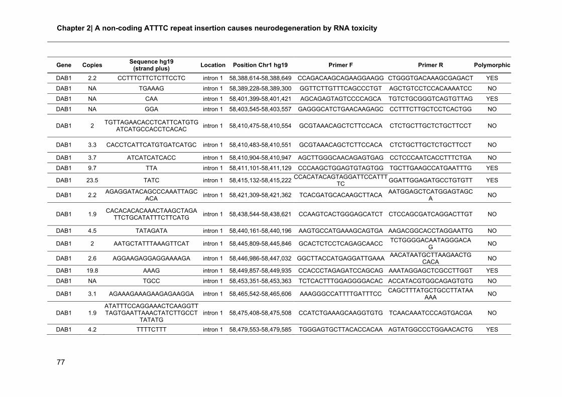
Chapter 2| A non-coding ATTTC repeat insertion causes neurodegeneration by RNA toxicity
77
Gene Copies Sequence hg19
(strand plus) Location Position Chr1 hg19 Primer F Primer R Polymorphic
DAB1 2.2 CCTTTCTTCTCTTCCTC intron 1 58,388,614-58,388,649 CCAGACAAGCAGAAGGAAGG CTGGGTGACAAAGCGAGACT YES
DAB1 NA TGAAAG intron 1 58,389,228-58,389,300 GGTTCTTGTTTCAGCCCTGT AGCTGTCCTCCACAAAATCC NO
DAB1 NA CAA intron 1 58,401,399-58,401,421 AGCAGAGTAGTCCCCAGCA TGTCTGCGGGTCAGTGTTAG YES
DAB1 NA GGA intron 1 58,403,545-58,403,557 GAGGGCATCTGAACAAGAGC CCTTTCTTGCTCCTCACTGG NO
DAB1 2 TGTTAGAACACCTCATTCATGTG
ATCATGCCACCTCACAC intron 1 58,410,475-58,410,554 GCGTAAACAGCTCTTCCACA CTCTGCTTGCTCTGCTTCCT NO
DAB1 3.3 CACCTCATTCATGTGATCATGC intron 1 58,410,483-58,410,551 GCGTAAACAGCTCTTCCACA CTCTGCTTGCTCTGCTTCCT NO
DAB1 3.7 ATCATCATCACC intron 1 58,410,904-58,410,947 AGCTTGGGCAACAGAGTGAG CCTCCCAATCACCTTTCTGA NO
DAB1 9.7 TTA intron 1 58,411,101-58,411,129 CCCAAGCTGGAGTGTAGTGG TGCTTGAAGCCATGAATTTG YES
DAB1 23.5 TATC intron 1 58,415,132-58,415,222 CCACATACAGTAGGATTCCATTT
TC GGATTGGAGATGCCTGTGTT YES
DAB1 2.2 AGAGGATACAGCCCAAATTAGC
ACA intron 1 58,421,309-58,421,362 TCACGATGCACAAGCTTACA
AATGGAGCTCATGGAGTAGCA
NO
DAB1 1.9 CACACACACAAACTAAGCTAGA
TTCTGCATATTTCTTCATG intron 1 58,438,544-58,438,621 CCAAGTCACTGGGAGCATCT CTCCAGCGATCAGGACTTGT NO
DAB1 4.5 TATAGATA intron 1 58,440,161-58,440,196 AAGTGCCATGAAAGCAGTGA AAGACGGCACCTAGGAATTG NO
DAB1 2 AATGCTATTTAAAGTTCAT intron 1 58,445,809-58,445,846 GCACTCTCCTCAGAGCAACC TCTGGGGACAATAGGGACA
G NO
DAB1 2.6 AGGAAGAGGAGGAAAAGA intron 1 58,446,986-58,447,032 GGCTTACCATGAGGATTGAAA AACATAATGCTTAAGAACTG
CACA NO
DAB1 19.8 AAAG intron 1 58,449,857-58,449,935 CCACCCTAGAGATCCAGCAG AAATAGGAGCTCGCCTTGGT YES
DAB1 NA TGCC intron 1 58,453,351-58,453,363 TCTCACTTTGGAGGGGACAC ACCATACGTGGCAGAGTGTG NO
DAB1 3.1 AGAAAGAAAGAAGAGAAGGA intron 1 58,465,542-58,465,606 AAAGGGCCATTTTGATTTCC CAGCTTTATGCTGCCTTATAA
AAA NO
DAB1 1.9 ATATTTCCAGGAAACTCAAGGTTTAGTGAATTAAACTATCTTGCCT
TATATG intron 1 58,475,408-58,475,508 CCATCTGAAAGCAAGGTGTG TCAACAAATCCCAGTGACGA NO
DAB1 4.2 TTTTCTTT intron 1 58,479,553-58,479,585 TGGGAGTGCTTACACCACAA AGTATGGCCCTGGAACACTG YES

Non-coding repeat insertion and RNA-mediated neurodegeneration
78
Gene Copies Sequence hg19
(strand plus) Location Position Chr1 hg19 Primer F Primer R Polymorphic
DAB1 2.4 GTGCTTTTTACTGCATTCTAA intron 1 58,483,749-58,483,798 GGTTCGCCAATTGACAGCTA GCAGAGAGAGTGAGGGAAC
G YES
DAB1 NA TCA intron 1 58,488,535-58,488,551 CCATGTGAGGCATTGTGTTC TAATTTGGCACTGCACTTCG NO
DAB1 1.9
TTCATAGATTTTATTTCTTTCTCTTGTCTCCTATTAAATGTAGATGAGCTATAAATATAATATTATATATA
TATAGAGAGAGAGCCT
intron 1 58,491,158-58,491,318 TCTCTAAACTTTCATCCCTTCAG
C ACCCCAAAGACAGAGGAGG
A YES
DAB1 2.1 CTTCTCCAGGTACGAAGTT intron 1 58,496,660-58,496,698 GGCCTTCAAGACATGCAATAA CCTCCATCCAGGAAGACATC NO
DAB1 2 TGTTGATTTTATTTTC intron 1 58,512,116-58,512,147 AAGCAAACTGAGGCAAAGGA TCCTAGTTTCAGAGGGCCTT
A NO
DAB1 2 ATATCCAAACTGTTATCATTTCA
AC intron 1 58,512,962-58,513,011 AAATTTGCCACTTAGCCACA GCCACACTTCACACCACACT NO
DAB1 NA CTG intron 1 58,513,278-58,513,290 CACATGTATGGAATTGCGTGA TGGCACTAGCCACATTTCAG NO
DAB1 7.2 TTTA intron 1 58,516,189-58,516,218 TGGTGGGGAGAACAAAGTGT TGGGTGACAGAGCAAGACT
G NO
DAB1 NA CAG intron 1 58,520,666-58,520,675 AGGGTCCAGTGAAAAGATGG GCTGTCAAGCAATGGTCCAG NO
DAB1 2.1 TCACTGCAACCTCTG intron 1 58,521,449-58,521,480 CACTATACATTTGACCAGTGATG
GA AATAATAATAATAACTCGGTG
TGATGG NO
DAB1 8.7 TTA intron 1 58,521,555-58,521,580 CCTCTGTCTTCTGGGCTCAA GTGGCTCACACCTGGAATTT NO
DAB1 15 ATTT intron 1 58,530,351-58,530,410 CCAGGCATTTAACTTGGACA CGGAGAGTCTGAGGCAGAA
G YES
DAB1 2 GGGGATAATGAGTGTTCCATAC
ACATAAGCTGCTGTATT intron 1 58,533,477-58,533,555 AGGCCTCAACCAGTTCATCT
TTCTTTCCTTCTGGCTAAGACTG
NO
DAB1 NA TGC intron 1 58,555,697-58,555,709 GCAACCTCAGTGGGCTGTAT ATTGCATCGGCAGTAAAAGC NO
DAB1 NA AGCC intron 1 58,555,716-58,555,728 GCAACCTCAGTGGGCTGTAT ATTGCATCGGCAGTAAAAGC NO
DAB1 5.8 AAAAT intron 1 58,566,991-58,567,019 AGGTCATGCCACTGCACTC TGATCCCCCTAGCAACTGAC NO
DAB1 7.3 CTTTTCTTT intron 1 58,571,781-58,571,844 AGCCATGATCCTACCACTGC TGCAAAGAATTTTCCAACAC
A YES
DAB1 28.5 TCCC intron 1 58,572,497-58,572,577 GCATAATGCCCAGCACATAG AGGGCCCAGCTGAAATAAAT YES

Chapter 2| A non-coding ATTTC repeat insertion causes neurodegeneration by RNA toxicity
79
Gene Copies Sequence hg19
(strand plus) Location Position Chr1 hg19 Primer F Primer R Polymorphic
DAB1 3.7 TGTTTTTGTTT intron 1 58,573,265-58,573,306 TTCAGCATGGGTGACAAGAG GGGTGACAGCAAGACTCCAT NO
DAB1 NA CAG intron 1 58,580,415-58,580,424 TGATTGAATATCAGCCTTGCTT AAAGCCACAAAGTGTAAAAC
ACA NO
DAB1 2.7 TTTTTTTTAATA intron 1 58,583,517-58,583,548 CCATGCTTCACTGTGTGCTT CACCACTGCAGCACTCTAGC NO
DAB1 50 ATCC intron 1 58,603,094-58,603,282 AACTGAATCAAAGAGAATGAGT
ATCC GGATGAATCAATGGTTGAAT
AAA NO
DAB1 NA ATCC intron 1 58,612,371-58,612,383 GCCAAGATATGGAAACAACCT GGCACAAAAGGCAGCATATC YES
DAB1 8.5 TTCA intron 1 58,620,125-58,620,158 TGCTTCTTACCTGTGGCAGA CACCAGGCTTGAAACAGACA YES
DAB1 2.1 AATTTTGGGTAC intron 1 58,626,402-58,626,426 TCCCTCCAACGATCTCAAAC AGTCTTGGGGCCCAGTATTC NO
DAB1 3.3 AAATGCACCAATCAGCACTCTGTGTCTAGCTAAAGGATTGTAAATGCACCAATCAGCACTCTGTAA
intron 1 58,632,972-58,633,185 AGGTGGGGACTTGGAAAACT TTATTTGACCCTGCCCACAT NO
DAB1 4
CTCTTGCCCAAGAACCTGCAACGGTCCCTGGACCCTGCTGATCGGAATAGTTGTGCTCACTGAACG
CAGCAGCAGAAACAC
intron 1 58,634,715-58,635,030 TGCCTGAATGTTTCCCATTT GCTGCTACGTTGATGAGTGC NO
DAB1 3 TGTGTCTAGCTAAAGGATTGTAA
ATGCACCAATCAGCGCTC intron 1 58,640,327-58,640,450 CCCTTATTTGGCCCCACTCA CCCTTCTTCACCCCATCCAG NO
DAB1 2.3 ATAATTATAGTGTTATTTTT intron 1 58,645,608-58,645,651 CCCATCTCTAGATTACTTATAGC
ACCT TGAATTCAGCCAACTGTGGA NO
DAB1 2.8 TTTCTTTTTTTTTTTTTTT intron 1 58,645,929-58,645,982 TGCTTTGGGTTTGAATTGCT GAGGCTGCAGTGAGCTGTTA YES
DAB1 NA CAG intron 1 58,646,575-58,646,584 GGCCAACTAGAAAGAAAATCAT
C TGCGATACTGGTTTTGTTGG NO
DAB1 3.5 AATATATAACATAAT intron 1 58,649,945-58,649,994 AAACACCCACTTGCCTACAGA TCCAGCTGAAAGAGGTTTGA NO
DAB1 5 GTTTT intron 1 58,651,226-58,651,250 GCCTGAAGCCCTATTGACAG GCCAGAAGTAGTGCCAGAG
G NO
DAB1 11 TAT intron 1 58,653,229-58,653,261 TGGTGGTGCACACCTGTAAT AGCCTGGGACCACACAGTAA YES
DAB1 4.1 AAGCAATGTGCTCAGC intron 1 58,673,101-58,673,167 AGGTAAACCAGGATGGCTGA GCATGGTTGGGAAAAGAGAA NO

Non-coding repeat insertion and RNA-mediated neurodegeneration
80
Gene Copies Sequence hg19
(strand plus) Location Position Chr1 hg19 Primer F Primer R Polymorphic
DAB1 NA CAG intron 1 58,677,746-58,677,755 GCACCTAGTGGGCATTCAA TTCCTGCTGGGTTAAGCATC NO
DAB1 NA TGGA intron 1 58,681,638-58,681,690 TTTAACCACAGCACCCATCA CAAACCTGCAAGTTCTGCAT NO
DAB1 2.3 CACCTGCACCACA intron 1 58,685,376-58,685,406 CACACAGCTGACTCCTTCCA ATGTGTGCAATGGAGGTGAA NO
DAB1 NA CTG intron 1 58,699,150-58,699,159 TCATGAGAATTAGAGAGAGAAT
GAAAA GGCCCTCCGATCACCATA NO
DAB1 NA ATTC intron 1 58,699,780-58,699,969 TGCCAGAAGGCAAGATCATA CTTCCGTGACACTGATCCAA NO
DAB1 NA CGGGG intron 1 58,701,770-58,701,800 CTTGGCCTTGGCTCATTAGT AAGGGAAAGCTGCTACAGGA NO
DAB1 NA CAG intron 1 58,702,430-58,702,439 GGCATTAGGAGCTTGGTTTG CATGCATCATCTCTTTGAAG
C NO
DAB1 NA CTG intron 1 58,714,013-58,714,022 AGTGACCTGACAAGGGTTGG CCAGCTGGGAAATCCCTATT NO
DAB1 NA CCAG intron 1 58,715,215-58,715,227 CCGGGGAAACAACTTCATC GGCGGGAGGGTTAAAATTAC NO
DAB1 NA GCC intron 1 58,715,526-58,715,535 AGTGGGGGAAAAGTCTGGAG GCTTGTCTCGGGGGATTC NO
DAB-end ROI
4.7 TTTTTG intergenic 58,727,381-58,727,408 GTGGAGAAAGGCCAGATTCA GAGGCCAAGACAGGAGGAT
T YES
DAB-end ROI
1.9 CTCAGTTTCCTCAT intergenic 58,728,279-58,728,305 AGGGCGAGACTGCATCTAAA GTGCCTAACGTGCCAAAAAT NO
DAB-end ROI
2.6 TTTTATTTTTT intergenic 58,734,672-58,734,701 GCAGCACTGCCTGAGAGAC ACATATGTAACTAACCTGCA
CAATG NO
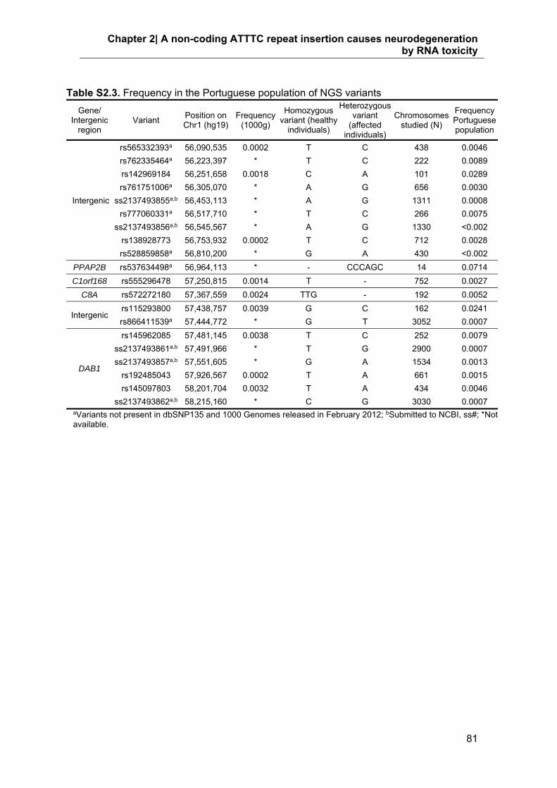
Chapter 2| A non-coding ATTTC repeat insertion causes neurodegeneration by RNA toxicity
81
Table S2.3. Frequency in the Portuguese population of NGS variants
Gene/ Intergenic
region Variant
Position on Chr1 (hg19)
Frequency (1000g)
Homozygous variant (healthy
individuals)
Heterozygous variant
(affected individuals)
Chromosomes studied (N)
Frequency Portuguese population
Intergenic
rs565332393a 56,090,535 0.0002 T C 438 0.0046
rs762335464a 56,223,397 * T C 222 0.0089
rs142969184 56,251,658 0.0018 C A 101 0.0289
rs761751006a 56,305,070 * A G 656 0.0030
ss2137493855a,b 56,453,113 * A G 1311 0.0008
rs777060331a 56,517,710 * T C 266 0.0075
ss2137493856a,b 56,545,567 * A G 1330 <0.002
rs138928773 56,753,932 0.0002 T C 712 0.0028
rs528859858a 56,810,200 * G A 430 <0.002
PPAP2B rs537634498a 56,964,113 * - CCCAGC 14 0.0714
C1orf168 rs555296478 57,250,815 0.0014 T - 752 0.0027
C8A rs572272180 57,367,559 0.0024 TTG - 192 0.0052
Intergenic rs115293800 57,438,757 0.0039 G C 162 0.0241
rs866411539a 57,444,772 * G T 3052 0.0007
DAB1
rs145962085 57,481,145 0.0038 T C 252 0.0079
ss2137493861a,b 57,491,966 * T G 2900 0.0007
ss2137493857a,b 57,551,605 * G A 1534 0.0013
rs192485043 57,926,567 0.0002 T A 661 0.0015
rs145097803 58,201,704 0.0032 T A 434 0.0046
ss2137493862a,b 58,215,160 * C G 3030 0.0007 aVariants not present in dbSNP135 and 1000 Genomes released in February 2012; bSubmitted to NCBI, ss#; *Not available.

Non-coding repeat insertion and RNA-mediated neurodegeneration
82
Table S2.4. Configuration of (ATTTC)n insertion alleles in the three additional families
Family (Haplotype)
Affected individuals
(ATTTT)n allele (ATTTC)n insertion allele
MS (B)
MS1 (ATTTT)19 (ATTTT)73(ATTTC)46(ATTTT)81
MS2 (ATTTT)19 (ATTTT)69(ATTTC)52(ATTTT)90
MS3 (ATTTT)19 (ATTTT)72(ATTTC)46(ATTTT)16b
MS4 (ATTTT)12 (ATTTT)67(ATTTC)61(ATTTT)70b
C (C) C1 (ATTTT)16 (ATTTT)81(ATTTC)64(ATTTT)64b
D (D) D1 (ATTTT)16 (ATTTT)52(ATTTC)71(ATTTT)83b
b3' ATTTTs not completely sequenced

Chapter 3. A workflow for SCA37 molecular
diagnosis
Adapted from A Repeat-Primed PCR Assay for Pentanucleotide Repeat Alleles in
Spinocerebellar Ataxia Type 37, Journal of Human Genetics: 63: 981–987, 2018.


Chapter 3| A workflow for SCA37 molecular diagnosis
85
A Repeat-Primed PCR Assay for Pentanucleotide Repeat Alleles in
Spinocerebellar Ataxia Type 37
Joana Rocha Loureiro1,2,3, Cláudia Louro Oliveira1,2, Jorge Sequeiros2,3,4,5, and Isabel Silveira1,2
1Genetics of Cognitive Dysfunction Laboratory, i3S-Instituto de Investigação e Inovação em Saúde, Universidade do
Porto, 4200-135 Porto, Portugal; 2IBMC- Institute for Molecular and Cell Biology, Universidade do Porto, 4200-135
Porto, Portugal; 3ICBAS, Universidade do Porto, 4050-313 Porto, Portugal; 4UnIGENe, i3S-Instituto de Investigação e
Inovação em Saúde, Universidade do Porto, 4200-135 Porto, Portugal; 5CGPP, i3S-Instituto de Investigação e Inovação
em Saúde, Universidade do Porto, 4200-135 Porto, Portugal;
Spinocerebellar ataxia 37 (SCA37) is caused by an (ATTTC)n insertion in a
polymorphic ATTTT repeat in the non-coding region of DAB1. The non-pathogenic
alleles have a configuration [(ATTTT)7-400], whereas pathogenic alleles have a complex
structure of [(ATTTT)60-79(ATTTC)31-75(ATTTT)58-90]. Molecular diagnosis of SCA37 is
laborious because about 7% of the pentanucleotide repeat alleles in DAB1 are larger
than 30 units and, thus, fail to amplify with standard PCR conditions, resulting in
apparently homoallelism or in complete lack of PCR amplification in several cases. The
molecular test currently available requires long-range PCR and sequencing analysis for
the detection and characterization of these large alleles. We developed a simple assay
capable of rapidly detecting the presence or absence of large pentanucleotide repeat
sizes. This assay is based on repeat-primed PCR followed by high-throughput capillary
electrophoresis. Combining the standard PCR with RP-PCR allows completion of the
diagnosis in more than 80% of individuals, minimizing the number of samples that require
long-range PCR followed by Sanger sequencing analysis. This assay meets many of the
requirements for pre-screening of large cohorts of affected individuals.

Non-coding repeat insertion and RNA-mediated neurodegeneration
86
3.1. Introduction
A large number of neurological and neuromuscular diseases are caused by unstable
repetitive tracts in coding or non-coding gene regions1-3. These include the expansion of
trinucleotide repeats in coding regions, the most common being CAG repeats that
encode toxic polyglutamine proteins4, 5. Tri-, tetra-, penta-, and hexanucleotide repeats
in non-coding gene regions, however, are also responsible for many of these disorders6-
11. In two of the unstable repeat diseases, spinocerebellar ataxia type 31 (SCA31) and
SCA37, there is a pentanucleotide repeat insertion in another polymorphic
pentanucleotide repeat, which is a challenge for routine diagnostic testing12, 13. This is
further complicated by the existence of very large non-pathogenic alleles comprised of
pentanucleotide repeats13, 14.
In SCA37, there is an (ATTTC)n insertion in the middle of a polymorphic ATTTT repeat
in the 5’UTR region of DAB1 (DAB1, reelin adaptor protein)13. The non-pathogenic alleles
have ATTTT repeats that can vary from 7 to 400 units and never contain ATTTC.
Standard PCR amplification of the pentanucleotide repeat in DAB1 can easily detect
alleles shorter than 30 units13; however, amplification of larger alleles is technically
challenging and requires long-range PCR13. By standard PCR, finding only one band of
potentially two normal alleles of the same size (homoallelism) is not conclusive because
there is the possibility of preferential amplification of the shorter allele in the presence of
a large allele that is not amplified. As the molecular diagnosis relies on the presence of
the ATTTC insertion and since 7% of non-pathological alleles are larger than 30
ATTTTs13, long-range PCR and sequencing analysis are frequently required.
To facilitate screening of large cohorts of patients with SCA, there is a need for simple
and inexpensive tests with high sensitivity and specificity. Expanded alleles in several of
the SCAs and in fragile-X syndrome can be detected using a fluorescent repeat-primed
PCR assay15, 16. In SCA37, the (ATTTC)n insertion has been successfully detected by a
similar repeat-primed PCR adapted to the specificity of the pentanucleotide repeat
insertion13. DNA samples with detected ATTTC repeat insertions are, then, amplified by
long-range PCR followed by sequencing analysis of the products obtained. This method
is adequate for repeat insertions flanked by ATTTT stretches of approximately 100 units;
however, for much larger ATTTT flanking repeats the ladder of peaks obtained would fall
out of the electropherogram detection.
For screening purposes, this limitation of the ATTTC RP-PCR negatively affects the
sensitivity of the test. To overcome this problem, we have developed an ATTTT RP-PCR
that allows detection of pentanucleotide repeat alleles in DAB1 larger than 30 units. With
this screening method both normal alleles larger than 30 ATTTTs and alleles with the

Chapter 3| A workflow for SCA37 molecular diagnosis
87
ATTTC repeat insertion flanked by ATTTTs are detected. The final step is always
sequencing analysis of the products of long-range PCR to identify alleles with the ATTTC
repeat insertion.
3.2. Subjects and Methods
3.2.1. DNA Samples from Affected Individuals and Controls
This study used the de-identified, previously collected DNA samples, of affected
individuals with SCA37 as well as anonymized DNA samples from control individuals;
they were described in a previous study13.
3.2.2. Repeat-primed PCR for the ATTTT repeat
The pentanucleotide repeat sequence in DAB1 was amplified by ATTTT RP-PCR with
primers 24F (6-FAM-5’-GAAGTGGTCCTCCCAAGTCA-3’), FLAG (5’-
TACGCATCCCAGTTTGAGACG-3’), and RP-AATAA (5’-
TACGCATCCCAGTTTGAGACGAATAAAATAAAATAAAATAAAATAA-3’). The forward
primer (24F) was a locus-specific primer, while the RP-AATAA primer was an ATTTT-
repeat specific. The RP-AATAA primer had a DNA tail sequence at the 5’ end (absent in
the human genome) and the reverse primer FLAG had the 5’ tail sequence of the RP-
AATAA primer. PCR was performed with 100 ng genomic DNA, 1X HotStarTaq Master
Mix (Qiagen), 0.8 µM primer 24F and primer FLAG, and 0.4 µM primer RP-AATAA, in
12.5 µL. The initial RP-PCR step was at 95ºC for 15 min and was followed by 40 cycles
(94ºC for 1 min, 54ºC for 1 min, and 72ºC for 2 min and 30 sec) and a final extension of
10 min at 72ºC. RP-PCR products were detected by capillary electrophoresis of 3 µl of
RP-PCR product mixed with 0.5 µl of GS500(-250)LIZ size standard in Hi-DiTM
formamide (Applied Biosystems). Samples were denatured at 95ºC, for 5 min, before
loading onto an ABI3730xl DNA Analyzer on a 36-cm capillary. Fragment sizes were
analysed with GeneMapper v.4.0 (Applied Biosystems).
3.2.3. Sequencing Analysis
The long-range PCR13 was performed in duplicate, for each sample with positive
detection by ATTTT RP-PCR; PCR products were separated by electrophoresis in a 1%

Non-coding repeat insertion and RNA-mediated neurodegeneration
88
agarose gel. For each sample, each DNA and corresponding duplicate band (of 0.9 kb
or larger) were sliced from the gel and the DNA was extracted together using the
QIAquick gel extraction kit (Qiagen) following the manufacturer instructions. The DNA
was analysed by Sanger sequencing. Sequencing analysis in the forward orientation
used 2 µL of DNA extracted from the duplicate bands together, 4 µL of BigDye terminator
v.1.1 (Applied Biosystems), 0.5X BigDye sequencing buffer, 2.5 µM of primer 24F (5’-
GAAGTGGTCCTCCCAAGTCA-3’) in a final volume of 20 µl. Sequencing analysis in the
reverse orientation used 2 µl of DNA, 8 µL of BigDye terminator v.1.1, 0.5X BigDye
sequencing buffer and 1.5 µM of primer 24R4 (5’- GAGACCAGCCTGGGCAAC-3’) in a
final volume of 20 µl. Sequencing reactions were performed, for both forward and reverse
orientations, with an initial denaturation of 5 min at 95ºC followed by 50 cycles of 30 sec
at 95ºC, 10 sec at 56ºC, and 4 min at 60ºC and a final extension of 10 min at 60ºC.
3.3. Results
3.3.1. RP-PCR of Pentanucleotide Repeat Alleles
Standard PCR analysis of the pentanucleotide repeat in DAB1 resulted in the
amplification of two, one or no alleles in samples from affected or control individuals
(Table 3.1); this method could not amplify successfully alleles larger than 30 repeats13.
To detect pentanucleotide repeats larger than 30 units in DAB1 for the screening of
SCA37 in large cohorts of individuals, we developed a simple ATTTT RP-PCR (Figure
3.1). After capillary electrophoresis and electropherogram analysis, we observed that
alleles larger than 30 repeats produced a ladder of multiple peaks 5-bp apart and
extending beyond the 250 bp, while this ladder ended below 250 bp for alleles smaller
than 30 repeats. Thus, we predefined a cut-off value of 250 bp below which the test was
considered negative, indicating the absence of the SCA37 pentanucleotide repeat
insertion. All the samples with long non-pathogenic ATTTT tracts of 33-400 (17 samples)
or with the pathogenic repeat insertion (41 samples) showed an ATTTT RP-PCR ladder
extending beyond 250 bp.

Chapter 3| A workflow for SCA37 diagnosis
89
Table 3.1. Genotypes of Affected Individuals and Controls at DAB1
Affected individuals (n=41) Controls (n=96)
A-1 (ATTTT)14 / (ATTTT)69(ATTTC)37(ATTTT)83a C-1 (ATTTT)7 / (ATTTT)7 C-42 (ATTTT)11 / (ATTTT)21 C-83 (ATTTT)13 / (ATTTT)13
A-2 (ATTTT)14 / (ATTTT)62(ATTTC)43(ATTTT)35a C-2 (ATTTT)7 / (ATTTT)8 C-43 (ATTTT)11 / (ATTTT)14 C-84 (ATTTT)14 / (ATTTT)14
A-3 (ATTTT)14 / (ATTTT)73(ATTTC)51(ATTTT)64 C-3 (ATTTT)7 / (ATTTT)19 C-44 (ATTTT)11 / (ATTTT)~400a C-85 (ATTTT)14 / (ATTTT)22
A-4 (ATTTT)14 / (ATTTT)75(ATTTC)55(ATTTT)63a C-4 (ATTTT)7 / (ATTTT)13 C-45 (ATTTT)12 / (ATTTT)46 C-86 (ATTTT)14 / (ATTTT)14
A-5 (ATTTT)8 / (ATTTT)77(ATTTC)51(ATTTT)43a C-5 (ATTTT)7 / (ATTTT)15 C-46 (ATTTT)12 / (ATTTT)16 C-87 (ATTTT)14 / (ATTTT)16
A-6 (ATTTT)14 / (ATTTT)66(ATTTC)60(ATTTT)67 C-6 (ATTTT)7 / (ATTTT)17 C-47 (ATTTT)12 / (ATTTT)14 C-88 (ATTTT)14 / (ATTTT)43
A-7 (ATTTT)25 / (ATTTT)72(ATTTC)53(ATTTT)49a C-7 (ATTTT)7 / (ATTTT)52 C-48 (ATTTT)12 / (ATTTT)~180
a C-89 (ATTTT)15 / (ATTTT)16
A-8 (ATTTT)11 / (ATTTT)79(ATTTC)51(ATTTT)86 C-8 (ATTTT)7 / (ATTTT)14 C-49 (ATTTT)12 / (ATTTT)23 C-90 (ATTTT)15 / (ATTTT)23
A-9 (ATTTT)14 / (ATTTT)65(ATTTC)36(ATTTT)73a C-9 (ATTTT)7 / (ATTTT)11 C-50 (ATTTT)12 / (ATTTT)17 C-91 (ATTTT)16 / (ATTTT)66
A-10 (ATTTT)8 / (ATTTT)69(ATTTC)40(ATTTT)89 C-10 (ATTTT)7 / (ATTTT)14 C-51 (ATTTT)12 / (ATTTT)14 C-92 (ATTTT)16 / (ATTTT)~300a
A-11 (ATTTT)14 / (ATTTT)66(ATTTC)31(ATTTT)59 C-11 (ATTTT)7 / (ATTTT)14 C-52 (ATTTT)12 / (ATTTT)14 C-93 (ATTTT)16 / (ATTTT)18
A-12 (ATTTT)52 / (ATTTT)71(ATTTC)50(ATTTT)57a C-12 (ATTTT)7 / (ATTTT)13 C-53 (ATTTT)12 / (ATTTT)20 C-94 (ATTTT)16 / (ATTTT)22
A-13 (ATTTT)52 / (ATTTT)72(ATTTC)56(ATTTT)73 C-13 (ATTTT)7 / (ATTTT)21 C-54 (ATTTT)12 / (ATTTT)14 C-95 (ATTTT)19 / (ATTTT)~180a
A-14 (ATTTT)52 / (ATTTT)69(ATTTC)52(ATTTT)73 C-14 (ATTTT)8 / (ATTTT)15 C-55 (ATTTT)12 / (ATTTT)127 C-96 (ATTTT)22 / (ATTTT)39
A-15 (ATTTT)14 / (ATTTT)75(ATTTC)57(ATTTT)60a C-15 (ATTTT)8 / (ATTTT)20 C-56 (ATTTT)12 / (ATTTT)14
A-16 (ATTTT)10 / (ATTTT)73(ATTTC)59(ATTTT)68a C-16 (ATTTT)8 / (ATTTT)13 C-57 (ATTTT)13 / (ATTTT)21
A-17 (ATTTT)12 / (ATTTT)73(ATTTC)58(ATTTT)75a C-17 (ATTTT)8 / (ATTTT)13 C-58 (ATTTT)13 / (ATTTT)15
A-18 (ATTTT)12 / (ATTTT)78(ATTTC)55(ATTTT)68 C-18 (ATTTT)8 / (ATTTT)21 C-59 (ATTTT)13 / (ATTTT)14 A-19 (ATTTT)14 / (ATTTT)69(ATTTC)59(ATTTT)69 C-19 (ATTTT)8 / (ATTTT)13 C-60 (ATTTT)13 / (ATTTT)21 A-20 (ATTTT)51 / (ATTTT)69(ATTTC)69(ATTTT)63
a C-20 (ATTTT)8 / (ATTTT)24 C-61 (ATTTT)13 / (ATTTT)15 A-21 (ATTTT)7 / (ATTTT)67(ATTTC)71(ATTTT)68
a C-21 (ATTTT)8 / (ATTTT)70 C-62 (ATTTT)13 / (ATTTT)16 A-22 (ATTTT)51 / (ATTTT)63(ATTTC)71(ATTTT)68
a C-22 (ATTTT)8 / (ATTTT)14 C-63 (ATTTT)13 / (ATTTT)54
A-23 (ATTTT)~400 / (ATTTT)66(ATTTC)60(ATTTT)61a C-23 (ATTTT)8 / (ATTTT)49 C-64 (ATTTT)13 / (ATTTT)16
A-24 (ATTTT)13 / (ATTTT)69(ATTTC)73(ATTTT)62a C-24 (ATTTT)8 / (ATTTT)16 C-65 (ATTTT)13 / (ATTTT)14
A-25 (ATTTT)20 / (ATTTT)66(ATTTC)60(ATTTT)81 C-25 (ATTTT)8 / (ATTTT)13 C-66 (ATTTT)13 / (ATTTT)20 A-26 (ATTTT)139 / (ATTTT)69(ATTTC)60(ATTTT)72
a C-26 (ATTTT)8 / (ATTTT)14 C-67 (ATTTT)13 / (ATTTT)14

Non-coding repeat insertion and RNA-mediated neurodegeneration
90
Table 3.1. (Continued)
Affected individuals (n=41) Controls (n=96)
A-27 (ATTTT)51 / (ATTTT)65(ATTTC)65(ATTTT)58 C-27 (ATTTT)8 / (ATTTT)8 C-68 (ATTTT)13 / (ATTTT)13 A-28 (ATTTT)8 / (ATTTT)64(ATTTC)67(ATTTT)64 C-28 (ATTTT)8 / (ATTTT)13 C-69 (ATTTT)13 / (ATTTT)13 A-29 (ATTTT)12 / (ATTTT)69(ATTTC)53(ATTTT)90 C-29 (ATTTT)8 / (ATTTT)19 C-70 (ATTTT)13 / (ATTTT)14 A-30 (ATTTT)14 / (ATTTT)67(ATTTC)62(ATTTT)79 C-30 (ATTTT)8 / (ATTTT)15 C-71 (ATTTT)13 / (ATTTT)~400
a
A-31 (ATTTT)14 / (ATTTT)60(ATTTC)75(ATTTT)75 C-31 (ATTTT)8 / (ATTTT)16 C-72 (ATTTT)13 / (ATTTT)14 A-32 (ATTTT)7 / (ATTTT)65(ATTTC)74(ATTTT)78 C-32 (ATTTT)8 / (ATTTT)13 C-73 (ATTTT)13 / (ATTTT)21
A-33 (ATTTT)7 / (ATTTT)63(ATTTC)61(ATTTT)74 C-33 (ATTTT)8 / (ATTTT)15 C-74 (ATTTT)13 / (ATTTT)14 A-34 (ATTTT)7 / (ATTTT)63(ATTTC)60(ATTTT)64
a C-34 (ATTTT)8 / (ATTTT)33 C-75 (ATTTT)13 / (ATTTT)13 A-35 (ATTTT)20 / (ATTTT)71(ATTTC)54(ATTTT)81 C-35 (ATTTT)8 / (ATTTT)10 C-76 (ATTTT)13 / (ATTTT)17 A-36 (ATTTT)16 / (ATTTT)81(ATTTC)64(ATTTT)64
a C-36 (ATTTT)8 / (ATTTT)14 C-77 (ATTTT)13 / (ATTTT)13 A-37 (ATTTT)16 / (ATTTT)52(ATTTC)71(ATTTT)83
a C-37 (ATTTT)8 / (ATTTT)14 C-78 (ATTTT)13 / (ATTTT)18 A-38 (ATTTT)12 / (ATTTT)67(ATTTC)61(ATTTT)70
a C-38 (ATTTT)8 / (ATTTT)8 C-79 (ATTTT)13 / (ATTTT)13 A-39 (ATTTT)19 / (ATTTT)69(ATTTC)52(ATTTT)90 C-39 (ATTTT)8 / (ATTTT)48 C-80 (ATTTT)13 / (ATTTT)14
A-40 (ATTTT)19 / (ATTTT)73(ATTTC)46(ATTTT)81 C-40 (ATTTT)9 / (ATTTT)12 C-81 (ATTTT)13 / (ATTTT)~180a
A-41 (ATTTT)19 / (ATTTT)72(ATTTC)46(ATTTT)16a C-41 (ATTTT)10 / (ATTTT)23 C-82 (ATTTT)13 / (ATTTT)22
a3’-ATTTTs not completely sequenced.

Chapter 3| A workflow for SCA37 diagnosis
91
3.3.2. Assay Validation and Specificity
One hundred and thirty seven samples previously analysed by sequence analysis13
were tested to validate the ATTTT RP-PCR assay. The samples showed full
concordance with previous results from sequencing analysis (Table 3.1 and Figure 3.2).
All samples with a negative result by ATTTT RP-PCR had pentanucleotide repeat alleles
shorter than 30, demonstrating the specificity of the assay (Figure 3.1).
Figure 3.1. ATTTT RP-PCR to detect large pentanucleotide alleles in DAB1. (A) Schematic representation of the ATTTT RP-PCR primers that anneal with the repetitive ATTTT region, resulting in DNA amplification in normal and mutant alleles, (B) Electropherograms showing the fluorescent ATTTT RP-PCR analysis in control individuals from the Table: C-75, C-88, C-91, C-95 and C-44; and in SCA37 affected individuals A-1 and A-9.
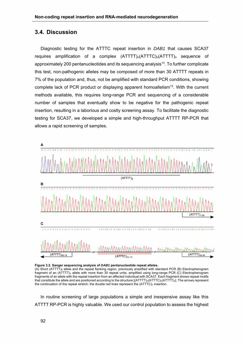
Non-coding repeat insertion and RNA-mediated neurodegeneration
92
3.4. Discussion
Diagnostic testing for the ATTTC repeat insertion in DAB1 that causes SCA37
requires amplification of a complex (ATTTT)n(ATTTC)n(ATTTT)n sequence of
approximately 200 pentanucleotides and its sequencing analysis13. To further complicate
this test, non-pathogenic alleles may be composed of more than 30 ATTTT repeats in
7% of the population and, thus, not be amplified with standard PCR conditions, showing
complete lack of PCR product or displaying apparent homoallelism13. With the current
methods available, this requires long-range PCR and sequencing of a considerable
number of samples that eventually show to be negative for the pathogenic repeat
insertion, resulting in a laborious and costly screening assay. To facilitate the diagnostic
testing for SCA37, we developed a simple and high-throughput ATTTT RP-PCR that
allows a rapid screening of samples.
Figure 3.2. Sanger sequencing analysis of DAB1 pentanucleotide repeat alleles. (A) Short (ATTTT)8 allele and the repeat flanking region, previously amplified with standard PCR (B) Electropherogram fragment of an (ATTTT)n allele with more than 30 repeat units, amplified using long-range PCR (C) Electropherogram fragments of an allele with the repeat insertion from an affected individual with SCA37. Each fragment shows repeat motifs that constitute the allele and are positioned according to the structure [(ATTTT)n(ATTTC)n(ATTTT)n]. The arrows represent the continuation of the repeat stretch; the double red lines represent the (ATTTC)n insertion.
In routine screening of large populations a simple and inexpensive assay like this
ATTTT RP-PCR is highly valuable. We used our control population to assess the highest

Chapter 3| A workflow for SCA37 diagnosis
93
effort needed in obtaining a conclusive molecular diagnostic. Standard PCR analysis can
easily detect pentanucleotide repeat alleles smaller than 30 in DAB1, allowing a
conclusive diagnosis in 71% of the samples based on the detection of both alleles (Figure
3.3). Larger alleles, however, are not successfully amplified by standard PCR conditions.
Therefore, in samples with apparent homoallelism ATTTT RP-PCR proves
advantageous by enabling a conclusive negative result in a total of 82% of the samples.
On the other hand, this method allows detection of pathogenic alleles with the
configuration [(ATTTT)60-79(ATTTC)31-75(ATTTT)58-90] despite the possibility of missing
alleles with very different configurations. Samples with a positive ATTTT RP-PCR need
to be amplified by long-range PCR followed by sequencing analysis.
Figure 3.3. Workflow for (ATTTC)n insertion screening. This workflow decreases the number of samples that require long-range PCR followed by Sanger sequencing. First, it is performed a standard PCR, followed by ATTTT RP-PCR in samples wherein only one allele is amplified. The ATTTT RP-PCR distinguishes homoallelism from large allele carriers. In samples with a positive result for the ATTTT RP-PCR long-range PCR and Sanger sequencing are performed to assess the size and composition of large alleles. In the control population studied, 71% of the samples required only standard PCR to complete the negative diagnosis; standard PCR followed by ATTTT RP-PCR in samples with one allele provided a conclusive negative result in 82% of the total samples; the remaining 18% required long-range PCR followed by Sanger sequencing.
In conclusion, we suggest that the ATTTT RP-PCR can be used as a rapid screening
test in the diagnostic protocol of SCA37 provided this is always followed by long-range
PCR amplification and sequencing analysis if a positive ATTTT RP-PCR is detected.

Non-coding repeat insertion and RNA-mediated neurodegeneration
94
3.5. Acknowledgments
This work was funded by Fundo Europeu de Desenvolvimento Regional-FEDER
funds through the COMPETE 2020 - Operational Programme for Competitiveness and
Internationalisation (POCI), Portugal 2020, and by funding from FCT - Fundação para a
Ciência e a Tecnologia/ Ministério da Ciência, Tecnologia e Inovação, Portugal, in the
framework of the project "Institute for Research and Innovation in Health Sciences"
(POCI-01-0145-FEDER-007274); by Grant PTDC/SAU-GMG/098305/2008, from FCT,
to I.S. J.R.L. was supported by scholarships from Grant PTDC/GMG-SAU/098305/2008,
FCT, PEst-C/SAU/LA0002/2013 and EMBO (ASTF494-2015). C.L.O was supported by
a scholarship from PEst-C/SAU/LA0002/2013. This work was also funded by Norte
Portugal Regional Operational Programme (NORTE 2020), under the PORTUGAL 2020
Partnership Agreement, through the European Regional Development Fund (FEDER),
Portugal, that supports the Norte-01-0145-FEDER-000008 - Porto Neurosciences and
Neurologic Disease Research Initiative at I3S.
3.6. References
1. Loureiro, J.R., Oliveira, C.L. & Silveira, I. (2016). Unstable repeat expansions in
neurodegenerative diseases: nucleocytoplasmic transport emerges on the scene.
Neurobiol Aging 39, 174-183.
2. Sequeiros, J., Martins, S. & Silveira, I. in Handb Clin Neurol, Vol. 103, 227-251,
(Netherlands 2012).
3. Huang, M. & Verbeek, D.S. (2018). Why do so many genetic insults lead to Purkinje
Cell degeneration and spinocerebellar ataxia? Neurosci Lett in press.
4. Paulson, H.L., Shakkottai, V.G., Clark, H.B. & Orr, H.T. (2017). Polyglutamine
spinocerebellar ataxias - from genes to potential treatments. Nat Rev Neurosci 18,
613-626.
5. Coutinho, P., Ruano, L., Loureiro, J., Cruz, V., Barros, J., Tuna, A. et al. (2013).
Hereditary Ataxia and Spastic Paraplegia in Portugal A Population-Based
Prevalence Study. JAMA Neurol 70, 746-755 .
6. Nelson, D.L., Orr, H.T. & Warren, S.T. (2013). The unstable repeats--three evolving
faces of neurological disease. Neuron 77, 825-843.

Chapter 3| A workflow for SCA37 diagnosis
95
7. Garcia-Murias, M., Quintans, B., Arias, M., Seixas, A.I., Cacheiro, P., Tarrio, R. et al.
'Costa da Morte' ataxia is spinocerebellar ataxia 36: clinical and genetic
characterization. (2012). Brain 135, 1423-1435.
8. Alonso, I., Jardim, L., Artigalas, O., Saraiva-Pereira, M., Matsuura, T., Ashizawa, T. et
al. (2006). Reduced penetrance of intermediate size alleles in spinocerebellar
ataxia type 10. Neurology 66, 1602-1604.
9. Silveira, I., Alonso, I., Guimarães, L., Mendonça, P., Santos, C., Maciel, P. et al. High
germinal instability of the (CTG)n at the SCA8 locus of both expanded and normal
alleles. (2000). Am J Hum Genet 66, 830-840.
10. Silveira, I., Miranda, C., Guimaraes, L., Moreira, M., Alonso, I., Mendonca, P. et al.
Trinucleotide repeats in 202 families with ataxia - A small expanded (CAG)(n) allele
at the SCA17 locus. (2002). Arch Neurol 59, 623-629.
11. Seixas, A.I., Vale, J., Jorge, P., Marques, I., Santos, R., Alonso, I. et al. FXTAS is
rare among Portuguese patients with movement disorders: FMR1 premutations
may be associated with a wider spectrum of phenotypes. (2011). Behav Brain
Funct 7, 19.
12. Sato, N., Amino, T., Kobayashi, K., Asakawa, S., Ishiguro, T., Tsunemi, T. et al.
Spinocerebellar ataxia type 31 is associated with "inserted" penta-nucleotide
repeats containing (TGGAA)n. (2009). Am J Hum Genet 85, 544-557.
13. Seixas, A.I., Loureiro, J.R., Costa, C., Ordonez-Ugalde, A., Marcelino, H., Oliveira,
C.L. et al. A Pentanucleotide ATTTC Repeat Insertion in the Non-coding Region of
DAB1, Mapping to SCA37, Causes Spinocerebellar Ataxia. (2017). Am J Hum
Genet 101, 87-103.
14. Ishikawa, K., Dürr, A., Klopstock, T., Müller, S., De Toffol, B., Vidailhet, M. et al.
Pentanucleotide repeats at the spinocerebellar ataxia type 31 (SCA31) locus in
Caucasians. (2011). Neurology 77, 1853-1855.
15. Cagnoli, C., Michielotto, C., Matsuura, T., Ashizawa, T., Margolis, R.L., Holmes, S.E.
et al. Detection of large pathogenic expansions in FRDA1, SCA10, and SCA12
genes using a simple fluorescent repeat-primed PCR assay. (2004). J Mol Diagn
6, 96-100.
16. Chen, L., Hadd, A., Sah, S., Filipovic-Sadic, S., Krosting, J., Sekinger, E. et al. An
information-rich CGG repeat primed PCR that detects the full range of fragile X
expanded alleles and minimizes the need for southern blot analysis (2010). J Mol
Diagn 12, 589-600.


Chapter 4. Mutational mechanism in the origin of the
non-coding (ATTTC)n insertion in DAB1
Adapted from Mutational Mechanism for DAB1 (ATTTC)n insertion in SCA37: ATTTT
Repeat Lengthening and Nucleotide substitution, Under review.


Chapter 4| Mutational mechanism in the origin of the non-coding (ATTTC)n insertion
99
Mutational Mechanism for DAB1 (ATTTC)n insertion in SCA37: ATTTT
Repeat Lengthening and Nucleotide substitution
Joana R. Loureiro1, 2, 3, Cláudia L. Oliveira1, 2, Carolina Mota1, 2, Ana F. Castro1, 2, Cristina Costa4, José
L. Loureiro2,5,6, Paula Coutinho2,5, Sandra Martins7, 8, Jorge Sequeiros2, 3, 5, Isabel Silveira1, 2
1Genetics of Cognitive Dysfunction Laboratory, i3S-Instituto de Investigação e Inovação em Saúde, Universidade do
Porto, 4200-135 Porto, Portugal; 2IBMC- Institute for Molecular and Cell Biology, Universidade do Porto, 4200-135
Porto, Portugal; 3ICBAS, Universidade do Porto, 4050-313 Porto, Portugal; 4Department of Neurology, Hospital Prof. Dr.
Fernando Fonseca, E.P.E, 2720-276 Amadora, Portugal; 5UnIGENe, i3S-Instituto de Investigação e Inovação em
Saúde, Universidade do Porto, 4200-135 Porto, Portugal; 6 Department of Neurology, Hospital São Sebastião, 4520-211
Feira, Portugal; 7Population Genetics & Evolution, i3S-Instituto de Investigação e Inovação em Saúde, Universidade do
Porto, 4200-135 Porto, Portugal; 8IPATIMUP - Institute of Molecular Pathology and Immunology, University of Porto,
Porto, Portugal.
Dynamic mutations by microsatellite instability are the molecular basis of a growing
number of neuromuscular and neurodegenerative diseases. Repetitive stretches in the
human genome may drive pathogenicity, either by expansion above a given threshold,
or by insertion of abnormal tracts in non-pathogenic polymorphic repetitive regions, as is
the case in spinocerebellar ataxia type 37 (SCA37). We have recently established that
this neurodegenerative disease is caused by an (ATTTC)n insertion within an (ATTTT)n
in a non-coding region of DAB1. We now investigated the mutational mechanism that
originated the (ATTTC)n insertion within an ancestral (ATTTT)n. Approximately 3% of
non-pathogenic (ATTTT)n alleles are interspersed by AT-rich motifs, contrarily to mutant
alleles that are composed of pure (ATTTT)n and (ATTTC)n stretches. Haplotype studies
in unaffected chromosomes suggested that the primary mutational mechanism, leading
to the (ATTTC)n insertion, was likely one or more T>C substitutions in an (ATTTT)n pure
allele of approximately 200 repeats. Then, the (ATTTC)n expanded in size, originating a
deleterious allele in DAB1 that leads to SCA37. This is likely the mutational mechanism
in three similar (TTTCA)n insertions responsible for familial myoclonic epilepsy. Because
(ATTTT)n tracts are frequent in the human genome, many loci could be at risk for this
mutational process.

Non-coding repeat insertion and RNA-mediated
100
4.1. Introduction
Spinocerebellar ataxia type 37 (SCA37; MIM# 615945) is an autosomal dominant
neurodegenerative disease, characterized by onset of dysarthria, followed by
progressive gait and limb ataxia1; 2. We have recently reported that SCA37 is caused by
an (ATTTC)n insertion in the middle of an ATTTT repeat located in a 5’UTR intron of
DAB1. Unaffected subjects carry (ATTTT)n alleles ranging from 7 to 400 units, while
affected individuals showed an allele with the configuration (ATTTT)60-79(ATTTC)31-
75(ATTTT)58-901. We have previously shown that pathogenic SCA37 alleles present
intergenerational instability towards expansion; the (ATTTC)n increases in size in 81% of
parent-offspring transmissions, whereas repeat insertion contractions have never been
detected in SCA37 pedigrees1. In pathogenic alleles, the flanking 5’-ATTTT repeat is
also unstable during transmission; the 3’ATTTT sequencing is incomplete, but for two
branches with full sequencing the ATTTT length shows instability upon transmission. In
SCA37 pedigrees, the large non-pathogenic (ATTTT)n alleles have also shown instability
upon transmission; in one family branch two siblings inherited from their unaffected
parent non-pathogenic alleles with different sizes1.
Over the last three decades, expansions of microsatellites with tri-, tetra-, penta- and
hexanucleotide repeats have been identified as the cause of more than 25 neurological
or neuromuscular diseases3. More recently, however, knowledge has been gathered
regarding a cryptic subclass of dynamic mutations consisting of an insertion of a
repetitive motif adjacent or within a polymorphic repeat in non-coding regions. In addition
to SCA37, this type of mutation underlies SCA31 (MIM# 117210) and benign adult
familial myoclonic epilepsy (BAFME1; MIM# 601068, 6 and 7)1; 4-6.
Microsatellite repetitive tracts are often interrupted by other nucleotide motifs. In some
diseases, as SCA1 (MIM# 164400), SCA2 (MIM# 183090), fragile-X syndrome (MIM#
300624), myotonic dystrophy type 2 (DM2; MIM# 602668) and Friedrich ataxia (FRDA;
MIM#229300), non-pathogenic alleles are more likely to be interrupted than expanded
alleles, whereas in SCA8 (MIM# 608768), SCA10 (MIM# 603516) and DM1 (MIM#
160900) interruptions are more expected in expanded alleles7-21. Depending on the gene,
interruptions in the repetitive tract may be randomly positioned, as in ATXN2, ATXN8OS
and ATXN10, or follow a pattern19. In FMR1, AGG interruptions are usually located every
nine or ten CGGs, whereas in ATXN1 interrupted alleles have one to three CAT
interruptions in the middle of the allele intercalated with one CAG, following a
configuration from (CAG)n CAT (CAG)n to (CAG)n CAT CAG CAT CAG CAT (CAG)n7; 12.
Repeat interruptions in non-pathogenic alleles stabilize the repeat tract7; 22; 23 in
expanded alleles at specific loci interruptions may decrease penetrance or delay age-of-

Chapter 4| Mutational mechanism in the origin of the non-coding (ATTTC)n insertion
101
onset, as in SCA1 and DM18; 9; 24; 25. In SCA10, however, interruptions in expanded alleles
may turn them more deleterious, lowering age-of-onset and associating with seizures26.
The pentanucleotide repeat associated with SCA37 is located in the middle poly-A of
an AluJb element1. Several other disease-causing repeats are in middle or 3’-end poly-
A regions of Alu elements, such as the repeat expansions responsible for FRDA, DM2
and SCA10, as well as the pentanucleotide repeat insertions associated with SCA31 and
two types of BAFME4-6; 15; 27-30.
Notably, loci containing repeat insertions are similar in that (1) they have an
ATTTT/TAAAA repeat in the reference genome database, (2) shorter repeats with the
insertion motif have not been detected in the general population, (3) non-pathogenic
ATTTT/TAAAA alleles are highly polymorphic (ranging from a few to hundreds of
repeats), and (4) pathogenic alleles have a complex configuration of both non-
pathogenic repeat and insertion motifs. Such a significant number of loci with repeat
insertions indicates that mutational events on the origin of these deleterious alleles are
recurrent in the human genome.
To gain insight into the mutational mechanisms responsible for origin, evolution and
instability of these pathogenic repeat insertions, we focused on the SCA37 locus. Thus,
we (1) characterized the complexity of the pentanucleotide repeat in non-pathogenic and
pathogenic alleles, (2) compared orthologous sequences along the primate lineage, and
(3) analyzed haplotypes flanking the repeat in humans. Our results suggest that the
mutational mechanism responsible for pathogenic repeat insertions involves ATTTT
repeat lengthening and nucleotide substitution, providing a simple explanation for the
birth of these dynamic mutations from ancestral alleles.
4.2. Materials and Methods
4.2.1. Subjects
We used DNA samples from 44 SCA37 affected individuals, 260 control subjects from
the Portuguese general population, and 394 individuals with neurodegenerative
diseases without the repeat insertion, as previously published1. Affected individuals were
referred for diagnostic purposes to the authorized Center for Predictive and Preventive
Genetics (CGPP) at the Institute for Molecular and Cell Biology. This study used the de-
identified, previously collected DNA samples that were stored at the CGPP biobank, as
well as anonymized DNA samples from internal lab controls.

Non-coding repeat insertion and RNA-mediated
102
4.2.2. Assessment of short pentanucleotide repeat alleles
Repeat size of short alleles was assessed by standard PCR with HotStartTaq Master
Mix Kit (QIAGEN), 0.4 µM of primers 24F and 24R1, and 30 ng of DNA, in a final volume
of 12.5 µL. PCR was performed with an initial denaturation of 15 min at 95ºC, followed
by 30 cycles of amplification (95ºC for 45 sec, 64ºC for 30 sec and 72ºC for 40 sec), and
a final extension at 72ºC for 10 min. The allele structure (AS) was determined by Sanger
sequencing of PCR products, with primers 24F and 24R1.
4.2.3. Identification of the allele structure in large alleles
To detect large alleles or confirm homoallelism (two alleles of the same size), in
samples where only one allele was detected by standard PCR, we carried out RP-PCR
with a specific primer to anneal with the (ATTTT)n tract, as previously described31.
Samples with a positive ATTTT RP-PCR were amplified with long range-PCR1. PCR
products were separated by electrophoresis, in 1% agarose gel. DNA fragments were
extracted from the gel, purified with QIAquick gel extraction kit (QIAGEN) and
sequencing analysis was performed with the internal primers 24F or 24R4, as published1;
31. For very large alleles, with over 200 repeat units or more than 1 kb, Sanger
sequencing in both forward and reverse orientations did not completely overlap, but a
partial overlap of the ATTTT repeat was safely achieved, and was in accordance with
the estimated repeat size in agarose gel. Each allele structure found in one chromosome
only was confirmed by PCR and sequencing replicates.
4.2.4. Haplotype analysis
Haplotypes were constructed with 8 SNPs, spanning a region of 723 kb flanking the
DAB1 pentanucleotide repeat, including (1) rs1043184969 and rs954450605 upstream,
and rs192485043, rs145097803 and rs929412570 downstream the repeat, as described
before1; (2) and three additional SNPs, presenting higher minor allele frequencies and
closer to the repetitive region, rs514412, rs2113453 and rs11207020; positions and
physical distances are according to GRCh37/hg19. SNP regions were PCR amplified
with GoTaq Green® Mastermix (Promega), 1.25 µM of primers F and R (Supp. Table
S4.1) and 30 ng DNA. The initial denaturation at 95ºC for 3 min was followed by 35
cycles of amplification at 95ºC for 45 sec, annealing temperature for 30 sec (Supp. Table
S4.1) and 72ºC for 40 sec, and a final extension at 72ºC for 10 min. Allele discrimination
was performed by RFLP or by Sanger sequencing (Supp. Table S4.1). Haplotype phases

Chapter 4| Mutational mechanism in the origin of the non-coding (ATTTC)n insertion
103
were assessed using PHASE v.2.1.1
(http://stephenslab.uchicago.edu/phase/download.html); only haplotypes with probability
higher than 0.6 were included in further analyses. Phylogenetic relationships among
unaffected short pure, large pure and interrupted alleles were assessed by median-
joining method, using the Network 5.0.0.3 software (www.fluxus-engineering.com).
4.2.5. Pentanucleotide repeat evolution
Time of divergence between human and primate species was calculated using the
node time searching data in the public knowledge-base TimeTree32; 33. Data from
nucleotide to pentanucleotide repeat evolution in DAB1 was obtained from Vertebrate
Multiz Alignment & Conservation, UCSC genome browser34. Data included whole
genome assemblies from shotgun sequencing on 454 GS FLX and GS FLX large read
platforms from Pan troglodytes (chimpanzee), Gorilla gorilla (gorilla), Pongo abelii
(orangutan), Nomascus leucogenys (gibbon), Macaca fascicularis (crab eating
macaque), Macaca mulatta (rhesus), Papio hamadryas (baboon), Chlorocebus sabaeus
(green monkey), Callithrix jacchus (marmoset), Saimiri boliviensis (squirrel monkey) and
Otolemur garnettii (bushbaby); the reference genome for each primate specie includes
a single allele.
4.3. Results
4.3.1. Unaffected alleles may be interrupted
To characterize the pentanucleotide repeat sequence in DAB1, we performed
Sanger sequencing. We found that both unaffected (ATTTT)n and pathogenic SCA37
(ATTTT)n(ATTTC)n(ATTTT)n alleles were always preceded, at the 5’-end, by two
ATTTs that were not polymorphic. From the 1,308 unaffected chromosomes studied,
we assessed the purity or interrupted nature in 1,293 alleles; 1,249 (96.6%) were
composed by pure repetitive stretches of (ATTTT)n, one of which had only one ACTTT
at the 3’-end of the repeat, while 44 alleles (3.4%) which ranged from 16 to about 400
repeat units had AT-rich interruptions (Table 4.1). The complete allele structure of
interrupted alleles was only assessed for 30 alleles (Figure 4.1). SCA37 alleles had no
interruptions in neither (ATTTT)n, nor (ATTTC)n tracts (Figure 4.1).

Non-coding repeat insertion and RNA-mediated
104
In non-disease associated (ATTTT)n chromosomes, we detected seven types of
interruption motifs in the 30 fully sequenced alleles. These interruptions included a single
A or A(T)n, varying from di- to tetra- or hexa- to octanucleotide (Figure 4.1); the most
common was ATTT (46.6%), followed by AT (29.3%) (Figure 4.2); an ATTTTTTT was
found only in one allele. Regarding the position of the interruptions, we found nine
different allele configurations; 17 alleles (56.7%) had an allele structure (AS) F; the G
and J structures were detected in three alleles each (10%), and two alleles (6.7%) had
the AS C (Figure 4.1). The remaining interrupted alleles did not follow any common
interruption pattern.
Figure 4.1. Schematic representation of structure of non-pathogenic and pathogenic (SCA37) alleles with nucleotide variations. Allele size is the number of pentanucleotide repeats in each allele and is represented by white and dashed boxes. The seven types of interruption motifs, shown by colored boxes, and the (ATTT)2 preceding the pentanucleotide repeat, indicated by two grey boxes, were not considered for repeat size assessment. The T>C substitution in pattern B is ss2137543903. The patterns of interruptions are shown from C to K for the 30 fully sequenced alleles at the 3’-end from the total of 44 interrupted alleles; number of (ATTTT)n and (ATTTC)n in each stretch are indicated inside the respective box. AS- Allele structure.
4.3.2. Interruptions are associated with ATTTT repeat size
To investigate whether repeat interruptions in unaffected chromosomes were
associated with specific allele sizes, we divided them in three subgroups based on repeat

Chapter 4| Mutational mechanism in the origin of the non-coding (ATTTC)n insertion
105
length: alleles with 30 ATTTTs or less, alleles ranging from 31 to 79 repeats and alleles
with 80 or more repeats (Table 4.1). In 1,226 alleles with 30 repeats or less, only two,
with 16 and 17 ATTTTs, had one interruption (0.2%); whereas in alleles of 31 to 79
ATTTTs, 27 had interruptions (69.2%); and in alleles with 80 repeats or more, 15 were
interrupted (53.6%) (Table 4.1). The frequency of interrupted alleles was higher in alleles
ranging from 31-79 repeats than in alleles with 30 repeats or less (Fisher’s exact test,
p<0.0001) and was also higher in alleles with 80 or more repeats than in alleles with 30
repeats or less (Fisher’s exact test, p<0.0001). The difference in frequency between
interrupted alleles ranging from 31-79 and with 80 repeats or more was not statistically
significant.
Regarding the number of allele interruptions (one to eight) in the fully sequenced
alleles, they increased with repeat size: interrupted alleles with less than 30 repeats had
only one, while larger alleles had from two to four or seven to eight interruptions for
repeat sizes below or 80 units and above, respectively (Figure 4.1 and 4.3).
Table 4.1. Frequency of pure and interrupted DAB1 alleles in 1,308a unaffected chromosomes
Allele size Allele type
Unaffected chromosomes
n %
≤30 Pure 1,224 99.8%
Interrupted 2 0.2%
Total 1,226
31-79 Pure 12 30.8%
Interrupted 27 69.2%
Total 39
≥80 Pure 13 46.4%
Interrupted 15 53.6%
Total 28
Total Pure 1,249 96.6%
Interruptedb 44 3.4%
Totala 1,293 a15 additional ATTTT alleles were studied, but could not be fully sequenced at the 3’-end for purity assessment; bThe allele structure was completely characterized at 3’-end in only 30 alleles.

Non-coding repeat insertion and RNA-mediated
106
Figure 4.2. Frequency of each AT-rich interruption in the interrupted alleles fully sequenced (n=30).
Two types of AT-rich interruptions were exclusively detected in alleles of 31-79
repeats (A and ATTTTT); and another three types of interruptions were only detected in
alleles over 80 repeats (ATT, ATTTTTT and ATTTTTTT) (Figure 4.1).
Figure 4.3. Number of interruptions associated with repeat size in the interrupted alleles fully sequenced (n=30). Haplotypes associated with each type of repeat alleles are represented with different colors; n.d.- not determined.
4.3.3. Evolution of pentanucleotide alleles in primate lineage
In DAB1, the ATTTT repeat is in the middle poly-A region of an AluJb element
encoded in the opposite strand1. To understand how the pentanucleotide emerged and
evolved, we compared the orthologous region of primates by using reference primate
databases (Figure 4.4). We found that the AluJb element was inserted in the DAB1-
opposite strand before New World monkeys divergence at approximately 43.2 million
years ago (mya). The Alu middle poly-A, in the DAB1-opposite strand, was composed of
a pure stretch of adenines, as observed in New World monkeys. Before the Old World
monkeys divergence, approximately 29.44 mya, the Alu middle poly-A increased its size

Chapter 4| Mutational mechanism in the origin of the non-coding (ATTTC)n insertion
107
and started mutating. In Old World monkeys, the (ATTTT)n in DAB1-oriented strand is
preceded by stretches of thymines and adenines and by an ATTT. The pure stretches of
Ts and As mutated before the Hominoids divergence, approximately 20.19 mya. In
Hominoids, the repeat is composed by one or two ATTTs, followed by a pure or
interrupted (ATTTT)n. We did not find ATTTC repeat motifs in any primate genome data
available.
Figure 4.4. Evolution of the DAB1 ATTTT repeat in primate lineages. AluJb retrotransposition event in DAB1-opposite strand is represented by a black box. For simplicity, nucleotide composition of Alu poly-A in primate species is represented in DAB1-oriented strand. The length of the phylogenetic tree branches represents the evolutionary distance among species; M- million years ago.
4.3.4. The SCA37 haplotype is very rare in Portuguese unaffected
chromosomes
SCA37 is associated with a rare haplotype shared by all affected individuals studied
so far1. To investigate mutational mechanisms leading to the origin of the (ATTTC)n
insertion, we constructed SNP haplotypes of non-pathogenic pure and interrupted
(ATTTT)n alleles and compared them to the single SCA37 haplotype.
We found eight haplotypes to be associated with non-pathogenic alleles, identified
from I to VIII (Table 4.2 and Supp. Table S4.2). Six (haplotypes I, II, III, V, VI and VII)
were associated with short pure alleles (<100 ATTTTs). Interrupted alleles shared three
haplotypes (I, IV and V) (Figure 4.3), whereas large pure alleles (>100 ATTTTs)
segregated with only two haplotypes (I and VIII). Haplotype I was shared by short pure,
large pure and interrupted alleles, while haplotype V was common to short pure and
interrupted alleles. Haplotype VIII, the one associated with the (ATTTC)n insertion in

Non-coding repeat insertion and RNA-mediated
108
SCA37, was seen only in two unaffected chromosomes, both carrying a pure allele with
approximately 200 ATTTT repeats.
To explore the origin of the SCA37 mutation, we designed a haplotype network to
visualize genetic distance among short pure, large pure and interrupted alleles (Figure
4.5). Short pure alleles were phylogenetically closer to each other, as interrupted alleles
were; large pure alleles with haplotype VIII were genetically closer to interrupted alleles
than to short pure repeats. As the SCA37 haplotype is very rare in the Portuguese
population and genetically distant from the haplotypes found in non-pathogenic alleles,
we hypothesized it could have been introduced from another population where it may
have a higher frequency. Therefore, we analyzed data from phase 3 database of The
1000 Genomes Project (The Genomes Project, 2015); although several SNP alleles from
the SCA37 haplotype have been observed in some persons of African, European and
Asian ancestry, the full SCA37-associated haplotype was not detected in those
populations (Supp. Table S4.3).
Figure 4.5. Close phylogenetic relationship among non-pathogenic DAB1 alleles. Haplotype network showing the phylogenetic relationship among non-pathogenic (short pure, large pure and interrupted) alleles. Circle size is proportional to the number of chromosomes; line length is proportional to the genetic distance among haplotypes. Short pure alleles (<100 ATTTTs) are represented in white; large pure alleles in black (>100 ATTTTs) and interrupted alleles in grey.
4.4. Discussion
This study provides a plausible mutational mechanism for the origin of the ATTTC
repeat insertion in SCA37. Haplotype analysis allowed us to identify a class of large non-
pathogenic alleles at DAB1 characterized by (1) the acquisition of A or A(T)n repeat
interruptions and (2) repeat size instability. This analysis showed a haplotype shared
among non-pathogenic short pure, interrupted and large pure alleles, indicating that both

Chapter 4| Mutational mechanism in the origin of the non-coding (ATTTC)n insertion
109
nucleotide changes and repeat size instability are likely in the origin of the polymorphic
pentanucleotide repeat at DAB1. The haplotype network (Figure 4.5) shows a clear
distance among short pure stable, large and interrupted unstable alleles, suggesting that
genetic variants flanking the mutant allele act as cis elements influencing repeat
instability. The evolution of the repetitive region in primates (Figure 4.4) further supports
this observation. Haplotype studies in non-pathogenic and pathogenic chromosomes
suggest that the mutational mechanism towards the acquisition of the deleterious ATTTC
repeat insertion in SCA37 was one or more T>C substitutions in a large pure ATTTT
allele.
The pure (ATTTT)n in DAB1-oriented strand (at the SCA37 locus) emerged after
several A>T substitutions in the Alu poly-A (Alu orientation), which originated the AT-rich
stretches identified in various species from the primate lineage. Then, the pure tracts of
adenines and thymines seem to have disappeared by the occurrence of more A>T or
T>A substitutions or indels, followed by an increased number of pentanucleotides,
originating the pure pentanucleotide alleles found in chimpanzee and humans. Thus, the
primates genomes show that the evolution of the pure (ATTTT)n resulted from numerous
mutational events in pure poly-A, to finally originate the pure highly unstable
pentanucleotide repeat found nowadays that later suffered T>C transition(s) which
increased in size and became the SCA37 causing variant.
Different cellular mechanisms have been associated with gain and loss of genetic
material in repeat loci36-39. The repetitive nature of these genomic regions burden normal
cell mechanisms like replication and transcription, leading to an increase in DNA breaks
in repeats40; 41. The mechanisms responsible for DNA break-repair have been largely
associated with repeat size instability36; 42-44 and may have had a role in the highly
polymorphic nature of this pentanucleotide repeat.
The ultimate mutational mechanism leading to a (ATTTC)n insertion seems to be
recurrent in the human genome. Similar (TTTCA)n mutations have been identified in
three types of BAFME5; 6. The primary mutational feature, common to these diseases, is
likely the T>C substitution(s) in the last thymine of the ATTTT or TTTTA repeats, followed
by size increase of the (ATTTC)n or (TTTCA)n, as a result of its unstable nature. This
new type of mutational mechanism should be taken into consideration when searching
new disease-causing variants.
In conclusion, the repeat region associated with SCA37 is highly polymorphic,
mutable and unstable, and the AluJb seems to have had a role in these features, as the
(ATTTC)n insertion was probably originated by one or more T>C substitutions in the Alu
element. Furthermore, this type of events seems to be recurrent in Alu ATTTT motifs in

Non-coding repeat insertion and RNA-mediated
110
the human genome, suggesting that other AT-rich repeats can be implicated in other
brain diseases.

Chapter 4| Mutational mechanism in the origin of the non-coding (ATTTC)n insertion
111
Table 4.2. Haplotypes in pure and interrupted DAB1 non-pathogenic alleles
Haplotypes
SNP Position
chr1 (hg19)
Distance from
(ATTTT)n
(bp)
Ancestral allele
MAF Alla MAF
EURa MAF PTb
(ATTTC)n insertion
Pure
(ATTTT)>100
Interrupted (ATTTT)n
Pure (ATTTT)<100
rs1043184969 57,491,966 -340,750 T * * G: 0.0007 G G T T T T T T T T T T rs514412 57,551,569 -281,147 G A: 0.1949 A: 0.1998 A: 0.2120 G G G G G A G G G A A A
rs954450605 57,551,605 -281,111 G * * A: 0.0013 A A G G G G G G G G G G (ATTTT)n 57,832,716 0
rs2113453 57,833,689 +973 C C: 0.4157 T: 0.4750 C: 0.2600 T T T T T T C T T T T C rs11207020 57,834,035 +1,319 C T: 0.2171 T: 0.2843 T: 0.4700 T T T T T T C C T C T C
rs192485043 57,926,567 +93,851 T A: 0.0002 A: 0.0010 A: 0.0015 A A T T T T T T T T T T rs145097803 58,201,704 +368,988 T A: 0.0032 * A: 0.0046 A A T T T T T T T T T T rs929412570 58,215,160 +382,444 C * * G: 0.0007 G G C C G C C C C C C C
Absolute frequency
44 2 11 10 8 3 25 17 11 10 2 1
Haplotype ID VIII VIII I I IV V III II I VI V VII aMinor allele frequency from 1000 genomes phase 3; bMinor allele frequency in Portugal: Seixas et al, 2017 and present study; EUR- Europe; PT- Portugal; *Not available or not found.
Non-coding repeat insertion and RNA-mediated
112
4.5. Acknowledgements
We are grateful to the families and individuals who participated in this work. We thank
Patricia Ribeiro for technical assistance. This study was financed by Fundo Europeu de
Desenvolvimento Regional (FEDER), through the COMPETE 2020 Operational Program
for Competitiveness and Internationalization (POCI) of Portugal 2020, and by the
Fundacão para a Ciência e a Tecnologia (FCT) and Ministério da Ciência, Tecnologia e
Inovação (Portugal), in the framework of the project Instituto de Investigação e Inovação
em Saúde (i3S) (POCI-01-0145-FEDER-007274); and by FCT grant PTDC/MED-
GEN/29255/2017 to I.S. J.R.L. and C.L.O. were supported by scholarships from PEst-
C/SAU/LA0002/2013. SM is funded by the project IF/00930/2013/ CP1184/CT0002 from
FCT. This work was also funded by the Porto Neurosciences and Neurologic Disease
Research Initiative at the Instituto de Investigação e Inovação em Saúde (Norte-01-
0145-FEDER-000008), supported by Norte Portugal Regional Operational Programme
(NORTE 2020), under the PORTUGAL 2020 Partnership Agreement with FEDER.
4.6. References
1. Seixas, A.I., Loureiro, J.R., Costa, C., Ordóñez-Ugalde, A., Marcelino, H., Oliveira,
C.L., Loureiro, J.L., Dhingra, A., Brandão, E., Cruz, V.T., et al. (2017). A
Pentanucleotide ATTTC Repeat Insertion in the Non-coding Region of DAB1,
Mapping to SCA37, Causes Spinocerebellar Ataxia. Am J of Hum Genet 101, 87-
103.
2. Serrano-Munuera, C., Corral-Juan, M., Stevanin, G., San Nicolas, H., Roig, C., Corral,
J., Campos, B., de Jorge, L., Morcillo-Suarez, C., Navarro, A., et al. (2013). New
subtype of spinocerebellar ataxia with altered vertical eye movements mapping to
chromosome 1p32. JAMA Neurol 70, 764-771.
3. Loureiro, J.R., Oliveira, C.L., and Silveira, I. (2016). Unstable repeat expansions in
neurodegenerative diseases: nucleocytoplasmic transport emerges on the scene.
Neurobiol Aging 39, 174-183.
4. Sato, N., Amino, T., Kobayashi, K., Asakawa, S., Ishiguro, T., Tsunemi, T., Takahashi,
M., Matsuura, T., Flanigan, K.M., Iwasaki, S., et al. (2009). Spinocerebellar ataxia
type 31 is associated with "inserted" penta-nucleotide repeats containing
(TGGAA)n. Am J Hum Genet 85, 544-557.

Chapter 4| Mutational mechanism in the origin of the non-coding (ATTTC)n insertion
113
5. Ishiura, H., Doi, K., Mitsui, J., Yoshimura, J., Matsukawa, M.K., Fujiyama, A.,
Toyoshima, Y., Kakita, A., Takahashi, H., Suzuki, Y., et al. (2018). Expansions of
intronic TTTCA and TTTTA repeats in benign adult familial myoclonic epilepsy. Nat
Genet 50, 581-590.
6. Cen, Z., Jiang, Z., Chen, Y., Zheng, X., Xie, F., Yang, X., Lu, X., Ouyang, Z., Wu, H.,
Chen, S., et al. (2018). Intronic pentanucleotide TTTCA repeat insertion in the
SAMD12 gene causes familial cortical myoclonic tremor with epilepsy type 1.
Brain, In Press.
7. Chung, M.Y., Ranum, L.P., Duvick, L.A., Servadio, A., Zoghbi, H.Y., and Orr, H.T.
(1993). Evidence for a mechanism predisposing to intergenerational CAG repeat
instability in spinocerebellar ataxia type I. Nat Genet 5, 254-258.
8. Menon, R.P., Nethisinghe, S., Faggiano, S., Vannocci, T., Rezaei, H., Pemble, S.,
Sweeney, M.G., Wood, N.W., Davis, M.B., Pastore, A., et al. (2013). The Role of
Interruptions in polyQ in the Pathology of SCA1. PLoS Genet 9, e1003648.
9. Zuhlke, C., Dalski, A., Hellenbroich, Y., Bubel, S., Schwinger, E., and Burk, K. (2002).
Spinocerebellar ataxia type 1 (SCA1): phenotype-genotype correlation studies in
intermediate alleles. Eur J Hum Genet 10, 204-209.
10. Imbert, G., Saudou, F., Yvert, G., Devys, D., Trottier, Y., Garnier, J.M., Weber, C.,
Mandel, J.L., Cancel, G., Abbas, N., et al. (1996). Cloning of the gene for
spinocerebellar ataxia 2 reveals a locus with high sensitivity to expanded
CAG/glutamine repeats. Nat Genet 14, 285-291.
11. Ramos, E.M., Martins, S., Alonso, I., Emmel, V.E., Saraiva-Pereira, M.L., Jardim,
L.B., Coutinho, P., Sequeiros, J., and Silveira, I. (2010). Common origin of pure
and interrupted repeat expansions in spinocerebellar ataxia type 2 (SCA2). Am J
Med Genet B 153, 524-531.
12. Yrigollen, C.M., Sweha, S., Durbin-Johnson, B., Zhou, L., Berry-Kravis, E.,
Fernandez-Carvajal, I., Faradz, S.M., Amiri, K., Shaheen, H., Polli, R., et al. (2014).
Distribution of AGG interruption patterns within nine world populations. Intractable
Rare Dis Res 3, 153-161.
13. Maia, N., Loureiro, J.R., Oliveira, B., Marques, I., Santos, R., Jorge, P., and Martins,
S. (2017). Contraction of fully expanded FMR1 alleles to the normal range:
predisposing haplotype or rare events? J Hum Genet 62, 269-275.
14. Liquori, C.L., Ikeda, Y., Weatherspoon, M., Ricker, K., Schoser, B.G.H., Dalton, J.C.,
Day, J.W., and Ranum, L.P.W. (2003). Myotonic Dystrophy Type 2: Human
Non-coding repeat insertion and RNA-mediated
114
Founder Haplotype and Evolutionary Conservation of the Repeat Tract. Am J Hum
Genet 73, 849-862.
15. Montermini, L., Andermann, E., Labuda, M., Richter, A., Pandolfo, M., Cavalcanti, F.,
Pianese, L., Iodice, L., Farina, G., Monticelli, A., et al. (1997). The Friedreich ataxia
GAA triplet repeat: premutation and normal alleles. Hum Mol Genet 6, 1261-1266.
16. Moseley, M.L., Schut, L.J., Bird, T.D., Koob, M.D., Day, J.W., and Ranum, L.P.W.
(2000). SCA8 CTG repeat: en masse contractions in sperm and intergenerational
sequence changes may play a role in reduced penetrance. Hum Mol Genet 9,
2125-2130.
17. Martins, S., Seixas, A.I., Magalhaes, P., Coutinho, P., Sequeiros, J., and Silveira, I.
(2005). Haplotype diversity and somatic instability in normal and expanded SCA8
alleles. Am J Med Genet B 139, 109-114.
18. Hu, Y., Hashimoto, Y., Ishii, T., Rayle, M., Soga, K., Sato, N., Okita, M., Higashi, M.,
Ozaki, K., Mizusawa, H., et al. (2017). Sequence configuration of spinocerebellar
ataxia type 8 repeat expansions in a Japanese cohort of 797 ataxia subjects. J
Neurol Sci 382, 87-90.
19. Landrian, I., McFarland, K.N., Liu, J., Mulligan, C.J., Rasmussen, A., and Ashizawa,
T. (2017). Inheritance patterns of ATCCT repeat interruptions in spinocerebellar
ataxia type 10 (SCA10) expansions. PloS One 12, e0175958.
20. Braida, C., Stefanatos, R.K.A., Adam, B., Mahajan, N., Smeets, H.J.M., Niel, F.,
Goizet, C., Arveiler, B., Koenig, M., Lagier-Tourenne, C., et al. (2010). Variant CCG
and GGC repeats within the CTG expansion dramatically modify mutational
dynamics and likely contribute toward unusual symptoms in some myotonic
dystrophy type 1 patients. Hum Mol Genet 19, 1399-1412.
21. Musova, Z., Mazanec, R., Krepelova, A., Ehler, E., Vales, J., Jaklova, R., Prochazka,
T., Koukal, P., Marikova, T., Kraus, J., et al. (2009). Highly unstable sequence
interruptions of the CTG repeat in the myotonic dystrophy gene. Am J Med Genet
A 149, 1365-1374.
22. Choudhry, S., Mukerji, M., Srivastava, A.K., Jain, S., and Brahmachari, S.K. (2001).
CAG repeat instability at SCA2 locus: anchoring CAA interruptions and linked
single nucleotide polymorphisms. Hum Mol Genet 10, 2437-2446.
23. Yrigollen, C.M., Durbin-Johnson, B., Gane, L., Nelson, D.L., Hagerman, R.,
Hagerman, P.J., and Tassone, F. (2012). AGG interruptions within the maternal

Chapter 4| Mutational mechanism in the origin of the non-coding (ATTTC)n insertion
115
FMR1 gene reduce the risk of offspring with fragile X syndrome. Genet Med 14,
729-736.
24. Botta, A., Rossi, G., Marcaurelio, M., Fontana, L., D'Apice, M.R., Brancati, F., Massa,
R., D, G.M., Sangiuolo, F., and Novelli, G. (2017). Identification and
characterization of 5' CCG interruptions in complex DMPK expanded alleles. Eur
J Hum Genet 25, 257-261.
25. Matsuyama, Z., Izumi, Y., Kameyama, M., Kawakami, H., and Nakamura, S. (1999).
The effect of CAT trinucleotide interruptions on the age at onset of spinocerebellar
ataxia type 1 (SCA1). J Med Genet 36, 546-548.
26. Matsuura, T., Fang, P., Pearson, C.E., Jayakar, P., Ashizawa, T., Roa, B.B., and
Nelson, D.L. (2006). Interruptions in the Expanded ATTCT Repeat of
Spinocerebellar Ataxia Type 10: Repeat Purity as a Disease Modifier? Am J Hum
Genet 78, 125-129.
27. Chauhan, C., Dash, D., Grover, D., Rajamani, J., and Mukerji, M. (2002). Origin and
instability of GAA repeats: insights from Alu elements. J Biomol Struct Dyn 20, 253-
263.
28. Clark, R.M., Dalgliesh, G.L., Endres, D., Gomez, M., Taylor, J., and Bidichandani,
S.I. (2004). Expansion of GAA triplet repeats in the human genome: unique origin
of the FRDA mutation at the center of an Alu. Genomics 83, 373-383.
29. Kurosaki, T., Ueda, S., Ishida, T., Abe, K., Ohno, K., and Matsuura, T. (2012). The
unstable CCTG repeat responsible for myotonic dystrophy type 2 originates from
an AluSx element insertion into an early primate genome. PLoS One 7, e38379.
30. Kurosaki, T., Matsuura, T., Ohno, K., and Ueda, S. (2009). Alu-mediated acquisition
of unstable ATTCT pentanucleotide repeats in the human ATXN10 gene. Mol Biol
Evol 26, 2573-2579.
31. Loureiro, J.R., Oliveira, C.L., Sequeiros, J., and Silveira, I. (2018). A repeat-primed
PCR assay for pentanucleotide repeat alleles in spinocerebellar ataxia type 37. J
Hum Genet 63 981-987.
32. Hedges, S.B., Dudley, J., and Kumar, S. (2006). TimeTree: a public knowledge-base
of divergence times among organisms. Bioinformatics 22, 2971-2972.
33. Kumar, S., Stecher, G., Suleski, M., and Hedges, S.B. (2017). TimeTree: A Resource
for Timelines, Timetrees, and Divergence Times. Mol Biol Evol 34, 1812-1819.

Non-coding repeat insertion and RNA-mediated
116
34. Blanchette, M., Kent, W.J., Riemer, C., Elnitski, L., Smit, A.F., Roskin, K.M.,
Baertsch, R., Rosenbloom, K., Clawson, H., Green, E.D., et al. (2004). Aligning
multiple genomic sequences with the threaded blockset aligner. Genome Res 14,
708-715.
35. Corral-Juan, M., Serrano-Munuera, C., Rábano, A., Cota-González, D., Segarra-
Roca, A., Ispierto, L., Cano-Orgaz, A.T., Adarmes, A.D., Méndez-del-Barrio, C.,
Jesús, S., et al. (2018). Clinical, genetic and neuropathological characterization of
spinocerebellar ataxia type 37. Brain 140 1987-1997.
36. Gomes-Pereira, M., Hilley, J.D., Morales, F., Adam, B., James, H.E., and Monckton,
D.G. (2014). Disease-associated CAGꞏ CTG triplet repeats expand rapidly in non-
dividing mouse cells, but cell cycle arrest is insufficient to drive expansion. Nucleic
Acids Res 42, 7047-7056.
37. Martins, S., Pearson, C.E., Coutinho, P., Provost, S., Amorim, A., Dube, M.P.,
Sequeiros, J., and Rouleau, G.A. (2014). Modifiers of (CAG)(n) instability in
Machado-Joseph disease (MJD/SCA3) transmissions: an association study with
DNA replication, repair and recombination genes. Hum Genet 133, 1311-1318.
38. Santos, D., Pimenta, J., Wong, V.C., Amorim, A., and Martins, S. (2014). Diversity in
the androgen receptor CAG repeat has been shaped by a multistep mutational
mechanism. Am J Med Genet B 165, 581-586.
39. Slean, M.M., Panigrahi, G.B., Castel, A.L., Pearson, A.B., Tomkinson, A.E., and
Pearson, C.E. (2016). Absence of MutSbeta leads to the formation of slipped-DNA
for CTG/CAG contractions at primate replication forks. DNA Repair (Amst) 42, 107-
118.
40. Krasilnikova, M.M., and Mirkin, S.M. (2004). Replication Stalling at Friedreich's
Ataxia (GAA)(n) Repeats In Vivo. Mol Cell Biol 24, 2286-2295.
41. Zhang, Y., Shishkin, Alexander A., Nishida, Y., Marcinkowski-Desmond, D., Saini,
N., Volkov, Kirill V., Mirkin, Sergei M., and Lobachev, Kirill S. (2012). Genome-
wide Screen Identifies Pathways that Govern GAA/TTC Repeat Fragility and
Expansions in Dividing and Nondividing Yeast Cells. Mol Cell 48, 254-265.
42. Axford, M.M., Wang, Y.H., Nakamori, M., Zannis-Hadjopoulos, M., Thornton, C.A.,
and Pearson, C.E. (2013). Detection of slipped-DNAs at the trinucleotide repeats
of the myotonic dystrophy type I disease locus in patient tissues. PLoS Genet 9,
e1003866.

Chapter 4| Mutational mechanism in the origin of the non-coding (ATTTC)n insertion
117
43. Lee, J.M., Zhang, J., Su, A.I., Walker, J.R., Wiltshire, T., Kang, K., Dragileva, E.,
Gillis, T., Lopez, E.T., Boily, M.J., et al. (2010). A novel approach to investigate
tissue-specific trinucleotide repeat instability. BMC Sys Biol 4, 29.
44. Polleys, E.J., House, N.C.M., and Freudenreich, C.H. (2017). Role of recombination
and replication fork restart in repeat instability. DNA Repair 56, 156-165.

Non-coding repeat insertion and RNA-mediated
118
4.7. Supplemental data
Supplemental Table S4.1. PCR conditions and primers for selected SNP
SNP ID Primers (5'-3') Anneling
temperature SNP detection
rs1043184969 F- gcccttactgacgtgaccat
60ºC RFLP: AjiI Thermo scientific R- cttgaatgcagccagaatca
rs954450605 F- gcccagcattttattgccta
62ºC Sanger sequencing R- ggggtggcagagttcagtaa
rs2113453 F- caggaactccccacatgc
60ºC RFLP: Eco130I Thermo Scientific R- ccagcactgagctgctctc
rs11207020 F- cacacactcagggcactgtt
60ºC RFLP: SsiI Thermo scientific R-ctgagcttggggcttttatg
rs192485043 F- gaggccccttcattctctct
64ºC Sanger sequencing R- tgggttgacaggtacagcaa
rs145097803 F- gggaggcattaatgtgctttt
60ºC RFLP: EcoRV New England Biolabs R- cccgggattaatgcacttct
rs929412570 F- ctgtggacaactggaaagca
60ºC RFLP: Eco130I Thermo scientific R- cctgaagccaggatttcaac

Chapter 4| Mutational mechanism in the origin of the non-coding (ATTTC)n insertion
119
Supplemental Table S4.2. Haplotypes in pure and interrupted non-pathogenic alleles Pure Interrupted
SNP Position chr1 (hg19)
(ATTTT)7-21 (ATTTT)66 (ATTTT)136-~300 (ATTTT)43-80 (ATTTT)100-400
rs1043184969 57491966 T T T T T T T T G T T T T T T
rs514412 57551569 G G A A G A G G G G G A G A G
rs954450605 57551605 G G G G G G G G A G G G G G G
rs2113453 57833689 T C T C T T T T T T T T T T T
rs11207020 57834035 T C C C C T T T T T T T T T T
rs192485043 57926567 T T T T T T T T A T T T T T T
rs145097803 58201704 T T T T T T T T A T T T T T T
rs929412570 58215160 C C C C C C C C G G C C G C C
Absolute frequency 10 25 10 1 17 2 1 11 2 6 8 2 2 1 2
Haplotype ID I III VI VII II V I I VIII IV I V IV V I

Non-coding repeat insertion and RNA-mediated
120
Supplemental Table S4.3. Haplotypes sharing the 5 rare SNP alleles from haplotype VIII (SCA37 haplotype) available in 1000g phase 3
African European Asian
Position chr1
(hg19) SNP
Afr
ican
Car
ibbe
an
in B
arba
dos
(AC
B)
Esa
n in
Nig
eria
Gam
bian
in w
este
rn
divi
sion
s of
Gam
bia
Men
de in
Sie
rra
Leon
e
UT
AH
res
iden
ts
(CE
PH
) E
urop
ean
ance
stry
Tus
cani
from
Ital
y
Sri
Lank
a T
amil
from
Uk
57491966 rs1043184969 T T T T T T T T T T T T T T T 57551569 rs514412 G A G A A G A A G G G G G G G 57551605 rs954450605 G G G G G G G G G G G G G G G 57832716 (ATTTT)n 57833689 rs2113453 C C T C T T C T C T C C T C T 57834035 rs11207020 T T C C C C T C C C C C C T C 57926567 rs192485043 T T T T T T T T T T A T T T T 58201704 rs145097803 T A A A A A A A A A T A A A A 58215160 rs929412570 C C C C C C C C C C C C C C C
Haplotypes (n) 1 1 1 1 1 3 1 1 1 1 1 1 1 1 2 Alleles (n) 192 198 226 170 198 214 204
Bold- Rare SNPs; Green- SNP alleles from SCA37 haplotype (VIII).

Chapter 5. General discussion


Chapter 5| General discussion
123
5.1. Thesis summary
This thesis addressed the establishment of a non-coding repeat insertion in DAB1 as the
cause of cerebellar degeneration, the molecular pathways triggered by the repeat insertion,
a strategy for its molecular diagnosis and the mutational mechanism responsible for its birth.
In Chapter 2, we identified an ATTTC repeat insertion, in a 5’UTR intron of DAB1,
segregating with a new type of SCA in six Portuguese families. The pathogenic repeat
insertion is located in the middle of a normal ATTTT repeat and was not found in the general
population. Normal alleles in the Portuguese population range from 7-400 ATTTT repeats,
whereas affected individuals carry a complex allele with the configuration [(ATTTT)60-
79(ATTTC)31-75(ATTTT)58-90]. The repeat insertion size explains approximately 50% of the
variation in age-at-onset. The RNA from the pathogenic allele aggregates in the nucleus of
cell lines, and when injected in 1- to 2-cell-stage zebrafish embryos increases the lethality
and malformation rate, showing that the AUUUC RNA is toxic in vivo.
In chapter 3, we established a workflow for the molecular diagnosis of the DAB1 repeat
insertion based on the combination of a PCR for sizing short normal alleles, an RP-PCR
specific for the ATTTT repeat to detect large alleles not amplifiable by standard PCR,
followed by long-range PCR and Sanger sequencing in samples with a positive RP-PCR
(alleles larger than 30 repeat units). This workflow decreases the number of samples that
require long-range PCR and Sanger sequencing to about 18%, providing an affordable
genetic testing for large cohorts of affected individuals.
In chapter 4, we suggested a plausible mutational mechanism for the birth of the
(ATTTC)n insertion that occurred in the middle of a pure allele with approximately 200
ATTTTs, likely by one or more T>C substitutions in the last thymine of the pentanucleotide
ATTTT. We propose this mutation mechanism is in the origin of similar TTTCA insertions in
three genes causing benign myoclonic epilepsy (BAFME) and threatens many (ATTTT)n
loci present in the human genome.
5.2. Future avenues
We established that an (ATTTC)n insertion in the non-coding region of DAB1, in
chromosomal region 1p32.2, causes cerebellar degeneration225. A few years before,
Serrano-Munuera and collaborators (2013) had reported a new subtype of SCA in Spanish
families, named SCA37, mapping to 1p32226. We proposed the (ATTTC)n as the molecular
basis for SCA37 because of the genetic overlap of the mapping region and the clinical

Non-coding repeat insertion and RNA-mediated neurodegeneration
124
similarities. Recently, Corral-Juan and colleagues (2018) have confirmed that this (ATTTC)n
insertion segregated with the disease in the Spanish pedigrees described with SCA37227.
The repeat insertion size in these families varied from 46 to 71 ATTTC repeats, within the
range reported by us in Portuguese pedigrees. Our finding will allow the molecular diagnosis
and genetic counselling of affected individuals and families with SCA37 worldwide228.
The (ATTTC)n repeat insertion causing SCA37 contributes to sustain the hypothesis that,
in addition to repeat expansions, other genetic variants harbouring dynamic repeats drive
pathogenicity. Our discovery placed SCA37, together with the previously identified SCA31,
in an emerging group of diseases caused by repeat insertions134. In 2018, this group was
further enlarged with the identification of three non-coding (TTTCA)n insertions in Japanese
families with BAFME1, 6 and 7229; 230(Figure 5.1).
The detection of pathogenic repeat insertions is very challenging which turns difficult the
molecular diagnosis of the diseases caused by them228. The complex repeats found in these
diseases can be very large and composed by, at least, two different repeat sequences which
precludes the use of fast techniques for their detection. An inexpensive and rapid solution
would be the combination of two RP-PCRs, one specific for the pathogenic insertion and
another for the flanking region. However, contrarily to RP-PCRs for flanking regions whose
repeat sequence is present in normal alleles, by repeat insertion RP-PCR a negative result
cannot be distinguished from technical amplification failure. Furthermore, as the size of the
flanking region is unstable, in a putative allele with large flanking regions, the RP-PCR would
give a false negative result due to the large repeat fragment size that would need to be
generated to detect the repeat insertion. In repeat insertion diseases, the pathogenicity has
been attributed to the repeat insertion motif and not to the total repeat size because up to
now short insertions have not been found in unaffected human populations to show
evidence of a size threshold for disease. Thus, only strategies for insertion detection have
been developed. One assay involves a long-range PCR followed by hybridization of PCR
products with a labelled repetitive DNA probe, predicted to hybridize with the pathogenic
motif231. Another approach, for very large repeats, uses Southern blot analysis with a
repetitive probe specific for the pathogenic repeat sequence, that contrary to traditional
probes, will hybridize several times in the same DNA fragment being the intensity of the
signal proportional to its repeat size73; 229. For both molecular diagnosis and genome
research of very large repeat insertions, sequencing is required and the new technology of
nanopore sequencing of very large reads holds promise for the identification of novel repeat
insertions in the human genome and diagnosis of new diseases229; 232.

Chapter 5| General discussion
125
Figure 5.1. Repeat insertion mutations. Schematic representations of the complex alleles containing pathogenic repeat insertions and normal alleles. n- number of studied chromosomes, repetitive motifs are indicated with coloured boxes.
The relationship between repeat size, nucleotide repeat composition and the pathogenic
phenotype is complex in repeat diseases. This work definitely shows evidence that repeat
size in a locus is not the crucial element leading to pathogenicity, but instead the nucleotide
composition of that repetitive tract is fundamental225. Our hypothesis is further supported by
the observation of a complete deletion of the (TTTCA)n insertion in a BAFME7 transmission
from an affected father to the unaffected daughter. This strongly favours the TTTCA repeat
as the pathogenic entity in this complex repeat. Though the inherited allele size is smaller
in the daughter than in the father229. In fact, there are very large repeats (expansions) in the
human genome presently not implicated in disease. The repeat expansion detection
technique enabled the identification of expanded CTG/CAG repeats in the human genome
that, initially, could not be clearly associated with disease. This was the case for the
CTG18.1, an intronic repeat in transcription factor 4 that could reach 2100 CTGs, found
during the search for genes of bipolar disease. At that time, a relationship with the
phenotype could not be established; however, several years after, this CTG repeat
expansion was found in 69.7% of individuals affected with Fuchs endothelial corneal
Non-coding repeat insertion and RNA-mediated neurodegeneration
126
dystrophy, an inherited disease affecting 5% of individuals older than 40 years in the US233-
235. The expanded repeat domain CAG/CTG1 or ERDA1 is another very large repeat,
identified in affected individuals with spastic paraplegia and ataxia, but that has never been
associated with a pathology236. Interestingly, sequencing of specific ERDA1 alleles showed
that intermediate-sized alleles (30-40 repeats) had CAT/CAC interruptions within the CAG
repeat array236. All these works, together with ours, reinforce the importance of completely
sequencing repeat expansions to assess their role in driving pathogenicity.
The STRs in the human genome may have a biological role because some STRs may
interfere with binding of transcription factors, chromatin organization and epigenetic
modifications.12-14; 237; 238 Interestingly, some microsatellite repeats when located within 100
kb of the transcriptional start site or end site of gene transcription, are able to affect gene
expression12. This influence in the gene transcription levels was detected in CSTB gene
with different sizes of the dodecamer CGGGGCGGGGCG repeat in the promoter14. The
DAB1 ATTTT repeat is located at less than 100 kb of the V1, V3 and V4 mRNA isoforms
transcript start site. Thus, with the advances in STR function studies, one may speculate
that the normal ATTTT allele in DAB1 might have a function in regulating the highly complex
DAB1 expression of mRNA variants and, in that case, perhaps the size of the ATTTT repeat
may also correlate with levels of gene expression, however, the expression levels of the
different DAB1 variants need to be assessed in normal individuals with different
pentanucleotide repeat sizes.
Several mechanisms have been suggested as responsible for dynamic mutations. In the
generation of repeat expansions, non-pathogenic repeats became pathogenic, first, by loss
of repeat size stability that is followed by repeat size expansion above a given threshold. In
the case of repeat insertions, the scenario seems to be clearly different. Repeat insertions
have flanking or adjacent ATTTT/TTTTA repeats, indicating that this ancestral repeat
contributed for repeat insertion birth (Figure 5.1). Four out of the five identified repeat
insertions are located in Alu poly-As (Table 1.1), suggesting that the mutable poly-A
contributes for the origin of these repeats134; 225; 229. Thus, two independent mutational
mechanisms, that may have some repair enzymes in common, act in repetitive regions,
leading to two types of dynamic mutations, one by repeat size increase only, and another
by the birth of a new repeat motif from an ancestral allele. For SCA37, both (ATTTT)n and
(ATTTC)n stretches compose the pathogenic alleles and both are unstable upon
transmission225. In light of what we learnt from other repeat diseases, one may speculate
that the lack of repeat interruptions in these stretches contributes to the observed instability
of SCA37 alleles. Furthermore, after performing haplotype studies in non-pathological
chromosomes, our results suggested that the repeat insertion likely originated after one or
more T>C substitutions in the ancestral allele, resulting in the birth of the ATTTC repeat that

Chapter 5| General discussion
127
expanded239. The genetic similarities among the loci with repeat insertions, led us to
propose that nucleotide substitutions in the ancestral allele was likely the mutational
mechanism for other repeat insertions in the human genome such as that in SCA31 and
BAFME1, 6 and 7.
Similarly to repeat expansions in non-coding regions, non-coding repeat insertions drive
RNA-mediated pathogenic mechanisms. Although some controversy exists about the role
of RNA foci in toxicity, they are a hallmark of non-coding repeat diseases and have been
shown in brain structures of individuals affected by repeat insertion diseases, as in Purkinge
cell from SCA31 individuals and in the cortex and cerebellum of individuals with BAFME1112;
134; 229; 240; 241. Translation of the UGGAA repeat in pentapeptides has also been detected in
Purkinge cells from SCA31 individuals240. These findings, together with the high toxicity rate
that we found in zebrafish embryos injected with the pathogenic RNA, points to an RNA
gain of function mediated by repeat insertion diseases. In SCA37, RNA foci formation and
RAN translation in brain tissue from affected individuals still needs to be investigated in a
suitable neuronal material, but RNA toxicity mediated by the (AUUUC)n was well stablished
in this thesis225. The RNA-mediated mechanism operating in SCA37 could likely extend to
neurological diseases resulting from (ATTTCATTTC)n insertion, such as BAFME. In fact,
RNA aggregates were found in BAFME1 neuronal material similarly to cell lines transfected
with DAB1 deleterious allele. The role of soluble pathogenic RNA was not assessed in
BAFME, but the toxicity that we observed in zebrafish embryos injected with the soluble
pathogenic RNA225 can be assumed for BAFME. Altogether, these results indicate RNA-
mediated pathology in BAFME. However, the gene expression context is expected to
modulate the toxic RNA levels in different tissues and cell stages and consequently this
differential expression is likely responsible for the different pathogenic phenotypes in
diseases caused by (ATTTCATTTC)n insertions.
The (ATTTC)n insertion is in 5’UTR introns of cerebellar-specific transcripts expressed
during fetal brain development and in adult cerebellum225. In our work, we identified
cerebellar expression of 4 coding full-length transcripts encompassing the repeat insertion.
In post mortem cerebella from 2 affected individuals with SCA37, Corral-Juan and
colleagues (2018) have reported the identification of 9 alternative non-coding DAB1
transcripts encompassing the repeat insertion intron, 5 of them, not previously described,
were dysregulated227. Unfortunately, only primers used to amplify fragments are provided
in this study and these results cannot be compared with transcript sequences in UCSC or
ENSEMBLE databases. To investigate their role in disease it is essential understanding
their normal function. In the same study, 4 partial alternative coding transcripts were
identified in post-mortem human cerebella, with 2 showing overexpression. Interestingly, a
fetal isoform of another transcript with abnormal retention of 2 evolutionary conserved

Non-coding repeat insertion and RNA-mediated neurodegeneration
128
alternative coding exons was uniquely present in SCA37 and undetected in control adult
cerebella. DAB1 is a key signal transducer of the reelin pathway, which plays an important
role during brain development in neuronal migration and in later stages in dendritic
outgrowth and spine development242. In all processes, reelin, ApoER2, VLDLR and DAB1
are the core players and the downstream cascade differs according with function in dendritic
outgrowth and spine development (Figure 5.2)242. The DAB1 alternative isoform, identified
in SCA37 individuals, containing two extra exons is normally expressed during early brain
development, for a very short time window, in inner-to-outer neuronal migration. The
retention of these 2 exons results in an isoform lacking crucial tyrosine motifs, inhibiting
DAB1 phosphorylation upon reelin signalling, turning DAB1 from a reelin on state to a reelin
off state, thus spatiotemporally controlling the activity of the DAB1-reelin pathway. During
brain development, alternative splicing of DAB1 modulates this tyrosine motif switch and
mediates a multipolar to bipolar neuronal morphology transition, which is required for proper
neuronal migration in cortical plate243; 244. Corral-Juan and colleagues (2018) have also
reported increased DAB1 and uncleaved reelin (responsible for inducing DAB1
phosphorylation) protein levels in cerebellar brain tissue from affected SCA37 individuals
compared with age matched controls227, possibly responsible for the ectopic Purkinge cells
found in the cerebellum of SCA37 affected individuals227.
Figure 5.2. Signal transduction from reelin through DAB1 and downstream players PI3K and AKT, adapted from Lee et al 2016242. Reelin binding to apolipoprotein E receptor 2 (ApoER2) and very low density lipoprotein receptor (VLDLR) activates Src and Fyn tyrosine kinases inducing DAB1 phosphorylation. Phosphorylated DAB1 activates (Phosphoinositide 3-kinase) PI3K/Akt pathway promoting protein translation, dendrite outgrowth and spine development in postnatal and adult stages.

Chapter 5| General discussion
129
How the ATTTC insertion triggers mRNA isoforms and protein levels dysregulation is not
known yet, but it has been speculated that the ATTTC would recruit the X-box binding
protein 1 (XBP1), a transcription factor involved in neuronal development that is only
expressed in embryos and during aging227. The RNABP Neuro-Oncological Ventral
Antigene 2 (NOVA2) binds YCAY clusters regulating retention of the fetal exons identified
in transcripts from cerebella of SCA37 affected individuals243. The recruitment of XBP1
would lead to upregulation of some DAB1 variants, resulting from NOVA2-dependent exon
retention, normally present in a fetal isoform227. Alternatively, the (ATTTC)n insertion could
create a stretch of new putative binding sites for NOVA2, leading to its aberrant recruitment.
The recruitment of NOVA2 by the large number of new binding sites could decrease the
amount of NOVA2 available to correctly splice out fetal exons 9b and 9c (according to
confirmed DAB1 transcript variants), resulting in an aberrant fetal isoform (Figure 5.3).
These speculative hypotheses need to be corroborated by experimental evidence.
Figure 5.3. Hypothesis for DAB1 splicing dysregulation in SCA37 affected individuals. (A) In unaffected cerebellum, NOVA2 binds to three YACY clusters in DAB1 pre-mRNA, NOVA2 binding upstream exons 9b and 9c is necessary to splice out DAB1 exons 9b and 9c. (B) In affected SCA37 cerebellum, the ATTTC repeat insertion originates a tract of new putative NOVA2 bindig sites. The NOVA2 recruitment to the new binding sites may lead to a decrease in NOVA2 available to correctly splice exons 9b and 9c; white boxes: non-coding exons, blue boxes: coding exons, yellow boxes; coding alternative exons.
Non-coding repeat insertion and RNA-mediated neurodegeneration
130
Contrarily to SCA37, in BAFME1 there is no change in SAMD12 mRNA expression
levels, but the protein is reduced in brain of affected individuals, suggesting that protein
levels might be involved in pathogenicity229. The expression levels of the genes associated
with SCA31, BAFME6 and BAFME7 repeat insertions have not been assessed, but similarly
to BAFME1, in non-coding repeat expansions when the gene mRNA is dysregulated it is
frequently down-regulated, as it happens in C9ORF72 repeat expansion and SCA36, or it
is upregulated with normal or borderline levels of protein, as in FXTAS25; 245; 246. Although
gene dysregulation occurs in diseases caused by non-coding dynamic mutations, it has
been generally accepted that RNA-mediated mechanisms are crucial for cellular toxicity. A
strong evidence of this toxicity is shown by the existence of several types of BAFME wherein
a similar phenotype is present caused by TTTCA repeat insertion in three genes229.
Presently, there is no therapeutic strategy able to stop or delay disease progression in
disorders caused by repeat insertions. For SCA37, it is expected that the identification of
the repeat insertion will be applied in the investigation of suitable therapies that rescue or
ameliorate the disease symptoms. One strategy that has been shown effective in the
treatment of repeat expansion diseases with RNA-mediated pathogenicity is the use of
antisense oligonucleotides (ASOs) specific for the repetitive motif. ASOs are chimeric
oligonucleotides containing 10 DNA nucleotides flanked by five 2’O-Methyl RNA residues.
In these molecules, the RNA nucleotides increase affinity to complementary RNAs, whereas
the middle DNA sequence serves as guide for RNase H-mediated degradation or
conformation alteration of the target repetitive RNA. In cellular models of the non-coding
repeat expansion diseases FTD/ALS and SCA36, the ASOs are able to reduce the amount
of RNA foci with no significant gene mRNA reduction, which indicates that the repetitive
RNA is being degraded or conformationaly altered and hence not able to trigger toxic
mechanisms 94; 123; 245. In polyglutamie diseases, the research studies with ASOs are more
advanced and presenting successful results as in the case of SCA2 and HD247. ASOs for
SCA2 were shown to be effective in mouse models, reducing mRNA and protein levels with
consequent improvement of the motor function247. On the other hand, ASOs for HD therapy
passed the clinical phase 1/2 study showing reduced levels of the mutant huntingtin protein
in cerebrospinal fluid248. These favourable results, observed with the use of ASOs targeting
other pathogenic repeats, strongly suggest that their use as a therapeutic strategy in
diseases caused by repeat insertion mutations would be valuable. In this case perhaps with
the advantage that the ASOs will be specific for the repeat insertion motif and hence, not
targeting the RNA containing the normal allele.
In conclusion, we found an (ATTTC)n repeat insertion causing SCA37, that similarly to
pathogenic non-coding repeat expansions drives RNA-mediated toxicity. The mutational
mechanism involved in SCA37 repeat insertion birth seems to be recurrent in AT-rich
Chapter 5| General discussion
131
repetitive regions from human genome suggesting that other (ATTTC)n insertions may be
implicated in neurological diseases with unknown genetic etiology.


References


References
135
References
1. Strachan, T., and Read, A. (2011). Human molecular genetics.(New York: Garland
Science).
2. Harshey, R.M., and Bukhari, A.I. (1981). A mechanism of DNA transposition. Proc Natl
Acad Sci USA 78, 1090-1094.
3. Boeke, J.D., Garfinkel, D.J., Styles, C.A., and Fink, G.R. (1985). Ty elements
transpose through an RNA intermediate. Cell 40, 491-500.
4. Wicker, T., Sabot, F., Hua-Van, A., Bennetzen, J.L., Capy, P., Chalhoub, B., Flavell,
A., Leroy, P., Morgante, M., Panaud, O., et al. (2007). A unified classification
system for eukaryotic transposable elements. Nat Rev Genet 8, 973-982.
5. Esnault, C., Maestre, J., and Heidmann, T. (2000). Human LINE retrotransposons
generate processed pseudogenes. Nat Genet 24, 363-367.
6. Lander, E.S., Linton, L.M., Birren, B., Nusbaum, C., Zody, M.C., Baldwin, J., Devon,
K., Dewar, K., Doyle, M., FitzHugh, W., et al. (2001). Initial sequencing and
analysis of the human genome. Nature 409, 860-921.
7. Batzer, M.A., Deininger, P.L., Hellmann-Blumberg, U., Jurka, J., Labuda, D., Rubin,
C.M., Schmid, C.W., Zietkiewicz, E., and Zuckerkandl, E. (1996). Standardized
nomenclature for Alu repeats. J Mol Evol 42, 3-6.
8. Bennett, E.A., Keller, H., Mills, R.E., Schmidt, S., Moran, J.V., Weichenrieder, O., and
Devine, S.E. (2008). Active Alu retrotransposons in the human genome. Genome
Res 18, 1875-1883.
9. Kazazian, H.H., Jr., Wong, C., Youssoufian, H., Scott, A.F., Phillips, D.G., and
Antonarakis, S.E. (1988). Haemophilia A resulting from de novo insertion of L1
sequences represents a novel mechanism for mutation in man. Nature 332, 164-
166.
10. de Boer, M., van Leeuwen, K., Geissler, J., Weemaes, C.M., van den Berg, T.K.,
Kuijpers, T.W., Warris, A., and Roos, D. (2014). Primary Immunodeficiency
Caused by an Exonized Retroposed Gene Copy Inserted in the CYBB Gene. Hum
Mutat 35, 486-496.
11. Ellegren, H. (2004). Microsatellites: simple sequences with complex evolution. Nat
Rev Genet 5, 435.

References
136
12. Gymrek, M., Willems, T., Guilmatre, A., Zeng, H., Markus, B., Georgiev, S., Daly,
M.J., Price, A.L., Pritchard, J.K., Sharp, A.J., et al. (2016). Abundant contribution
of short tandem repeats to gene expression variation in humans. Nat Genet 48,
22-29.
13. Gebhardt, F., Zanker, K.S., and Brandt, B. (1999). Modulation of epidermal growth
factor receptor gene transcription by a polymorphic dinucleotide repeat in intron 1.
J Biol Chem 274, 13176-13180.
14. Borel, C., Migliavacca, E., Letourneau, A., Gagnebin, M., Bena, F., Sailani, M.R.,
Dermitzakis, E.T., Sharp, A.J., and Antonarakis, S.E. (2012). Tandem repeat
sequence variation as causative cis-eQTLs for protein-coding gene expression
variation: the case of CSTB. Hum Mutat 33, 1302-1309.
15. Almeida, S., Gascon, E., Tran, H., Chou, H.J., Gendron, T.F., Degroot, S., Tapper,
A.R., Sellier, C., Charlet-Berguerand, N., Karydas, A., et al. (2013). Modeling key
pathological features of frontotemporal dementia with C9ORF72 repeat expansion
in iPSC-derived human neurons. Acta Neuropathol 126, 385-399.
16. Freibaum, B.D., Lu, Y., Lopez-Gonzalez, R., Kim, N.C., Almeida, S., Lee, K.H.,
Badders, N., Valentine, M., Miller, B.L., Wong, P.C., et al. (2015). GGGGCC repeat
expansion in C9orf72 compromises nucleocytoplasmic transport. Nature 525, 129-
133.
17. La Spada, A.R., and Taylor, J.P. (2010). Repeat expansion disease: progress and
puzzles in disease pathogenesis. Nat Rev Genet 11, 247-258.
18. Todd, P.K., and Paulson, H.L. (2010). RNA-mediated neurodegeneration in repeat
expansion disorders. Ann Neurol 67, 291-300.
19. Batra, R., Charizanis, K., and Swanson, M.S. (2010). Partners in crime: bidirectional
transcription in unstable microsatellite disease. Hum Mol Genet 19, R77-82.
20. Cleary, J.D., and Ranum, L.P. (2013). Repeat-associated non-ATG (RAN) translation
in neurological disease. Hum Mol Genet 22, R45-51.
21. Cooper, T.A., Wan, L., and Dreyfuss, G. (2009). RNA and disease. Cell 136, 777-
793.
22. O'Hearn, E.E., Hwang, H.S., Holmes, S.E., Rudnicki, D.D., Chung, D.W., Seixas,
A.I., Cohen, R.L., Ross, C.A., Trojanowski, J.Q., Pletnikova, O., et al. (2015).
Neuropathology and Cellular Pathogenesis of Spinocerebellar Ataxia Type 12.
Mov Disord 30, 1813-1824.

References
137
23. Sun, X., Li, P.P., Zhu, S., Cohen, R., Marque, L.O., Ross, C.A., Pulst, S.M., Chan,
H.Y., Margolis, R.L., and Rudnicki, D.D. (2015). Nuclear retention of full-length
HTT RNA is mediated by splicing factors MBNL1 and U2AF65. Sci Rep 5, 12521.
24. Zhang, K., Donnelly, C.J., Haeusler, A.R., Grima, J.C., Machamer, J.B., Steinwald,
P., Daley, E.L., Miller, S.J., Cunningham, K.M., Vidensky, S., et al. (2015). The
C9orf72 repeat expansion disrupts nucleocytoplasmic transport. Nature 525, 56-
61.
25. DeJesus-Hernandez, M., Mackenzie, I.R., Boeve, B.F., Boxer, A.L., Baker, M.,
Rutherford, N.J., Nicholson, A.M., Finch, N.A., Flynn, H., Adamson, J., et al.
(2011). Expanded GGGGCC hexanucleotide repeat in noncoding region of
C9ORF72 causes chromosome 9p-linked FTD and ALS. Neuron 72, 245-256.
26. Garcia-Murias, M., Quintans, B., Arias, M., Seixas, A.I., Cacheiro, P., Tarrio, R.,
Pardo, J., Millan, M.J., Arias-Rivas, S., Blanco-Arias, P., et al. (2012). 'Costa da
Morte' ataxia is spinocerebellar ataxia 36: clinical and genetic characterization.
Brain 135, 1423-1435.
27. Kobayashi, H., Abe, K., Matsuura, T., Ikeda, Y., Hitomi, T., Akechi, Y., Habu, T., Liu,
W., Okuda, H., and Koizumi, A. (2011). Expansion of intronic GGCCTG
hexanucleotide repeat in NOP56 causes SCA36, a type of spinocerebellar ataxia
accompanied by motor neuron involvement. Am J Hum Genet 89, 121-130.
28. Pearson, C.E. (2011). Repeat associated non-ATG translation initiation: one DNA,
two transcripts, seven reading frames, potentially nine toxic entities! PLoS Genet
7, e1002018.
29. Renton, A.E., Majounie, E., Waite, A., Simon-Sanchez, J., Rollinson, S., Gibbs, J.R.,
Schymick, J.C., Laaksovirta, H., van Swieten, J.C., Myllykangas, L., et al. (2011).
A hexanucleotide repeat expansion in C9ORF72 is the cause of chromosome
9p21-linked ALS-FTD. Neuron 72, 257-268.
30. Silveira, I., Miranda, C., Guimaraes, L., Moreira, M., Alonso, I., Mendonca, P., Ferro,
A., Pinto-Basto, J., Coelho, J., Ferreirinha, F., et al. (2002). Trinucleotide repeats
in 202 families with ataxia - A small expanded (CAG)(n) allele at the SCA17 locus.
Arch Neurol 59, 623-629.
31. Alonso, I., Jardim, L.B., Artigalas, O., Saraiva-Pereira, M.L., Matsuura, T., Ashizawa,
T., Sequeiros, J., and Silveira, I. (2006). Reduced penetrance of intermediate size
alleles in spinocerebellar ataxia type 10. Neurology 66, 1602-1604.

References
138
32. Matsuura, T., Fang, P., Lin, X., Khajavi, M., Tsuji, K., Rasmussen, A., Grewal, R.P.,
Achari, M., Alonso, M.E., Pulst, S.M., et al. (2004). Somatic and germline instability
of the ATTCT repeat in spinocerebellar ataxia type 10. Am J Hum Genet 74, 1216-
1224.
33. Moseley, M.L., Schut, L.J., Bird, T.D., Koob, M.D., Day, J.W., and Ranum, L.P.W.
(2000). SCA8 CTG repeat: en masse contractions in sperm and intergenerational
sequence changes may play a role in reduced penetrance. Hum Mol Genet 9,
2125-2130.
34. Ramos, E.M., Martins, S., Alonso, I., Emmel, V.E., Saraiva-Pereira, M.L., Jardim,
L.B., Coutinho, P., Sequeiros, J., and Silveira, I. (2010). Common origin of pure
and interrupted repeat expansions in spinocerebellar ataxia type 2 (SCA2). Am J
Med Genet B 153, 524-531.
35. Silveira, I., Alonso, I., Guimaraes, L., Mendonca, P., Santos, C., Maciel, P., Fidalgo
De Matos, J.M., Costa, M., Barbot, C., Tuna, A., et al. (2000). High germinal
instability of the (CTG)n at the SCA8 locus of both expanded and normal alleles.
Am J Hum Genet 66, 830-840.
36. Nelson, D.L., Orr, H.T., and Warren, S.T. (2013). The unstable repeats--three
evolving faces of neurological disease. Neuron 77, 825-843.
37. Lopes-Cendes, I., Maciel, P., Kish, S., Gaspar, C., Robitaille, Y., Clark, H.B.,
Koeppen, A.H., Nance, M., Schut, L., Silveira, I., et al. (1996). Somatic mosaicism
in the central nervous system in spinocerebellar ataxia type 1 and Machado-
Joseph disease. Ann Neurol 40, 199-206.
38. Martins, S., Seixas, A.I., Magalhaes, P., Coutinho, P., Sequeiros, J., and Silveira, I.
(2005). Haplotype diversity and somatic instability in normal and expanded SCA8
alleles. Am J Med Genet B 139, 109-114.
39. Charles, P., Camuzat, A., Benammar, N., Sellal, F., Destee, A., Bonnet, A.M.,
Lesage, S., Le Ber, I., Stevanin, G., Durr, A., et al. (2007). Are interrupted SCA2
CAG repeat expansions responsible for parkinsonism? Neurology 69, 1970-1975.
40. Coutinho, P., Ruano, L., Loureiro, J., Cruz, V., Barros, J., Tuna, A., Barbot, C.,
Guimaraes, J., Alonso, I., Silveira, I., et al. (2013). Hereditary Ataxia and Spastic
Paraplegia in Portugal A Population-Based Prevalence Study. JAMA Neurol 70,
746-755.

References
139
41. Seixas, A.I., Maurer, M.H., Lin, M., Callahan, C., Ahuja, A., Matsuura, T., Ross, C.A.,
Hisama, F.M., Silveira, I., and Margolis, R.L. (2005). FXTAS, SCA10, and SCA17
in American patients with movement disorders. Am J Med Genet A 136, 87-89.
42. Seixas, A.I., Vale, J., Jorge, P., Marques, I., Santos, R., Alonso, I., Fortuna, A.M.,
Pinto-Basto, J., Coutinho, P., Margolis, R.L., et al. (2011). FXTAS is rare among
Portuguese patients with movement disorders: FMR1 premutations may be
associated with a wider spectrum of phenotypes. Behav Brain Funct 7, 19.
43. Vale, J., Bugalho, P., Silveira, I., Sequeiros, J., Guimaraes, J., and Coutinho, P.
(2010). Autosomal dominant cerebellar ataxia: frequency analysis and clinical
characterization of 45 families from Portugal. Eur J Neurol 17, 124-128.
44. Yu, Z., Zhu, Y., Chen-Plotkin, A.S., Clay-Falcone, D., McCluskey, L., Elman, L., Kalb,
R.G., Trojanowski, J.Q., Lee, V.M., Van Deerlin, V.M., et al. (2011). PolyQ repeat
expansions in ATXN2 associated with ALS are CAA interrupted repeats. PLoS One
6, e17951.
45. Kremer, E.J., Pritchard, M., Lynch, M., Yu, S., Holman, K., Baker, E., Warren, S.T.,
Schlessinger, D., Sutherland, G.R., and Richards, R.I. (1991). Mapping of DNA
instability at the fragile X to a trinucleotide repeat sequence p(CCG)n. Science 252,
1711-1714.
46. La Spada, A.R., Wilson, E.M., Lubahn, D.B., Harding, A.E., and Fischbeck, K.H.
(1991). Androgen receptor gene mutations in X-linked spinal and bulbar muscular
atrophy. Nature 352, 77-79.
47. Liquori, C.L., Ricker, K., Moseley, M.L., Jacobsen, J.F., Kress, W., Naylor, S.L., Day,
J.W., and Ranum, L.P. (2001). Myotonic dystrophy type 2 caused by a CCTG
expansion in intron 1 of ZNF9. Science 293, 864-867.
48. Verkerk, A.J., Pieretti, M., Sutcliffe, J.S., Fu, Y.H., Kuhl, D.P., Pizzuti, A., Reiner, O.,
Richards, S., Victoria, M.F., and Zhang, F.P. (1991). Identification of a gene (FMR-
1) containing a CGG repeat coincident with a breakpoint cluster region exhibiting
length variation in fragile X syndrome. Cell 65, 905-914.
49. Holmes, S.E., O'Hearn, E., Rosenblatt, A., Callahan, C., Hwang, H.S., Ingersoll-
Ashworth, R.G., Fleisher, A., Stevanin, G., Brice, A., Potter, N.T., et al. (2001). A
repeat expansion in the gene encoding junctophilin-3 is associated with Huntington
disease-like 2. Nat Genet 29, 377-378.
50. Ranum, L.P., and Cooper, T.A. (2006). RNA-mediated neuromuscular disorders.
Annu Rev Neurosci 29, 259-277.

References
140
51. Ross, C.A., and Tabrizi, S.J. (2011). Huntington's disease: from molecular
pathogenesis to clinical treatment. Lancet Neurol 10, 83-98.
52. Sequeiros, J., Martins, S., and Silveira, I. (2012). Epidemiology and population
genetics of degenerative ataxias. In Handb Clin Neurol (Netherlands), 227-251.
53. Buchan, J.R. (2014). mRNP granules. Assembly, function, and connections with
disease. RNA Biol 11, 1019-1030.
54. Chan, H.Y. (2014). RNA-mediated pathogenic mechanisms in polyglutamine
diseases and amyotrophic lateral sclerosis. Front Cell Neurosci 8, 431.
55. Wojciechowska, M., and Krzyzosiak, W.J. (2011). CAG repeat RNA as an auxiliary
toxic agent in polyglutamine disorders. RNA Biol 8, 565-571.
56. Wojciechowska, M., and Krzyzosiak, W.J. (2011). Cellular toxicity of expanded RNA
repeats: focus on RNA foci. Human Mol Genet 20, 3811-3821.
57. Jimenez-Sanchez, M., Thomson, F., Zavodszky, E., and Rubinsztein, D.C. (2012).
Autophagy and polyglutamine diseases. Prog Neurobiol 97, 67-82.
58. Cleary, J.D., and Ranum, L.P. (2014). Repeat associated non-ATG (RAN)
translation: new starts in microsatellite expansion disorders. Curr Opin Genet Dev
26, 6-15.
59. Zu, T., Gibbens, B., Doty, N.S., Gomes-Pereira, M., Huguet, A., Stone, M.D.,
Margolis, J., Peterson, M., Markowski, T.W., Ingram, M.A., et al. (2011). Non-ATG-
initiated translation directed by microsatellite expansions. Proc Natl Acad Sci USA
108, 260-265.
60. Zu, T., Liu, Y., Bañez-Coronel, M., Reid, T., Pletnikova, O., Lewis, J., Miller, T.M.,
Harms, M.B., Falchook, A.E., Subramony, S.H., et al. (2013). RAN proteins and
RNA foci from antisense transcripts in C9ORF72 ALS and frontotemporal
dementia. Proc Natl Acad Sci USA 110, E4968-E4977.
61. Fugier, C., Klein, A.F., Hammer, C., Vassilopoulos, S., Ivarsson, Y., Toussaint, A.,
Tosch, V., Vignaud, A., Ferry, A., Messaddeq, N., et al. (2011). Misregulated
alternative splicing of BIN1 is associated with T tubule alterations and muscle
weakness in myotonic dystrophy. Nat Med 17, 720-725.
62. Mizielinska, S., and Isaacs, A.M. (2014). C9orf72 amyotrophic lateral sclerosis and
frontotemporal dementia: gain or loss of function? Curr Opin Neurol 27, 515-523.
63. Mizielinska, S., Lashley, T., Norona, F.E., Clayton, E.L., Ridler, C.E., Fratta, P., and
Isaacs, A.M. (2013). C9orf72 frontotemporal lobar degeneration is characterised

References
141
by frequent neuronal sense and antisense RNA foci. Acta Neuropathol 126, 845-
857.
64. Moseley, M.L., Zu, T., Ikeda, Y., Gao, W., Mosemiller, A.K., Daughters, R.S., Chen,
G., Weatherspoon, M.R., Clark, H.B., Ebner, T.J., et al. (2006). Bidirectional
expression of CUG and CAG expansion transcripts and intranuclear polyglutamine
inclusions in spinocerebellar ataxia type 8. Nat Genet 38, 758-769.
65. Wilburn, B., Rudnicki, D.D., Zhao, J., Weitz, T.M., Cheng, Y., Gu, X., Greiner, E.,
Park, C.S., Wang, N., Sopher, B.L., et al. (2011). An antisense CAG repeat
transcript at JPH3 locus mediates expanded polyglutamine protein toxicity in
Huntington's disease-like 2 mice. Neuron 70, 427-440.
66. Wen, X., Tan, W., Westergard, T., Krishnamurthy, K., Markandaiah, S.S., Shi, Y.,
Lin, S., Shneider, N.A., Monaghan, J., Pandey, U.B., et al. (2014). Antisense
proline-arginine RAN dipeptides linked to C9ORF72-ALS/FTD form toxic nuclear
aggregates that initiate in vitro and in vivo neuronal death. Neuron 84, 1213-1225.
67. Seixas, A.I., Holmes, S.E., Takeshima, H., Pavlovich, A., Sachs, N., Pruitt, J.L.,
Silveira, I., Ross, C.A., Margolis, R.L., and Rudnicki, D.D. (2012). Loss of
junctophilin-3 contributes to huntington disease-like 2 pathogenesis. Ann Neurol
71, 245-257.
68. Chio, A., Borghero, G., Restagno, G., Mora, G., Drepper, C., Traynor, B.J., Sendtner,
M., Brunetti, M., Ossola, I., Calvo, A., et al. (2012). Clinical characteristics of
patients with familial amyotrophic lateral sclerosis carrying the pathogenic
GGGGCC hexanucleotide repeat expansion of C9ORF72. Brain 135, 784-793.
69. He, J., Tang, L., Benyamin, B., Shah, S., Hemani, G., Liu, R., Ye, S., Liu, X., Ma, Y.,
Zhang, H., et al. (2015). C9orf72 hexanucleotide repeat expansions in Chinese
sporadic amyotrophic lateral sclerosis. Neurobiol Aging 36, 2660.e1-2660.e8.
70. Heutink, P., Jansen, I.E., and Lynes, E.M. (2014). C9orf72; abnormal RNA
expression is the key. Exp Neurol 262 Pt B, 102-110.
71. Majounie, E., Renton, A.E., Mok, K., Dopper, E.G., Waite, A., Rollinson, S., Chio, A.,
Restagno, G., Nicolaou, N., Simon-Sanchez, J., et al. (2012). Frequency of the
C9orf72 hexanucleotide repeat expansion in patients with amyotrophic lateral
sclerosis and frontotemporal dementia: a cross-sectional study. Lancet Neurol 11,
323-330.
72. Sabatelli, M., Conforti, F.L., Zollino, M., Mora, G., Monsurro, M.R., Volanti, P.,
Marinou, K., Salvi, F., Corbo, M., Giannini, F., et al. (2012). C9ORF72

References
142
hexanucleotide repeat expansions in the Italian sporadic ALS population.
Neurobiol Aging 33, 1848.e15-1848.e20.
73. Beck, J., Poulter, M., Hensman, D., Rohrer, J.D., Mahoney, C.J., Adamson, G.,
Campbell, T., Uphill, J., Borg, A., Fratta, P., et al. (2013). Large C9orf72
hexanucleotide repeat expansions are seen in multiple neurodegenerative
syndromes and are more frequent than expected in the UK population. Am J Hum
Genet 92, 345-353.
74. Cooper-Knock, J., Hewitt, C., Highley, J.R., Brockington, A., Milano, A., Man, S.,
Martindale, J., Hartley, J., Walsh, T., Gelsthorpe, C., et al. (2012). Clinico-
pathological features in amyotrophic lateral sclerosis with expansions in C9ORF72.
Brain 135, 751-764.
75. Cooper-Knock, J., Walsh, M.J., Higginbottom, A., Robin Highley, J., Dickman, M.J.,
Edbauer, D., Ince, P.G., Wharton, S.B., Wilson, S.A., Kirby, J., et al. (2014).
Sequestration of multiple RNA recognition motif-containing proteins by C9orf72
repeat expansions. Brain 137, 2040-2051.
76. Lee, Y.B., Chen, H.J., Peres, J.N., Gomez-Deza, J., Attig, J., Stalekar, M., Troakes,
C., Nishimura, A.L., Scotter, E.L., Vance, C., et al. (2013). Hexanucleotide repeats
in ALS/FTD form length-dependent RNA foci, sequester RNA binding proteins, and
are neurotoxic. Cell Rep 5, 1178-1186.
77. Rohrer, J.D., Isaacs, A.M., Mizielinska, S., Mead, S., Lashley, T., Wray, S., Sidle, K.,
Fratta, P., Orrell, R.W., Hardy, J., et al. (2015). C9orf72 expansions in
frontotemporal dementia and amyotrophic lateral sclerosis. Lancet Neurol 14, 291-
301.
78. Xi, Z., van Blitterswijk, M., Zhang, M., McGoldrick, P., McLean, Jesse R., Yunusova,
Y., Knock, E., Moreno, D., Sato, C., McKeever, Paul M., et al. (2015). Jump from
Pre-mutation to Pathologic Expansion in C9orf72. Am J Hum Genet 96, 962-970.
79. Nordin, A., Akimoto, C., Wuolikainen, A., Alstermark, H., Jonsson, P., Birve, A.,
Marklund, S.L., Graffmo, K.S., Forsberg, K., Brännström, T., et al. (2015).
Extensive size variability of the GGGGCC expansion in C9orf72 in both neuronal
and non-neuronal tissues in 18 patients with ALS or FTD. Hum Mol Genet 24,
3133-3142.
80. Akimoto, C., Volk, A.E., van Blitterswijk, M., Van den Broeck, M., Leblond, C.S.,
Lumbroso, S., Camu, W., Neitzel, B., Onodera, O., van Rheenen, W., et al. (2014).
A blinded international study on the reliability of genetic testing for GGGGCC-
References
143
repeat expansions in C9orf72 reveals marked differences in results among 14
laboratories. J Med Genet 51, 419-424.
81. Rollinson, S., Bennion Callister, J., Young, K., Ryan, S.J., Druyeh, R., Rohrer, J.D.,
Snowden, J., Richardson, A., Jones, M., Harris, J., et al. (2015). A small deletion
in C9orf72 hides a proportion of expansion carriers in FTLD. Neurobiol Aging 36,
1601.e1-1601.e5.
82. Rutherford, N.J., Heckman, M.G., Dejesus-Hernandez, M., Baker, M.C., Soto-
Ortolaza, A.I., Rayaprolu, S., Stewart, H., Finger, E., Volkening, K., Seeley, W.W.,
et al. (2012). Length of normal alleles of C9ORF72 GGGGCC repeat do not
influence disease phenotype. Neurobiol Aging 33, 2950.e5-2950.e7.
83. Mok, K., Traynor, B.J., Schymick, J., Tienari, P.J., Laaksovirta, H., Peuralinna, T.,
Myllykangas, L., Chiò, A., Shatunov, A., Boeve, B.F., et al. (2012). The
chromosome 9 ALS and FTD locus is probably derived from a single founder.
Neurobiol Aging 33, 209.e3-209.e8.
84. Ratti, A., Corrado, L., Castellotti, B., Del Bo, R., Fogh, I., Cereda, C., Tiloca, C.,
D'Ascenzo, C., Bagarotti, A., Pensato, V., et al. (2012). C9ORF72 repeat
expansion in a large Italian ALS cohort: evidence of a founder effect. Neurobiol
Aging 33, 2528.e7-2528.e14.
85. Almeida, T., Alonso, I., Martins, S., Ramos, E.M., Azevedo, L., Ohno, K., Amorim,
A., Saraiva-Pereira, M.L., Jardim, L.B., Matsuura, T., et al. (2009). Ancestral origin
of the ATTCT repeat expansion in spinocerebellar ataxia type 10 (SCA10). PLoS
One 4, e4553.
86. Gaspar, C., Lopes-Cendes, I., Hayes, S., Goto, J., Arvidsson, K., Dias, A., Silveira,
I., Maciel, P., Coutinho, P., Lima, M., et al. (2001). Ancestral Origins of the
Machado-Joseph Disease Mutation: A Worldwide Haplotype Study. Am J Hum
Genet 68, 523-528.
87. Martins, S., Calafell, F., Gaspar, C., Wong, V.C., Silveira, I., Nicholson, G.A., Brunt,
E.R., Tranebjaerg, L., Stevanin, G., Hsieh, M., et al. (2007). Asian origin for the
worldwide-spread mutational event in Machado-Joseph disease. Arch Neurol 64,
1502-1508.
88. Martins, S., Matama, T., Guimaraes, L., Vale, J., Guimaraes, J., Ramos, L., Coutinho,
P., Sequeiros, J., and Silveira, I. (2003). Portuguese families with
dentatorubropallidoluysian atrophy (DRPLA) share a common haplotype of Asian
origin. Eur J Hum Genet 11, 808-811.

References
144
89. Yanagisawa, H., Fujii, K., Nagafuchi, S., Nakahori, Y., Nakagome, Y., Akane, A.,
Nakamura, M., Sano, A., Komure, O., Kondo, I., et al. (1996). A unique origin and
multistep process for the generation of expanded DRPLA triplet repeats. Hum Mol
Genet 5, 373-379.
90. Levine, T.P., Daniels, R.D., Gatta, A.T., Wong, L.H., and Hayes, M.J. (2013). The
product of C9orf72, a gene strongly implicated in neurodegeneration, is structurally
related to DENN Rab-GEFs. Bioinformatics 29, 499-503.
91. Farg, M.A., Sundaramoorthy, V., Sultana, J.M., Yang, S., Atkinson, R.A., Levina, V.,
Halloran, M.A., Gleeson, P.A., Blair, I.P., Soo, K.Y., et al. (2014). C9ORF72,
implicated in amytrophic lateral sclerosis and frontotemporal dementia, regulates
endosomal trafficking. Hum Mol Genet 23, 3579-3595.
92. Atkinson, R.A., Fernandez-Martos, C.M., Atkin, J.D., Vickers, J.C., and King, A.E.
(2015). C9ORF72 expression and cellular localization over mouse development.
Acta Neuropathol Commun 3, 59.
93. Waite, A.J., Bäumer, D., East, S., Neal, J., Morris, H.R., Ansorge, O., and Blake, D.J.
(2014). Reduced C9orf72 protein levels in frontal cortex of amyotrophic lateral
sclerosis and frontotemporal degeneration brain with the C9ORF72
hexanucleotide repeat expansion. Neurobiol Aging 35, 1779.e5-1779.e13.
94. Donnelly, C.J., Zhang, P.W., Pham, J.T., Haeusler, A.R., Mistry, N.A., Vidensky, S.,
Daley, E.L., Poth, E.M., Hoover, B., Fines, D.M., et al. (2013). RNA toxicity from
the ALS/FTD C9ORF72 expansion is mitigated by antisense intervention. Neuron
80, 415-428.
95. Belzil, V.V., Bauer, P.O., Gendron, T.F., Murray, M.E., Dickson, D., and Petrucelli, L.
(2014). Characterization of DNA hypermethylation in the cerebellum of c9FTD/ALS
patients. Brain Res 1584, 15-21.
96. Xi, Z., Zinman, L., Moreno, D., Schymick, J., Liang, Y., Sato, C., Zheng, Y., Ghani,
M., Dib, S., Keith, J., et al. (2013). Hypermethylation of the CpG island near the
G4C2 repeat in ALS with a C9orf72 expansion. Am J Hum Genet 92, 981-989.
97. Liu, E.Y., Russ, J., Wu, K., Neal, D., Suh, E., McNally, A.G., Irwin, D.J., Van Deerlin,
V.M., and Lee, E.B. (2014). C9orf72 hypermethylation protects against repeat
expansion-associated pathology in ALS/FTD. Acta Neuropathol 128, 525-541.
98. Xi, Z., Zhang, M., Bruni, A., Maletta, R., Colao, R., Fratta, P., Polke, J., Sweeney,
M., Mudanohwo, E., Nacmias, B., et al. (2015). The C9orf72 repeat expansion itself
is methylated in ALS and FTLD patients. Acta Neuropathol 129, 715-727.

References
145
99. Belzil, V.V., Bauer, P.O., Prudencio, M., Gendron, T.F., Stetler, C.T., Yan, I.K.,
Pregent, L., Daughrity, L., Baker, M.C., Rademakers, R., et al. (2013). Reduced
C9orf72 gene expression in c9FTD/ALS is caused by histone trimethylation, an
epigenetic event detectable in blood. Acta Neuropathol 126, 895-905.
100. Koppers, M., Blokhuis, A.M., Westeneng, H.J., Terpstra, M.L., Zundel, C.A., Vieira
de Sa, R., Schellevis, R.D., Waite, A.J., Blake, D.J., Veldink, J.H., et al. (2015).
C9orf72 ablation in mice does not cause motor neuron degeneration or motor
deficits. Ann Neurol 78, 426-438.
101. Chew, J., Gendron, T.F., Prudencio, M., Sasaguri, H., Zhang, Y.J., Castanedes-
Casey, M., Lee, C.W., Jansen-West, K., Kurti, A., Murray, M.E., et al. (2015).
C9ORF72 repeat expansions in mice cause TDP-43 pathology, neuronal loss, and
behavioral deficits. Science 348, 1151-1154.
102. Cooper-Knock, J., Higginbottom, A., Stopford, M.J., Highley, J.R., Ince, P.G.,
Wharton, S.B., Pickering-Brown, S., Kirby, J., Hautbergue, G.M., and Shaw, P.J.
(2015). Antisense RNA foci in the motor neurons of C9ORF72-ALS patients are
associated with TDP-43 proteinopathy. Acta Neuropathol 130, 63-75.
103. Cooper-Knock, J., Bury, J.J., Heath, P.R., Wyles, M., Higginbottom, A., Gelsthorpe,
C., Highley, J.R., Hautbergue, G., Rattray, M., Kirby, J., et al. (2015). C9ORF72
GGGGCC Expanded Repeats Produce Splicing Dysregulation which Correlates
with Disease Severity in Amyotrophic Lateral Sclerosis. PLoS One 10, e0127376.
104. Prudencio, M., Belzil, V.V., Batra, R., Ross, C.A., Gendron, T.F., Pregent, L.J.,
Murray, M.E., Overstreet, K.K., Piazza-Johnston, A.E., Desaro, P., et al. (2015).
Distinct brain transcriptome profiles in C9orf72-associated and sporadic ALS. Nat
Neurosci 18, 1175-1182.
105. Schludi, M.H., May, S., Grasser, F.A., Rentzsch, K., Kremmer, E., Kupper, C.,
Klopstock, T., Arzberger, T., and Edbauer, D. (2015). Distribution of dipeptide
repeat proteins in cellular models and C9orf72 mutation cases suggests link to
transcriptional silencing. Acta Neuropathol 130, 537-555.
106. Gendron, T.F., Bieniek, K.F., Zhang, Y.J., Jansen-West, K., Ash, P.E., Caulfield, T.,
Daughrity, L., Dunmore, J.H., Castanedes-Casey, M., Chew, J., et al. (2013).
Antisense transcripts of the expanded C9ORF72 hexanucleotide repeat form
nuclear RNA foci and undergo repeat-associated non-ATG translation in
c9FTD/ALS. Acta Neuropathol 126, 829-844.

References
146
107. Mori, K., Lammich, S., Mackenzie, I.R., Forne, I., Zilow, S., Kretzschmar, H.,
Edbauer, D., Janssens, J., Kleinberger, G., Cruts, M., et al. (2013). hnRNP A3
binds to GGGGCC repeats and is a constituent of p62-positive/TDP43-negative
inclusions in the hippocampus of patients with C9orf72 mutations. Acta
Neuropathol 125, 413-423.
108. Xu, Z., Poidevin, M., Li, X., Li, Y., Shu, L., Nelson, D.L., Li, H., Hales, C.M., Gearing,
M., Wingo, T.S., et al. (2013). Expanded GGGGCC repeat RNA associated with
amyotrophic lateral sclerosis and frontotemporal dementia causes
neurodegeneration. Proc Natl Acad Sci USA 110, 7778-7783.
109. Fratta, P., Mizielinska, S., Nicoll, A.J., Zloh, M., Fisher, E.M., Parkinson, G., and
Isaacs, A.M. (2012). C9orf72 hexanucleotide repeat associated with amyotrophic
lateral sclerosis and frontotemporal dementia forms RNA G-quadruplexes. Sci Rep
2, 1016.
110. Haeusler, A.R., Donnelly, C.J., Periz, G., Simko, E.A.J., Shaw, P.G., Kim, M.-S.,
Maragakis, N.J., Troncoso, J.C., Pandey, A., Sattler, R., et al. (2014). C9orf72
nucleotide repeat structures initiate molecular cascades of disease. Nature 507,
195-200.
111. Reddy, K., Zamiri, B., Stanley, S.Y., Macgregor, R.B., and Pearson, C.E. (2013).
The disease-associated r(GGGGCC)n repeat from the C9orf72 gene forms tract
length-dependent uni- and multimolecular RNA G-quadruplex structures. J Biol
Chem 288, 9860-9866.
112. Tran, H., Almeida, S., Moore, J., Gendron, T.F., Chalasani, U., Lu, Y., Du, X.,
Nickerson, J.A., Petrucelli, L., Weng, Z., et al. (2015). Differential Toxicity of
Nuclear RNA Foci versus Dipeptide Repeat Proteins in a Drosophila Model of
C9ORF72 FTD/ALS. Neuron 87, 1207-1214.
113. May, S., Hornburg, D., Schludi, M.H., Arzberger, T., Rentzsch, K., Schwenk, B.M.,
Grässer, F.A., Mori, K., Kremmer, E., Banzhaf-Strathmann, J., et al. (2014).
C9orf72 FTLD/ALS-associated Gly-Ala dipeptide repeat proteins cause neuronal
toxicity and Unc119 sequestration. Acta Neuropathol 128, 485-503.
114. Mori, K., Arzberger, T., Grasser, F.A., Gijselinck, I., May, S., Rentzsch, K., Weng,
S.M., Schludi, M.H., van der Zee, J., Cruts, M., et al. (2013). Bidirectional
transcripts of the expanded C9orf72 hexanucleotide repeat are translated into
aggregating dipeptide repeat proteins. Acta Neuropathol 126, 881-893.

References
147
115. Mori, K., Weng, S.M., Arzberger, T., May, S., Rentzsch, K., Kremmer, E., Schmid,
B., Kretzschmar, H.A., Cruts, M., Van Broeckhoven, C., et al. (2013). The C9orf72
GGGGCC repeat is translated into aggregating dipeptide-repeat proteins in
FTLD/ALS. Science 339, 1335-1338.
116. Zhang, Y.J., Jansen-West, K., Xu, Y.F., Gendron, T.F., Bieniek, K.F., Lin, W.L.,
Sasaguri, H., Caulfield, T., Hubbard, J., Daughrity, L., et al. (2014). Aggregation-
prone c9FTD/ALS poly(GA) RAN-translated proteins cause neurotoxicity by
inducing ER stress. Acta Neuropathol 128, 505-524.
117. Porta, S., Kwong, L.K., Trojanowski, J.Q., and Lee, V.M. (2015). Drosha Inclusions
Are New Components of Dipeptide-Repeat Protein Aggregates in FTLD-TDP and
ALS C9orf72 Expansion Cases. J Neuropathol Exp Neurol 74, 380-387.
118. Kwon, I., Xiang, S., Kato, M., Wu, L., Theodoropoulos, P., Wang, T., Kim, J., Yun,
J., Xie, Y., and McKnight, S.L. (2014). Poly-dipeptides encoded by the C9orf72
repeats bind nucleoli, impede RNA biogenesis, and kill cells. Science 345, 1139-
1145.
119. Mizielinska, S., Grönke, S., Niccoli, T., Ridler, C.E., Clayton, E.L., Devoy, A.,
Moens, T., Norona, F.E., Woollacott, I.O., Pietrzyk, J., et al. (2014). C9orf72 repeat
expansions cause neurodegeneration in Drosophila through arginine-rich proteins.
Science 345, 1192-1194.
120. Tao, Z., Wang, H., Xia, Q., Li, K., Li, K., Jiang, X., Xu, G., Wang, G., and Ying, Z.
(2015). Nucleolar stress and impaired stress granule formation contribute to
C9orf72 RAN translation-induced cytotoxicity. Hum Mol Genet 24, 2426-2441.
121. Jovicic, A., Mertens, J., Boeynaems, S., Bogaert, E., Chai, N., Yamada, S.B., Paul,
J.W., 3rd, Sun, S., Herdy, J.R., Bieri, G., et al. (2015). Modifiers of C9orf72
dipeptide repeat toxicity connect nucleocytoplasmic transport defects to FTD/ALS.
Nat Neurosci 18, 1226-1229.
122. Xiao, S., MacNair, L., McGoldrick, P., McKeever, P.M., McLean, J.R., Zhang, M.,
Keith, J., Zinman, L., Rogaeva, E., and Robertson, J. (2015). Isoform-specific
antibodies reveal distinct subcellular localizations of C9orf72 in amyotrophic lateral
sclerosis. Ann Neurol 78, 568-583.
123. Lagier-Tourenne, C., Baughn, M., Rigo, F., Sun, S., Liu, P., Li, H.-R., Jiang, J., Watt,
A.T., Chun, S., Katz, M., et al. (2013). Targeted degradation of sense and
antisense C9orf72 RNA foci as therapy for ALS and frontotemporal degeneration.
Proc Natl Acad Sci 110, E4530-E4539.
References
148
124. Sareen, D., O'Rourke, J.G., Meera, P., Muhammad, A.K., Grant, S., Simpkinson,
M., Bell, S., Carmona, S., Ornelas, L., Sahabian, A., et al. (2013). Targeting RNA
foci in iPSC-derived motor neurons from ALS patients with a C9ORF72 repeat
expansion. Sci Transl Med 5, 208ra149.
125. Evers, M.M., Toonen, L.J., and van Roon-Mom, W.M. (2015). Antisense
oligonucleotides in therapy for neurodegenerative disorders. Adv Drug Deliv Rev
87, 90-103.
126. Oliveira, C., Silveira, I., Veiga, F., and Ribeiro, A.J. (2015). Recent advances in
characterization of nonviral vectors for delivery of nucleic acids: impact on their
biological performance. Exp Opin Drug Deliv 12, 27-39.
127. Jurka, J., and Pethiyagoda, C. (1995). Simple repetitive DNA sequences from
primates: Compilation and analysis. J Mol Evol 40, 120-126.
128. Kelkar, Y.D., Tyekucheva, S., Chiaromonte, F., and Makova, K.D. (2008). The
genome-wide determinants of human and chimpanzee microsatellite evolution.
Genome Res 18, 30-38.
129. Arcot, S.S., Wang, Z., Weber, J.L., Deininger, P.L., and Batzer, M.A. (1995). Alu
Repeats: A Source for the Genesis of Primate Microsatellites. Genomics 29, 136-
144.
130. Kelkar, Y.D., Eckert, K.A., Chiaromonte, F., and Makova, K.D. (2011). A matter of
life or death: how microsatellites emerge in and vanish from the human genome.
Genome Res 21, 2038-2048.
131. Kurosaki, T., Matsuura, T., Ohno, K., and Ueda, S. (2009). Alu-mediated acquisition
of unstable ATTCT pentanucleotide repeats in the human ATXN10 gene. Mol Biol
Evol 26, 2573-2579.
132. Clark, R.M., Dalgliesh, G.L., Endres, D., Gomez, M., Taylor, J., and Bidichandani,
S.I. (2004). Expansion of GAA triplet repeats in the human genome: unique origin
of the FRDA mutation at the center of an Alu. Genomics 83, 373-383.
133. Kurosaki, T., Ueda, S., Ishida, T., Abe, K., Ohno, K., and Matsuura, T. (2012). The
unstable CCTG repeat responsible for myotonic dystrophy type 2 originates from
an AluSx element insertion into an early primate genome. PLoS One 7, e38379.
134. Sato, N., Amino, T., Kobayashi, K., Asakawa, S., Ishiguro, T., Tsunemi, T.,
Takahashi, M., Matsuura, T., Flanigan, K.M., Iwasaki, S., et al. (2009).
References
149
Spinocerebellar ataxia type 31 is associated with "inserted" penta-nucleotide
repeats containing (TGGAA)n. Am J Hum Genet 85, 544-557.
135. Justice, C.M., Den, Z., Nguyen, S.V., Stoneking, M., Deininger, P.L., Batzer, M.A.,
and Keats, B.J.B. (2001). Phylogenetic Analysis of the Friedreich Ataxia GAA
Trinucleotide Repeat. J Mol Evol 52, 232-238.
136. van de Warrenburg, B.P., Frenken, C.W., Ausems, M.G., Kleefstra, T., Sinke, R.J.,
Knoers, N.V., and Kremer, H.P. (2001). Striking anticipation in spinocerebellar
ataxia type 7: the infantile phenotype. J Neurol 248, 911-914.
137. Orr, H.T., Chung, M.Y., Banfi, S., Kwiatkowski, T.J., Jr., Servadio, A., Beaudet, A.L.,
McCall, A.E., Duvick, L.A., Ranum, L.P., and Zoghbi, H.Y. (1993). Expansion of an
unstable trinucleotide CAG repeat in spinocerebellar ataxia type 1. Nat Genet 4,
221-226.
138. Cancel, G., Durr, A., Didierjean, O., Imbert, G., Burk, K., Lezin, A., Belal, S.,
Benomar, A., Abada-Bendib, M., Vial, C., et al. (1997). Molecular and clinical
correlations in spinocerebellar ataxia 2: a study of 32 families. Hum Mol Genet 6,
709-715.
139. Maciel, P., Gaspar, C., DeStefano, A.L., Silveira, I., Coutinho, P., Radvany, J.,
Dawson, D.M., Sudarsky, L., Guimaraes, J., Loureiro, J.E., et al. (1995).
Correlation between CAG repeat length and clinical features in Machado-Joseph
disease. Am J Hum Genet 57, 54-61.
140. David, G., Durr, A., Stevanin, G., Cancel, G., Abbas, N., Benomar, A., Belal, S.,
Lebre, A.S., Abada-Bendib, M., Grid, D., et al. (1998). Molecular and clinical
correlations in autosomal dominant cerebellar ataxia with progressive macular
dystrophy (SCA7). Hum Mol Genet 7, 165-170.
141. Stine, O.C., Pleasant, N., Franz, M.L., Abbott, M.H., Folstein, S.E., and Ross, C.A.
(1993). Correlation between the onset age of Huntington's disease and length of
the trinucleotide repeat in IT-15. Hum Mol Genet 2, 1547-1549.
142. Hunter, A., Tsilfidis, C., Mettler, G., Jacob, P., Mahadevan, M., Surh, L., and
Korneluk, R. (1992). The correlation of age of onset with CTG trinucleotide repeat
amplification in myotonic dystrophy. J Med Genet 29, 774-779.
143. Matsuura, T., Yamagata, T., Burgess, D.L., Rasmussen, A., Grewal, R.P., Watase,
K., Khajavi, M., McCall, A.E., Davis, C.F., Zu, L., et al. (2000). Large expansion of
the ATTCT pentanucleotide repeat in spinocerebellar ataxia type 10. Nat Genet
26, 191-194.

References
150
144. Nolin, S.L., Lewis, F.A., 3rd, Ye, L.L., Houck, G.E., Jr., Glicksman, A.E., Limprasert,
P., Li, S.Y., Zhong, N., Ashley, A.E., Feingold, E., et al. (1996). Familial
transmission of the FMR1 CGG repeat. Am J Hum Genet 59, 1252-1261.
145. Jarem, D.A., Huckaby, L.V., and Delaney, S. (2010). AGG Interruptions in (CGG)(n)
DNA Repeat Tracts Modulate the Structure and Thermodynamics of Non-B
Conformations in vitro. Biochemistry 49, 6826-6837.
146. Eichler, E.E., Holden, J.J., Popovich, B.W., Reiss, A.L., Snow, K., Thibodeau, S.N.,
Richards, C.S., Ward, P.A., and Nelson, D.L. (1994). Length of uninterrupted CGG
repeats determines instability in the FMR1 gene. Nat Genet 8, 88-94.
147. Yrigollen, C.M., Sweha, S., Durbin-Johnson, B., Zhou, L., Berry-Kravis, E.,
Fernandez-Carvajal, I., Faradz, S.M., Amiri, K., Shaheen, H., Polli, R., et al. (2014).
Distribution of AGG interruption patterns within nine world populations. Intractable
Rare Dis Res 3, 153-161.
148. Yrigollen, C.M., Durbin-Johnson, B., Gane, L., Nelson, D.L., Hagerman, R.,
Hagerman, P.J., and Tassone, F. (2012). AGG interruptions within the maternal
FMR1 gene reduce the risk of offspring with fragile X syndrome. Genet Med 14,
729-736.
149. Goldfarb, L.G., Vasconcelos, O., Platonov, F.A., Lunkes, A., Kipnis, V., Kononova,
S., Chabrashvili, T., Vladimirtsev, V.A., Alexeev, V.P., and Gajdusek, D.C. (1996).
Unstable triplet repeat and phenotypic variability of spinocerebellar ataxia type 1.
Ann Neurol 39, 500-506.
150. Chung, M.Y., Ranum, L.P., Duvick, L.A., Servadio, A., Zoghbi, H.Y., and Orr, H.T.
(1993). Evidence for a mechanism predisposing to intergenerational CAG repeat
instability in spinocerebellar ataxia type I. Nat Genet 5, 254-258.
151. Menon, R.P., Nethisinghe, S., Faggiano, S., Vannocci, T., Rezaei, H., Pemble, S.,
Sweeney, M.G., Wood, N.W., Davis, M.B., Pastore, A., et al. (2013). The Role of
Interruptions in polyQ in the Pathology of SCA1. PLoS Genet 9, e1003648.
152. Botta, A., Rossi, G., Marcaurelio, M., Fontana, L., D'Apice, M.R., Brancati, F.,
Massa, R., D, G.M., Sangiuolo, F., and Novelli, G. (2017). Identification and
characterization of 5' CCG interruptions in complex DMPK expanded alleles. Eur
J Hum Genet 25, 257-261.
153. Braida, C., Stefanatos, R.K.A., Adam, B., Mahajan, N., Smeets, H.J.M., Niel, F.,
Goizet, C., Arveiler, B., Koenig, M., Lagier-Tourenne, C., et al. (2010). Variant CCG
and GGC repeats within the CTG expansion dramatically modify mutational

References
151
dynamics and likely contribute toward unusual symptoms in some myotonic
dystrophy type 1 patients. Hum Mol Genet 19, 1399-1412.
154. Musova, Z., Mazanec, R., Krepelova, A., Ehler, E., Vales, J., Jaklova, R.,
Prochazka, T., Koukal, P., Marikova, T., Kraus, J., et al. (2009). Highly unstable
sequence interruptions of the CTG repeat in the myotonic dystrophy gene. Am J
Med Genet A 149, 1365-1374.
155. Santoro, M., Fontana, L., Masciullo, M., Bianchi, M.L.E., Rossi, S., Leoncini, E.,
Novelli, G., Botta, A., and Silvestri, G. (2015). Expansion size and presence of
CCG/CTC/CGG sequence interruptions in the expanded CTG array are
independently associated to hypermethylation at the DMPK locus in myotonic
dystrophy type 1 (DM1). Biochim Biophys Acta 1852, 2645-2652.
156. Tome, S., Dandelot, E., Dogan, C., Bertrand, A., Genevieve, D., Pereon, Y., Simon,
M., Bonnefont, J.P., Bassez, G., and Gourdon, G. (2018). Unusual association of
a unique CAG interruption in 5' of DM1 CTG repeats with intergenerational
contractions and low somatic mosaicism. Hum Mutat 39, 970-982.
157. Landrian, I., McFarland, K.N., Liu, J., Mulligan, C.J., Rasmussen, A., and Ashizawa,
T. (2017). Inheritance patterns of ATCCT repeat interruptions in spinocerebellar
ataxia type 10 (SCA10) expansions. PloS One 12, e0175958.
158. Matsuura, T., Fang, P., Pearson, C.E., Jayakar, P., Ashizawa, T., Roa, B.B., and
Nelson, D.L. (2006). Interruptions in the Expanded ATTCT Repeat of
Spinocerebellar Ataxia Type 10: Repeat Purity as a Disease Modifier? Am J Hum
Genet 78, 125-129.
159. McFarland, K.N., Liu, J., Landrian, I., Gao, R., Sarkar, P.S., Raskin, S., Moscovich,
M., Gatto, E.M., Teive, H.A., Ochoa, A., et al. (2013). Paradoxical effects of repeat
interruptions on spinocerebellar ataxia type 10 expansions and repeat instability.
Eur J Hum Genet 21, 1272-1276.
160. Maia, N., Loureiro, J.R., Oliveira, B., Marques, I., Santos, R., Jorge, P., and Martins,
S. (2017). Contraction of fully expanded FMR1 alleles to the normal range:
predisposing haplotype or rare events? J Hum Genet 62, 269-275.
161. Libby, R.T., Monckton, D.G., Fu, Y.H., Martinez, R.A., McAbney, J.P., Lau, R.,
Einum, D.D., Nichol, K., Ware, C.B., Ptacek, L.J., et al. (2003). Genomic context
drives SCA7 CAG repeat instability, while expressed SCA7 cDNAs are
intergenerationally and somatically stable in transgenic mice. Hum Mol Genet 12,
41-50.

References
152
162. Imbert, G., Kretz, C., Johnson, K., and Mandel, J.L. (1993). Origin of the expansion
mutation in myotonic dystrophy. Nat Genet 4, 72-76.
163. Chiurazzi, P., Macpherson, J., Sherman, S., and Neri, G. (1996). Significance of
linkage disequilibrium between the fragile X locus and its flanking markers. Am J
Med Genet 64, 203-208.
164. Koob, M.D., Moseley, M.L., Schut, L.J., Benzow, K.A., Bird, T.D., Day, J.W., and
Ranum, L.P.W. (1999). An untranslated CTG expansion causes a novel form of
spinocerebellar ataxia (SCA8). Nat Genet 21, 379-384.
165. Giunti, P., Sabbadini, G., Sweeney, M.G., Davis, M.B., Veneziano, L., Mantuano,
E., Federico, A., Plasmati, R., Frontali, M., and Wood, N.W. (1998). The role of the
SCA2 trinucleotide repeat expansion in 89 autosomal dominant cerebellar ataxia
families. Frequency, clinical and genetic correlates. Brain 121, 459-467.
166. Maruyama, H., Nakamura, S., Matsuyama, Z., Sakai, T., Doyu, M., Sobue, G., Seto,
M., Tsujihata, M., Oh-i, T., Nishio, T., et al. (1995). Molecular features of the CAG
repeats and clinical manifestation of Machado-Joseph disease. Hum Mol Genet 4,
807-812.
167. Trottier, Y., Biancalana, V., and Mandel, J.L. (1994). Instability of CAG repeats in
Huntington's disease: relation to parental transmission and age of onset. J Med
Genet 31, 377-382.
168. Koide, R., Ikeuchi, T., Onodera, O., Tanaka, H., Igarashi, S., Endo, K., Takahashi,
H., Kondo, R., Ishikawa, A., Hayashi, T., et al. (1994). Unstable expansion of CAG
repeat in hereditary dentatorubral-pallidoluysian atrophy (DRPLA). Nat Genet 6, 9-
13.
169. Nagafuchi, S., Yanagisawa, H., Sato, K., Shirayama, T., Ohsaki, E., Bundo, M.,
Takeda, T., Tadokoro, K., Kondo, I., Murayama, N., et al. (1994). Dentatorubral
and pallidoluysian atrophy expansion of an unstable CAG trinucleotide on
chromosome 12p. Nat Genet 6, 14-18.
170. Brunner, H.G., Bruggenwirth, H.T., Nillesen, W., Jansen, G., Hamel, B.C., Hoppe,
R.L., de Die, C.E., Howeler, C.J., van Oost, B.A., Wieringa, B., et al. (1993).
Influence of sex of the transmitting parent as well as of parental allele size on the
CTG expansion in myotonic dystrophy (DM). Am J Hum Genet 53, 1016-1023.
171. Harley, H.G., Rundle, S.A., MacMillan, J.C., Myring, J., Brook, J.D., Crow, S.,
Reardon, W., Fenton, I., Shaw, D.J., and Harper, P.S. (1993). Size of the unstable

References
153
CTG repeat sequence in relation to phenotype and parental transmission in
myotonic dystrophy. Am J Hum Genet 52, 1164-1174.
172. Zeesman, S., Carson, N., and Whelan, D.T. (2002). Paternal transmission of the
congenital form of myotonic dystrophy type 1: a new case and review of the
literature. Am J Med Genet 107, 222-226.
173. Jansen, G., Willems, P., Coerwinkel, M., Nillesen, W., Smeets, H., Vits, L., Howeler,
C., Brunner, H., and Wieringa, B. (1994). Gonosomal mosaicism in myotonic
dystrophy patients: involvement of mitotic events in (CTG)n repeat variation and
selection against extreme expansion in sperm. Am J Hum Genet 54, 575-585.
174. Ashley-Koch, A.E., Robinson, H., Glicksman, A.E., Nolin, S.L., Schwartz, C.E.,
Brown, W.T., Turner, G., and Sherman, S.L. (1998). Examination of factors
associated with instability of the FMR1 CGG repeat. Am J Hum Genet 63, 776-
785.
175. Nolin, S.L., Houck, G.E., Jr., Gargano, A.D., Blumstein, H., Dobkin, C.S., and
Brown, W.T. (1999). FMR1 CGG-repeat instability in single sperm and
lymphocytes of fragile-X premutation males. Am J Hum Genet 65, 680-688.
176. Carbonell, P., Lopez, I., Gabarron, J., Bernabe, M.J., Lucas, J.M., Guitart, M.,
Gabau, E., and Glover, G. (1996). FRAXE mutation analysis in three Spanish
families. Am J Med Genet 64, 434-440.
177. Pianese, L., Cavalcanti, F., De Michele, G., Filla, A., Campanella, G., Calabrese,
O., Castaldo, I., Monticelli, A., and Cocozza, S. (1997). The effect of parental
gender on the GAA dynamic mutation in the FRDA gene. Am J Hum Genet 60,
460-463.
178. Zhao, X.N., and Usdin, K. (2018). Timing of Expansion of Fragile X Premutation
Alleles During Intergenerational Transmission in a Mouse Model of the Fragile X-
Related Disorders. Front Genet 9, 314.
179. Chong, S.S., McCall, A.E., Cota, J., Subramony, S.H., Orr, H.T., Hughes, M.R., and
Zoghbi, H.Y. (1995). Gametic and somatic tissue-specific heterogeneity of the
expanded SCA1 CAG repeat in spinocerebellar ataxia type 1. Nat Genet 10, 344-
350.
180. Telenius, H., Kremer, B., Goldberg, Y.P., Theilmann, J., Andrew, S.E., Zeisler, J.,
Adam, S., Greenberg, C., Ives, E.J., Clarke, L.A., et al. (1994). Somatic and
gonadal mosaicism of the Huntington disease gene CAG repeat in brain and
sperm. Nat Genet 6, 409-414.

References
154
181. Martorell, L., Monckton, D.G., Gamez, J., and Baiget, M. (2000). Complex patterns
of male germline instability and somatic mosaicism in myotonic dystrophy type 1.
Euro J Hum Genet 8, 423-430.
182. Zhang, Y., Monckton, D.G., Siciliano, M.J., Connor, T.H., and Meistrich, M.L.
(2002). Age and insertion site dependence of repeat number instability of a human
DM1 transgene in individual mouse sperm. Hum Mol Genet 11, 791-798.
183. Sato, T., Oyake, M., Nakamura, K., Nakao, K., Fukusima, Y., Onodera, O., Igarashi,
S., Takano, H., Kikugawa, K., Ishida, Y., et al. (1999). Transgenic Mice Harboring
a Full-Length Human Mutant DRPLA Gene Exhibit Age-Dependent
Intergenerational and Somatic Instabilities of CAG repeats Comparable with Those
in DRPLA patients. Hum Mol Genet 8, 99-106.
184. Nolin, S.L., Sah, S., Glicksman, A., Sherman, S.L., Allen, E., Berry-Kravis, E.,
Tassone, F., Yrigollen, C., Cronister, A., Jodah, M., et al. (2013). Fragile X AGG
analysis provides new risk predictions for 45-69 repeat alleles. Am J Med Genet A
161, 771-778.
185. Matsuura, T., Sasaki, H., Yabe, I., Hamada, K., Hamada, T., Shitara, M., and
Tashiro, K. (1999). Mosaicism of unstable CAG repeats in the brain of
spinocerebellar ataxia type 2. J Neurol 246, 835-839.
186. Ranen, N.G., Stine, O.C., Abbott, M.H., Sherr, M., Codori, A.-M., Franz, M.L., Chao,
N.I., Chung, A.S., Pleasant, N., Callahan, C., et al. (1995). Anticipation and
Instability of IT-15 (CAG)N Repeats in Parent-Offspring Pairs with Huntington
Disease. Am J Hum Genet 57, 593-602.
187. Pratte, A., Prevost, C., Puymirat, J., and Mathieu, J. (2015). Anticipation in myotonic
dystrophy type 1 parents with small CTG expansions. Am J Med Genet A 167, 708-
714.
188. Gomes-Pereira, M., Hilley, J.D., Morales, F., Adam, B., James, H.E., and Monckton,
D.G. (2014). Disease-associated CAGꞏ CTG triplet repeats expand rapidly in non-
dividing mouse cells, but cell cycle arrest is insufficient to drive expansion. Nucleic
Acids Res 42, 7047-7056.
189. De Biase, I., Rasmussen, A., Monticelli, A., Al-Mahdawi, S., Pook, M., Cocozza, S.,
and Bidichandani, S.I. (2007). Somatic instability of the expanded GAA triplet-
repeat sequence in Friedreich ataxia progresses throughout life. Genomics 90, 1-
5.

References
155
190. Long, A., Napierala, J.S., Polak, U., Hauser, L., Koeppen, A.H., Lynch, D.R., and
Napierala, M. (2017). Somatic instability of the expanded GAA repeats in
Friedreich's ataxia. PLoS One 12, e0189990.
191. Géraldine, C., Isabelle, G.-A., Giovanni, S., Olivier, D., Nacer, A., Etienne, H., Yves,
A., and Alexis, B. (1998). Somatic mosaicism of the CAG repeat expansion in
spinocerebellar ataxia type 3/Machado-Joseph disease. Hum Mutat 11, 23-27.
192. Martorell, L., Johnson, K., Boucher, C.A., and Baiget, M. (1997). Somatic Instability
of the Myotonic Dystrophy (CTG)n Repeat during Human Fetal Development. Hum
Mol Genet 6, 877-880.
193. Martorell, L., Monckton, D.G., Gamez, J., Johnson, K.J., Gich, I., de Munain, A.L.,
and Baiget, M. (1998). Progression of Somatic CTG Repeat Length Heterogeneity
in the Blood Cells of Myotonic Dystrophy Patients. Hum Mol Genet 7, 307-312.
194. Kennedy, L., Evans, E., Chen, C.M., Craven, L., Detloff, P.J., Ennis, M., and
Shelbourne, P.F. (2003). Dramatic tissue-specific mutation length increases are an
early molecular event in Huntington disease pathogenesis. Hum Mol Genet 12,
3359-3367.
195. Watase, K., Venken, K.J., Sun, Y., Orr, H.T., and Zoghbi, H.Y. (2003). Regional
differences of somatic CAG repeat instability do not account for selective neuronal
vulnerability in a knock-in mouse model of SCA1. Hum Mol Genet 12, 2789-2795.
196. Swami, M., Hendricks, A.E., Gillis, T., Massood, T., Mysore, J., Myers, R.H., and
Wheeler, V.C. (2009). Somatic expansion of the Huntington's disease CAG repeat
in the brain is associated with an earlier age of disease onset. Hum Mol Genet 18,
3039-3047.
197. Branzei, D., and Foiani, M. (2008). Regulation of DNA repair throughout the cell
cycle. Nat Rev Mol Cell Biol 9, 297-308.
198. Kim, J.C., Harris, S.T., Dinter, T., Shah, K.A., and Mirkin, S.M. (2016). The role of
break-induced replication in large-scale expansions of (CAG)n/(CTG)n repeats.
Nat Struct Mol Biol 24, 55-60.
199. Freudenreich, C.H., Kantrow, S.M., and Zakian, V.A. (1998). Expansion and length-
dependent fragility of CTG repeats in yeast. Science 279, 853-856.
200. Mirkin, S.M., and Smirnova, E.V. (2002). Positioned to expand. Nat Genet 31, 5-6.

References
156
201. Cleary, J.D., Nichol, K., Wang, Y.H., and Pearson, C.E. (2002). Evidence of cis-
acting factors in replication-mediated trinucleotide repeat instability in primate cells.
Nat Genet 31, 37-46.
202. Mirkin, S.M. (2007). Expandable DNA repeats and human disease. Nature 447,
932-940.
203. Viguera, E., Canceill, D., and Ehrlich, S. (2001). Replication slippage involves DNA
polymerase pausing and dissociation. EMBO J 20, 2587-2595.
204. Pearson, C.E., Wang, Y.H., Griffith, J.D., and Sinden, R.R. (1998). Structural
analysis of slipped-strand DNA (S-DNA) formed in (CTG)n. (CAG)n repeats from
the myotonic dystrophy locus. Nucleic Acids Res 26, 816-823.
205. Richard, G.F., Kerrest, A., and Dujon, B. (2008). Comparative Genomics and
Molecular Dynamics of DNA Repeats in Eukaryotes. Microbiol Mol Biol Rev 72,
686-727.
206. Cherng, N., Shishkin, A.A., Schlager, L.I., Tuck, R.H., Sloan, L., Matera, R., Sarkar,
P.S., Ashizawa, T., Freudenreich, C.H., and Mirkin, S.M. (2011). Expansions,
contractions, and fragility of the spinocerebellar ataxia type 10 pentanucleotide
repeat in yeast. Proc Natl Acad Sci USA 108, 2843-2848.
207. Shishkin, A.A., Voineagu, I., Matera, R., Cherng, N., Chernet, B.T., Krasilnikova,
M.M., Narayanan, V., Lobachev, K.S., and Mirkin, S.M. (2009). Large-scale
expansions of Friedreich's ataxia GAA repeats in yeast. Mol Cell 35, 82-92.
208. Shah, K.A., Shishkin, A.A., Voineagu, I., Pavlov, Y.I., Shcherbakova, P.V., and
Mirkin, S.M. (2012). Role of DNA polymerases in repeat-mediated genome
instability. Cell Rep 2, 1088-1095.
209. Shah, K.A., McGinty, R.J., Egorova, V.I., and Mirkin, S.M. (2014). Coupling
transcriptional state to large-scale repeat expansions in yeast. Cell Rep 9, 1594-
1602.
210. Eichler, E.E., Kunst, C.B., Lugenbeel, K.A., Ryder, O.A., Davison, D., Warren, S.T.,
and Nelson, D.L. (1995). Evolution of the cryptic FMR1 CGG repeat. Nature
genetics 11, 301-308.
211. Warren, S.T., Muragaki, Y., Mundlos, S., Upton, J., and Olsen, B.R. (1997).
Polyalanine Expansion in Synpolydactyly Might Result from Unequal Crossing-
Over of HOXD13. Science 275, 408-409.

References
157
212. Kononenko, A.V., Ebersole, T., Vasquez, K.M., and Mirkin, S.M. (2018).
Mechanisms of genetic instability caused by (CGG)n repeats in an experimental
mammalian system. Nat Struct Mol Biol 25, 669-676.
213. Nakamori, M., Pearson, C.E., and Thornton, C.A. (2011). Bidirectional transcription
stimulates expansion and contraction of expanded (CTG)*(CAG) repeats. Hum Mol
Genet 20, 580-588.
214. Neil, A.J., Liang, M.U., Khristich, A.N., Shah, K.A., and Mirkin, S.M. (2018). RNA-
DNA hybrids promote the expansion of Friedreich's ataxia (GAA)n repeats via
break-induced replication. Nucleic Acids Res 46, 3487-3497.
215. Ginno, P.A., Lim, Y.W., Lott, P.L., Korf, I., and Chedin, F. (2013). GC skew at the 5'
and 3' ends of human genes links R-loop formation to epigenetic regulation and
transcription termination. Genome Res 23, 1590-1600.
216. Lin, Y., Dent, S.Y.R., Wilson, J.H., Wells, R.D., and Napierala, M. (2010). R loops
stimulate genetic instability of CTGꞏCAG repeats. Proc Natl Acad Sci 107, 692-
697.
217. Massey, T.H., and Jones, L. (2018). The central role of DNA damage and repair in
CAG repeat diseases. Dis Model Mech 11.
218. Bettencourt, C., Hensman-Moss, D., Flower, M., Wiethoff, S., Brice, A., Goizet, C.,
Stevanin, G., Koutsis, G., Karadima, G., Panas, M., et al. (2016). DNA repair
pathways underlie a common genetic mechanism modulating onset in
polyglutamine diseases. Ann Neurol 79, 983-990.
219. Hensman Moss, D.J., Pardinas, A.F., Langbehn, D., Lo, K., Leavitt, B.R., Roos, R.,
Durr, A., Mead, S., Holmans, P., Jones, L., et al. (2017). Identification of genetic
variants associated with Huntington's disease progression: a genome-wide
association study. Lancet Neurol 16, 701-711.
220. Zhao, X.N., and Usdin, K. (2018). FAN1 protects against repeat expansions in a
Fragile X mouse model. DNA Repair (Amst) 69, 1-5.
221. Lee, J.-M., Wheeler, Vanessa C., Chao, Michael J., Vonsattel, Jean Paul G., Pinto,
Ricardo M., Lucente, D., Abu-Elneel, K., Ramos, Eliana M., Mysore,
Jayalakshmi S., Gillis, T., et al. (2015). Identification of Genetic Factors that Modify
Clinical Onset of Huntington’s Disease. Cell 162, 516-526.
222. Pinto, R.M., Dragileva, E., Kirby, A., Lloret, A., Lopez, E., St Claire, J., Panigrahi,
G.B., Hou, C., Holloway, K., Gillis, T., et al. (2013). Mismatch repair genes Mlh1

References
158
and Mlh3 modify CAG instability in Huntington's disease mice: genome-wide and
candidate approaches. PLoS Genet 9, e1003930.
223. van den Broek, W.J., Nelen, M.R., Wansink, D.G., Coerwinkel, M.M., te Riele, H.,
Groenen, P.J., and Wieringa, B. (2002). Somatic expansion behaviour of the
(CTG)n repeat in myotonic dystrophy knock-in mice is differentially affected by
Msh3 and Msh6 mismatch-repair proteins. Hum Mol Genet 11, 191-198.
224. Gomes-Pereira, M., Fortune, M.T., Ingram, L., McAbney, J.P., and Monckton, D.G.
(2004). Pms2 is a genetic enhancer of trinucleotide CAG.CTG repeat somatic
mosaicism: implications for the mechanism of triplet repeat expansion. Hum Mol
Genet 13, 1815-1825.
225. Seixas, A.I., Loureiro, J.R., Costa, C., Ordóñez-Ugalde, A., Marcelino, H., Oliveira,
C.L., Loureiro, J.L., Dhingra, A., Brandão, E., Cruz, V.T., et al. (2017). A
Pentanucleotide ATTTC Repeat Insertion in the Non-coding Region of DAB1,
Mapping to SCA37, Causes Spinocerebellar Ataxia. Am J Hum Genet 101, 87-
103.
226. Serrano-Munuera, C., Corral-Juan, M., Stevanin, G., San Nicolas, H., Roig, C.,
Corral, J., Campos, B., de Jorge, L., Morcillo-Suarez, C., Navarro, A., et al. (2013).
New subtype of spinocerebellar ataxia with altered vertical eye movements
mapping to chromosome 1p32. JAMA Neurol 70, 764-771.
227. Corral-Juan, M., Serrano-Munuera, C., Rábano, A., Cota-González, D., Segarra-
Roca, A., Ispierto, L., Cano-Orgaz, A.T., Adarmes, A.D., Méndez-del-Barrio, C.,
Jesús, S., et al. (2018). Clinical, genetic and neuropathological characterization of
spinocerebellar ataxia type 37. Brain 7, 1981-1997.
228. Loureiro, J.R., Oliveira, C.L., Sequeiros, J., and Silveira, I. (2018). A repeat-primed
PCR assay for pentanucleotide repeat alleles in spinocerebellar ataxia type 37. J
Hum Genet 63, 981-987.
229. Ishiura, H., Doi, K., Mitsui, J., Yoshimura, J., Matsukawa, M.K., Fujiyama, A.,
Toyoshima, Y., Kakita, A., Takahashi, H., Suzuki, Y., et al. (2018). Expansions of
intronic TTTCA and TTTTA repeats in benign adult familial myoclonic epilepsy. Nat
Genet 50, 581-590.
230. Cen, Z., Jiang, Z., Chen, Y., Zheng, X., Xie, F., Yang, X., Lu, X., Ouyang, Z., Wu,
H., Chen, S., et al. (2018). Intronic pentanucleotide TTTCA repeat insertion in the
SAMD12 gene causes familial cortical myoclonic tremor with epilepsy type 1. Brain
8, 2280-2288.

References
159
231. Sakai, H., Yoshida, K., Shimizu, Y., Morita, H., Ikeda, S.-i., and Matsumoto, N.
(2010). Analysis of an insertion mutation in a cohort of 94 patients with
spinocerebellar ataxia type 31 from Nagano, Japan. Neurogenetics 11, 409-415.
232. Madoui, M.-A., Engelen, S., Cruaud, C., Belser, C., Bertrand, L., Alberti, A.,
Lemainque, A., Wincker, P., and Aury, J.-M. (2015). Genome assembly using
Nanopore-guided long and error-free DNA reads. BMC Genomics 16, 327.
233. Breschel, T.S., McInnis, M.G., Margolis, R.L., Sirugo, G., Corneliussen, B.,
Simpson, S.G., McMahon, F.J., MacKinnon, D.F., Xu, J.F., Pleasant, N., et al.
(1997). A Novel, Heritable, Expanding CTG Repeat in an Intron of the SEF2-1
Gene on Chromosome 18q21.1. Hum Mol Genet 6, 1855-1863.
234. Wieben, E.D., Aleff, R.A., Eckloff, B.W., Atkinson, E.J., Baheti, S., Middha, S.,
Brown, W.L., Patel, S.V., Kocher, J.P., and Baratz, K.H. (2014). Comprehensive
assessment of genetic variants within TCF4 in Fuchs' endothelial corneal
dystrophy. Invest Ophthalm Vis Sci 55, 6101-6107.
235. Soliman, A.Z., Xing, C., Radwan, S.H., Gong, X., and Mootha, V.V. (2015).
Correlation of Severity of Fuchs Endothelial Corneal Dystrophy With Triplet Repeat
Expansion in TCF4. JAMA Ophthalmol 133, 1386-1391.
236. Deka, R., Guangyun, S., Wiest, J., Smelser, D., Chunhua, S., Zhong, Y., and
Chakraborty, R. (1999). Patterns of instability of expanded CAG repeats at the
ERDA1 locus in general populations. Am J Hum Genet 65, 192-198.
237. Contente, A., Dittmer, A., Koch, M.C., Roth, J., and Dobbelstein, M. (2002). A
polymorphic microsatellite that mediates induction of PIG3 by p53. Nat Genet 30,
315-320.
238. Krishnan, J., Athar, F., Rani, T.S., and Mishra, R.K. (2017). Simple sequence
repeats showing 'length preference' have regulatory functions in humans. Gene
628, 156-161.
239. Loureiro, J.R., Oliveira, C.L., Mota, C., Castro, A.F., Costa, C., Loureiro, J.L.,
Coutinho, P., Martins, S., Sequeiros, J., and Silveira, I. Mutational Mechanism for
DAB1 (ATTTC)n insertion in SCA37: ATTTT Repeat Lengthening and Nucleotide
substitution. Under review.
240. Ishiguro, T., Sato, N., Ueyama, M., Fujikake, N., Sellier, C., Kanegami, A., Tokuda,
E., Zamiri, B., Gall-Duncan, T., Mirceta, M., et al. (2017). Regulatory Role of RNA
Chaperone TDP-43 for RNA Misfolding and Repeat-Associated Translation in
SCA31. Neuron 94 108-124.
References
160
241. DeJesus-Hernandez, M., Finch, N.A., Wang, X., Gendron, T.F., Bieniek, K.F.,
Heckman, M.G., Vasilevich, A., Murray, M.E., Rousseau, L., Weesner, R., et al.
(2017). In-depth clinico-pathological examination of RNA foci in a large cohort of
C9ORF72 expansion carriers. Acta Neuropathol 134, 255-269.
242. Lee, G.H., and D’Arcangelo, G. (2016). New Insights into Reelin-Mediated Signaling
Pathways. Frontiers Cell Neurosci 10, 122.
243. Yano, M., Hayakawa-Yano, Y., Mele, A., and Darnell, R.B. (2010). Nova2 regulates
neuronal migration through an RNA switch in disabled-1 signaling. Neuron 66, 848-
858.
244. Zhang, B., Wang, W., Zhang, Z., Hu, Y., Meng, F., Wang, F., Lou, H., Zhu, L.,
Godbout, R., Duan, S., et al. (2017). Alternative Splicing of Disabled-1 Controls
Multipolar-to-Bipolar Transition of Migrating Neurons in the Neocortex. Cereb
Cortex 28, 3457-2467.
245. Matsuzono, K., Imamura, K., Murakami, N., Tsukita, K., Yamamoto, T., Izumi, Y.,
Kaji, R., Ohta, Y., Yamashita, T., Abe, K., et al. (2017). Antisense Oligonucleotides
Reduce RNA Foci in Spinocerebellar Ataxia 36 Patient iPSCs. Mol Ther Nucleic
Acids 8, 211-219.
246. Hagerman, R.J., Leehey, M., Heinrichs, W., Tassone, F., Wilson, R., Hills, J.,
Grigsby, J., Gage, B., and Hagerman, P.J. (2001). Intention tremor, parkinsonism,
and generalized brain atrophy in male carriers of fragile X. Neurology 57, 127.
247. Scoles, D.R., Meera, P., Schneider, M.D., Paul, S., Dansithong, W., Figueroa, K.P.,
Hung, G., Rigo, F., Bennett, C.F., Otis, T.S., et al. (2017). Antisense
oligonucleotide therapy for spinocerebellar ataxia type 2. Nature 544, 362-366.
248. van Roon-Mom, W.M.C., Roos, R.A.C., and de Bot, S.T. (2018). Dose-Dependent
Lowering of Mutant Huntingtin Using Antisense Oligonucleotides in Huntington
Disease Patients. Nucleic Acid Ther 28, 59-62.