
Business Considerations for the FASB IASB Lease Accounting Changes
Annu. Rev. Immunol. 2000. 18:53–81Copyright q 2000 by Annual Reviews. All rights reserved
0732–0582/00/0410–0053$14.00 53
MOLECULAR BASIS OF CELIAC DISEASE
Ludvig M. SollidInstitute of Immunology, Rikshospitalet, University of Oslo, N-0027 Oslo, Norway;e-mail: [email protected]
Key Words celiac disease, gluten, T cells, HLA, DQ2, peptides
Abstract Celiac disease (CD) is an intestinal disorder with multifactorial eti-ology. HLA and non-HLA genes together with gluten and possibly additional envi-ronmental factors are involved in disease development. Evidence suggests that CD4`
T cells are central in controlling an immune response to gluten that causes the immu-nopathology, but the actual mechanisms responsible for the tissue damage are as yetonly partly characterized. CD provides a good model for HLA-associated diseases,and insight into the mechanism of this disease may well shed light on oral tolerancein humans. The primary HLA association in the majority of CD patients is with DQ2and in the minority of patients with DQ8. Gluten-reactive T cells can be isolated fromsmall intestinal biopsies of celiac patients but not of non-celiac controls. DQ2 or DQ8,but not other HLA molecules carried by patients, are the predominant restrictionelements for these T cells. Lesion-derived T cells predominantly recognize deami-dated gluten peptides. A number of distinct T cell epitopes within gluten exist. DQ2and DQ8 bind the epitopes so that the glutamic acid residues created by deamidationare accommodated in pockets that have a preference for negatively charged sidechains. Evidence indicates that deamidation in vivo is mediated by the enzyme tissuetransglutaminase (tTG). Notably, tTG can also cross-link glutamine residues of pep-tides to lysine residues in other proteins including tTG itself. This may result in theformation of complexes of gluten-tTG. These complexes may permit gluten-reactiveT cells to provide help to tTG-specific B cells by a mechanism of intramolecular help,thereby explaining the occurrence of gluten-dependent tTG autoantibodies that is acharacteristic feature of active CD.
INTRODUCTION
Celiac disease (CD), or gluten sensitive enteropathy, is a condition in whichingested wheat gluten or related proteins from rye and barley are not tolerated(1). CD, like type 1 diabetes, rheumatoid arthritis, and multiple sclerosis, has achronic nature where particular HLA alleles are overrepresented among thepatients (2). Commonly these disorders are multifactorial; HLA genes and othergenes together with environmental factors are involved in disease development.The expression of CD is strictly dependent on dietary exposure to gluten and

54 SOLLID
similar cereal proteins (1). Patients go into complete remission when they are puton a gluten-free diet, and they relapse when gluten is reintroduced into the diet.CD is in this respect unique among the chronic inflammatory HLA- associateddiseases in that a critical environmental factor has been identified.
CD is primarily a disease of caucasians (1). It is most frequently recognizedamong Europeans, although there is an increasing awareness of this disorder inthe United States. CD commonly presents in early childhood with classic symp-toms including chronic diarrhea, abdominal distension, and failure to thrive (3).The general condition of these children is severely impaired. The disease mayalso present later in life with symptoms that tend to be more vague and includeanemia, fatigue, weight loss, diarrhea, constipation, and neurological symptoms(4).
CD patients on a gluten-containing diet have increased levels of serum anti-bodies to a variety of antigens, including gluten and the autoantigen tissue trans-glutaminase (tTG) (5, 6). The presence of antibodies to gluten and tTG is strictlydependent on dietary exposure to gluten. Testing of serum antibodies to glutenand tissue tTG is utilized to predict CD, and this provides a great aid in clinicalpractice (5, 7, 8). The final diagnosis of CD, nevertheless, rests on the demon-stration of typical mucosal pathology by histological examination of small intes-tinal biopsies. The reported prevalence of disease with overt symptoms variesenormously in the populations of Europe and North America. Assessment of theprevalence by biopsy examination of individuals identified by antibody screeninghas however demonstrated surprisingly similar prevalence rates of about 1:200to 1:400 throughout Europe and North America (9). Many of the patients iden-tified in these studies have no symptoms or only mild symptoms that are oftenassociated with decreased psychophysical well-being and anemia (4, 9).
The clinical expression of CD is probably influenced by environmental factors.In Sweden an ‘‘epidemic’’ of CD in children under the age of two years produceda dramatic fourfold increase in incidence rates in the period 1985–1987 and asimilar rapid decline in the incidence rates from 1995–1997 (10). These changesin incidence concur with changes in infant feeding practices and suggest that theamount and timing of the gluten introduction (perhaps in conjuction with thebreast feeding duration) is important for precipitation of the disease in children(10). Whether the pattern of gluten feeding in infants affects only the age of onsetof the disease or whether it ultimately changes the overall population prevalenceis still an open question.
Current treatment of CD is a lifelong exclusion of gluten from the diet. Poordiet compliance by patients and undiscovered disease are associated with com-plications including increased risk of anemia, infertility, osteoporosis, and intes-tinal lymphoma (4). Notably, untreated CD is associated with increased mortality.Research into the molecular basis of the disease has already lead to improv-ed diagnosis, and it is hoped this research will lead to better treatment in thefuture.

MOLECULAR BASIS OF CELIAC DISEASE 55
THE CELIAC LESION
The lesion in CD is localized in the proximal part of the small intestine. Villousatrophy, crypt cell hyperplasia, lymphocytic infiltration of the epithelium, andincreased density of various leukocytes in the lamina propria characterize theclassic textbook type of lesion (1). These alterations represent one end of a spec-trum of mucosal pathology that Marsh (11) has classified into three stages: theinfiltrative, the hyperplastic, and the destructive lesions. The infiltrative lesion ischaracterized by infiltration of small nonmitotic lymphocytes in the villous epi-thelium without any other sign of mucosal pathology. The hyperplastic lesion issimilar to the infiltrate lesion but in addition has hypertrophic crypts whose epi-thelium may be infiltrated by lymphocytes. The destructive lesion is synonymousto the classic lesion described in textbooks. Oral challenge experiments withgluten have demonstrated that these stages are dynamically related (12). Theexistence of a spectrum of pathological stages in CD is interesting when consid-ering the polygenic nature of CD. In the NOD mouse model of autoimmunediabetes, where at least 14 different loci are involved in the control of the disease,nearly all NOD mice develop insulitis, but many animals do not go on to developdiabetes (13). Notably fewer susceptibility genes are required to produce insulitisthan diabetes (13). It is conceivable that in CD different susceptibility genescontribute at different stages to the development of the end-stage disease.
The pathological alterations and the type of cellular infiltrates found in theclassical, flat-destructive lesion are well characterized, and the major features aresummarized in the following.
Enterocytes
In CD there is an increased loss of epithelial cells and increased proliferation ofepithelial cells in the crypts. Both these factors have been used to explain thevillus atrophy found in CD (14, 15). It is not clear whether the two phenomenaare causally linked, and if so, which of them is primary or secondary. Theincreased epithelial cell loss probably reflects increased apoptosis of enterocytes(16), whereas the increased enterocyte proliferation appears to be due to anincreased production of keratinocyte growth factor (KGF) by stromal cells (17).
Several molecules with immune function are known to have an altered expres-sion in CD. There is an increased epithelial expression of HLA class II moleculeswith strong expression of DR and DP molecules, but with little or no expressionof DQ molecules (18, 19). The expression of the polymeric Ig receptor is alsoupregulated (20). Notably, this enhanced expression of the polymeric Ig receptoris accompanied by increased transport of IgA and IgM into the gut lumen (21).
Intraepithelial Lymphocytes
Three major lineages of intraepithelial lymphocytes (IELs) occur in the normalhuman small intestine; the most prominent is the TCRab` CD8`CD41 popu-

56 SOLLID
lation, while the TCRab` CD81CD4` population and the TCRcd`CD81CD41 population are also present. Both the TCRab` CD8`CD41 andthe TCRcd` CD81CD41 populations are expanded in CD. In contrast to theTCRab` CD8` IELs that return to normal when gluten is removed from thediet, the TCRcd` IELs appear to remain at an elevated level (22). However,IELs of both the TCRab` CD8` and TCRcd` lineages express the Ki67 pro-liferation marker, suggesting intraepithelial proliferation of both populations inCD (23). Interestingly, the majority of TCRcd` IELs express the Vd1 TCRvariable region (24, 25). Spies and co-workers have demonstrated that cd T cellsexpressing this variable region recognize MICA and MICB molecules (26)—molecules that are mainly expressed by intestinal epithelial cells (27). Activatedhuman IELs are able to produce a number of cytokines including IFN-c, IL-2,IL-8 and TNF-a and are known to have a lytic potential (28). Furthermore, inCD, but not in giardiasis, the IELs stain positive for granzyme B and TiA (amarker characteristic for cytotoxic lymphocytes), indicating that some IELs inthe celiac lesion may be activated cytotoxic T cells (29).
Lamina Propria Leukocytes
A marked infiltration of TCRab` T cells appears in the lamina propria in theactive lesion. These T cells are mostly CD4` and carry a memory phenotype(CD45RO`) (30). Notably, an increased percentage of these lamina propria Tcells express the CD25 (IL2R a-chain) activation marker but lack the Ki67 markerassociated with proliferation (23). Thus, gluten appears to induce a nonprolifer-ative activation of CD4` lamina propria T cells. This fits well with the results ofseveral studies reporting increased cytokine production by T cells in the laminapropria (31–33). There seems to be a particular increase in cells producing IFN-c, whereas no increase appears in cells producing IL-4 or IL-10 (33, 34). mRNAfor IFN-c has been found to be increased more than 1000-fold in untreated diseaserelated to a small increase in the message for IL-2, IL-4, IL-6, and TNF-a (33).Furthermore, the IFN-c mRNA level of biopsies of treated patients has beendemonstrated to reach that of untreated patients by in vitro stimulation with gluten(33). Altogether, these results are consistent with the conception that gluten-reactive T cells in the lamina propria have a cytokine profile dominated by pro-duction of IFN-c.
A characteristic of the CD lesion is an accumulation of IgA-, IgM-, and IgG-producing plasma cells (35). The specificities of the antibodies produced by thesecells have been only partly characterized; however, in vitro culture of biopsieshas demonstrated that antibodies to gliadin (36) and endomysium (i.e. tTG) (37)are produced.
Just beneath the epithelium in the normal mucosa a high number of macro-phage/dendritic-like cells stain positive for CD68 (38). It is conceivable that thesecells are involved in sampling of luminal antigens. The expression of the HLA

MOLECULAR BASIS OF CELIAC DISEASE 57
class II, ICAM-1, and CD25 molecules is increased in these macrophage/dendritic-like cells, suggesting that they are activated in the disease state (18, 23,39).
The Extracellular Matrix
In the normal small intestine extracellular matrix formation (ECM) by stromalcells balances ECM degradation mediated by matrix metalloproteinases (MMPs).Increased ECM degradation has been suggested to play a role in the villous atro-phy of CD. This is supported by the demonstration of a decreased ratio of cellsexpressing collagen I and tissue inhibitor of metalloproteinases (TIMP)-1 mRNAto those expressing matrix metalloproteinase (MMP)-1 and -3 mRNA in untreatedCD (40). Expression of MMP-1 and MMP-3 mRNA is mainly localized to subep-ithelial fibroblasts and macrophages. It is likely that the increased expression ofmetalloproteinases is related to activation of mucosal T cells (see later).
THE GENETICS OF CELIAC DISEASE
A high prevalence rate (10%) among first degree relatives of CD patients indicatesa strong genetic influence on susceptibility to develop CD (41). Familial cluster-ing can be expressed as the ratio of the prevalence in relatives of affected indi-viduals over the prevalence within the population as a whole (42). The ratio ks
based on the sibpair risk is the most commonly used. If this ratio is close to 1,then there is no evidence for genetic factors in susceptibility. In contrast, the ks
value for CD is estimated to be 30–60 (42, 43), which is high compared withother multifactorial disorders like rheumatoid arthritis, type 1 diabetes, and mul-tiple sclerosis. The strong genetic influence in CD is further supported by a highconcordance rate of 70% in monozygotic twins (44). The sibship aggregationattributable to HLA (ks HLA) is estimated to be 2.3–5.5 (42, 43). Using theseestimates and assuming a multiplicative model of disease predisposing genes, theoverall importance of non-HLA genes has been calculated to be greater than thatof HLA genes (42, 43). However, attempts to map predisposing genes by linkageanalysis have, with the exception of the HLA, failed to reveal unambiguous can-didate genes or chromosomal regions (45–48). This suggests that each of the yet-unmapped predisposing CD genes has only a minor genetic influence. Indicationsfor susceptibility regions at 5qter and 11qter are weak (47). As with other poly-genic inflammatory diseases, little is known about the non-HLA susceptibilitygenes. Conceivably, however, the gene products of many of these genes haveimmune-related functions. In the case of CD, the HLA genes (see later) and thenon-HLA genes shape the immune response to gluten so that immunopathologyis produced in the small intestine. Relevant to this are the recent reports that theCTLA-4/CD28 gene region contains a CD susceptibility gene (49, 50), althoughthis finding is not consistent in all populations (51).
58 SOLLID
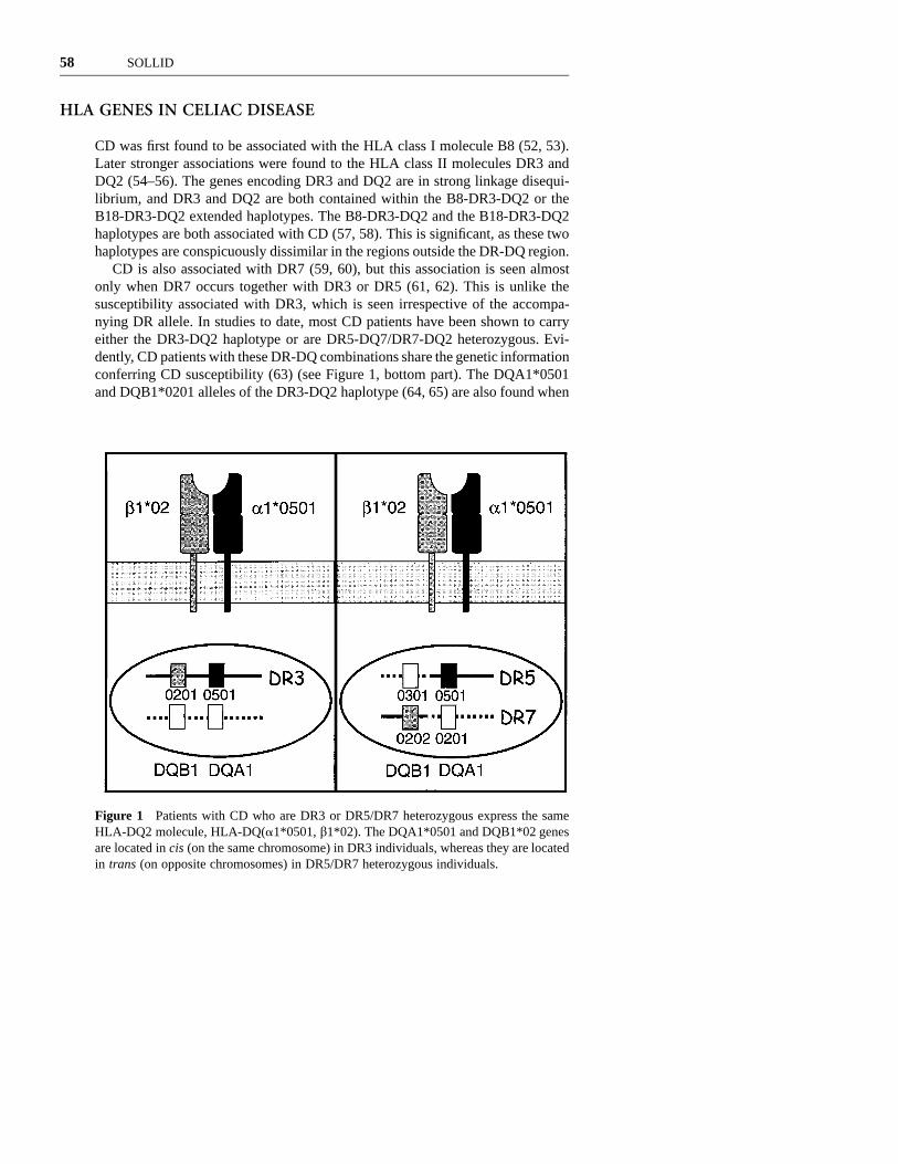
Figure 1 Patients with CD who are DR3 or DR5/DR7 heterozygous express the sameHLA-DQ2 molecule, HLA-DQ(a1*0501, b1*02). The DQA1*0501 and DQB1*02 genesare located in cis (on the same chromosome) in DR3 individuals, whereas they are locatedin trans (on opposite chromosomes) in DR5/DR7 heterozygous individuals.
HLA GENES IN CELIAC DISEASE
CD was first found to be associated with the HLA class I molecule B8 (52, 53).Later stronger associations were found to the HLA class II molecules DR3 andDQ2 (54–56). The genes encoding DR3 and DQ2 are in strong linkage disequi-librium, and DR3 and DQ2 are both contained within the B8-DR3-DQ2 or theB18-DR3-DQ2 extended haplotypes. The B8-DR3-DQ2 and the B18-DR3-DQ2haplotypes are both associated with CD (57, 58). This is significant, as these twohaplotypes are conspicuously dissimilar in the regions outside the DR-DQ region.
CD is also associated with DR7 (59, 60), but this association is seen almostonly when DR7 occurs together with DR3 or DR5 (61, 62). This is unlike thesusceptibility associated with DR3, which is seen irrespective of the accompa-nying DR allele. In studies to date, most CD patients have been shown to carryeither the DR3-DQ2 haplotype or are DR5-DQ7/DR7-DQ2 heterozygous. Evi-dently, CD patients with these DR-DQ combinations share the genetic informationconferring CD susceptibility (63) (see Figure 1, bottom part). The DQA1*0501and DQB1*0201 alleles of the DR3-DQ2 haplotype (64, 65) are also found when

MOLECULAR BASIS OF CELIAC DISEASE 59
the DR5-DQ7 and DR7-DQ2 haplotypes are combined. The DR5-DQ7 haplotypecarries the DQA1*0501 and DQB1*0301 alleles (66), and the DR7-DQ2 haplo-type carries the DQA1*0201 and DQB1*0202 alleles (67, 68). Notably, theDQB1*0201 and DQB1*0202 alleles are identical except for the codon of residue135 located in the membrane proximal domain of the DQb chain (69). Recom-bination (crossing over) seems to be an important mechanism for the generationof HLA haplotypes (70). Accumulating evidence suggests that the DR3-DQ2,DR7-DQ2, and the DR5-DQ7 haplotypes have a close evolutionary relationship.Based on microsatellite analysis, fragments of DNA flanking the DQA1 gene ofthe DR3-DQ2 haplotype have been identified on the DR5-DQ7 haplotype, andfragments of DNA flanking the DQB1 gene of the DR3-DQ2 haplotype havebeen identified on the DR7-DQ2 haplotype (71, 72). Thus, the genetic informationin the DQ subregion of the DR3-DQ2 haplotype is reestablished in DR5-DQ7/DR7-DQ2 heterozygotes, although the sequence information is split between twochromosomes.
It can be argued that susceptibility for CD depends on an interaction betweenat least two genes on the DR3-DQ2 haplotype that are reunited in DR5-DQ7/DR7-DQ2 heterozygous individuals. Theoretically this gene interaction couldinvolve any HLA-linked genes in the DQ region. However, complete sequencingof an 86-kb genomic fragment spanning the DQ subregion of the DR3-DQ2haplotype failed to identify genes other than the DQA1 and the DQB1 genes inthis region (73). Furthermore, the DQA1 and DQB1 are very good candidatesbecause their products interact by forming a class II heterodimer and because theyare situated close to the putative recombination site. This evolutionary consid-eration together with the fact that most CD patients share a particular pair ofDQA1 and DQB1 genes located either in cis or in trans are strong arguments thatthe DQA1*0501 and DQB1*0201 alleles jointly confer susceptibility to CD bycoding for the DQ(a1*0501, b1*02) heterodimer (Figure 1, top part).
In most populations studied, 90% or more of the CD patients carry theDQ(a1*0501, b1*02) heterodimer, compared to 20%–30% in healthy controls(63). The fraction of patients in different populations that encode this DQ het-erodimer by genes in cis or in trans position depends on the haplotype frequenciesof DR3-DQ2, DR5-DQ7, and DR7-DQ2 haplotypes in the given populations (74).In a few patients the DQ(a1*0501, b1*02) heterodimer may be found to beencoded in cis position by haplotypes other than DR3-DQ2 or in trans positionby individuals being heterozygous for combinations other than DR5-DQ7/DR7-DQ2 (63). There is no increase of the DQ(a1*0501, b1*0301) or DQ(a1*0201,b1*02) heterodimers alone in CD demonstrating that susceptibility is dependenton both the DQa and DQb chains in the DQ(a1*0501, b1*02) heterodimer.
Many studies have reported a particular increased risk for CD among individ-uals who are DR3-DQ2 homozygous and DR3-DQ2/DR7-DQ2 heterozygous (forreferences, see 63). This could be explained by a gene dosage effect of theDQB1*02 allele possibly caused by an increased expression of the DQ(a1*0501,b1*0201) heterodimer in such individuals (75). A gene dosage effect of DQB1*02
60 SOLLID
could also provide an explanation of the high degree of HLA haplotype identityobserved among affected siblings (74, 76).
Depending on the populations studied, about 2%–10% of CD patients do notcarry the DQ(a1*0501, b1*02) heterodimer. The great majority of these patientscarry different subtypes of DR4. The genetic determinant responsible for the HLAassociation in these individuals is likely to be different from that of theDQ(a1*0501, b1*02)-expressing individuals. To unequivocally identify theresponsible molecule encoded by the DR4 haplotype by a genetic approach is,however, difficult. Notably, there is a clear skewing in the representation of theDR4-DQ8 vs the DR4-DQ7 haplotype among these patients (77–79). This impliesthat DQ8, i.e. DQ(a1*0301, b1*0302), is most probably the molecule responsiblefor susceptibility. An opposing view is that the susceptibility is mediated by theDR53, i.e. DR(a*, b4*0101), molecule that is carried on most of the DR4, DR7,and DR9 haplotypes (80). The majority of DQ(a1*0501, b1*02)-negative patientswould fit into this category. Importantly, however, this model does not accountfor the observed skewing of the DR4-DQ8 vs. the DR4-DQ7 haplotypes. More-over, DQ(a1*0501, b1*02)-negative CD patients who carry the DRB1*0701-DQB1*03032 haplotype exist (79), and this haplotype is reported to carry anon-expressed null allele at the DRB4 locus (81). Further studies including typingfor the DRB4 null allele are needed to clarify the role of DR53 as a susceptibilitymolecule in CD.
Genes located in the HLA gene complex other than DQ might also contributeto CD susceptibility. Associations to particular DP alleles have been reported indifferent populations, but many of these associations can be explained by linkagedisequilibrium between the involved DP allele(s) and the DQA1*0501 andDQB1*02 alleles (for further discussion, see 63). Moreover, no independent asso-ciations to alleles at the TAP1 and TAP2 loci have been found (82–84). Severalstudies have consistently indicated that DQA1*0501/DQB1*02-positive individ-uals carrying the DR5/DR7 genotype have a higher risk to develop disease thando those of the DR3/DRX genotype (X ? DR7 and DR3) (84–86). Furthermore,it has been indicated that the risk of the DR3/DR7 genotype is higher than thatof the DR3/DR3 genotype (84, 86), although this is not a consistent finding (75,87). This has led to the suggestion that a gene on the DR7-DQ2 haplotype confersan additive effect to that of the DQA1*0501/DQB1*02 genes (86). To note, alocus with a protective allele of the DR3-DQ2 haplotype would produce the sameeffect. Studies of Irish CD patients have indicated an additional predisposing roleof TNF genes, an association independent of DQ2 that has been demonstratedusing a microsatellite polymorphism situated near the TNF genes (88). Moreover,a polymorphism of the TNF-a gene promoter has been demonstrated to be acomponent of the DR3-DQ2 haplotype (89). A Finnish study failed to reproducethe finding of a DQ2-independent association of the TNF microsattelites (90).These discrepant results may relate to population differences.
Recently, an allele of a locus (D6S2223) that is located 2, 5 Mb telomeric tothe HLA-F locus was found by Lie et al (91) to be less frequent among DR3-
MOLECULAR BASIS OF CELIAC DISEASE 61
DQ2 homozygous CD patients compared to DR3-DQ2 homozygous controls. Thesame allele of the D6S2223 locus was also found to be underrepresented amongDR3-DQ2 homozygous type 1 diabetes patients, and it was transmitted less oftenthan expected from DR3-DQ2 homozygous parents to diabetic siblings (92).These findings suggest that a gene(s) in the vicinity of D6S2223 is involved inthe pathogenesis of both CD and type 1 diabetes. In addition, the MIC-A andMIC-B genes are interesting candidate susceptibility genes in CD, as the MICmolecules are ligands for TCRcd T cells. The MIC genes are located near theHLA-B locus, and the MIC-A*008 (5.1) allele is in strong positive linkage dis-equilibrium with HLA-B8 (93, 94). This allele is particularly interesting since itbears a frameshift and a premature stop codon in exon 5 (95) that might affectthe expression of the molecule.
Taken together, available data strongly suggest that susceptibility to developCD is primarily associated to two conventional peptide-presenting DQ molecules:i.e. DQ(a1*0501, b1*02) (4DQ2) or to a lesser extent DQ(a1*03, b1*0302)(4DQ8). An issue still to be clarified is whether there are additional moleculesencoded by unidentified genes in the HLA gene complex that also contribute tothe genetic predisposition for CD. However, any effect of these additional genesis likely to be moderate. A key question for the understanding of the molecularbasis for CD is therefore to define the functional role of the DQ2 and DQ8molecules.
PEPTIDE BINDING MOTIF OF DISEASE-ASSOCIATEDDQ MOLECULES
Peptides binding to DQ2 have anchor residues in the relative positions P1, P4,P6, P7, and P9 (96–100). This is the same spacing as previously found for DRmolecules, suggesting that DQ2 bound peptides adopt to a conformation similarto that of peptides bound to DR molecules. The peptide-binding motif of DQ2illustrated in Figure 2 is quite different from other class II–binding motifs thathave been identified (101). Notably, the preference for negatively charged resi-dues for the three anchor positions in the middle seems to be unique for DQ2.The binding motif of DQ8 is different from that of DQ2, but DQ8 also displaysa preference for binding negatively charged residues at several positions (i.e. P1,P4, and P9) (102, 103). Hence, both the DQ2 and DQ8 molecules share a pref-erence for negatively charged residues at some of their anchor positions.
The peptide-binding motif of DQ2, i.e. DQ(a1*0501, b1*02), is different fromthe motifs of the closely related DQ(a1*0501, b1*0301) and DQ(a1*0201,b1*02) molecules (96, 99, 104), which do not confer susceptibility to CD (seeabove). The binding motif of the DQ(a1*0501, b1*0301) molecule is clearlydifferent from that of DQ(a1*0501, b1*02) with differences at the P4, P7 andP9 pockets (96), whereas the differences between DQ(a1*0501, b1*02) and

62 SOLLID
Figure 2 Schematic depiction of the peptide binding groove of HLA-DQ2 (i.e. HLA-DQ(a1*0501, b1*02) with the peptide-binding motif displayed with the one letter codefor the amino acids. A bound peptide and a TCR recognizing the peptide/HLA complexare also indicated. This motif description is based on a compilation of results from refer-ences 96–100.
DQ(a1*0201, b1*02) are more subtle (96, 99). The molecules have similar bind-ing motifs with the most apparent difference being an additional anchor residueat P3 for DQ(a1*0201, b1*02) (99, 105).
PREFERENTIAL PRESENTATION OF GLUTEN-DERIVEDPEPTIDES BY DISEASE-ASSOCIATED HLA MOLECULESTO INTESTINAL T CELLS
The DQ2 and DQ8 molecules could confer susceptibility to CD by presentingdisease-related peptides in the target organ or alternatively by shaping the T cellrepertoire during T cell development in the thymus. This issue has been addressedby studies of T cells derived from the celiac lesion. Stimulation of small intestinalbiopsy specimens with a peptic/tryptic digest of gluten induces rapid activation(i.e. expression of CD25, the IL-2 receptor a-chain) of the T cells in the laminapropria of CD patients, but not of non-CD control subjects (106). Gluten-reactiveT cells can be isolated and propagated from intestinal biopsies of CD patients but

MOLECULAR BASIS OF CELIAC DISEASE 63
not from non-CD controls (107–109). These T cells are CD4` and use the abTCR. Importantly, T cells isolated from biopsy specimens of patients carryingthe DR3-DQ2 haplotype typically recognize gluten fragments presented by theDQ2 molecule rather than the other HLA molecules carried by the patients (107).Both DR3-DQ2-positive and DR5-DQ7/DR7-DQ2-positive antigen-presentingcells (i.e. carrying the DQA1*0501 and DQB1*02 genes in cis or in trans posi-tion) are able to present the gluten antigen to these T cells (107, 110). Likewise,T cells isolated from small intestinal biopsies of DQ2-negative, DR4-DQ8-posi-tive patients predominantly recognize gluten-derived peptides when presented bythe DQ8 molecule (111). It is notable that no DR(a, b1*01)-restricted intestinalT cells specific for gluten have been reported supporting a role of DQ8 ratherthan the DR(a, b1*01) molecule in conferring susceptibility to CD. Takentogether, these results allude to presentation of gluten peptides in the small intes-tine as the mechanism by which DQ2 and DQ8 confer susceptibility to CD. Athymic effect of the same DQ molecules on the TCR repertoire selection is,however, not excluded by these results.
The DQ2 and DQ8 molecules are not preferential antigen-presenting moleculesin the intestinal mucosa irrespective of antigen. T cells specific for astrovirus (acommon gastroenteritis virus) are predominantly DR restricted (109), which sug-gests that the peculiar HLA restriction pattern of the gliadin-specific T cells ofthe intestine must be related to the antigen. Interestingly, gluten-specific T cellscan also be found in the peripheral blood (112). These T cells are restricted eitherby DR, DP, or DQ molecules, and they do not therefore display the the predom-inant DQ2 or DQ8 restriction observed for gluten-specific T cells from the intes-tinal mucosa (112). One explanation for this could be that the majority ofgluten-specific T cells of peripheral blood recognize epitopes different from thoserecognized by T cells of the small intestine.
Studies of lamina propria T cells in situ have, as mentioned above, indicatedthat gluten reactive T cells have a cytokine profile dominated by IFN-c. Thisnotion is sustained by the characterization of gut-derived DQ2 and DQ8-restrictedgluten-specific T cell clones. These T cells uniformly secrete IFN-c at high con-centrations, and some produce IL-4, IL-5, IL-6, IL-10, TNF-a, or TGF-b in addi-tion (113).
T CELL RECOGNITION OF DEAMIDATED GLUTENPEPTIDES
Wheat gluten is a mixture of numerous proteins grouped into the gliadin andglutenin fractions. These proteins serve as a source of nitrogen and carbon forthe growing seedling during germination. A vast sequence heterogeneity amonggliadin and glutenin proteins probably reflects that these proteins have been sub-jected to few structural constraints during evolution. Generally, gluten proteins

64 SOLLID
TABLE 1
EpitopeDerived from
proteinPresentation
elementRecognizedby patients Reference
DQ2-c-I-gliadin c-gliadin DQ2 Infrequently (118)
DQ2-a-I-gliadin a-gliadin DQ2 Frequently (119)
DQ2-a-II-gliadin a-gliadin DQ2 Frequently (119)
DQ8-a-I-gliadin a-gliadin DQ8 Frequently? (120)
DQ8-I-glutenin Glutenin DQ8 Not known (121)
contain a large percentage of proline and glutamine residues, while many otheramino acids, including glutamic and aspartic acid, are unusually scarce. Feedingexperiments have demonstrated that the gliadin fraction can precipitate CD (114),whereas the role of glutenins is still inconclusive. Proteins of the gliadin fractioncan be subdivided according to their sequence into the a-, c-, and x-gliadins(115). A large number of different gliadins exist within each of these gliadinfamilies. Estimates suggest that as many as 50 to 150 different a-gliadin genesmay be present in a single wheat cultivar (116). For the work of identifyingpeptide fragments recognized by T cells, the complexity of this antigen presentsa big challenge.
Initially it was difficult to reconcile the DQ2 (and DQ8) binding motifs withpresentation of gluten peptides because gluten proteins have an unusual scarcityof negatively charged residues. A clue to help explain this paradox came fromthe observation that the stimulatory capacity of gliadin preparations for gliadin-specific intestinal T cells was significantly enhanced following treatment at hightemperatures and low pH (117). These conditions are known to cause nonspecificdeamidation of glutamines to glutamic acid and may thus convert gliadin from aprotein with very few peptides with the potential to bind to DQ2/DQ8 into onewith many such. An important and general role for deamidation of gluten for Tcell recognition was sustained by analysis of the response pattern of a panel ofpolyclonal, gliadin-specific T cell lines derived from biopsies (118). All the linesresponded poorly to a gliadin antigen prepared under conditions of minimal deam-idation (chymotrypsin-digestion), compared to the same antigen when furtherheat-treated in an acidic environment.
The characterization of gluten epitopes recognized by intestinal T cells hasextended the knowledge about the importance of deamidation for their T cellrecognition. So far five unique epitopes of gluten that are recognized by gut Tcells have been identified; three restricted by DQ2 (118, 119) (Table 1 and Figure3) and two restricted by DQ8 (120, 121) (Table 1). The three DQ2-restrictedpeptides, one from c-gliadin and two from a-gliadins (DQ2-c-gliadin-I, DQ2-a-gliadin-I and DQ2-a-gliadin-II), fail to stimulate T cells in their native form butare potent antigens when a single glutamine residue is exchanged with glutamic
MOLECULAR BASIS OF CELIAC DISEASE 65
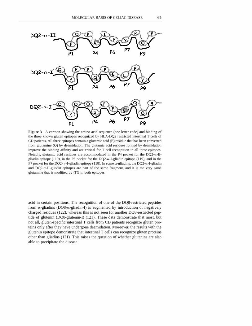
Figure 3 A cartoon showing the amino acid sequence (one letter code) and binding ofthe three known gluten epitopes recognized by HLA-DQ2 restricted intestinal T cells ofCD patients. All three epitopes contain a glutamic acid (E) residue that has been convertedfrom glutamine (Q) by deamidation. The glutamic acid residues formed by deamidationimprove the binding affinity and are critical for T cell recognition in all three epitopes.Notably, glutamic acid residues are accommodated in the P4 pocket for the DQ2-a-II-gliadin epitope (119), in the P6 pocket for the DQ2-a-I-gliadin epitope (119), and in theP7 pocket for the DQ2- c-I-gliadin epitope (118). In some a-gliadins, the DQ2-a-I-gliadinand DQ2-a-II-gliadin epitopes are part of the same fragment, and it is the very sameglutamine that is modified by tTG in both epitopes.
acid in certain positions. The recognition of one of the DQ8-restricted peptidesfrom a-gliadins (DQ8-a-gliadin-I) is augmented by introduction of negativelycharged residues (122), whereas this is not seen for another DQ8-restricted pep-tide of glutenin (DQ8-glutenin-I) (121). These data demonstrate that most, butnot all, gluten-specific intestinal T cells from CD patients recognize gluten pro-teins only after they have undergone deamidation. Moreover, the results with theglutenin epitope demonstrate that intestinal T cells can recognize gluten proteinsother than gliadins (121). This raises the question of whether glutenins are alsoable to precipitate the disease.
66 SOLLID
DEAMIDATION IN VIVO IS LIKELY TO BEENZYMATICALLY MEDIATED BY TISSUETRANSGLUTAMINASE
The deamidation of gliadin may take place in the acidic environment in the stom-ach (118). Alternatively, it can be mediated by the enzyme tissue transglutaminase(tTG) as demonstrated by Molberg et al (123) and later also by van de Wal et al(122). tTG is expressed in many different tissues and organs; in the small intestineit is expressed just beneath the epithelium in the gut wall (123). Notably theactivity of tTG is elevated in the small intestinal mucosa of CD patients in boththe active disease phase and in remission (123a). The enzyme is present bothintracellularly and extracellulary, and in the extracellular environment tTG playsa role in extracellular matrix assembly, cell adhesion, and wound healing (124).The calcium-dependent transglutaminase activity of tTG catalyzes selective cross-linking or deamidation of protein-bound glutamine residues (125). Notably, tTGis the same protein that Dieterich et al found to be a major focus of the autoan-tibody response in CD (6). In contrast to the nonenzymatically mediated deami-dation that results in a near random deamidation of the often numerous glutamineresidues in gliadin peptides, tTG appears to carry out an ordered deamidation ofsome few specific glutamines (123). For all the three DQ2-restricted gliadin epi-topes recognized by gut T cells and the DQ8-a-gliadin-I epitope, the residuescritical for T cell recognition are all specifically targeted by tTG (119, 122, 123).Interestingly, the deamidation of glutamines that are not targeted by tTG (e.g. byacid treatment) can be deleterious for T cell recognition (105, 122). Additionalevidence for a role of tTG comes from experiments where T cell lines have beenestablished from biopsies challenged with a minimally deamidated gliadin antigen(chymotrypsin-digested) and then tested for recognition of this antigen or thesame antigen treated with tTG (Ø Molberg, S McAdam, KEA Lundin, C Kris-tiansen, K Kett, EH Arentz-Hansen, LM Sollid, manuscript in preparation). In 14out of 15 patients, the T cell lines responded better to the antigen that had beensubjected to treatment with tTG. Similarly, T cell lines established from twoDQ2` patients by stimulating biopsies with a chymotrypsin-digested recombi-nant a-gliadin were found to recognize synthetic peptides representing the DQ2-a-gliadin-I and DQ2-a-gliadin-II epitopes, but not the corresponding non-deamidated peptides (Ø Molberg, S McAdam, KEA Lundin, C Kristiansen, EHArentz-Hansen, K Kett, LM Sollid, manuscript in preparation). Taken together,these results indicate that deamidation in vivo is mediated by tTG.
It is intriguing to hypothesize that tTG plays a central role in the selection ofgliadin T cell epitopes. Credence to this idea comes from the observation that theintestinal T cell response to a-gliadin in adults is focused on a single deamidatedglutamine (in the related DQ2-a-gliadin-I and DQ2-a-gliadin-II epitopes) that istargeted by tTG (119). Knowledge of the substrate recognition sites of tTG shouldallow further testing of this hypothesis. Unfortunately, the available information

MOLECULAR BASIS OF CELIAC DISEASE 67
on sequences targeted by tTG is not presently sufficient to establish the overallsubstrate specificity of the enzyme.
HIERARCHIES OF GLUTEN T CELL EPITOPES?
The existence of multiple epitopes in gluten that are recognized by small intestinalT cells of CD patients raises several interesting questions: Are only some of theepitopes pathogenic and thereby relevant to explain the HLA association? Areresponses toward some of the epitopes generated during the early phases of dis-ease development, while the responses to others are a result of epitope spreading?Are different epitopes recognized by distinct groups of patients (e.g. children vs.adults)? Are some epitopes more relevant to disease as responses to them arefound in the majority of the patients or because there is a higher precursor fre-quency of T cells in the lesion specific for these epitopes? The answers to mostof these questions must await further investigations. At present we know that forthe DQ2-a-gliadin-I and DQ2-a-gliadin-II epitopes, intestinal T cell reactivity isfound in most if not all adult DQ2` patients (119), whereas for the DQ2-c-gliadin-I epitope, intestinal T cell reactivity is found in only a minority of DQ2`patients (118). Less is known about the DQ8-restricted epitopes because fewDQ8-positive patients have been tested so far. However, the DQ8-a-gliadin-Iappears to be frequently recognized (120). What causes the variance in respon-siveness to the different epitopes and whether this reflects qualitative or quanti-tative differences between the patients are presently unclear.
Epitope spreading (126) may be a mechanism relevant to CD that could explainthe existence of several gluten epitopes. In experimental autoimmune encepha-lomyelitis where epitope spreading occurs, along with spreading of new antigenicepitopes there is also a ‘‘spreading’’ of MHC class II molecules involved inepitope presentation (127, 128). The strict restriction of DQ2 and DQ8 as pre-sentation elements for gluten-reactive T cells of the disease lesion clearly deviatesfrom the picture found in experimental autoimmune encephalomyelitis and maysuggest that other mechanisms are operating. Further studies are clearly neededto sort out this question.
The mapping of epitopes of gluten proteins recognized by intestinal T cells isincomplete; the actual number of distinct epitopes is currently a matter of specu-lation. However, recent results from testing intestinal T cells of Norwegian adultsagainst a panel of recombinant a-gliadins suggest that the number of epitopesmight be more limited than initially thought (119). From the sequences repre-sented in a panel of full-length recombinant a-gliadins, there seems to be only asingle immunodominant fragment that contains the two related epitopes DQ2-a-gliadin-I and DQ2-a-gliadin-II.
The disease relevance of epitopes defined using peripheral blood T cells mustalso be questioned because peripheral blood T cell gliadin epitopes do not appearto be limited in their presentation by DQ2 or DQ8 (112) nor to be enhanced by

68 SOLLID
treatment with tTG (123). Furthermore, a DQ2-restricted epitope of a-gliadin,which was defined by peripheral blood T cells of a CD patient (129) and whichinduces mucosal changes in peptide feeding experiments (130), fails to be rec-ognized by gluten-reactive polyclonal intestinal T cell lines from six patients evenafter tTG treatment (119).
GLUTEN-SPECIFIC T CELLS MAY PROVIDE HELP FORAUTOANTIBODY PRODUCTION
The IgG and IgA serum antibodies to tTG (also termed anti-endomysial antibod-ies) are a hallmark of CD, and detection of serum IgA tTG-antibodies is utilizedto predict the disease (7, 8). As the B cells producing the tTG antibodies haveundergone an isotype switch, it is likely that these are T cell-dependent antibodyresponses. This poses a problem since the existence of T cells recognizing tTGis doubtful. tTG is expressed ubiquitously in the human body, and staining withsera from untreated CD patients indicates that the antigen is also expressed infetal thymus (131). Most likely, T cells reactive with tTG are therefore deletedby negative selection in thymus, and if they should exist they would likely haveinduced serious systemic autoimmunity. Interestingly, gluten seems to drive theantibody production, as the presence of tTG antibodies is strictly dependent ondietary gluten exposure (8). This raises the possibility that gluten-reactive T cellsprovide help for tTG-specific B cells by a mechanism of intramolecular help (132)analogous to the hapten-carrier system (133). As mentioned earlier, an importantphysiological role of tTG is the catalysis of isopeptide bond formation betweenglutamine and lysine residues (125). Indeed, it is the substitution of water ratherthan lysine in this reaction that results in deamidation. In vitro treatment of gliadinfragments with tTG leads to some gliadin fragments becoming covalently attachedto tTG by autocatalysis (6, 123). tTG-specific B cells may selectively bind andinternalize gliadin-tTG complexes via specific surface immunoglobulins. The gli-adin fragment may finally be processed and presented by DQ2 or DQ8 to thegliadin-specific T cells, thereby providing cognate help for B cell maturation,isotype switching, and antibody secretion. This model can explain why tTG anti-body levels in CD are dependent on the presence of gliadin in the diet becauseits removal will also abolish the T cell help needed for antibody production.
Autocatalysis by tTG should be more likely to occur when the concentrationof other amine donors (lysine containing proteins/primary amines) is low. In fact,deamidation is also likely to happen when the amount of primary amines is lowor absent (125). Formation of gliadin-tTG complexes and deamidation of gliadinmay thus reflect an altered microenvironment in the gut mucosa. The unusualability of gliadins to act as excellent amine acceptor substrates for tTG may resultin a local depletion of lysine/polyamines, and the altered microenvironment mayhence be established in situations where increased levels of gluten proteins getaccess to the subepithelial area.

MOLECULAR BASIS OF CELIAC DISEASE 69
PERTURBED ORAL TOLERANCE TO GLUTEN INCELIAC DISEASE?
Although the concept of oral tolerance is not as firmly established in humans asit is in rodents, it is clearly necessary that mechanisms that allow for tolerance tosoluble food antigens exist in humans (134). In keeping with this thinking, oraltolerance to gluten in patients with CD either is not established properly or isbroken. A deeper understanding of this issue should shed new light on the mech-anism behind oral tolerance in humans. Given the preferential intestinal T cellresponse to deamidated gluten fragments in CD patients, it is conceivable thatdeamidation is central to the perturbation of the oral tolerance. Deamidationincreases the binding affinity of gliadin peptides for DQ2 from poor but significantbinders to epitopes with reasonable, but by no means exceptional, affinity (118,119). The moderate binding affinity of these epitopes concurs with the findingthat they do not carry optimal anchors in all the anchor positions. It is interestingthat the modified glutamine residues for the three defined DQ2-restricted gliadinepitopes recognized by intestinal T cells occupy different pockets within DQ2(Figure 3). This suggests that the altered affinity of the gliadin peptides for DQ2is a critical factor involved in loss of tolerance rather than recognition of a single‘‘pathogenic’’ motif that binds to DQ2 (119). Concurrent with the increase inaffinity for DQ2 caused by deamidation of the gliadin peptide is a change inconformation of the gliadin/DQ2 complex. This is apparent by the failure of theT cells to recognize the unmodified peptides even at higher concentrations thatshould compensate for their lower affinity for DQ2. However, the simple modi-fication of glutamine residues that act as major T cell receptor contact residuesappears not to be sufficient to break tolerance as none of the modified glutaminesare found in such positions (105, 119).
Gliadin fragments containing two glutamine residues targeted by tTG may wellbe deamidated and cross-linked to other proteins that contain lysine. Conditionsmay exist in the gut, where T cell epitopes are both created and trapped locallyby tTG, that prevent the epitopes from being presented by antigen-presentingcells that induce tolerance in the gut. Alternatively, it may prevent these epitopesfrom spreading systemically, a factor thought to be important in the establishmentof oral tolerance (135). In this regard it is interesting that the motif targeted bytTG and shared in the DQ2-a-gliadin-I and DQ2-a-gliadin-II epitopes is repeatedwithin many of the a-gliadins (119).
MECHANISMS INVOLVED IN FORMATION OF THECELIAC LESION
The evidence discussed above provides strong evidence that CD4` TCRab` Tcells in the lamina propria are central for controlling the immune response togluten that produces the immunopathology of CD. The knowledge of the events

70 SOLLID
downstream of T cell activation is, however, still incomplete. The characterizationof mechanisms operating in the model of human fetal gut explant cultures, whereactivation of T cells induces villous atrophy and hyperplasia of the crypts, hasprovided interesting clues and indicated some major pathways (136). However,knowing how the immune system usually utilizes a multitude of effector mech-anisms for fighting its opponents, it is reasonable to believe that multiple effectormechanisms may well be involved in the creation of the celiac lesion. Adding tothe complexity, recent in vitro organ culture studies have indicated that glutenexerts additional immune relevant effects independent of T cell activation (137,138). Some of these effects have rapid kinetics, and conceivably the direct effectsof gluten may facilitate subsequent T cell responses.
Cytokines produced by lamina propria CD4` T cells may be involved in theincreased crypt cell proliferation and the increased loss of epithelial cells. IFN-cinduces macrophages to produce TNF-a. TNF-a activates stromal cells to pro-duce KGF, and KGF causes epithelial proliferation and crypt cell hyperplasia(17). IFN-c and TNF-a can jointly have a direct cytotoxic effect on intestinalepithelial cells (139). It is also conceivable that IELs and in particular cd T cellsplay a role in the epithelial cell destruction by recognizing MIC molecules inducedby stress (26).
Alterations of the extracellular matrix can also distort the epithelial arrange-ment, as the extracellular matrix provides the scaffold on which the epitheliumlies. Enterocytes adhere to basement membrane through extracellular matrixreceptors so that modification or loss of the basement membrane can result inenterocyte shedding. Evidence for increased extracellular matrix degeneration inCD exists, and this degeneration may be important for the mucosal transformationfound in CD (40). The increased production of metalloproteinases by subepithelialfibroblasts and macrophages is likely to be directly or indirectly induced by cyto-kines that are released from activated T cells.
Do the autoantibodies play a role in the pathogenesis of CD, or are they justan epiphenomenon? The significant increase in prevalence of CD among IgA-deficient individuals (1) speaks against a role of the antibodies. However, mostCD patients also have elevated levels of serum IgG endomysial (i.e. tTG) anti-bodies (5), and little is known about the antibodies found locally in the mucosaof IgA-deficient CD patients. Interestingly, the endomysial (i.e. tTG) antibodiescan, as suggested by Maki and coworkers (140), be involved in the disease devel-opment by blocking interactions between mesenchymal cells and epithelial cellsduring the migration of epithelial cells and fibroblasts from the crypts to the tipsof the villi. tTG is necessary for activation of transforming growth factor-b (TGF-b) (141). Indirect inhibition of TGF-b activation by anti-tTG antibodies can beenvisaged to have broad effects as TGF-b is known to affect the differentiationof the intestinal epithelium (140), to stimulate extracellular matrix formation(142), and to regulate the function of many immune competent cells within thegut microenvironment (143). In addition, tTG has been demonstrated to beinvolved in attachment of fibroblasts to the extracellular matrix (144), suggesting

MOLECULAR BASIS OF CELIAC DISEASE 71
that the autoantibodies could also be involved in lesion formation by perturbingimportant contacts between fibroblasts and extracellular matrix components. ThetTG antibodies may in addition modulate the deamidating activity of tTG in eitheran inhibiting or a promoting fashion (145). Further research is clearly needed toestablish whether and how the tTG antibodies play a role in CD pathogenesis.
HLA ASSOCIATION WITH DISEASE: LESSONS TO BELEARNED FROM CD
Strong evidence suggests that the primary HLA association in CD is to the clas-sical peptide presenting HLA molecules DQ2 and DQ8. These HLA moleculespredispose to disease by presenting gluten peptides to CD4` T cells in the affectedorgan, although an effect mediated by shaping of the T cell repertoire in thethymus cannot yet be excluded. This has clear relevance for studies of other HLA-associated diseases where the identity of the HLA molecules involved are lesswell defined and where the triggering antigens have not been identified.
The DQ2 and DQ8 molecules bind gluten peptides that after specific deami-dation become good peptide ligands for DQ2 and DQ8. Exactly why no otherclass II molecules are able to present gluten peptides in the gut that result indisease is not yet fully understood. Likely related is that peptides that becomedeamidated in the gut mucosa are particularly effective in inducing a pathologicimmune response, and that the DQ2 and DQ8 molecules are especially suited tobind deamidated peptides. The DQ(a1*0501, b1*0301) and DQ(a1*0201,b1*0202) molecules which are related to the predisposing DQ(a1*0501,b1*0201) molecule and which do not predispose to CD have different bindingmotifs, although the binding motif of DQ(a1*0201, b1*0202) is very similar.Interestingly, DQ(a1*0501, b1*0201) and DQ(a1*0201, b1*0202) expressing Blymphoblastoid cell lines exhibit abilities to present the DQ2-a-gliadin-II epitopethat differ according to when the epitope is incorporated into a complex antigenthat requires processing as compared with the peptide that is processing indepen-dent (105). This might suggest that factors involved in processing and peptidebinding act differentially for loading of the gliadin peptides to two DQ moleculesand that this is relevant for explaining the HLA association.
Modification of self-proteins analogous to gliadin in CD would create epitopesrecognized as nonself. This could be a more general mechanism for breaking ofimmunological tolerance and precipitation of autoimmune disease. Perhaps asmany as 50% to 90% of the proteins in the human body are posttranslationallymodified (P Roepstorff, personal communication), and the degree and type ofmodification are likely to be altered in an inflamed microenvironment. Epitopesharboring a posttranslational modification may go unreported, as the standard useof recombinant proteins and synthetic peptides for the characterization of T cellepitopes means that most in vivo modified epitopes would escape detection. Thisclass of epitopes should not be overlooked, and it will be important to devise

72 SOLLID
strategies that will identify modified T cell epitopes of potential autoantigens sothat their role in autoimmune disease can be clarified.
Another important point illustrated from the studies of CD is how a foreignantigen drives autoantibody production. For most autoimmune diseases, autoan-tigens have been defined by use of the autoantibodies. It is often inferred that Tcells must exist that are reactive with the autoantigen because the antibodies areof the IgG or IgA isotypes whose formation is dependent on T cell help. In somecases this assumption may turn out to be unjustified. It is in my opinion appro-priate to intensify the search for unknown foreign agents that might be hosted bythe human body (virus, bacteria, etc) and that are capable, after combining witha self-protein, of providing help for autoimmune responses similar to that foundin CD.
This review illustrates that the molecular basis of CD is complex. Given themultifactorial etiology of the disease with involvement of several genes and envi-ronmental factors, this is not unexpected. Despite the recent advances in under-standing of critical steps in disease development, there is much still to be learnedabout the disease. Several predisposing genes are yet to be identified. Given thedifficulty in defining susceptibility genes with modest effects, a combined func-tional and genetic approch will be required for their identification. The full under-standing of multifactorial inflammatory diseases is surely a formidable challengefor scientists. Compared with the other diseases of this nature, however, CD standsout as a disease for which it should be easier to decipher both the actions of thepredisposing gene products and how they interact with other gene products andenvironmental factors. In this situation it is justified to call for intensified researchon CD as this can serve as an illuminator for the other multifactorial inflammatorydiseases.
ACKNOWLEDGMENTS
I thank Knut E. A. Lundin, Stephen N. McAdam, Øyvind Molberg, and FrodeVartdal for critically reading the manuscript. I also thank members of the ‘‘celiacdisease research group’’ at my Institute for stimulating discussions and ErikThorsby for continous support. The author’s research is supported by grants fromthe Research Council of Norway and the European Commission (BMH4-CT98–3087).
Visit the Annual Reviews home page at www.AnnualReviews.org.
LITERATURE CITED
1. Trier JS. 1991. Celiac sprue. N. Engl. J.Med. 325:1709–19
2. Thorsby E. 1997. Invited anniversary
review: HLA associated diseases. Hum.Immunol. 53:1–11
3. Schmitz J. 1992. Coelic disease in child-

MOLECULAR BASIS OF CELIAC DISEASE 73
hood. In Coeliac Disease, ed. MNMarsh, pp. 17–48. Oxford: Blackwell
4. Maki M, Collin P. 1997. Coeliac disease.Lancet 349:1755–59
5. Maki M. 1995. The humoral immunesystem in coeliac disease. In Coeliac Dis-ease, ed. PD Howdle, pp. 231–49. Lon-don: Bailliere Tindall
6. Dieterich W, Ehnis T, Bauer M, DonnerP, Volta U, Riecken EO, Schuppan D.1997. Identification of tissue transgluta-minase as the autoantigen of celiac dis-ease. Nat. Med. 3:797–801
7. Dieterich W, Laag E, Schopper H, VoltaU, Ferguson A, Gillett H, Riecken EO,Schuppan D. 1998. Autoantibodies to tis-sue transglutaminase as predictors ofceliac disease. Gastroenterology 115:1317–21
8. Sulkanen S, Halttunen T, Laurila K,Kolho KL, Korponay-Szabo IR, SarnestoA, Savilahti E, Collin P, Maki M. 1998.Tissue transglutaminase autoantibodyenzyme-linked immunosorbent assay indetecting celiac disease. Gastroenterol-ogy 115:1322–28
9. Parnell N, Ciclitira PJ. 1999. Celiac dis-ease. Curr. Opin. Gastroenterol. 15:120–24
10. Ivarsson A, Persson LA, Hernell O,Ascher H, Cavell B, Danielsson L, Dan-naeus A, Lindberg T, Lindquist B, Sten-hammar L. 1999. The ‘‘epidemic’’ ofcoeliac disease in Swedish children. ActaPaediatr. In press
11. Marsh MN. 1992. Mucosal pathology ingluten sensitivity. In Coeliac Disease, ed.MN Marsh, pp. 136–91. Oxford:Blackwell
12. Leigh RJ, Marsh MN, Crowe P, Kelly C,Garner V, Gordon D. 1985. Studies ofintestinal lymphoid tissue. IX. Dose-dependent, gluten-induced lymphoidinfiltration of coeliac jejunal epithelium.Scand. J. Gastroenterol. 20:715–19
13. Wicker LS, Todd JA, Peterson LB. 1995.Genetic control of autoimmune diabetes
in the NOD mouse. Annu. Rev. Immunol.13:179–200
14. Booth CC. 1970. Enterocyte in coeliacdisease. Br. Med. J. 3:725–31
15. Walker-Smith J, MacDonald T. 1989.Insights provided by the study of thesmall intestine in the child and the foetus.Gut 30(Spec. No):11–16
16. Moss SF, Attia L, Scholes JV, WaltersJR, Holt PR. 1996. Increased small intes-tinal apoptosis in coeliac disease. Gut39:811–17
17. Bajaj-Elliott M, Poulsom R, Pender SL,Wathen NC, MacDonald TT. 1998. Inter-actions between stromal cell-derivedkeratinocyte growth factor and epithelialtransforming growth factor in immune-mediated crypt cell hyperplasia. J. Clin.Invest. 102:1473–80
18. Scott H, Sollid LM, Fausa O, BrandtzaegP, Thorsby E. 1987. Expression of majorhistocompatibility complex class II sub-region products by jejunal epithelium inpatients with coeliac disease. Scand. J.Immunol. 26:563–71
19. Marley NJ, Macartney JC, Ciclitira PJ.1987. HLA-DR, DP and DQ expressionin the small intestine of patients withcoeliac disease. Clin. Exp. Immunol.70:386–93
20. Scott H, Brandtzaeg P, Solheim BG,Thorsby E. 1981. Relation betweenHLA-DR-like antigens and secretorycomponent (SC) in jejunal epithelium ofpatients with coeliac disease or dermati-tis herpetiformis. Clin. Exp. Immunol.44:233–38
21. Colombel JF, Mascart-Lemone F,Nemeth J, Vaerman JP, Dive C, RambaudJC. 1990. Jejunal immunoglobulin andantigliadin antibody secretion in adultcoeliac disease. Gut 31:1345–49
22. Kutlu T, Brousse N, Rambaud C, LeDeist F, Schmitz J, Cerf-Bensussan N.1993. Numbers of T cell receptor (TCR)ab` but not of TcR cd` intraepitheliallymphocytes correlate with the grade of

74 SOLLID
villous atrophy in coeliac patients on along term normal diet. Gut 34:208–14
23. Halstensen TS, Brandtzaeg P. 1993. Acti-vated T lymphocytes in the celiac lesion:non-proliferative activation (CD25) ofCD4` a/b cells in the lamina propriabut proliferation (Ki-67) of a/b and c/dcells in the epithelium. Eur. J. Immunol.23:505–10
24. Spencer J, Isaacson PG, Diss TC, Mac-Donald TT. 1989. Expression ofdisulfide-linked and non-disulfide-linkedforms of the T cell receptor c/d hetero-dimer in human intestinal intraepitheliallymphocytes. Eur. J. Immunol. 19:1335–38
25. Halstensen TS, Scott H, Brandtzaeg P.1989. Intraepithelial T cells of theTcRcd` CD8- and Vd1/Jd1` pheno-types are increased in coeliac disease.Scand. J. Immunol. 30:665–72
26. Groh V, Steinle A, Bauer S, Spies T.1998. Recognition of stress-inducedMHC molecules by intestinal epithelialcd T cells. Science 279:1737–40
27. Groh V, Bahram S, Bauer S, Herman A,Beauchamp M, Spies T. 1996. Cellstress-regulated human major histocom-patibility complex class I gene expressedin gastrointestinal epithelium. Proc. Natl.Acad. Sci. USA 93:12445–50
28. Lundqvist C, Melgar S, Yeung MM,Hammarstrom S, Hammarstrom ML.1996. Intraepithelial lymphocytes inhuman gut have lytic potential and acytokine profile that suggest T helper 1and cytotoxic functions. J. Immunol.157:1926–34
29. Oberhuber G, Vogelsang H, Stolte M,Muthenthaler S, Kummer AJ, Radasz-kiewicz T. 1996. Evidence that intestinalintraepithelial lymphocytes are activatedcytotoxic T cells in celiac disease but notin giardiasis. Am. J. Pathol. 148:1351–57
30. Halstensen TS, Farstad IN, Scott H,Fausa O, Brandtzaeg P. 1990. Intraepi-thelial TcRa/b` lymphocytes expressCD45RO more often than the TcRc/d`
counterparts in coeliac disease. Immu-nology 71:460–66
31. Kontakou M, Sturgess RP, PrzemiosloRT, Limb GA, Nelufer JM, Ciclitira PJ.1994. Detection of interferon-c mRNAin the mucosa of patients with coeliacdisease by in situ hybridisation. Gut35:1037–41
32. Kontakou M, Przemioslo RT, SturgessRP, Limb AG, Ciclitira PJ. 1995. Expres-sion of tumour necrosis factor-a, inter-leukin-6, and interleukin-2 mRNA in thejejunum of patients with coeliac disease.Scand. J. Gastroenterol. 30:456–63
33. Nilsen EM, Jahnsen FL, Lundin KEA,Johansen FE, Fausa O, Sollid LM, Jahn-sen J, Scott H, Brandtzaeg P. 1998. Glu-ten induces an intestinal cytokineresponse strongly dominated by inter-feron-c in patients with celiac disease.Gastroenterology 115:551–63
34. Beckett CG, Dell’Olio D, Kontakou M,Przemioslo RT, Rosen-Bronson S, Cicli-tira PJ. 1996. Analysis of interleukin-4and interleukin-10 and their associationwith the lymphocytic infiltrate in thesmall intestine of patients with coeliacdisease. Gut 39:818–23
35. Baklien K, Fausa O, Thune PO, Gjone E.1977. Immunoglobulins in jejunalmucosa and serum from patients withdermatitis herpetiformis. Scand. J. Gas-troenterol. 12:161–68
36. Falchuk ZM, Strober W. 1974. Gluten-sensitive enteropathy: synthesis of anti-gliadin antibody in vitro. Gut 15:947–52
37. Picarelli A, Maiuri L, Frate A, Greco M,Auricchio S, Londei M. 1996. Produc-tion of antiendomysial antibodies afterin-vitro gliadin challenge of small intes-tine biopsy samples from patients withcoeliac disease. Lancet 348:1065–67
38. Nagashima R, Maeda K, Imai Y, Taka-hashi T. 1996. Lamina propria macro-phages in the human gastrointestinalmucosa: their distribution, immunohis-tological phenotype, and function. J. His-tochem. Cytochem. 44:721–31

MOLECULAR BASIS OF CELIAC DISEASE 75
39. Sturgess RP, Macartney JC, MakgobaMW, Hung CH, Haskard DO, CiclitiraPJ. 1990. Differential upregulation ofintercellular adhesion molecule-1 in coe-liac disease. Clin. Exp. Immunol.82:489–92
40. Daum S, Bauer U, Foss HD, SchuppanD, Stein H, Riecken EO, Ullrich R. 1999.Increased expression of mRNA formatrix metalloproteinases-1 and -3 andtissue inhibitor of metalloproteinases-1in intestinal biopsy specimens frompatients with coeliac disease. Gut 44:17–25
41. Ellis A. 1981. Coeliac disease: previousfamily studies. In The Genetics of Coe-liac Disease, ed. RB McConnell, pp.197–99. Lancaster: MTP
42. Risch N. 1987. Assessing the role ofHLA-linked and unlinked determinantsof disease. Am. J. Hum. Genet. 40:1–14
43. Petronzelli F, Bonamico M, Ferrante P,Grillo R, Mora B, Mariani P, ApollonioI, Gemme G, Mazzilli MC. 1997.Genetic contribution of the HLA regionto the familial clustering of coeliac dis-ease. Ann. Hum. Genet. 61:307–17
44. Polanco I, Biemond I, van Leeuwen A,Schreuder I, Meera Khan P, Guerrero J,D’Amaro J, Vazquez C, van Rood JJ,Pena AS. 1981. Gluten sensitive enter-opathy in Spain: genetic and environ-mental factors. In The Genetics ofCoeliac Disease, ed. RB McConnell, pp.211–31. Lancaster: MTP
45. Zhong F, McCombs CC, Olson JM, Els-ton RC, Stevens FM, McCarthy CF,Michalski JP. 1996. An autosomal screenfor genes that predispose to celiac dis-ease in the western counties of Ireland.Nat. Genet. 14:329–33
46. Houlston RS, Tomlinson IP, Ford D, SealS, Marossy AM, Ferguson A, HolmesGK, Hosie KB, Howdle PD, Jewell DP,Godkin A, Kerr GD, Kumar P, Logan RF,Love AH, Johnston S, Marsh MN, Mit-ton S, O’Donoghue D, Roberts A,Walker-Smith JA, Stratton MF. 1997.
Linkage analysis of candidate regions forcoeliac disease genes. Hum. Mol. Genet.6:1335–39
47. Greco L, Corazza G, Babron MC, Clot F,Fulchignoni-Lataud MC, Percopo S,Zavattari P, Bouguerra F, Dib C, Tosi R,Troncone R, Ventura A, Mantavoni W,Magazz, Gatti R, Lazzari R, Giunta A,Perri F, Iacono G, Cardi E, De VirgiliisS, Cataldo F, De Angelis G, MusumeciS, Ferrari R, Balli F, Bardella MT, VoltaU, Catassi C, Torre G, Eliaou JF, SerreJL, Clerget-Darpoux F. 1998. Genomesearch in celiac disease. Am. J. Hum.Genet. 62:669–75
48. Brett PM, Yiannakou JY, Morris MA,Bronson SR, Mathew C, Curtis D, Cicli-tira PJ. 1998. A pedigree-based linkagestudy of coeliac disease: failure to repli-cate previous positive findings. Ann.Hum. Genet. 62:25–32
49. Djilali-Saiah I, Schmitz J, Harfouch-Hammoud E, Mougenot JF, Bach JF,Caillat-Zucman S. 1998. CTLA-4 genepolymorphism is associated with predis-position to coeliac disease. Gut 43:187–89
50. Holopainen P, Arvas M, Sistonen P, Col-lin P, Maki M, Partanen J. 1999. CD28/CTLA4 gene region on chromosome2q33 confers genetic susceptibility toceliac disease. A linkage and family-based association study. Tissue Antigens53:470–75
51. Clot F, Fulchignoni-Lataud MC, PercopoS, Bouguerra F, Babron MC, Djilali-Saiah I, Caillat-Zucman S, Clerget-Darpoux F, Greco L, Serre JL. 1999.Linkage and association study of CTLA-4 region in coeliac disease for Italian andTunisian populations. Tissue Antigens. Inpress
52. Falchuk ZM, Rogentine GN, Strober W.1972. Predominance of histocompatibil-ity antigen HL-A8 in patients with glu-ten-sensitive enteropathy. J. Clin. Invest.51:1602–5
53. Stokes PL, Asquith P, Holmes GK,

76 SOLLID
Mackintosh P, Cooke WT. 1972. Histo-compatibility antigens associated withadult coeliac disease. Lancet 2:162–64
54. Keuning JJ, Pena AS, van Leeuwen A,van Hooff JP, van Rood JJ. 1976. HLA-DW3 associated with coeliac disease.Lancet 1:506–8
55. Solheim BG, Ek J, Thune PO, BaklienK, Bratlie A, Rankin B, Thoresen AB,Thorsby E. 1976. HLA antigens in der-matitis herpetiformis and coeliac disease.Tissue Antigens 7:57–59
56. Tosi R, Vismara D, Tanigaki N, FerraraGB, Cicimarra F, Buffolano W, Follo D,Auricchio S. 1983. Evidence that celiacdisease is primarily associated with a DClocus allelic specificity. Clin. Immunol.Immunopathol. 28:395–404
57. Alper CA, Fleischnick E, Awdeh Z, KatzAJ, Yunis EJ. 1987. Extended major his-tocompatibility complex haplotypes inpatients with gluten-sensitive entero-pathy. J. Clin. Invest. 79:251–56
58. Congia M, Frau F, Lampis R, Frau R,Mele R, Cucca F, Muntoni F, Porcu S,Boi F, Contu L, et al. 1992. A high fre-quency of the A30, B18, DR3, DRw52,DQw2 extended haplotype in Sardinianceliac disease patients: further evidencethat disease susceptibility is conferred byDQ A1*0501, B1*0201. Tissue Antigens39:78–83
59. DeMarchi M, Borelli I, Olivetti E,Richiardi P, Wright P, Ansaldi N, BarberaC, Santini B. 1979. Two HLA-D and DRalleles are associated with coeliac dis-ease. Tissue Antigens 14:309–16
60. Betuel H, Gebuhrer L, Descos L, Perce-bois H, Minaire Y, Bertrand J. 1980.Adult celiac disease associated withHLA-DRw3 and -DRw7. Tissue Anti-gens 15:231–38
61. Mearin ML, Biemond I, Pena AS,Polanco I, Vazquez C, Schreuder GT, deVries RR, van Rood JJ. 1983. HLA-DRphenotypes in Spanish coeliac children:their contribution to the understanding of
the genetics of the disease. Gut 24:532–37
62. Trabace S, Giunta A, Rosso M, Marzo-rati D, Cascino I, Tettamanti A, MazzilliMC, Gandini E. 1984. HLA-ABC andDR antigens in celiac disease. A study ina pediatric Italian population. Vox Sang.46:102–6
63. Sollid LM, Thorsby E. 1993. HLA sus-ceptibility genes in celiac disease:genetic mapping and role in pathogene-sis. Gastroenterology 105:910–22
64. Boss JM, Strominger JL. 1984. Cloningand sequence analysis of the humanmajor histocompatibility complex geneDC-3b. Proc. Natl. Acad. Sci. USA81:5199–5203
65. Schenning L, Larhammar D, Bill P,Wiman K, Jonsson AK, Rask L, PetersonPA. 1984. Both a and b chains of HLA-DC class II histocompatibility antigensdisplay extensive polymorphism in theiramino-terminal domains. EMBO J.3:447–52
66. Schiffenbauer J, Didier DK, KlearmanM, Rice K, Shuman S, Tieber VL, Kit-tlesen DJ, Schwartz BD. 1987. Completesequence of the HLA DQa and DQbcDNA from a DR5/DQw3 cell line. J.Immunol. 139:228–33
67. Chang HC, Moriuchi T, Silver J. 1983.The heavy chain of human B-cell alloan-tigen HLA-DS has a variable N-terminalregion and a constant immunoglobulin-like region. Nature 305:813–15
68. Karr RW, Gregersen PK, Obata F, Gold-berg D, Maccari J, Alber C, Silver J.1986. Analysis of DRb and DQb chaincDNA clones from a DR7 haplotype. J.Immunol. 137:2886–90
69. Hall MA, Lanchbury JS, Lee JS, WelshKI, Ciclitira PJ. 1993. HLA-DQ2 sec-ond-domain polymorphisms may explainincreased trans-associated risk in celiacdisease and dermatitis herpetiformis.Hum. Immunol. 38:284–92
70. Carrington M. 1999. Recombination

MOLECULAR BASIS OF CELIAC DISEASE 77
within the human MHC. Immunol. Rev.167:245–56
71. Lin L, Jin L, Kimura A, Carrington M,Mignot E. 1997. DQ microsatellite asso-ciation studies in three ethnic groups.Tissue Antigens 50:507–20
72. Lin L, Jin L, Lin X, Voros A, UnderhillP, Mignot E. 1998. Microsatellite singlenucleotide polymorphisms in the HLA-DQ region. Tissue Antigens 52:9–18
73. Ellis MC, Hetisimer AH, Ruddy DA,Hansen SL, Kronmal GS, McClelland E,Quintana L, Drayna DT, Aldrich MS,Mignot E. 1997. HLA class II haplotypeand sequence analysis support a role forDQ in narcolepsy. Immunogenetics46:410–17
74. Sollid LM, Thorsby E. 1990. The pri-mary association of celiac disease to agiven HLA-DQ a/b heterodimerexplains the divergent HLA-DR associ-ations observed in various Caucasianpopulations. Tissue Antigens 36:136–37
75. Ploski R, Ek J, Thorsby E, Sollid LM.1993. On the HLA-DQ(a1*0501,b1*0201)-associated susceptibility inceliac disease: a possible gene dosageeffect of DQB1*0201. Tissue Antigens41:173–77
76. Greenberg DA, Hodge SE, Rotter JI.1982. Evidence for recessive and againstdominant inheritance at the HLA-“linked” locus in coeliac disease. Am. J.Hum. Genet. 34:263–77
77. Spurkland A, Sollid LM, Polanco I, Vart-dal F, Thorsby E. 1992. HLA-DR and-DQ genotypes of celiac disease patientsserologically typed to be non-DR3 ornon-DR5/7. Hum. Immunol. 35:188–92
78. Tighe MR, Hall MA, Ashkenazi A, Sie-gler E, Lanchbury JS, Ciclitira PJ. 1993.Celiac disease among Ashkenazi Jewsfrom Israel. A study of the HLA class IIalleles and their associations with diseasesusceptibility. Hum. Immunol. 38:270–76
79. Polvi A, Arranz E, Fernandez-ArqueroM, Collin P, Maki M, Sanz A, Calvo C,
Maluenda C, Westman P, de la ConchaEG, Partanen J. 1998. HLA-DQ2-negative celiac disease in Finland andSpain. Hum. Immunol. 59:169–75
80. Clot F, Gianfrani C, Babron MC, Bou-guerra F, Southwood S, Kagnoff MF,Troncone R, Percopo S, Eliaou JF,Clerget-Darpoux F, Sette A, Greco L.1999. HLA-DR53 molecules are associ-ated with susceptibility to celiac diseaseand selectively bind gliadin-derived pep-tides. Immunogenetics 49:800–7
81. O’Neill CM, Bunce M, Welsh KI. 1996.Detection of the DRB4 null gene,DRB4*0101102N, by PCR-SSP and itsdistinction from other DRB4 genes. Tis-sue Antigens 47:245–48
82. Colonna M, Bresnahan M, Bahram S,Strominger JL, Spies T. 1992. Allelicvariants of the human putative peptidetransporter involved in antigen process-ing. Proc. Natl. Acad. Sci. USA 89:3932–36
83. Powis SH, Rosenberg WM, Hall M,Mockridge I, Tonks S, Ivinson A, Cicli-tira PJ, Jewell DP, Lanchbury JS, Bell JI,et al. 1993. TAP1 and TAP2 polymor-phism in coeliac disease. Immunogenet-ics 38:345–50
84. Meddeb-Garnaoui A, Zeliszewski D,Mougenot JF, Djilali-Saiah I, Caillat-Zucman S, Dormoy A, Gaudebout C,Tongio MM, Baudon JJ, Sterkers G.1995. Reevaluation of the relative riskfor susceptibility to celiac disease ofHLA-DRB1, -DQA1, -DQB1, -DPB1,and -TAP2 alleles in a French popula-tion. Hum. Immunol. 43:190–99
85. Mazzilli MC, Ferrante P, Mariani P, Mar-tone E, Petronzelli F, Triglione P, Bon-amico M. 1992. A study of Italianpediatric celiac disease patients confirmsthat the primary HLA association is tothe DQ(a1*0501, b1*0201) heterodimer.Hum. Immunol. 33:133–39
86. Fernandez-Arquero M, Figueredo MA,Maluenda C, de la Concha EG. 1995.HLA-linked genes acting as additive sus-

78 SOLLID
ceptibility factors in celiac disease. Hum.Immunol. 42:295–300
87. Howell WM, Leung ST, Jones DB, Nak-shabendi I, Hall MA, Lanchbury JS,Ciclitira PJ, Wright DH. 1995. HLA-DRB, -DQA, and -DQB polymorphismin celiac disease and enteropathy-associated T-cell lymphoma. Commonfeatures and additional risk factors formalignancy. Hum. Immunol. 43:29–37
88. McManus R, Wilson AG, Mansfield J,Weir DG, Duff GW, Kelleher D. 1996.TNF2, a polymorphism of the tumournecrosis-alpha gene promoter, is a com-ponent of the celiac disease major his-tocompatibility complex haplotype. Eur.J. Immunol. 26:2113–18
89. McManus R, Moloney M, Borton M,Finch A, Chuan YT, Lawlor E, Weir DG,Kelleher D. 1996. Association of celiacdisease with microsatellite polymor-phisms close to the tumor necrosis factorgenes. Hum. Immunol. 45:24–31
90. Polvi A, Maki M, Collin P, Partanen J.1998. TNF microsatellite alleles a2 andb3 are not primarily associated withceliac disease in the Finnish population.Tissue Antigens 51:553–55
91. Lie BA, Sollid LM, Ascher H, Ek J,Akselsen HE, Rønningen KS, Thorsby E,Undlien DE. 1999. A gene telomeric ofthe HLA class I region is involved in pre-disposition to type 1 diabetes and coeliacdisease. Tissue Antigens 54:162–68
92. Lie BA, Todd JA, Pociot F, Nerup J,Akselsen HE, Joner G, Dahl-JorgensenK, Rønningen KS, Thorsby E, UndlienDE. 1999. The predisposition to type 1diabetes linked to the human leukocyteantigen complex includes at least onenon-class II gene. Am. J. Hum. Genet.64:793–800
93. Fodil N, Pellet P, Laloux L, HauptmannG, Theodorou I, Bahram S. 1999. MICAhaplotype diversity. Immunogenetics49:557–60
94. Petersdorf EW, Schuler KB, Lanton GM,Spies T, Hansen JA. 1999. Population
study of alleleic diversity of the humanMHC class I-related MIC-A gene. Immu-nogenetics 49:605–12
95. Mizuki N, Ota M, Kimura M, Ohno S,Ando H, Katsuyama Y, Yamazaki M,Watanabe K, Goto K, Nakamura S, Bah-ram S, Inoko H. 1997. Triplet repeatpolymorphism in the transmembraneregion of the MICA gene: a strong asso-ciation of six GCT repetitions with Beh-cet disease. Proc. Natl. Acad. Sci. USA94:1298–1303
96. Johansen BH, Vartdal F, Eriksen JA,Thorsby E, Sollid LM. 1996. Identifica-tion of a putative motif for binding ofpeptides to HLA-DQ2. Int. Immunol.8:177–82
97. Vartdal F, Johansen BH, Friede T, ThorpeC, Stevanovic S, Eriksen JA, Sletten K,Thorsby E, Rammensee HG, Sollid LM.1996. The peptide binding motif of thedisease associated HLA-DQ(a1*0501,b1*0201) molecule. Eur. J. Immunol.26:2764–72
98. van de Wal Y, Kooy YMC, Drijfhout JW,Amons R, Koning F. 1996. Peptide bind-ing characteristics of the coeliac disease-associated DQ(a1*0501, b1*0201)molecule. Immunogenetics 44:246–53
99. van de Wal Y, Kooy YMC, Drijfhout JW,Amons R, Papadopoulos GK, Koning F.1997. Unique peptide binding character-istics of the disease-associatedDQ(a1*0501, b1*0201) vs the non-dis-ease-associated DQ(a1*0201, b1*0202)molecule. Immunogenetics 46:484–92
100. Quarsten H, Paulsen G, Johansen BH,Thorpe CJ, Holm A, Buus S, Sollid LM.1998. The P9 pocket of HLA-DQ2 (non-Aspb57) has no particular preference fornegatively charged anchor residuesfound in other type 1 diabetes-predispos-ing non-Aspb57 MHC class II mole-cules. Int. Immunol. 10:1229–36
101. Rammensee HG, Friede T, Stevanovic S.1995. MHC ligands and peptide motifs:first listing. Immunogenetics 41:178–228
102. Godkin A, Friede T, Davenport M, Ste-

MOLECULAR BASIS OF CELIAC DISEASE 79
vanovic S, Willis A, Jewell D, Hill A,Rammensee HG. 1997. Use of elutedpeptide sequence data to identify thebinding characteristics of peptides to theinsulin-dependent diabetes susceptibilityallele HLA-DQ8 (DQ 3.2). Int. Immunol.9:905–11
103. Kwok WW, Domeier ML, Raymond FC,Byers P, Nepom GT. 1996. Allele-specific motifs characterize HLA-DQinteractions with a diabetes-associatedpeptide derived from glutamic aciddecarboxylase. J. Immunol. 156:2171–77
104. Khalil-Daher I, Boisgerault F, FeugeasJP, Tieng V, Toubert A, Charron D. 1998.Naturally processed peptides from HLA-DQ7 (a1*0501-b1*0301): influence ofboth a and b chain polymorphism in theHLA-DQ peptide binding specificity.Eur. J. Immunol. 28:3840–49
105. Quarsten H, Molberg Ø, Fugger L,McAdam SN, Sollid LM. 1999. HLAbinding and T cell recognition of a tissuetransglutaminase modified gliadin epi-tope. Eur. J. Immunol. 29:2506–14
106. Halstensen TS, Scott H, Fausa O, Brandt-zaeg P. 1993. Gluten stimulation of coe-liac mucosa in vitro induces activation(CD25) of lamina propria CD4` T cellsand macrophages but no crypt-cellhyperplasia. Scand. J. Immunol. 38:581–90
107. Lundin KEA, Scott H, Hansen T, PaulsenG, Halstensen TS, Fausa O, Thorsby E,Sollid LM. 1993. Gliadin-specific, HLA-DQ(a1*0501,b1*0201) restricted T cellsisolated from the small intestinal mucosaof celiac disease patients. J. Exp. Med.178:187–96
108. Molberg Ø, Kett K, Scott H, Thorsby E,Sollid LM, Lundin KEA. 1997. Gliadinspecific, HLA DQ2-restricted T cells arecommonly found in small intestinal biop-sies from coeliac disease patients, but notfrom controls. Scand. J. Immunol.46:103–9
109. Molberg Ø, Lundin KEA, Nilsen EM,Scott H, Kett K, Brandtzaeg P, Thorsby
E, Sollid LM. 1998. HLA restriction pat-terns of gliadin- and astrovirus-specificCD4` T cells isolated in parallel fromthe small intestine of celiac diseasepatients. Tissue Antigens 52:407–15
110. Lundin KEA, Gjertsen HA, Scott H, Sol-lid LM, Thorsby E. 1994. Function ofDQ2 and DQ8 as HLA susceptibilitymolecules in celiac disease. Hum. Immu-nol. 41:24–27
111. Lundin KEA, Scott H, Fausa O, ThorsbyE, Sollid LM. 1994. T cells from thesmall intestinal mucosa of a DR4, DQ7/DR4, DQ8 celiac disease patient prefer-entially recognize gliadin whenpresented by DQ8. Hum. Immunol.41:285–91
112. Gjertsen HA, Sollid LM, Ek J, ThorsbyE, Lundin KEA. 1994. T cells from theperipheral blood of coeliac diseasepatients recognize gluten antigens whenpresented by HLA-DR, -DQ, or -DPmolecules. Scand. J. Immunol. 39:567–74
113. Nilsen EM, Lundin KEA, Krajci P, ScottH, Sollid LM, Brandtzaeg P. 1995. Glu-ten specific, HLA-DQ restricted T cellsfrom coeliac mucosa produce cytokineswith Th1 or Th0 profile dominated byinterferon gamma. Gut 37:766–76
114. van de Kamer JH, Weijers HA, DickeWK. 1953. Coeliac disease. IV. Aninvestigation into the injurious constitu-ents of wheat in connection with theiraction on patients with coeliac disease.Acta Paediatr. 42:223–31
115. Shewry PR, Tatham AS, Kasarda DD.1992. Cereal proteins and coeliac dis-ease. In Coeliac Disease, ed. M Marsh,pp. 305–48. Oxford: Blackwell
116. Anderson OD, Litts JC, Greene FC.1997. The a-gliadin gene family. I. Char-acterization of ten new wheat a-gliadingenomic clones, evidence for limitedsequence conservation of flanking DNA,and Southern analysis of the gene family.Theor. Appl. Genet. 95:50–58
117. Lundin KEA, Sollid LM, Noren O,

80 SOLLID
Anthonsen D, Molberg Ø, Thorsby E,Sjostrom H. 1997. Heterogenous reactiv-ity patterns of HLA-DQ-restricted smallintestinal T-cell clones from patients withceliac disease. Gastroenterology 112:752–59
118. Sjostrom H, Lundin KEA, Molberg Ø,Korner R, McAdam SN, Anthonsen D,Quarsten H, Noren O, Roepstorff P,Thorsby E, Sollid LM. 1998. Identifica-tion of a gliadin T-cell epitope in coeliacdisease: general importance of gliadindeamidation for intestinal T-cell recog-nition. Scand. J. Immunol. 48:111–15
119. Arentz-Hansen EH, Korner R, QuarstenH, Vader W, Kooy YMC, Lundin KEA,Koning F, Roepstorff P, Sollid LM,McAdam S. 1999. The intestinal T cellresponse to a-gliadin in adult celiac dis-ease is focused on a single deamidatedglutamine targeted by tissue transgluta-minase. Submitted
120. van de Wal Y, Kooy YM, van Veelen PA,Pena SA, Mearin LM, Molberg Ø, Lun-din KEA, Sollid LM, Mutis T, Benck-huijsen WE, Drijfhout JW, Koning F.1998. Small intestinal T cells of celiacdisease patients recognize a natural pep-sin fragment of gliadin. Proc. Natl. Acad.Sci. USA 95:10050–54
121. van de Wal Y, Kooy YMC, van Veelen P,Vader W, August SA, Drijfhout JW, PenaSA, Koning F. 1999. Glutenin isinvolved in the gluten-driven mucosal Tcell response. Eur. J. Immunol. In press
122. van de Wal Y, Kooy Y, van Veelen P,Pena S, Mearin L, Papadopoulos G, Kon-ing F. 1998. Selective deamidation by tis-sue transglutaminase strongly enhancesgliadin-specific T cell reactivity. J.Immunol. 161:1585–88
123. Molberg Ø, McAdam SN, Korner R,Quarsten H, Kristiansen C, Madsen L,Fugger L, Scott H, Roepstorff P, LundinKEA, Sjostrom H, Sollid LM. 1998. Tis-sue transglutaminase selectively modi-fies gliadin peptides that are recognized
by gut-derived T cells. Nat. Med. 4:713–17
123a. Bruce SE, Bjarnason I, Peters TJ. 1985.Human jejunal transglutaminase: dem-onstration of activity, enzyme kineticsand substrate specificity with specialrelation to gliadin and coeliac disease.Clin. Sci. 68:573–79
124. Aeschlimann D, Paulsson M. 1994.Transglutaminases: protein cross-linkingenzymes in tissues and body fluids.Thromb. Haemost. 71:402–15
125. Folk JE. 1983. Mechanism and basis forspecificity of transglutaminase-catalyzede-(c-glutamyl) lysine bond formation.Adv. Enzymol. Relat. Areas Mol. Biol.54:1–56
126. Lehmann PV, Sercarz EE, Forsthuber T,Dayan CM, Gammon G. 1993. Deter-minant spreading and the dynamics ofthe autoimmune T-cell repertoire. Immu-nol. Today 14:203–8
127. Perry LL, Barzaga ME. 1987. Kineticsand specificity of T and B cell responsesin relapsing experimental allergicencephalomyelitis. J. Immunol. 138:1434–41
128. McCarron RM, Fallis RJ, McFarlin DE.1990. Alterations in T cell antigen spec-ificity and class II restriction during thecourse of chronic relapsing experimentalallergic encephalomyelitis. J. Neuroim-munol. 29:73–79
129. Gjertsen HA, Lundin KEA, Sollid LM,Eriksen JA, Thorsby E. 1994. T cells rec-ognize a peptide derived from a-gliadinpresented by the celiac disease-associ-ated HLA-DQ(a1*0501, b1*0201) het-erodimer. Hum. Immunol. 39:243–52
130. Sturgess R, Day P, Ellis HJ, LundinKEA, Gjertsen HA, Kontakou M, Cicli-tira PJ. 1994. Wheat peptide challenge incoeliac disease. Lancet 343:758–61
131. Hallstrom O. 1989. Comparison of IgA-class reticulin and endomysium antibod-ies in coeliac disease and dermatitisherpetiformis. Gut 30:1225–32
132. Sollid LM, Molberg Ø, McAdam S, Lun-

MOLECULAR BASIS OF CELIAC DISEASE 81
din KEA. 1997. Autoantibodies in coe-liac disease: tissue transglutaminase—guilt by association? Gut 41:851–52
133. Lake P, Mitchison NA. 1977. Regulatorymechanisms in the immune response tocell-surface antigens. Cold Spring Har-bor Symp. Quant. Biol. 41:589–95
134. Garside P, Mowat AM, Khoruts A. 1999.Oral tolerance in disease. Gut 44:137–42
135. Gutgemann I, Fahrer AM, Altman JD,Davis MM, Chien YH. 1998. Inductionof rapid T cell activation and toleranceby systemic presentation of an orallyadministered antigen. Immunity 8:667–73
136. Pender SL, Tickle SP, Docherty AJ,Howie D, Wathen NC, MacDonald TT.1997. A major role for matrix metallo-proteinases in T cell injury in the gut. J.Immunol. 158:1582–90
137. Maiuri L, Picarelli A, Boirivant M, Col-etta S, Mazzilli MC, De Vincenzi M,Londei M, Auricchio S. 1996. Definitionof the initial immunologic modificationsupon in vitro gliadin challenge in thesmall intestine of celiac patients. Gastro-enterology 110:1368–78
138. Maiuri L, Auricchio S, Coletta S, DeMarco G, Picarelli A, Di Tola M, Quar-atino S, Londei M. 1998. Blockage of T-cell costimulation inhibits T-cell actionin celiac disease. Gastroenterology115:564–72
139. Deem RL, Shanahan F, Targan SR. 1991.
Triggered human mucosal T cells releasetumour necrosis factor- alpha and inter-feron-gamma which kill human colonicepithelial cells. Clin. Exp. Immunol.83:79–84
140. Halttunen T, Maki M. 1999. Serumimmunoglobulin A from patients withceliac disease inhibits human T84 intes-tinal crypt epithelial cell differentiation.Gastroenterology 116:566–72
141. Nunes I, Gleizes PE, Metz CN, RifkinDB. 1997. Latent transforming growthfactor-b binding protein domainsinvolved in activation and transglutamin-ase-dependent cross-linking of latenttransforming growth factor-b. J. CellBiol. 136:1151–63
142. Bonewald LF. 1999. Regulation and reg-ulatory activities of transforming growthfactor ß. Crit. Rev. Eukaryo. Gene Expr.9:33–44
143. Letterio JJ, Roberts AB. 1998. Regula-tion of immune responses by TGF-b.Annu. Rev. Immunol. 16:137–61
144. Verderio E, Nicholas B, Gross S, GriffinM. 1998. Regulated expression of tissuetransglutaminase in Swiss 3T3 fibro-blasts: effects on the processing of fibro-nectin, cell attachment, and cell death.Exp. Cell Res. 239:119–38
145. Sollid LM, Scott H. 1998. New tool topredict celiac disease on its way to theclinics. Gastroenterology 115:1584–86