Secondary Raynaud’s Phenomenon and Skin Necrosis … · old man with paraplegia presented with...
5
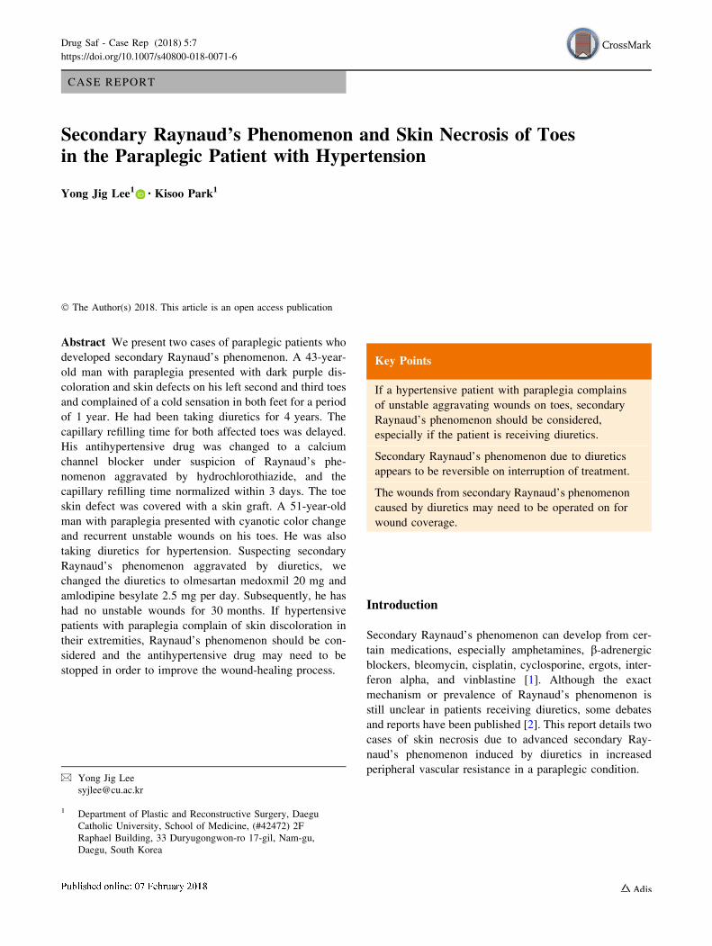
CASE REPORT Secondary Raynaud’s Phenomenon and Skin Necrosis of Toes in the Paraplegic Patient with Hypertension Yong Jig Lee 1 • Kisoo Park 1 Ó The Author(s) 2018. This article is an open access publication Abstract We present two cases of paraplegic patients who developed secondary Raynaud’s phenomenon. A 43-year- old man with paraplegia presented with dark purple dis- coloration and skin defects on his left second and third toes and complained of a cold sensation in both feet for a period of 1 year. He had been taking diuretics for 4 years. The capillary refilling time for both affected toes was delayed. His antihypertensive drug was changed to a calcium channel blocker under suspicion of Raynaud’s phe- nomenon aggravated by hydrochlorothiazide, and the capillary refilling time normalized within 3 days. The toe skin defect was covered with a skin graft. A 51-year-old man with paraplegia presented with cyanotic color change and recurrent unstable wounds on his toes. He was also taking diuretics for hypertension. Suspecting secondary Raynaud’s phenomenon aggravated by diuretics, we changed the diuretics to olmesartan medoxmil 20 mg and amlodipine besylate 2.5 mg per day. Subsequently, he has had no unstable wounds for 30 months. If hypertensive patients with paraplegia complain of skin discoloration in their extremities, Raynaud’s phenomenon should be con- sidered and the antihypertensive drug may need to be stopped in order to improve the wound-healing process. Key Points If a hypertensive patient with paraplegia complains of unstable aggravating wounds on toes, secondary Raynaud’s phenomenon should be considered, especially if the patient is receiving diuretics. Secondary Raynaud’s phenomenon due to diuretics appears to be reversible on interruption of treatment. The wounds from secondary Raynaud’s phenomenon caused by diuretics may need to be operated on for wound coverage. Introduction Secondary Raynaud’s phenomenon can develop from cer- tain medications, especially amphetamines, b-adrenergic blockers, bleomycin, cisplatin, cyclosporine, ergots, inter- feron alpha, and vinblastine [1]. Although the exact mechanism or prevalence of Raynaud’s phenomenon is still unclear in patients receiving diuretics, some debates and reports have been published [2]. This report details two cases of skin necrosis due to advanced secondary Ray- naud’s phenomenon induced by diuretics in increased peripheral vascular resistance in a paraplegic condition. & Yong Jig Lee [email protected] 1 Department of Plastic and Reconstructive Surgery, Daegu Catholic University, School of Medicine, (#42472) 2F Raphael Building, 33 Duryugongwon-ro 17-gil, Nam-gu, Daegu, South Korea Drug Saf - Case Rep (2018) 5:7 https://doi.org/10.1007/s40800-018-0071-6
Transcript of Secondary Raynaud’s Phenomenon and Skin Necrosis … · old man with paraplegia presented with...
CASE REPORT
Secondary Raynaud’s Phenomenon and Skin Necrosis of Toesin the Paraplegic Patient with Hypertension
Yong Jig Lee1• Kisoo Park1
� The Author(s) 2018. This article is an open access publication
Abstract We present two cases of paraplegic patients who
developed secondary Raynaud’s phenomenon. A 43-year-
old man with paraplegia presented with dark purple dis-
coloration and skin defects on his left second and third toes
and complained of a cold sensation in both feet for a period
of 1 year. He had been taking diuretics for 4 years. The
capillary refilling time for both affected toes was delayed.
His antihypertensive drug was changed to a calcium
channel blocker under suspicion of Raynaud’s phe-
nomenon aggravated by hydrochlorothiazide, and the
capillary refilling time normalized within 3 days. The toe
skin defect was covered with a skin graft. A 51-year-old
man with paraplegia presented with cyanotic color change
and recurrent unstable wounds on his toes. He was also
taking diuretics for hypertension. Suspecting secondary
Raynaud’s phenomenon aggravated by diuretics, we
changed the diuretics to olmesartan medoxmil 20 mg and
amlodipine besylate 2.5 mg per day. Subsequently, he has
had no unstable wounds for 30 months. If hypertensive
patients with paraplegia complain of skin discoloration in
their extremities, Raynaud’s phenomenon should be con-
sidered and the antihypertensive drug may need to be
stopped in order to improve the wound-healing process.
Key Points
If a hypertensive patient with paraplegia complains
of unstable aggravating wounds on toes, secondary
Raynaud’s phenomenon should be considered,
especially if the patient is receiving diuretics.
Secondary Raynaud’s phenomenon due to diuretics
appears to be reversible on interruption of treatment.
The wounds from secondary Raynaud’s phenomenon
caused by diuretics may need to be operated on for
wound coverage.
Introduction
Secondary Raynaud’s phenomenon can develop from cer-
tain medications, especially amphetamines, b-adrenergic
blockers, bleomycin, cisplatin, cyclosporine, ergots, inter-
feron alpha, and vinblastine [1]. Although the exact
mechanism or prevalence of Raynaud’s phenomenon is
still unclear in patients receiving diuretics, some debates
and reports have been published [2]. This report details two
cases of skin necrosis due to advanced secondary Ray-
naud’s phenomenon induced by diuretics in increased
peripheral vascular resistance in a paraplegic condition.& Yong Jig Lee
1 Department of Plastic and Reconstructive Surgery, Daegu
Catholic University, School of Medicine, (#42472) 2F
Raphael Building, 33 Duryugongwon-ro 17-gil, Nam-gu,
Daegu, South Korea
Drug Saf - Case Rep (2018) 5:7
https://doi.org/10.1007/s40800-018-0071-6
Cases
Case 1
A 43-year-old man with paraplegia for 17 years presented
with dark purple discoloration of left second and third toes
beginning 3 days earlier. He complained of having a cold
sensation in both feet for 1 year. He was diagnosed with
hypertension 4 years earlier and was taking
hydrochlorothiazide-valsartan. He had also been taking
kallidinogenase, ranitidine, levosulpiride, phloroglucinol,
and loxoprofen. Physical examination showed that both of
the patient’s feet were cold. However, the pulse was pal-
pable on ankles and heels on both sides. A portable Dop-
pler device (Minidop ES-100VX, Hadeco, Japan) showed
that the pulse signal intensity of the left and right dorsalis
pedis artery was 4–6 and 10 cm/s each, and that of the
posterior tibial artery was 6–8 and 10 cm/s each. In addi-
tion, the capillary refilling time for both toes was [10 s.
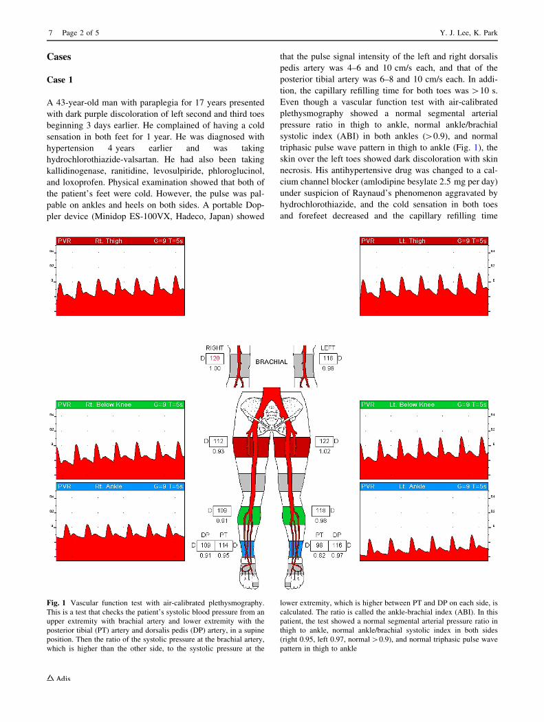
Even though a vascular function test with air-calibrated
plethysmography showed a normal segmental arterial
pressure ratio in thigh to ankle, normal ankle/brachial
systolic index (ABI) in both ankles ([0.9), and normal
triphasic pulse wave pattern in thigh to ankle (Fig. 1), the
skin over the left toes showed dark discoloration with skin
necrosis. His antihypertensive drug was changed to a cal-
cium channel blocker (amlodipine besylate 2.5 mg per day)
under suspicion of Raynaud’s phenomenon aggravated by
hydrochlorothiazide, and the cold sensation in both toes
and forefeet decreased and the capillary refilling time
Fig. 1 Vascular function test with air-calibrated plethysmography.
This is a test that checks the patient’s systolic blood pressure from an
upper extremity with brachial artery and lower extremity with the
posterior tibial (PT) artery and dorsalis pedis (DP) artery, in a supine
position. Then the ratio of the systolic pressure at the brachial artery,
which is higher than the other side, to the systolic pressure at the
lower extremity, which is higher between PT and DP on each side, is
calculated. The ratio is called the ankle-brachial index (ABI). In this
patient, the test showed a normal segmental arterial pressure ratio in
thigh to ankle, normal ankle/brachial systolic index in both sides
(right 0.95, left 0.97, normal[0.9), and normal triphasic pulse wave
pattern in thigh to ankle
7 Page 2 of 5 Y. J. Lee, K. Park
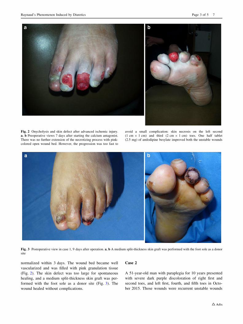
normalized within 3 days. The wound bed became well
vascularized and was filled with pink granulation tissue
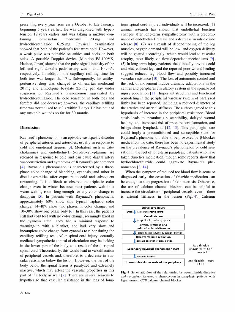
(Fig. 2). The skin defect was too large for spontaneous
healing, and a medium split-thickness skin graft was per-
formed with the foot sole as a donor site (Fig. 3). The
wound healed without complications.
Case 2
A 51-year-old man with paraplegia for 10 years presented
with severe dark purple discoloration of right first and
second toes, and left first, fourth, and fifth toes in Octo-
ber 2015. Those wounds were recurrent unstable wounds
Fig. 2 Onycholysis and skin defect after advanced ischemic injury.
a, b Preoperative views 7 days after starting the calcium antagonist.
There was no further extension of the necrotizing process with pink-
colored open wound bed. However, the progression was too fast to
avoid a small complication: skin necrosis on the left second
(1 cm 9 1 cm) and third (2 cm 9 1 cm) toes. One half tablet
(2.5 mg) of amlodipine besylate improved both the unstable wounds
Fig. 3 Postoperative view in case 1, 9 days after operation. a, b A medium split-thickness skin graft was performed with the foot sole as a donor
site
Raynaud’s Phenomenon Induced by Diuretics Page 3 of 5 7
presenting every year from early October to late January,
beginning 5 years earlier. He was diagnosed with hyper-
tension 12 years earlier and was taking a mixture con-
taining olmesartan medoxmil 20 mg and
hydrochlorothiazide 6.25 mg. Physical examination
showed that both of the patient’s feet were cold. However,
a weak pulse was palpable on ankles and heels on both
sides. A portable Doppler device (Minidop ES-100VX,
Hadeco, Japan) showed that the pulse signal intensity of the
left and right dorsalis pedis artery was 4 and 10 cm/s,
respectively. In addition, the capillary refilling time for
both toes was longer than 7 s. Subsequently, his antihy-
pertensive drug was changed to olmesartan medoxmil
20 mg and amlodipine besylate 2.5 mg per day under
suspicion of Raynaud’s phenomenon aggravated by
hydrochlorothiazide. The cold sensation in both toes and
forefeet did not decrease; however, the capillary refilling
time was normalized to\2 s within 7 days. He has not had
any unstable wounds so far for 30 months.
Discussion
Raynaud’s phenomenon is an episodic vasospastic disorder
of peripheral arteries and arterioles, usually in response to
cold and emotional triggers [3]. Mediators such as cate-
cholamines and endothelin-1, 5-hydroxytryptamine are
released in response to cold and can cause digital artery
vasoconstriction and symptoms of Raynaud’s phenomenon
[4]. Raynaud’s phenomenon is characterized by a three-
phase color change of blanching, cyanosis, and rubor in
distal extremities after exposure to cold and subsequent
rewarming. It is difficult to observe the triphasic color
change even in winter because most patients wait in a
warm waiting room long enough for any color change to
disappear [5]. In patients with Raynaud’s phenomena,
approximately 60% show this typical triphasic color
change, 14–40% show two phases in color change, and
10–30% show one phase only [6]. In this case, the patients
still had cold feet with no color change, seemingly fixed in
the cyanosis state. They had a minimal response to
warming-up with a blanket, and had very slow and
incomplete color change from cyanosis to rubor during the
capillary refilling test. After spinal-cord injury, centrally
mediated sympathetic control of circulation may be lacking
in the lower part of the body as a result of the disrupted
spinal cord. Theoretically, this would lead to vasodilatation
of peripheral vessels and, therefore, to a decrease in vas-
cular resistance below the lesion. However, the part of the
body below the spinal lesion is paralyzed and extremely
inactive, which may affect the vascular properties in this
part of the body as well [7]. There are several reasons to
hypothesize that vascular resistance in the legs of long-
term spinal-cord–injured individuals will be increased: (1)
animal research has shown that endothelial function
changes after long-term sympathectomy with a predomi-
nance of endothelin-1 release and a decrease in nitric oxide
release [8]. (2) As a result of deconditioning of the leg
muscles, oxygen demand will be low, and oxygen delivery
will be geared accordingly, which would lead to vascular
atrophy, most likely via flow-dependent mechanisms [9].
(3) In long-term injury patients, the clinically obvious cold
and blue-colored legs and the reported poor wound healing
suggest reduced leg blood flow and possibly increased
vascular resistance [10]. The loss of autonomic control and
the lack of movement induce dramatic adaptations in the
central and peripheral circulatory system in the spinal-cord
injury population [11]. Important structural and functional
remodeling in the peripheral vascular system of paralyzed
limbs has been reported, including a reduced diameter of
the arteries and arterial stiffness. The authors agreed to this
hypothesis of increase in the peripheral resistance. Blood
stasis leads to thrombosis susceptibility, delayed wound
healing, and increased risk of pressure sore formation, and
brings about lymphedema [12, 13]. This paraplegic state
could imply a preconditioned and susceptible state for
Raynaud’s phenomenon, able to be provoked by b-blocker
medication. To date, there has been no experimental study
on the prevalence of Raynaud’s phenomenon or cold sen-
sation in the feet of long-term paraplegic patients who have
taken diuretics medication, though some reports show that
hydrochlorothiazide could aggravate Raynaud’s phe-
nomenon [2, 14].
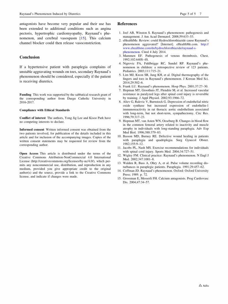
When the symptom of reduced toe blood flow is acute or
diagnosed early, the cessation of thiazide medication can
be enough to stop progression of skin necrosis. Otherwise,
the use of calcium channel blockers can be helpful to
increase the circulation of peripheral vessels, even if there
is arterial stiffness in the lesion (Fig. 4). Calcium
Fig. 4 Schematic flow of the relationship between thiazide diuretics
and secondary Raynaud’s phenomenon in paraplegic patients with
hypertension. CCB calcium channel blocker
7 Page 4 of 5 Y. J. Lee, K. Park
antagonists have become very popular and their use has
been extended to additional conditions such as angina
pectoris, hypertrophic cardiomyopathy, Raynaud’s phe-
nomenon, and cerebral vasospasm [15]. This calcium
channel blocker could then release vasoconstriction.
Conclusion
If a hypertensive patient with paraplegia complains of
unstable aggravating wounds on toes, secondary Raynaud’s
phenomenon should be considered, especially if the patient
is receiving diuretics.
Funding This work was supported by the sabbatical research grant of
the corresponding author from Daegu Catholic University in
2016-2017.
Compliance with Ethical Standards
Conflict of interest The authors, Yong Jig Lee and Kisoo Park have
no competing interests to declare.
Informed consent Written informed consent was obtained from the
two patients involved, for publication of the details included in this
article and for inclusion of the accompanying images. Copies of the
written consent statements may be requested for review from the
corresponding author.
Open Access This article is distributed under the terms of the
Creative Commons Attribution-NonCommercial 4.0 International
License (http://creativecommons.org/licenses/by-nc/4.0/), which per-
mits any noncommercial use, distribution, and reproduction in any
medium, provided you give appropriate credit to the original
author(s) and the source, provide a link to the Creative Commons
license, and indicate if changes were made.
References
1. Joel AB, Winston S. Raynaud’s phenomenon: pathogenesis and
management. J Am Acad Dermatol. 2008;59:633–53.
2. eHealthMe. Review: could Hydrochlorothiazide cause Raynaud’s
phenomenon aggravated? [Internet]. eHealthMe.com. http://
www.ehealthme.com/ds/hydrochlorothiazide/raynaud-s-
phenomenon. Cited 4 July 2014.
3. Mammen EF. Pathogenesis of venous thrombosis. Chest.
1992;102:640S–4S.
4. Nigrovic PA, Fuhlbrigge RC, Sundel RP. Raynaud’s phe-
nomenon in children: a retrospective review of 123 patients.
Pediatrics. 2003;111:715–21.
5. Lim MJ, Kwon SR, Jung KH, et al. Digital thermography of the
fingers and toes in Raynaud’s phenomenon. J Korean Med Sci.
2014;29:502–6.
6. Frank LU. Raynaud’s phenomenon. Hosp Phys. 2001;37:27–30.
7. Hopman MT, Groothuis JT, Flendrie M, et al. Increased vascular
resistance in paralyzed legs after spinal cord injury is reversible
by training. J Appl Physiol. 2002;93:1966–72.
8. Aliev G, Ralevic V, Burnstock G. Depression of endothelial nitric
oxide synthase but increased expression of endothelin-1
immunoreactivity in rat thoracic aortic endothelium associated
with long-term, but not short-term, sympathectomy. Circ Res.
1996;79:317–23.
9. Hopman MT, van Asten WN, Oeseburg B. Changes in blood flow
in the common femoral artery related to inactivity and muscle
atrophy in individuals with long-standing paraplegia. Adv Exp
Med Biol. 1996;388:379–83.
10. Basson MD, Burney RE. Defective wound healing in patients
with paraplegia and quadriplegia. Surg Gynecol Obstet.
1982;155:9–12.
11. Jacobs PL, Nash MS. Exercise recommendations for individuals
with spinal cord injury. Sports Med. 2004;34:727–51.
12. Wigley FM. Clinical practice: Raynaud’s phenomenon. N Engl J
Med. 2002;347:1001–8.
13. Walden R, Bass A, Ohry A, et al. Pulse volume recording dis-
turbances in paraplegic patients. Paraplegia. 1991;29:457–62.
14. Coffman JD. Raynaud’s phenomenon. Oxford: Oxford University
Press; 1989. p. 72.
15. Grossman E, Messerli FH. Calcium antagonists. Prog Cardiovasc
Dis. 2004;47:34–57.
Raynaud’s Phenomenon Induced by Diuretics Page 5 of 5 7