Secondary Causes of Osteoporosis and Low Bone Density · Secondary Causes of Osteoporosis and Low...
76
Secondary Causes of Osteoporosis and Low Bone Density Michael Maricic, M.D. Catalina Pointe Rheumatology Clinical Associate Professor University of Arizona, Tucson
-
Upload
nguyenngoc -
Category
Documents
-
view
217 -
download
2
Transcript of Secondary Causes of Osteoporosis and Low Bone Density · Secondary Causes of Osteoporosis and Low...
Secondary Causes of
Osteoporosis and Low Bone
Density
Michael Maricic, M.D.
Catalina Pointe Rheumatology
Clinical Associate Professor
University of Arizona, Tucson
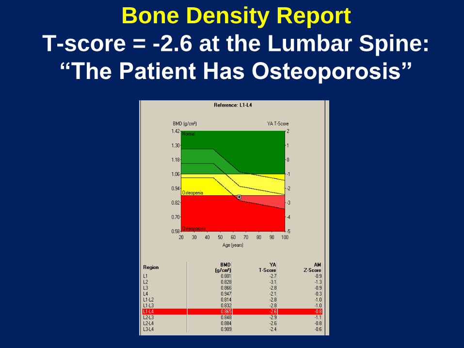
Bone Density Report
T-score = -2.6 at the Lumbar Spine:
“The Patient Has Osteoporosis”
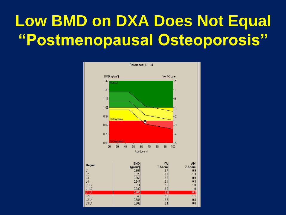
Low BMD on DXA Does Not Equal
“Postmenopausal Osteoporosis”
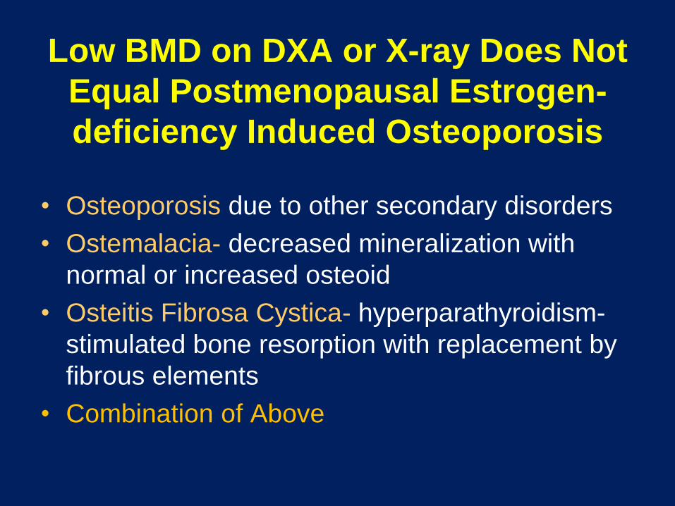
Low BMD on DXA or X-ray Does Not
Equal Postmenopausal Estrogen-
deficiency Induced Osteoporosis
• Osteoporosis due to other secondary disorders
• Ostemalacia- decreased mineralization with
normal or increased osteoid
• Osteitis Fibrosa Cystica- hyperparathyroidism-
stimulated bone resorption with replacement by
fibrous elements
• Combination of Above
Secondary Osteoporosis
• Secondary osteoporosis is defined as
osteoporosis or low bone density for
which there is an identifiable causal
factor other than menopause and
aging.
Evaluating for Secondary Causes
of Osteoporosis
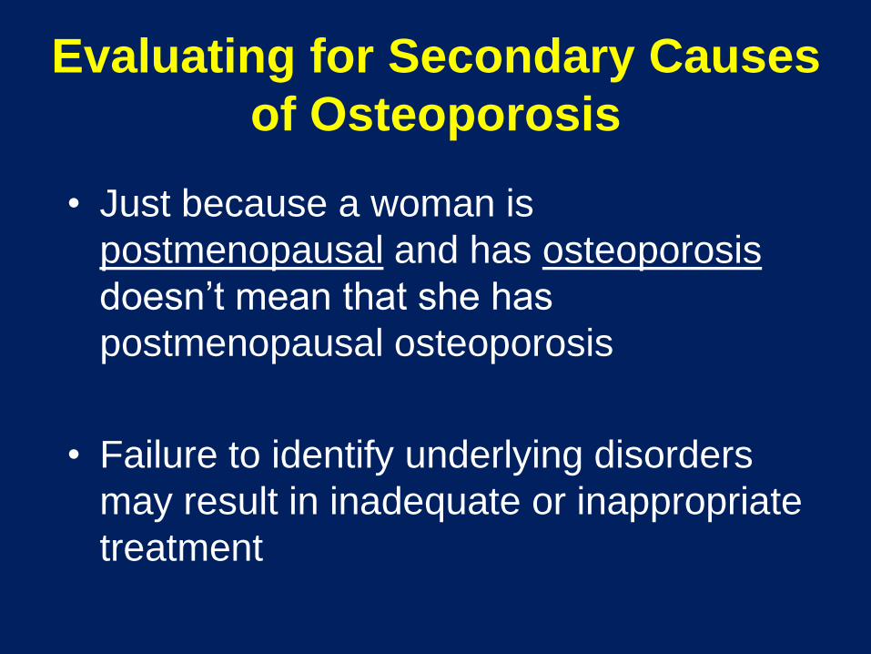
• Just because a woman is
postmenopausal and has osteoporosis
doesn’t mean that she has
postmenopausal osteoporosis
• Failure to identify underlying disorders
may result in inadequate or inappropriate
treatment
Secondary Causes of Osteoporosis
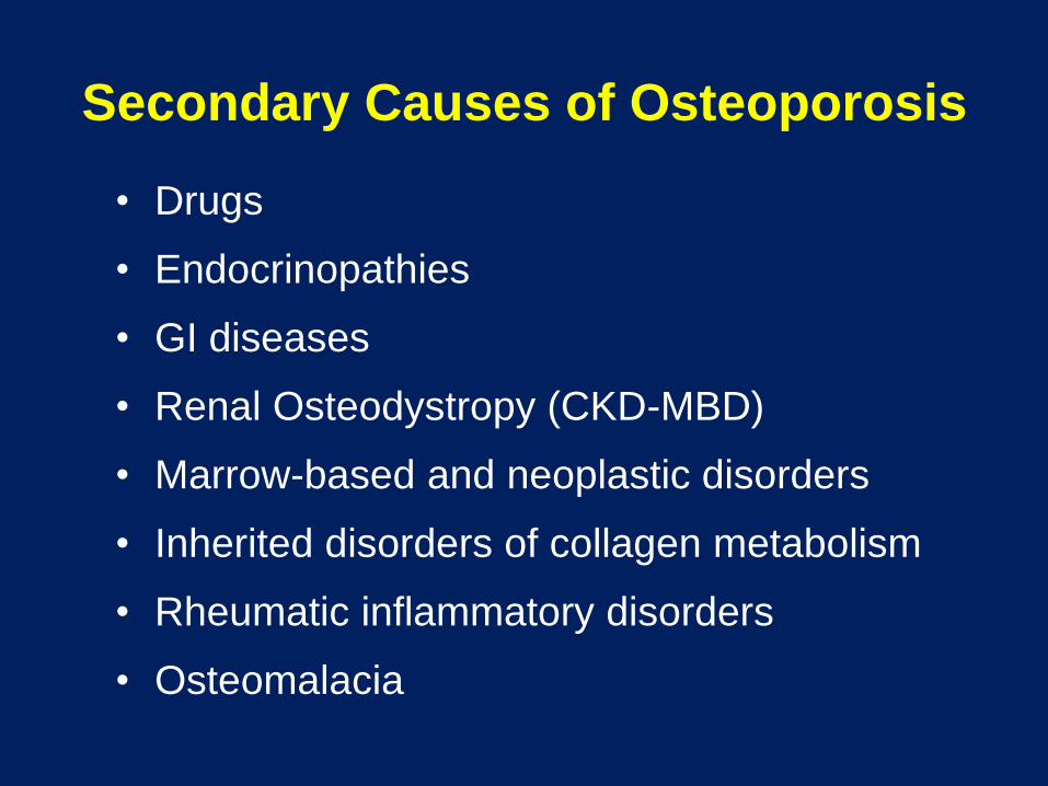
• Drugs
• Endocrinopathies
• GI diseases
• Renal Osteodystropy (CKD-MBD)
• Marrow-based and neoplastic disorders
• Inherited disorders of collagen metabolism
• Rheumatic inflammatory disorders
• Osteomalacia

Endocrinopathies
Associated with Osteoporosis
• Hypogonadism
• Primary Hyperparathyroidism
• Diabetes
• Hyperthyroidism
• Hypercortisolism
• Hypercalciuria with or without renal
stones

Gastrointestinal Disorders
Associated with Osteoporosis
• Celiac disease (Sprue)
• Gastrectomy
• Intestinal bypass surgery
• Primary biliary cirrhosis
• Pancreatic insufficiency
Marrow-based and Neoplastic Disorders
Associated with Osteoporosis
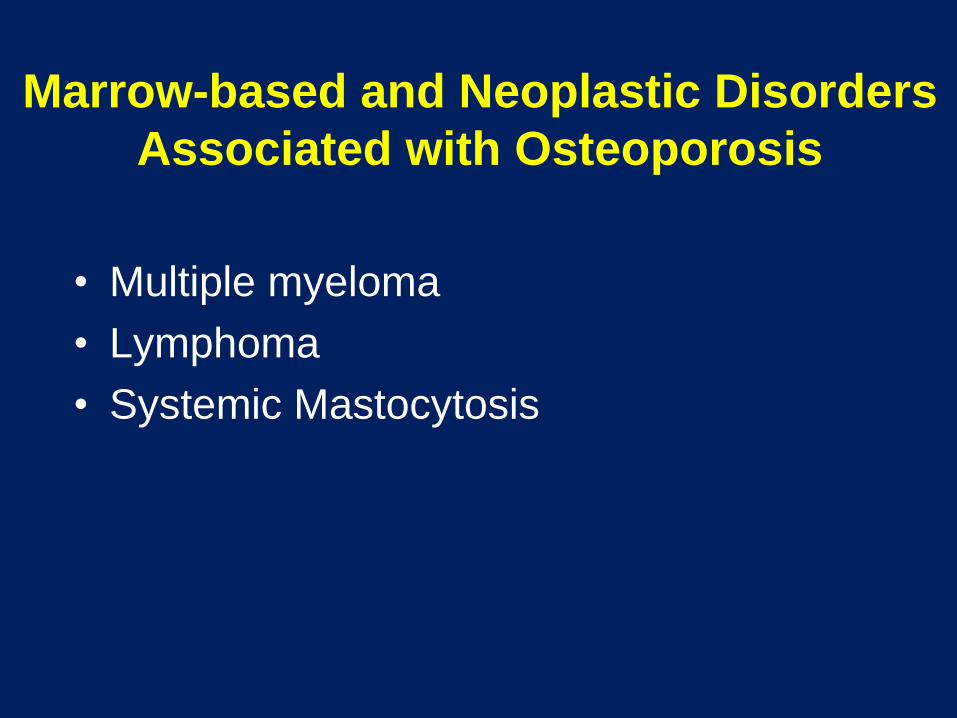
• Multiple myeloma
• Lymphoma
• Systemic Mastocytosis

Rheumatic Inflammatory Disorders
Associated with Osteoporosis
• Rheumatoid Arthritis
• Ankylosing Spondylitis
• Systemic Lupus

Genetic Disorders Associated with
Osteoporosis
Collagen/Metabolic
• Collagen Disorders
– Osteogenesis imperfecta
– Ehlers-Danlos syndrome
– Marfan syndrome
• Metabolic Disorders
– Hypophosphatasia
– Homocystinuria

How Often are Secondary
Causes Found?
Laboratory Yield-
Secondary Causes of Osteoporosis
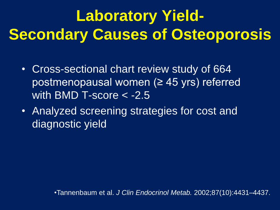
• Cross-sectional chart review study of 664
postmenopausal women (≥ 45 yrs) referred
with BMD T-score < -2.5
• Analyzed screening strategies for cost and
diagnostic yield
•Tannenbaum et al. J Clin Endocrinol Metab. 2002;87(10):4431–4437.
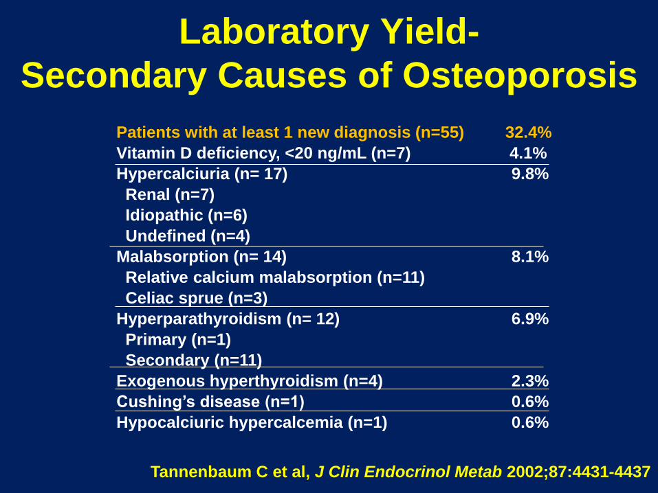
Laboratory Yield-
Secondary Causes of Osteoporosis
Tannenbaum C et al, J Clin Endocrinol Metab 2002;87:4431-4437
Patients with at least 1 new diagnosis (n=55) 32.4%
Vitamin D deficiency, <20 ng/mL (n=7) 4.1%
Hypercalciuria (n= 17) 9.8%
Renal (n=7)
Idiopathic (n=6)
Undefined (n=4)
Malabsorption (n= 14) 8.1%
Relative calcium malabsorption (n=11)
Celiac sprue (n=3)
Hyperparathyroidism (n= 12) 6.9%
Primary (n=1)
Secondary (n=11)
Exogenous hyperthyroidism (n=4) 2.3%
Cushing’s disease (n=1) 0.6%
Hypocalciuric hypercalcemia (n=1) 0.6%

Basic LabTests When Considering
Secondary Causes of Low Bone Mass
• Serum calcium (chemistry screen)
• 25-OH Vitamin D
• 24 hour urine calcium
• TSH (in women receiving thyroid supplementation)
• These tests identify 92% of patients with 2nd causes
• Cost of $ 118 per patient
Tannenbaum et al. J Clin Endocrinol Metab. 2002;87(10):4431–4437.

24 Hour Urine Calcium
• Low urine calcium = malabsorption
• High urine calcium = renal calcium wasting
• Lab reference range 100-300 mg/day (1-4 mg/kg/day)
Should be collected when vitamin D is adequate and
calcium intake is within target of 1200-1500 mg daily
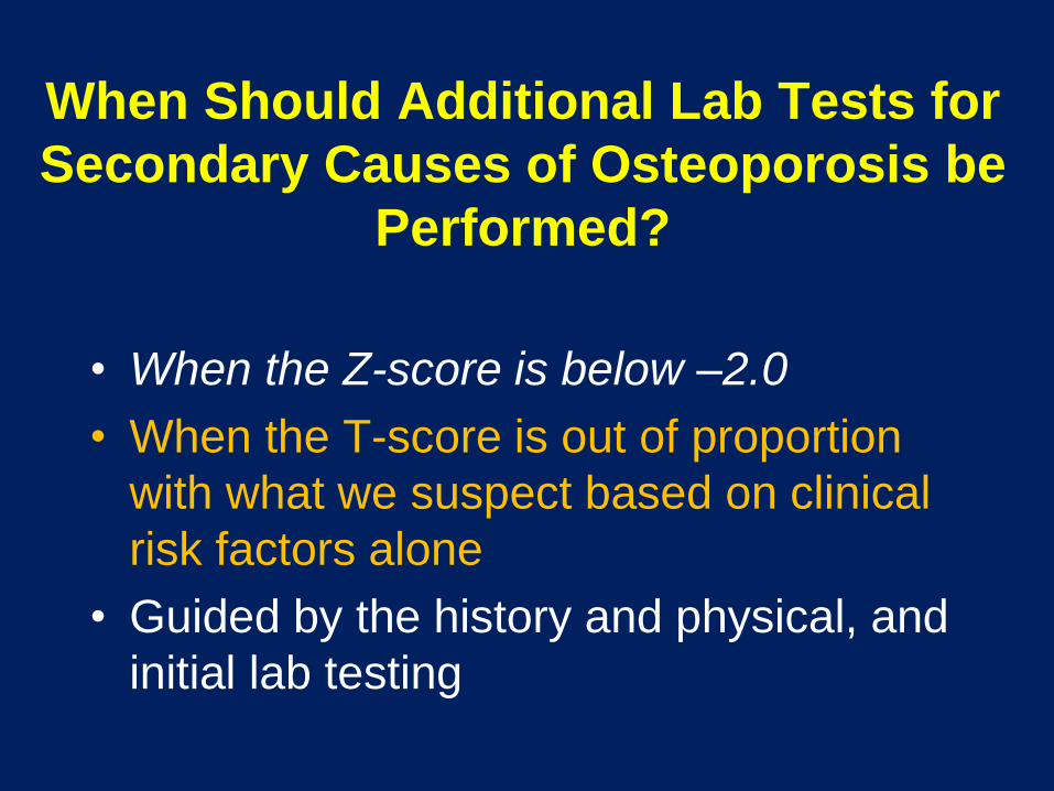
When Should Additional Lab Tests for
Secondary Causes of Osteoporosis be
Performed?
• When the Z-score is below –2.0
• When the T-score is out of proportion
with what we suspect based on clinical
risk factors alone
• Guided by the history and physical, and
initial lab testing

• Intact PTH ( if serum calcium is high)
• Celiac Panel
• SPEP, UPEP
• Serum Tryptase/Urine Histamine (mastocytosis)
• Total and Free Testosterone in Males
• Bone Biopsy in CKD-MBD
• Skin Biopsy for Collagen Disorder
Additional Lab Tests for Secondary
Causes of Osteoporosis

Differential Diagnosis of
Hypercalcemia
• Primary hyperparathyroidism
• Malignancy
• Hypervitaminosis D
• Thiazides
• Sarcoid, granulomatous disorders
• Lithium
• Hypervitaminosis A
• Milk alkali syndome
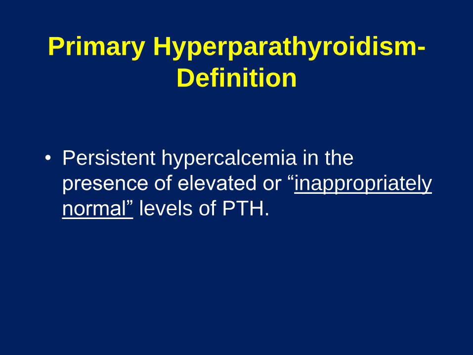
Primary Hyperparathyroidism-
Definition
• Persistent hypercalcemia in the
presence of elevated or “inappropriately
normal” levels of PTH.

iPTH- Calcium Relationship

BMD Recovery Following Parathyroidectomy
for Primary Hyperparathyroidism
Rubin MR. J Clin Endocrinol Metab 93: 3462–3470, 2008

Celiac Disease
• One of the most common genetic disorders
• Clinically evident in 1% of northern Europeans
• May be found in up to 3.4% of patients with osteoporosis
• Present in 5-20% of first-degree relatives of known celiac patients
Stenson WF, et al. Arch Intern Med. 2005;165:393-399

Antibodies Associated with
Celiac Disease
Fasano A. N Engl J Med 2012;367:2419-26.
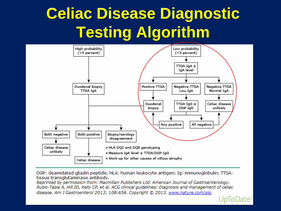
Celiac Disease Diagnostic
Testing Algorithm

Disease Associations with
Celiac Disease
• Dermatitis Herpetiformis (high correlation)
–Most with DH have celiac disease
• 3% of CD patients have Selective IgA deficiency
• 10% of patients with IgA deficiency have CD
• Patients with symptomatic iron deficiency anemia (10-15%)
• Type 1 diabetes (3-6%)
• Downs syndrome (5-12%)
Rostom A. Gastroenterology 2006 Dec;131(6):1981-2002
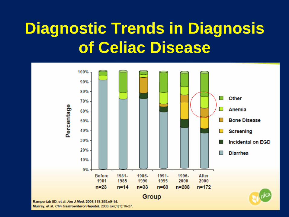
Diagnostic Trends in Diagnosis
of Celiac Disease

Summary- Celiac Disease
• Serologic and biopsy testing must be done on
a gluten-containing diet
• Both a positive serologic test and intestinal
biopsy are necessary to make a presumptive
diagnosis
• Definitive Dx requires a + response of
symptoms and/or signs of CD on gluten-free
diet
• Beware of patients with IgA deficiency
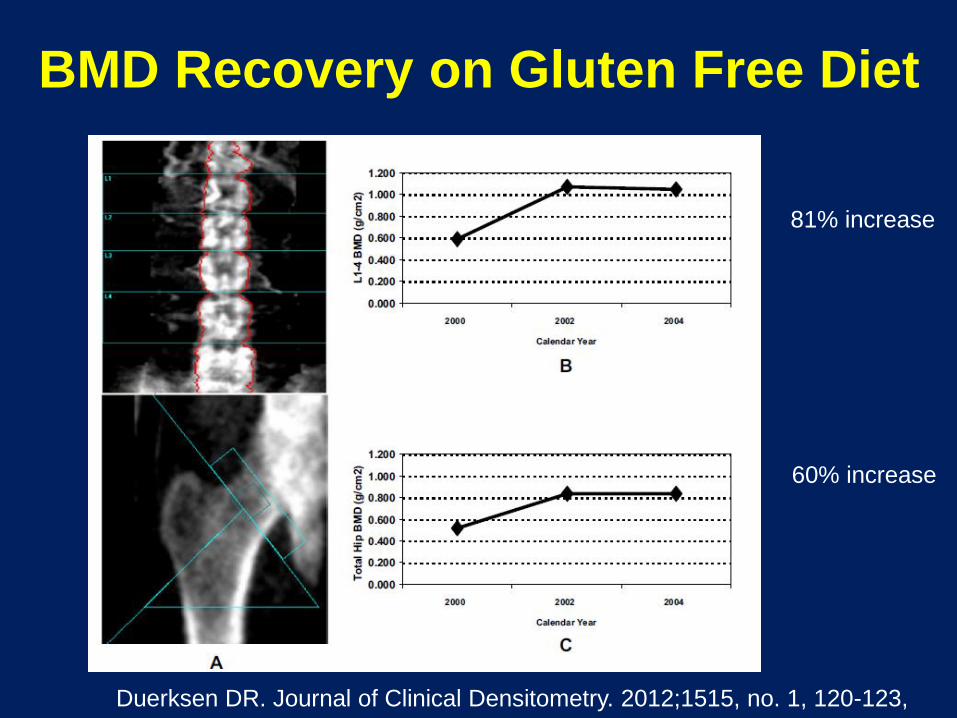
BMD Recovery on Gluten Free Diet
Duerksen DR. Journal of Clinical Densitometry. 2012;1515, no. 1, 120-123,
81% increase
60% increase
Drug-induced Osteoporosis • Glucocorticoids
• Aromatase inhibitors
• Proton pump inhibitors (PPIs)
• Selective serotonin reuptake inhibitors (SSRIs)
• Antiseizure medicines
• Thiazolidinediones
• Long-term Heparin
• Tamoxifen (premenopausal use)
• Gonadotropin releasing hormone agonists (GnRH)
• Thyroid hormones in excess
• Antiretroviral Therapy
• Lithium
• Chemotherapeutic / immunosuppressives (MTX, CsA, Tacrolimus)
• Medroxyprogesterone acetate for contraception

Glucocorticoid-Induced
Osteoporosis: Epidemiology
• GIOP is the most common iatrogenic cause of
osteoporosis
• Approximately 1% of the adult population is
receiving oral glucocorticoids at any given time
(up to 3% of patients over age 70)

Fractures Are the Most Common
Serious Adverse Event of Glucocorticoids
User Nonuser
Adverse Events (AEs) (112) (112)
Fracture 21 (19%) 8 (7%)
Cataract 17 5
Serious Infection 14 4
GI Bleed or Ulcer 11 4
Diabetic Complication 8 3
Herpes Zoster 8 1
Stroke 6 1
Death 2 0
Saag KG, et al. Am J Med. 1994;96(2):115-123.
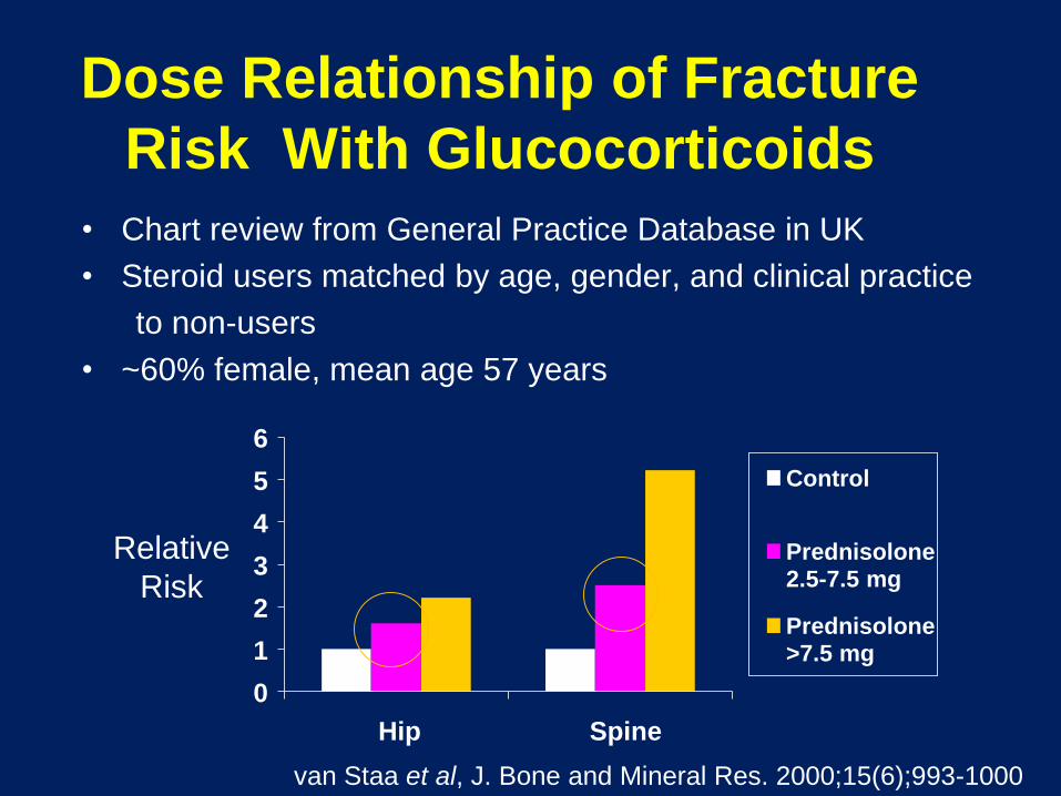
Dose Relationship of Fracture
Risk With Glucocorticoids
• Chart review from General Practice Database in UK
• Steroid users matched by age, gender, and clinical practice
to non-users
• ~60% female, mean age 57 years
0
1
2
3
4
5
6
Hip Spine
Control
Prednisolone2.5-7.5 mg
Prednisolone>7.5 mg
Relative
Risk
van Staa et al, J. Bone and Mineral Res. 2000;15(6);993-1000
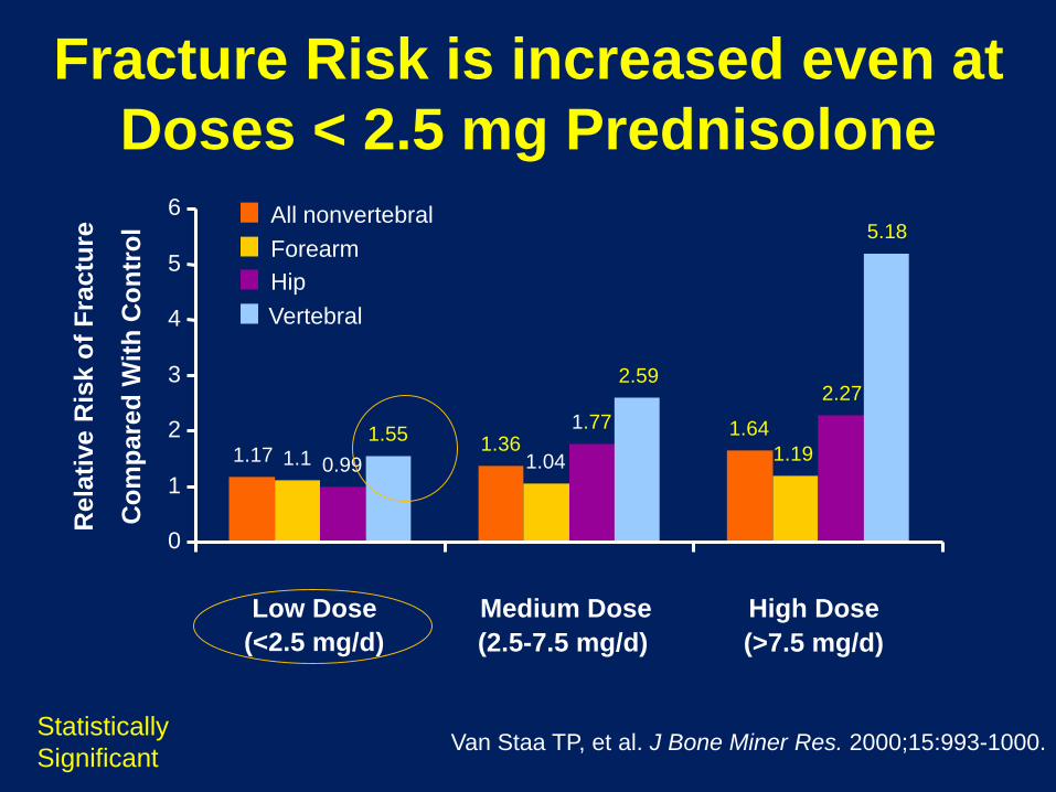
Fracture Risk is increased even at
Doses < 2.5 mg Prednisolone
1.171.36
1.64
1.1 1.04 1.190.99
1.77
2.27
1.55
2.59
5.18
0
1
2
3
4
5
6
Low Dose Medium Dose High Dose
All nonvertebral
Forearm
Hip
Vertebral
Rela
tive R
isk o
f F
ractu
re
Co
mp
are
d W
ith
Co
ntr
ol
Van Staa TP, et al. J Bone Miner Res. 2000;15:993-1000.
(<2.5 mg/d) (2.5-7.5 mg/d) (>7.5 mg/d)
Statistically
Significant
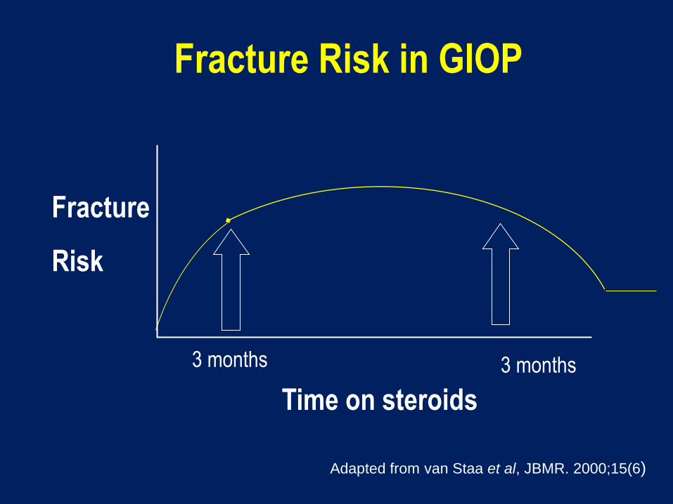
Fracture Risk in GIOP
Time on steroids
3 months 3 months
Adapted from van Staa et al, JBMR. 2000;15(6)
Fracture
Risk
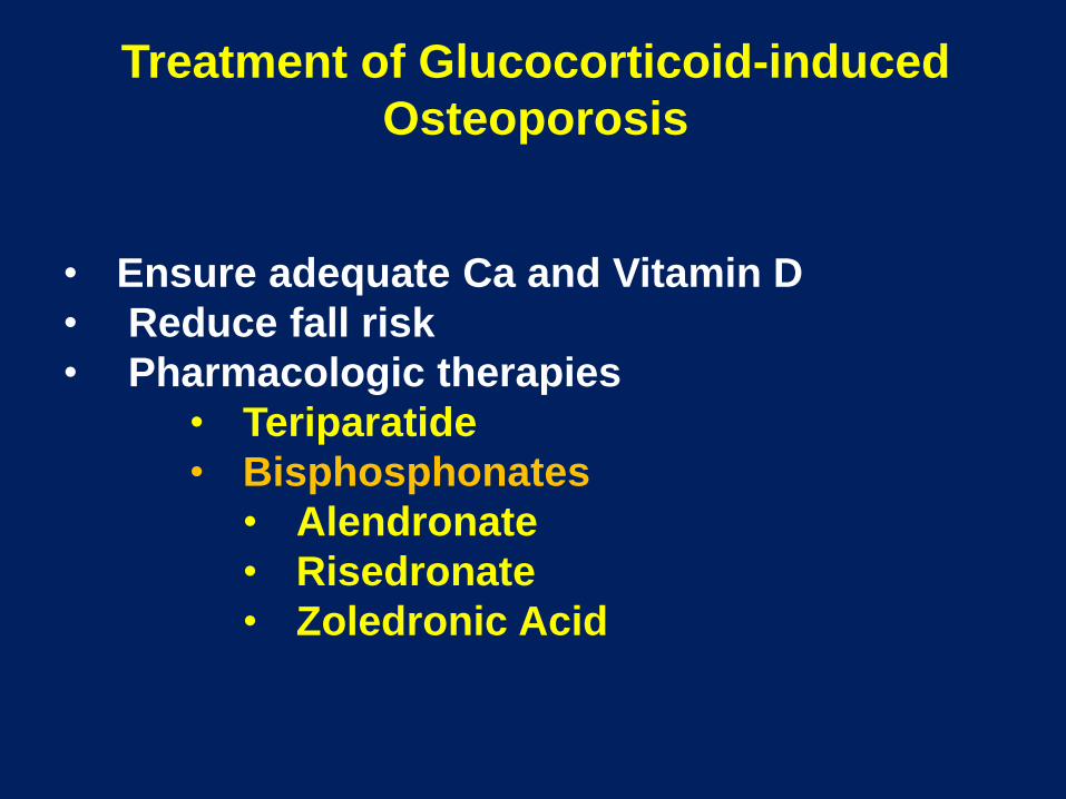
Treatment of Glucocorticoid-induced
Osteoporosis
• Ensure adequate Ca and Vitamin D
• Reduce fall risk
• Pharmacologic therapies
• Teriparatide
• Bisphosphonates
• Alendronate
• Risedronate
• Zoledronic Acid
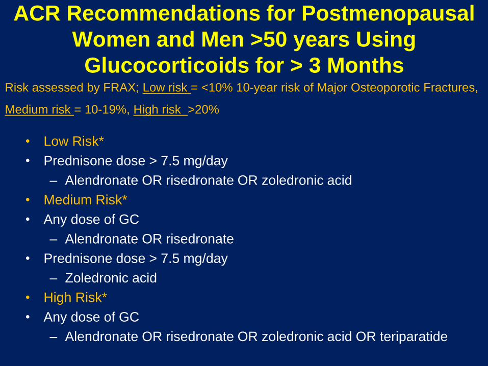
ACR Recommendations for Postmenopausal
Women and Men >50 years Using
Glucocorticoids for > 3 Months
• Low Risk*
• Prednisone dose > 7.5 mg/day
– Alendronate OR risedronate OR zoledronic acid
• Medium Risk*
• Any dose of GC
– Alendronate OR risedronate
• Prednisone dose > 7.5 mg/day
– Zoledronic acid
• High Risk*
• Any dose of GC
– Alendronate OR risedronate OR zoledronic acid OR teriparatide
Risk assessed by FRAX; Low risk = <10% 10-year risk of Major Osteoporotic Fractures,
Medium risk = 10-19%, High risk >20%
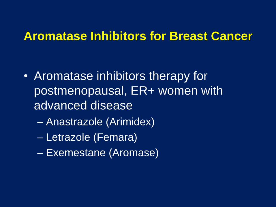
Aromatase Inhibitors for Breast Cancer
• Aromatase inhibitors therapy for
postmenopausal, ER+ women with
advanced disease
– Anastrazole (Arimidex)
– Letrazole (Femara)
– Exemestane (Aromase)
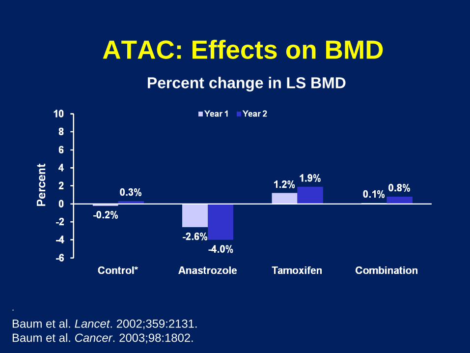
ATAC: Effects on BMDPercent change in LS BMD
.
Baum et al. Lancet. 2002;359:2131.
Baum et al. Cancer. 2003;98:1802.

45
ATAC: Bone Fractures
RR=1.59
RR=1.61
RR = relative risk.
Baum et al. Lancet. 2002;359:2131.
Baum et al. Cancer. 2003;98:1802.
n=219
n=137
n=183
n=115
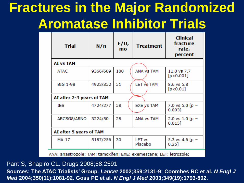
Fractures in the Major Randomized
Aromatase Inhibitor Trials
Pant S, Shapiro CL. Drugs 2008;68:2591
Sources: The ATAC Trialists’ Group. Lancet 2002;359:2131-9; Coombes RC et al. N Engl J
Med 2004;350(11):1081-92. Goss PE et al. N Engl J Med 2003;349(19):1793-802.

Source: Critical Reviews in Oncology/Hematology 2009; 69:73-82 (DOI:10.1016/j.critrevonc.2008.07.013 )
Copyright © 2009 Elsevier Ireland Ltd Terms and Conditions
Risk of Bone Fracture Correlates with
Undetectable Estrogen Levels
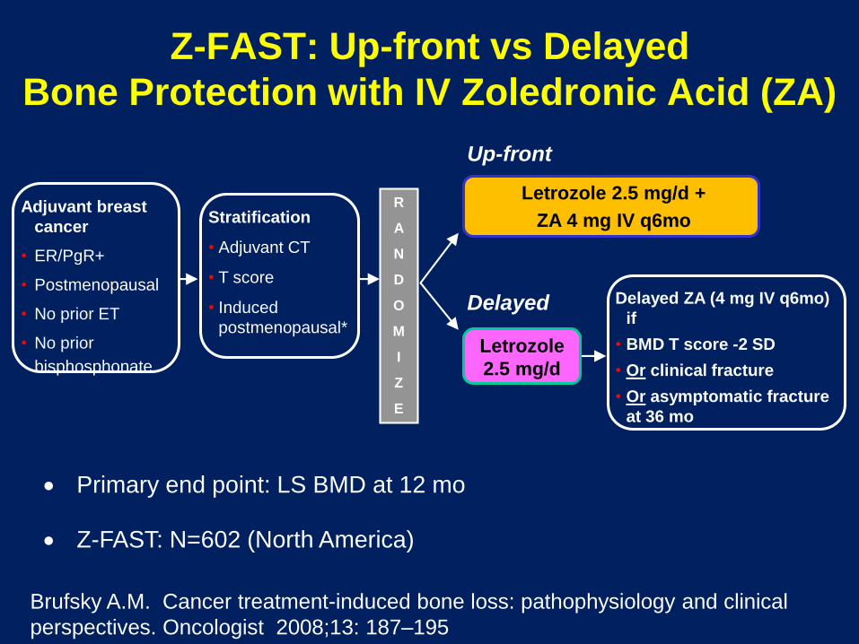
Z-FAST: Up-front vs Delayed
Bone Protection with IV Zoledronic Acid (ZA)
Adjuvant breast
cancer
• ER/PgR+
• Postmenopausal
• No prior ET
• No prior
bisphosphonate
R
A
N
D
O
M
I
Z
E
Letrozole 2.5 mg/d +
ZA 4 mg IV q6mo
Delayed ZA (4 mg IV q6mo)
if
• BMD T score -2 SD
• Or clinical fracture
• Or asymptomatic fracture
at 36 mo
Primary end point: LS BMD at 12 mo
Z-FAST: N=602 (North America)
Stratification
• Adjuvant CT
• T score
• Induced
postmenopausal*Letrozole
2.5 mg/d
Up-front
Delayed
Brufsky A.M. Cancer treatment-induced bone loss: pathophysiology and clinical
perspectives. Oncologist 2008;13: 187–195

Z-FAST: Mean Percent
Change in BMD with Zoledronic Acid
Lumbar Spine Total Hip
P<0.0001
P<0.0001
Month 6 Month 12 Month 6 Month 12
Me
an
(±
SD
)
% c
ha
ng
e in
BM
D, g
/cm
2
Brufsky A.M. Cancer treatment-induced bone loss: pathophysiology and clinical
perspectives. Oncologist 2008;13: 187–195

Randomized Trial of Denosumab in Patients
Receiving Adjuvant Aromatase Inhibitors
for Nonmetastatic Breast Cancer
Ellis GK et al. J Clin Oncol 2008; 26:
4875-822

Denosumab in Patients Receiving Adjuvant
Aromatase Inhibitors
Ellis GK et al. J Clin Oncol 2008; 26: 4875-822
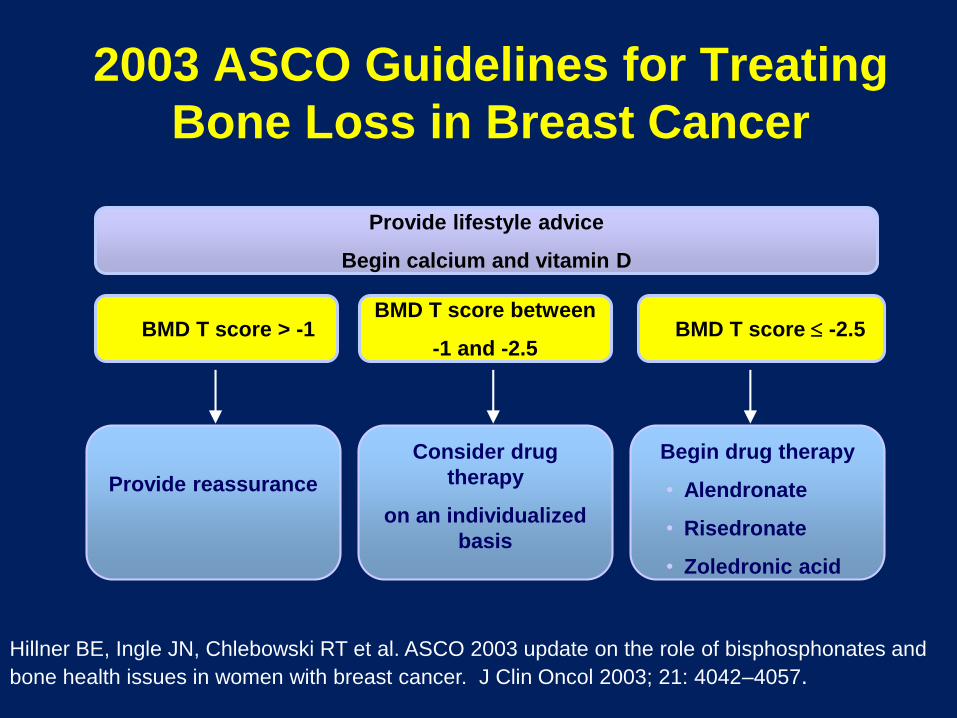
2003 ASCO Guidelines for Treating
Bone Loss in Breast Cancer
BMD T score > -1BMD T score between
-1 and -2.5BMD T score -2.5
Consider drug
therapy
on an individualized
basis
Begin drug therapy
• Alendronate
• Risedronate
• Zoledronic acid
Provide reassurance
Provide lifestyle advice
Begin calcium and vitamin D
Hillner BE, Ingle JN, Chlebowski RT et al. ASCO 2003 update on the role of bisphosphonates and
bone health issues in women with breast cancer. J Clin Oncol 2003; 21: 4042–4057.

Proton Pump Inhibitors and Increased
Fracture Risk: 2010 FDA Warning
• Revised warning for PPI: possible increased risk of
hip, wrist, & spine fractures.
• Based on 7 epidemiologic studies & claims data
base analysis ( no randomized trials)
• Increased risk after 1-7 years of treatment
– ( note: OTC label for 14 days treatment)
• Risk include age >50, “high dose”, longer duration
• Calcium carbonate absorption?
www.fda.gov safety communication 5/25/10
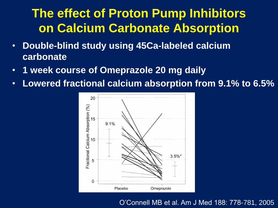
The effect of Proton Pump Inhibitors
on Calcium Carbonate Absorption
• Double-blind study using 45Ca-labeled calcium
carbonate
• 1 week course of Omeprazole 20 mg daily
• Lowered fractional calcium absorption from 9.1% to 6.5%
O’Connell MB et al. Am J Med 188: 778-781, 2005

PPIs and Hip Fractures
• PPI therapy 1st linked to an increased risk for hip
fractures in 2006
• UK General Practice Research Database
– Cases included all patients with an incident hip
fracture (n = 13,556), and 135,386 controls
• The strength of the association between hip
fracture and PPI therapy increased with
increasing duration of PPI therapy .
• Adjusted OR=
– 1 year, 1.22 [95% C I, 1.15 - 1.30];
– 2 years, 1.41 [95% C I, 1.28 - 1.56];
– 3 years, 1.54 [95% C I, 1.37 - 1.73]; and
– 4 years, 1.59 [95% C I, 1.39 - 1.80]; (P < .001)
Yang Y, et al. JAMA. 2006;296:2947-2953.

PPIs and Hip Fractures
• Retrospective, case–control study matched
15,792 cases of osteoporosis-related fractures
with 47,289 controls
• Long-term exposure to PPI therapy, (7 or more
years), was significantly associated with an
increased risk of any osteoporosis-related
fractures (OR 1.92 [1.16–3.18], P = 0.011)
• Hip fracture risk was increased after only 5
years of continuous use
Targownik et al CMAJ 2008;179(4):319
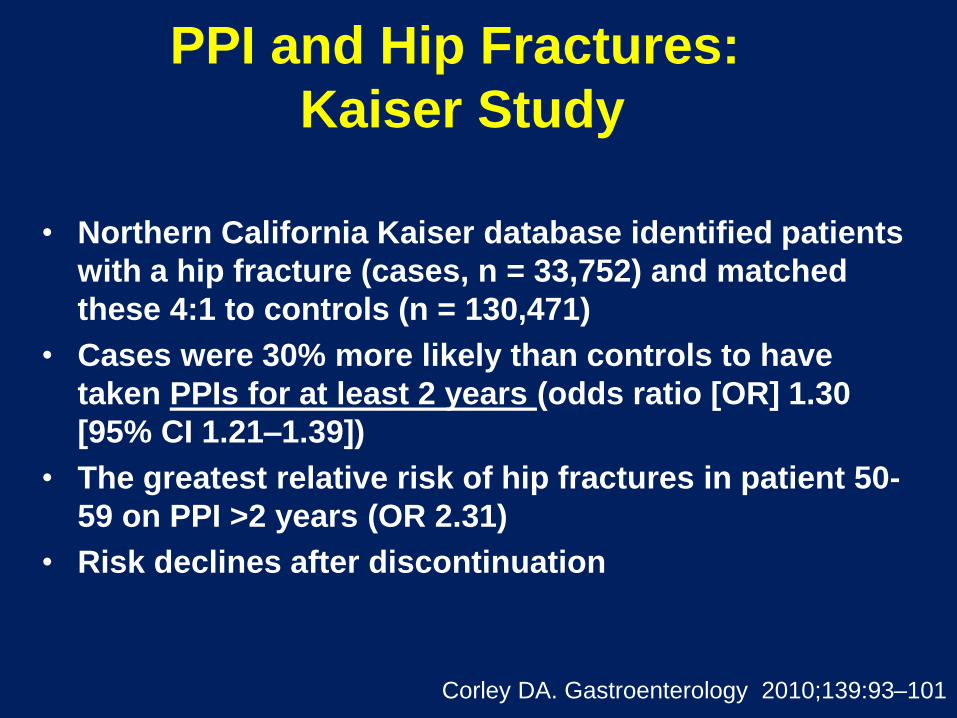
PPI and Hip Fractures:
Kaiser Study
• Northern California Kaiser database identified patients
with a hip fracture (cases, n = 33,752) and matched
these 4:1 to controls (n = 130,471)
• Cases were 30% more likely than controls to have
taken PPIs for at least 2 years (odds ratio [OR] 1.30
[95% CI 1.21–1.39])
• The greatest relative risk of hip fractures in patient 50-
59 on PPI >2 years (OR 2.31)
• Risk declines after discontinuation
Corley DA. Gastroenterology 2010;139:93–101

PPIs and hip fracture in the WHI
• The Women’s Health Initiative, with more
than 1 million person-years of follow-up,
found no association between PPI use and
hip fracture
• There was a modest association between PPI
use and spine, arm, wrist and total fractures
Gray SL, et al. Arch Intern Med. 2010;170(9):765-71.

Proton Pump Inhibitors and Risk of Fractures:
A Meta-Analysis of 11 International Studies
Yu, et al. The American Journal of Medicine (2011) 124, 519-526
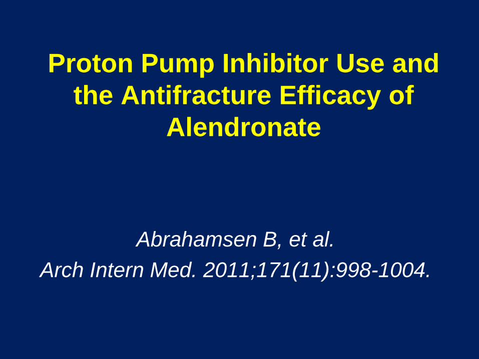
Proton Pump Inhibitor Use and
the Antifracture Efficacy of
Alendronate
Abrahamsen B, et al.
Arch Intern Med. 2011;171(11):998-1004.

Proton Pump Inhibitor Use and
the Antifracture Efficacy of Alendronate
• Population-based, national register–based, open cohort
study of 38,088 new alendronate sodium users (mean
duration of follow-up 3.5 yrs.
• The hip fracture risk reduction associated with
complete refill compliance to alendronate was a
39% risk reduction [HR], 0.61; 95% [CI], 0.52- 0.71; P.001)
in patients who were not PPI users
• The risk reduction in concurrent PPI users was
non-significant (19%; HR, 0.81; 95% CI, 0.64-1.01; P=.06).
Abrahamsen B, et al. Arch Intern Med. 2011;171(11):998-1004

PPI Use and the Risk Reduction in
Hip Fractures on Alendronate
Abrahamsen B, et al. Arch Intern Med. 2011;171(11):998-1004

Use of selective serotonin-reuptake
inhibitors or tricyclic
antidepressants and risk of hip
fractures in elderly people
Liu et al.
Lancet 1998;351 (9112). 1303-1307

Odds Ratio for Hip Fracture by Timing of
Exposure to Anti-depressant
Liu et al. Lancet 1998;351 (9112). 1303-1307

Depression-induced Structural
Impairment of Mice Exposed to Chronic
Mild Stress for 4 Weeks
Yimaya et al Proc Natl Acad Sci U S A 2006, 103:16876–16881.
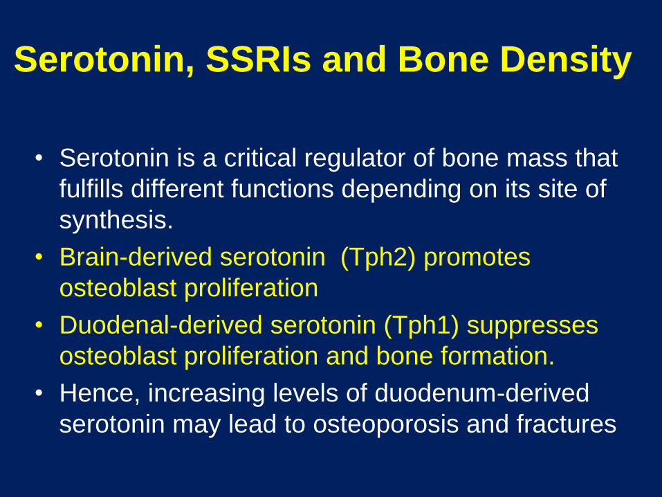
Serotonin, SSRIs and Bone Density
• Serotonin is a critical regulator of bone mass that
fulfills different functions depending on its site of
synthesis.
• Brain-derived serotonin (Tph2) promotes
osteoblast proliferation
• Duodenal-derived serotonin (Tph1) suppresses
osteoblast proliferation and bone formation.
• Hence, increasing levels of duodenum-derived
serotonin may lead to osteoporosis and fractures
Bone – Gut ConnectionMechanism
With permission from Yadav VK, et al. Cell. 2008;135:825-837.
Abbreviation: Creb, cyclic AMP-responsive element-binding protein.
Serotonin
Bone
Duodenum
LRP5
Tph1Enterochromaffin
Cell
Decreased
Osteoblast
Proliferation
Creb
Htr1b
Osteoblast

SSRIs and Fracture:
Canadian Multicenter Osteoporosis Study
Prospective cohort of 5008 adults 50 years old or greater, followed over 5 years for fractures
• 137 were on SSRIs
• Risk of fracture was 2.1 times higher in people
≥50 on SSRIs
• Odds of falling for people on SSRIs is 2.2 times
greater
• BMD in the hips of SSRI users 4% lower than for
non-users
• Effects among SSRI users are dose-dependent
JB Richards et al, Arch. Int. Med. 2007 167(2):188-94

Fracture-free Survival by SSRI Use
JB Richards et al, Arch. Int. Med. 2007 167(2):188-94

Fracture Risk From
Psychotropic Medications
A Population-Based Analysis
Leslie W, et al.
J Clin Psychopharmacol
2008;28:384–391

Odds Ratio for Fracture According to
Medication and Underlying Disorder
Leslie W, et al. J Clin Psychopharmacol 2008;28:384–391

Selective serotonin reuptake
inhibitor treatment and risk
of fractures: a meta-analysis of
cohort and case–control studies
Wu Q et al.
Osteoporos Int (2012) 23:365–375
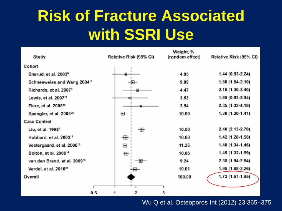
Risk of Fracture Associated
with SSRI Use
Wu Q et al. Osteoporos Int (2012) 23:365–375

Anticonvulsant-induced bone loss
• Some, not all, anticonvulsants induce hepatic
cytochrome P450 activity and accelerate
catabolism of 25-OH vitamin D
• Low 25-OH vitamin D levels
• Inhibition of calcium absorption
• 2nd hyperparathyroidism
• Osteomalacia
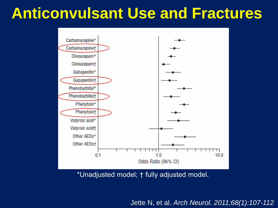
Anticonvulsant Use and Fractures
Jette N, et al. Arch Neurol. 2011;68(1):107-112
*Unadjusted model; † fully adjusted model.

Risk Factors for
Anticonvulsant Bone Disease
• High-dose, multiple drug regimen
• Long-term therapy
• Low vitamin D intake
• Limited sunlight exposure
• Elderly or institutionalized patients

Treatment of Anticonvulsant
Bone Disease
• Calcium 1200 mg QD
• Vitamin D in dosages necessary to maintain
normal 25-OH vitamin D level
• Mean 2400 mg/day*
• Consider switching to other anticonvulsants
which do not cause this problem
*A prospective study to evaluate the dose of vitamin D required to correct low 25-
hydroxyvitamin D levels, calcium, and alkaline phosphatase in patients at risk of
developing antiepileptic drug-induced osteomalacia.
Collins N; Maher J; Cole M; Baker M; Callaghan N. Q J Med 1991;78(286):113-22

Drug-induced Osteoporosis • Glucocorticoids
• Aromatase inhibitors
• Medroxyprogesterone acetate for contraception
• Proton pump inhibitors (PPIs)
• Selective serotonin reuptake inhibitors (SSRIs)
• Antiseizure medicines
• Thiazolidinediones
• Long-term Heparin
• Tamoxifen (premenopausal use)
• Gonadotropin releasing hormone agonists (GnRH)
• Thyroid hormones in excess
• Antiretroviral Therapy
• Lithium
• Chemotherapeutic / immunosuppressives (MTX, CsA, Tacrolimus)