
second scientific national conference for iraqi dental colleges
50
SECOND SCIENTIFIC NATIONAL CONFERENCE FOR IRAQI DENTAL COLLEGES BAGHDAD 10-11/4/2013
Transcript of second scientific national conference for iraqi dental colleges

SECOND SCIENTIFIC NATIONAL
CONFERENCE FOR IRAQI DENTAL
COLLEGES BAGHDAD 10-11/4/2013

تشايذ صفم افتتاس انؤتش انعه انىط انخا نكهاخ طة الاعا
ف انعشاق
صثاصا ف لاعح تىص. 9.03الافتتاس انغاعح *
انشذ انىط.*
انزكش انضكى ولشاءج عىسج انفاتضح وانىلىف صذادا ح يتلاوج يثاسكح ي أ*
.عه اسواس شهذاء انعشاق
سئظ انؤتش.*كهح انغذ
كهح يعان انغذ وصش انتعهى انعان وانثضج انعه.*
*كهح انغذ سئظ انزايعح انغتصشح.
*كهح انغذ يخم كهاخ طة الاعا ف انعشاق.
تعشف تغشج كهاخ طة *فهى وحائم نثغذاد عاصح انخمافح انعشتح و
الاعا ف انعشاق.
.سكانشا*تكشى عذد ي انغادج انضضىس و
.الأعا ف لاعح انىسكاءطة وأرهضجافتتاس انعشض انتخصص نىاد *
.(11.33-11.03) اعتشاصح صف عاعح*
.(1.33-11.33*تذء انهاد انعه نهؤتش وعه كافح انماعاخ )
.(1.03-1.33نهشاسك )غذاء ج *دعى
(2-1.2*إتاو انهاد انعه )
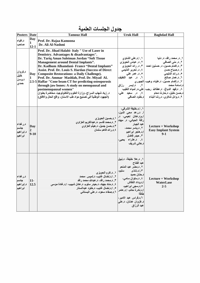
انعهح رذول انزهغاخPosters Date Tamouz Hall Uruk Hall Baghdad Hall
د.افاء
صاصة
Day
1
12-1
Prof. Dr. Rajaa Kamoona
Dr. Ali Al-Nashmi
د.كشوا
خهم
د.وع
صذ
Day
1
2.5-5
Prof. Dr. Jihad Halabi- Italy " Use of Laser in
Dentistry. Advantages & disadvantages".
Dr. Tariq Aman Suleiman Jordan “Soft Tissue
Management around Dental Implants”.
Dr. Kadhum Alhamdani- France “Dental Implants”
Assist. Prof. Dr. Louis S. Hardan (Success of Direct
Composite Restorations: a Daily Challenge).
Prof. Dr. Ammar Mashlah, Prof. Dr. Miyad AL
Haffar "Cone beam CT for predicting osteoporosis
through jaw bones: A study on menopausal and
postmenopausal women"
يضاضشج تعىا -وصاسج انعهىو وانتكىنىرا -د. صاد شهاب انغشاد
)انزهىد انىطح ف تصع يىاد طة الاعا، والع انضال والافاق(
ا.د.عه انشاو. 1
د. عثاط انضىض. 1
د. سائذ انضىض. 0
د. تضشش انذن. 4
عه د. عش. 2
د. عثذ انهطف . 6
انزثىس
سصاق د.اغش. 7
،د. ناء انمةعه
ععذ عه، د.. 8
خهىد انصافد.
د. يضذ سافذ، د. دا .1
. د. ي انصاف1
صغ اصذ، د. اكاو صغ. د.0
د.صثاس صغ. 4
د.سائذ انذن .2
د.عاس صانش .6
، وهة . د.اكاو صغ، د.عهاء7
يضذ د.اعايح
، سرة ، د.عطااللهانذتاغ . د.فهذ8
دصاو ، د.صاسثخهم د.صغ
انثاء ، د.سشاشلاو . د.وائم9
د.سغذاء
راعى
د.اتشاهى
اتشاهى
Day
2
9-10
.د.صغ انضىض1
د.عثذانكشى انعضاو. د.يضذ لاعى ،1
.د.صغ رم، د.هخى انعضاو0
.د.سائذ فاهى عها4
،انششل د.خهفحا. .1
، يض انذ د.سعذ ا.
، د.ع د.عادلا.و.
سافح انضا، د. يهذ
عثذ انزثاس
، يضذ .اعشد. 1
اتشاهى د.هارشا.
صذس فاضم .0
، ض عزساءد. .4
ششف د.ها
Lecture + Workshop
Easy Implant System
9-1
د.سغذاء
راعى
د.اتشاهى
اتشاهى
11-
12.5
. د.اكشو انضىض1
يضذ ، د.نظ غاة د.ضالا. .1
د.يضذ سافذ، د.عثذالله يضذ سافذ .0
يىع د.غادجا.، شثة ، د.عادلعهىو ، د.صذسرهاد . د.ياد4
عثذانغتاس ، د.خهىدغاة د.ضالا. .2
، د.عه انثغتاععىد .د.صفاء6
. د.علا خهفح، د.ثم 1
عثذ انفتاس
عثذ انعى . د.يضش1
، عهى د.شزيا..0
صذ د.صا
، عاي د.عهىا .4
د.وداد انماػا.
، اتشاهى د.عز.2
د.عايشا.، صاتى د.ادسجا.
طالح
، عثذ الايش د.فشاط.6
، د.عهعذا د.غضوا
عثذ انشصاق
Lecture + Workshop
WaterLase
2-5

د.ساض
ىس
د.اصلاو
صذ
2.5-5
انغايشائ .د.دانا خضش، ا. د.علافح1
.د.سا سشذ، ا.و. د.تا صاصة1
. ا.و.د.عه هاد0
.د.را اناط، ا.و.د.تا راب4
ض، د.شاء عثذ الله عزساء. ا.د.تا عه، ا.د.2
.د.وعاو صذ، ا.د.عىلافح انغايشائ6
.د.عه انىعى، ا.د.وصال انعثذ7
. ا.د.عثذانكشى انعضاو، د.صايذ عثاط8
.د.يهذ انفشعى، د.عهاد رثاس، د.عاء عثذ انشصاق9
. ا.د. ساض انمغ1
. ا.د.وفاء يضذ،1
ا. د.سراء هاد،
ا.د.دا انفاض، عارذ
يزذ.
. د.يصطف صلاس،0
ا. و.د.تتىل انغشات
د.عاسج تىفك .4
. د.اعاء عاي2
د.صهشاء شاكش، . 6
ا.و.د.تتىل انغشات
.د.ىس ععذ، 7
ا.د.تغشذ صذا
. د.عف انذ 8
انزثىس، د.اوط عثذ
انعضض
. د.صة انغشات 9
سؤوف، .د.ايم 13
د.فات كاظى، د.هاد
عهىا
د.يادج
عثذ انغفىس
د.سفا
عادل
د.انهاو
هاد

01/4/3102الارتعاء
انجهسح انعهح الاون
0:11 ----03:11 تىسقاعح
رئس انجهسح: ا.د.ثم عثذ انفتاح
د.عثذ انىهاب اناصزا.يقزر انجهسح:
1. Prof. Dr. Rajaa Kamoona "Biological Reconstruction of
Temporomandibular Joint"
2. Dr. Ali Al-Nashmi " انطة و طة الاسا ف انعصىر الاسلايح"
01/4/3102الارتعاء
انجهسح انعهح انثاح
0:11 ----3:21 تىسقاعح
رئس انجهسح: ا.د.وائم الانىس
ا.د.سلافح انسايزائيقزر انجهسح:
1. Prof Dr. Jihad Halabi- Italy " Use of Laser in Dentistry. Advantages &
disadvantages".
2. Dr. Tariq Aman Suleiman Jordan (soft tissue management around dental
implants)
3. Dr. Kadhum Alhamdani- France (Dental Implants)
4. Assist. Prof. Dr. Louis S. Hardan (Success of Direct Composite
Restorations: a Daily Challenge).
5. Prof. Dr. Ammar Mashlah, Prof. Dr. M iyad AL Haffar
"Cone beam CT for predicting osteoporosis through jaw bones: A study
on menopausal and postmenopausal women"
" انجهىد انىطح ف تصع يىاد –وسارج انعهىو وانتكىنىجا –ذ انسزاج د.ساد شهاب اح .6
طة الاسا, واقع انحال والافاق "

01/4/31الارتعاء
انجهسح انعهح انثانثح
0:11 ----3:21 اوروكقاعح
احذ د.سىرا.رئس انجهسح:
يقزر انجهسح: ا.د.وداد انقاش
1. Dr. Ali Alshawi BDS, FDSRCS, FFDRCSI. Consultant Maxillofacial
Surgeon"Oro-facial-cervical infective swellings
2.Assiss. Prof. Dr. Abbas Faisal Al-Huwaizi Ph.D. Prosthodontics. Dean of
Kufa Dental College " DentalVeneers"
3.Raed Faisal Al-Huwaizi "Preventive measures in implantology"
4. Tahrir N.N. Aldelaimi BDS, CDI, MSc, FIBMS, FIHTS
"Bony syngnathia congenital fusion of maxilla and mandible"
5Omar Ali*, Margarita Makou*, Triantafillos Papadopoulos** and George
Eliades University of mosul/ Departments of Orthodontics, IRAQ/
*Orthodontics and **Biomaterials, School of Dentistry, University of Athens,
Athens, Greece "Laboratory evaluation of modern plastic brackets" **
6.Pr.Dr. Abdullatif A.H. Aljuboury "Esthetic utility and stem cell implication
of a new surgical procedure AL J Technique"
7. Aysar Razzaq Ali, Lamia H. Al-Nakib "The Value of Lateral Cephalometric
Image in Sex Identification
8. Dr. Saeed Ali, Prof. Dr. Khulood A. AlSafi "A new Method for Treatment
of Dentin Hypersensitivity by Using Nano fluor-Hydroxyapatite and Nd:YAG
laser: A Fluorescent Light Microscope Study"

01/4/3102الارتعاء
انجهسح انعهح انزاتعح
0:11 ----3:21 تغذادقاعح
د.عادل انخاطا.رئس انجهسح:
د.رجاء انجثىرا.يقزر انجهسح:
1. Dr. Mohammed Rafid Abdulammer (AlMustansiria University- COD)Dr.
Dunia Ahmed K. Al Dulayme (AlMustansiria University- COD)
"Non Extraction therapy for a unilateral malocclusion by intra-oral molars
distalizer"
2. Assist. Prof. Dr. Mona A. Alsafi. Dean of dental department of Al-Yarmouk
University College."Comparison between panoramic radiographs and intraoral
full mouth surveys in epidemiological studies of dental health"
3. Akmam H. Al-Mahdi, Hassanien Ahmed Hadi Al-Jumaily, "The changes in
overbite, overjet, and midline shift following mandibular DO in Iraqi patients"
4. Prof. Dr. Sabah Hasan Yarmouk University College /Baghdad-Iraq
"Aesthetic Contour of non Functional Bones Of the Face Due to Facial Trauma"
5.Ra'ed Mohammed Ayoub Al-Delayme, Senior Lecturer at oral and
Maxillofacial Surgery Dept., Dentistry Dept., AL-Yarmuk University College
,Baghdad , Iraq. Senior Specialist at Oral and Maxillofacial Surgery Dept., AL-
Yarmuk Teaching Hospital, Baghdad, Iraq. "Glass displaced into the
infratemporal region from submandibular injury: case report"
6. Dr. Ammar Salih Al-Alawi Specialized dentist. Department of maxillofacial
surgery.Al-Kadhymia Teaching Hospital.Ministry of Health. Baghdad, Iraq.
"Surgical Diode Laser: An Effective Therapy for Oral Soft Tissue Lesions"
7. Akmam H. Al-Mahdi, Aliaa M.Waheeb, Usama Mohammed Al-Daghir,
"Evaluate the effectiveness of mucogingival buccal sliding flap in the
reconstruction of alveolar cleft"
8. Fahad M. Al Dabbagh, Attallah F. Rajab, Hasan Khalil and Harith
Daham , "Effect of Suturing Stitch Position on Probing Pocket Depth and
Relative Attachment Level of Lower 2nd Molar after Surgical Extraction of
Lower 3rd Molar: Clinical Study"

9. Wael Sh. Shallawi, Rasha F. Albannaa "Restoring Implant Bed Alveolar
Defect with Autologous Bone Graft Taken from the Patient’s Chin: a Case
Report for the Surgical Procedure"
00/4/3102انخس
انجهسح انعهح انخايسح
01:11----0:11 تىسقاعح
انشاو ا.د. عهرئس انجهسح:
د. يحذ انقسا. يقزر انجهسح:
1. Prof. Dr. Hussain F. Al-Huwaizi, College of Dentistry-University of
Baghdad.
"Photoactivated Intracanal Medication-Recent trends in disinfecting canals"
2. Dr. Mohammad Qasim Al-bagdaly, Prof. Dr. Abdul-karim j. Al-azzawi
College of Dentistry, University of Baghdad "Evaluation of simulated lateral
canal obturation using three different techniques"(A comparative Study)
3. Dr. Hassan Jameel Abdul-Wahed, Prof. Dr. Haitham J. Al-Azzawi. "The
influence of two chelating agents used in two different working times on the
microleakage of packable composite resin used in post space (in vitro study) "
4. Raid F. Salman. Assistant Professor, College of Dentistry, Hawler
Medical University. "3-Years in vivo evaluation of use of Bio-dentin compared
with other different materials as therapeutic agent for apex-genesis"

00/4/3102انخس
انجهسح انعهح انسادسح
01:11 ----0:11 اوروكقاعح
د.دا انفاضا.رئس انجهسح:
د.وس حذا.يقزر انجهسح:
1.Professor Khalifa E. Sharquie, Prof. Raad Muhi Aldeen Helmi, Assist. Prof.
Adil A. Noiami, Dr. Raafa K. Al-Hayani, Dr. Mohanad Abduljabbar Kadhim,
"Therapeutic Role of Isotretinoin in Management of Recurrent Aphthous
Stomatitis: Single Blind Controlled therapeutic study"
2.Dr. Yassir Mohammed, Dr. Hajer Ibrahem, "The Effects of Cytotoxic Drugs
on the Vitality of Dental Pulp in Patients with Breast Cancer Undergoing
Chemotherapy"
3.Dr. Haidar Fadhil Al-Rubay’e "The Antibiotic Prophylaxis of Infective
Endocarditis: The Changed Guideline"
4.Prof. Dr. Athraa Yahya Al-Hijazi, Dr. Hani Shareef Mohammed
"Evaluation of the Effect of Nigella Sativa Oil and Powder on Healing Process
,Histologically and Radiographically" (An Experimental Study on Rabbit)
00/4/3102انخس
انجهسح انعهح انساتعح
03:21 ----00:11 تىسانقاعح
رئس انجهسح: ا.د. رافم حذ
د. عذراء يصطفا.يقزر انجهسح:
1.Prof. Dr. Akram F. Al-Huwaizi, Department of Orthodontics, College of
Dentistry, University of Baghdad "Role of TADs in modern orthodontics"

2.Prof. Dr.Nidhal H. Ghaib, Lamis K. Mohammed, “The Effect of Low
Intensity Pulsed Ultra Sound (LIPUS) Therapy on the Relapse Rate and Bone
Remodeling Post-Orthodontic Tooth Movement" (An Experimental Study on
Rabbits) part II
3.Dr. Mohammed Rafid Abdulammer (AlMustansiria University- COD)
Dr. Abdullah M.R. Abdulameer "No more Monobloc and Frankle appliances,
Welcome Twin bloc"
4.Monad J. Al-Duliamy, Hayder F. Saloom, Adil H. Shebeeb, Ghada M.
Mustafa "The effect of local injection of strontium on inhibition and repair of
orthodontically induced root resorption in rats" (An experimental study)
5.Prof. Nidhel H. Ghaib, Dr. Khulood Abdal Sattar, College of dentistry,
university of Baghdad, "Evaluation of frictional forces generated by different
brackets with orthodontic wires"
6.Safaa Saud Abed, Ali I. Albustani, "Surgically assisted orthodontic canine
retraction"
00/4/3102انخس
انجهسح انعهح انثايح
03:21 ----00:11 اوروكانقاعح
سهم جى د. رجاءا.رئس انجهسح:
عادل فزحاا.د.يقزر انجهسح:
1. Dr. Ola Khaleefah Ahmed AL-Husayni, Prof. Dr.Nabeel Abdul Fatah,
College of Dentistry, University of Baghdad. "Effect of silver nitrate
incorporation into heat polymerized acrylic resin on some mechanical
properties"
2.Dr. Mudher Abdulmun’im. Lecturer,College of Dentistry- Almustansiriyah
University "Traumatic Challenges in cosmetic Dentistry"
3.Prof.Dr. Shatha Al-ameer, Dr. Hanan A. Hameed "The corrosion behavior of
commercially pure titanium and Ti-6Al-4V alloy with and without coating"
4.Dr. Salwan Sami Abdulwahhab, Specialized Dental Health Center in
Baqubaa, Diyala, Ministry of Health, IRAQ, Prof. Dr. Widad A.H. Alnakkash.

College of Dentistry University of Baghdad, "The effect of autoclave processing
on some properties of heat cured denture base materials"
5. Saja. N. Ebraheem, Prof. Nadira A. Hatim, Prof. Amer A. Taqa "An
Evaluation of Microwave Radiation Effects on PMMA Powder"
6.Firas A.F., Ghazwan A.A., Ali A.M. Assistant Lecturer University of
Baghdad, College of dentistry, Prosthodontic department "Evaluation of
bonding strength of repaired nylon denture base material by cold or heat cure
acrylic resin"
00/4/3102انخس
انجهسح انعهح انتاسعح
0:11 ----3:21 تىسانقاعح
رئس انجهسح: ا.د. ي انصاف
حجاسد. عذراء ا.يقزر انجهسح:
1.Dr. Dalia Kudier Abbas , Department of Preventive dentistry, College of
Dentistry-Al-Mustansiriya University, Prof. Dr. Sulafa K. El-Samarrai,
Department of Preventive dentistry, College of Dentistry-Baghdad University
"Infection Control in Dentistry"
2.Dr. Raya Rashid Abid, College of Dentistry, University of Baghdad, Dr. Ban
Sahib College of Dentistry, University of Baghdad "Pollution in dentistry and
its prevention"
3.Ali Hadi Fahad Al-Fatlawi, Assistant Lecturer. Department of Pedodontics,
Orthodontics and Preventive Dentistry, Dental College, University of Kufa
"Prevalence and factors associated with traumatized dental injuries to
permanent anterior teeth among 7-12 years old children in Najaf city"
4.Jenan O.Almaas, Ban S.Diab, Ali Y. Al-Rubaii, "The effect of Intelligence
Quotient status (IQ) on caries experience in relation to salivary lead among 6
years old school children in Baghdad- Iraq"
5.Dr. Ban Ali Salih, Dr. Athraa Yahya Al-Hijazi, Dr. Shayma Abdullah
Hanoon, "Effect of Salts Supplemented to Citric Acid on the Surface Roughness
and Microscopical Feature of the Dentin of Permanent Teeth (In Vitro Study)"

6.Assistant lecturer: Dr. wisam Hameid Al-janabi, Prof. Dr. Sulafa El-
Samarrai. "Techniques of sterilization"
7.Ali M. El-mosawi, Wesal A. Al-Obaidi, University of Baghdad, College of
Dentistry, Pedodontic and Preventive Dentistry Department. "An evaluation of
three fissure sealants microleakage with presence or absence of bonding agent
through time intervals" (In vitro study)
8.Prof. Dr. Abdul-alkareem Jassim alazzawi, Dr. Hamid Abbas Hamid
College of dentistry, university of Baghdad "The effect of smear layer on Push-
out bond strength of bioceramic sealer: an in vitro study"
9.Mahdi A.S. Al-faraoon, Suhad Jabbar Hamed, Sana Abdul-Razak Ibraheem,
"Evaluation of the Effect of Diode Laser 810NM on the Diffusion of the
Hydroxyl Ion from Calcium Hydroxide Intracanal Medicament Paste Through
the Dentinal Tubules"(In Vitro Comparative Study)
00/4/3102انخس
انعهح انعاشزجانجهسح
0:11 ----3:21 اوروكانقاعح
د. عثذ انهطف انجثىرا.رئس انجهسح:
يقزر انجهسح: ا.د. شذي سهى
1.Prof. Dr. Riyadh Othman AlQaysi, College of Dentistry/Baghdad University
"The leadership rule of Ancient IRAQ in the establishment &progress of
Dentistry& Medicine"
2.Prof. Dr. Wafaa Mohamed Attoof, Prof. Dr. Raja Hadi Abbas , Prof. Dr.
Dunia W. AL-Fayad, Sajid Majeed Hameed"Oral Complications associated
with Chemotherapy in Children's with Lymphoma"
3.Mustafa M. Salah, Forensic Medicine Institute/Baghdad, Batool H. Al-
Ghurabi, College of Dentistry/ University of Baghdad "Possible Role of
Salivary Tumor Necrosis Factor-alpha in Pathogenesis of Recurrent Aphthous
Stomatitis"

4.Sara M. Tawfeeq, "Maxillary and Mandibular alveolar and palatal bone
densities for adolescents"
5.Dr. Asmaa Sami, "Evaluate the immunohistochemical expression of E-
cadherin and CD44 adhesion molecules in oral squamous cell carcinoma and to
correlate the expression of either marker with lymph node metastasis and tumor
grade"
6.Zahraa F. Shaker, Dr. Batool H. Al-Ghurabei, Department of Basic Scince,
College of Dentistry, University of Baghdad. "Evaluation of Serum Interleukin-
2 Produce from T-Helper 1 in Periodontitis"
7.Dr. Noor Saad Mohammed Ali, Prof. Dr Taghreed F. Zaidan "Oral
manifestations, oral health status and saliva composition changes in a sample of
Iraqi systemic lupus erythematosus patients"
8.Dr. Saifeddin Al-jubory, Dr. Aous Abdul-Aziz Abdul-jaleal "Relation
between bad oral hygiene and patients fearing from dentistry"
9.Zainab H. AL-Ghurabi, College of Dentistry, University of Baghdad “(Cone
Beam Computed Tomography) localization of impacted Maxillary canine pre
orthodontic treatment"
10.Dr. Amal R.S. Mohammed, Dr. Fatin Kh. Abbas, Dr. Nuhad Al.Hassan
"Diagnostic efficacy of mandibular cortical thickness on panoramic radiographs
to identify postmenopausal women with low bone mineral densities"(Iraqi
Population)

Conservative
Dentistry
"Photoactivated Intracanal Medication-Recent trends in disinfecting canals"
Prof. Dr. Hussain F. Al-Huwaizi
College of Dentistry-University of Baghdad.
ABSTRACT
Disinfection of the root canal is performed by “chemomechanical” approach that involves cleaning
and shaping of the root canal system by the application of a chemical disinfectant and mechanical
instrumentation. This technique often fails to eradicate bacterial biofilms completely, mostly because
of various microbiological and anatomical factors. Chemo-mechanical preparation of the root canal
reduces endodontic infection, but microorganisms are able to survive within the complex anatomy of
the root canal system. Many antimicrobial intracanal medicaments are used to complement the
disinfection of the root canal system. Methods of chemical disinfection supporting the instrumental
endodontic debridement have a strong bactericidal effect, but commonly used irrigants, such as
sodium hypochlorite (NaOCl) and chlorhexidine digluconate, and calcium hydroxide as an
interappointment dressing do not always eradicate the entire microbial flora in infected root canals.
Although biomechanical preparation and root canal shaping effectively reduce microbiota, these
procedures do not completely eliminate bacteria in the lateral and accessory root canals, isthmi, and
apical deltas.
Phototactivated disinfection (PAD) is an antimicrobial strategy that combines a nontoxic
photosensitizer and low-energy light to produce highly reactive singlet oxygen species, which results
in microbial elimination. PAD has emerged as a promising approach to eradicate endodontic
pathogens.
Dental Veneers
Assist. Prof. Dr. Abbas Faisal Al-Huwaizi
Ph.D. Prosthodontics
Dean of Kufa Dental College
ABSTRACT
Veneers are very thin pieces of composite or porcelain used to create brilliant, beautiful smiles. They
are bond to the teeth and improve the shape and/or color of your teeth. They have a variety of shapes,
extensions and modifications to gain maximum retention and prevent dislodgement under the occlusal
load. The veneer can be placed on the tooth either by a direct or indirect method. Each of which has
its own advantages and disadvantages.

"EVALUATION OF THE EFFECT OF DIODE LASER 810NM ON THE DIFFUSION OF
THE HYDROXYL IONS FROM CALCIUM HYDROXIDE INTRACANAL MEDICAMENT
PASTE THROUGH THE DENTINAL TUBULES."
(In Vitro Comparative Study)
Mahdi A.S. Al-faraoon, 1BDS, M.Sc., PhD
Suhad Jabbar Hamed, BDS, M.Sc
Sana Abdul-Razak Ibraheem, BDS, M.Sc.
ABSTRACT Purpose: The main known benefits of calcium hydroxide in endodontology lies in the bactericidal,
antimicrobial, and anti-inflammatory. Calcium hydroxide to be more effective should be diffuse
through the dentinal tubules. However, laser has a promising effect to enhance that by removing of
the smear layer in the inner walls of root canals.
Aim: This study aimed to evaluate the effect of laser Diode 810nm on the dentine permeability for
calcium hydroxide endodontic dressing.
Materials and Methods: Fifty extracted lower first premolar teeth were prepared and randomly
divided into two groups (A &B). The root canals of the group A were filled with calcium hydroxide
medicament and in group B were filled with calcium hydroxide after irradiated with different laser
output energies. Diode laser 810 nm wavelength was used in this study. Fiber optic 200 micrometer
diameter is the delivery system to the root canal. The output laser energy was 1, 2, 3, and 4 Jules. The
frequency was 25Hz and the time was fixed in 30 second each root canal treatment. The fluencies
were 0.3, 0.6, 0.9, 0.12 J/Cm2.The apical foramen and the coronal orifice for each samples was sealed
and stored in glass tube containing 5ml of distil water ,then the pH of the surrounding medium was
measured at 1 Hour (H), 1Day (D), 1 Week (W), 2 Weeks, 3 Weeks to the both groups.
Result: The level of pH in each group increased significantly with time, indicating that the hydroxyl
ions were continuously diffused through the dentinal tubules to the surrounding medium. There was
statistical significant difference between the two groups in the level of pH, that revealed there is
increasing effect when using laser irradiation on the pH of the surrounding media of the root that
filled by calcium hydroxide paste specially with 1 and 2 J.
Conclusion: The using laser diode before calcium hydroxide paste will accelerate the alkalinity of the
medium surrounding the root which was filled with the paste which means the laser is effect in the
cleaning the dentinal tubules from the smear layer leading to increase the chance of the calcium
hydroxide paste ions to flew out and increase the alkalinity of the surrounding media.
Key words: laser; permeability; endodontology; calcium hydroxide; dentinal tubules.
"3-Years in vivo evaluation of use of Bio-dentin compared with other different materials as
therapeutic agent for apex-genesis"
Raid F. Salman* B.D.S., M.Sc., Ph.D.
Assistant Professor, College of Dentistry, Hawler Medical University
ABSTRACT
Background & Aim: Apex-genesis is treatment of vital pulp in immature teeth to permit continued
dentin formation & apical closure. This done after trauma, & shallow pulpatomy had done when pulp
exposure occur without inflammation. A new material had been marketed with the aim of optimum
therapy of remaining pulp of the canal system & continued dentin formation & apical closure,
however, no information present about the quality of closure compared to conventional materials.
This study investigated 3-years in vivo evaluation of apical closure when the canal treated by Bio-
dentin compared with MTA & Calcium hydroxide by clinical follow up & radiographs.
Methods: Twenty one cases attended for treating traumatized different teeth with pulp exposure were
utilized for this study; and randomly divided into 3 groups. In the first group, the root canals were
treated by using MTA. In the second group, the root canals were treated by using Calcium hydroxide.
In the third group, the root canals were treated by using Bio-dentin. Special including & excluding
criteria had been designed for standardizing the cases. After shallow pulpatomy had been executed for
each case with the same procedure for each, every case had received a material as therapeutic agent &
follow up radiographs & clinical examination scores had been recorded.
Result: For MTA, a mean of 2.3 clinical scoring & a mean of 3.5 radiographic scoring were recorded
after 3-years of follow up with 6 months intervals, for Calcium hydroxide, a mean of 3.6 clinical
scoring & a mean of 3.2 radiographic scoring were examined. For Bio-dentin, a mean of 2.5 clinical
scoring & a mean of 2.8 radiographic scoring were recorded. However, there was non-significant
difference between them (p=0.52) using ANOVA test.
Conclusion: Bio-dentin material had comparable results clinically & radiographically with
conventional agents (MTA & Calcium hydroxide) used for executing successful apex-genesis.
"The effect of smear layer on Push-out bond strength of bioceramic sealer: an in vitro study"
Prof. Dr. Abdul-alkareem Jassim alazzawi
Dr. Hamid Abbas Hamid
College of dentistry, university of Baghdad
ABSTRACT
Aim of study: To compare the bond strength of three different endodontic sealers in presence or
absence of smear layer on three levels coronal, middle and apical.
Materials and Methods: Sixty freshly extracted mandibles premolars teeth were used in this study,
all canals were instrumented using ProTaper rotary instruments to size F3 to achieve tapered canal
walls. Irrigation was performed using 5 mL 5.25% NaOCl between each instrument, roots were
randomly divided into three groups according types of endodontic sealer and then subdivided
according to the effect of different irrigate solution remove smear layer.
A1, use apexit plus sealer and no attempt to remove smear layer.
A2, use apexit plus and remove smear layer by 17%EDTA.
B1, use AH plus sealer and no attempt to remove smear layer.
B2, use AH plus and remove smear layer by 17%EDTA.
C1, use I root sp sealer and no attempt to remove smear layer.
C2, use I root sp sealer and remove smear layer by 17%EDTA.
ALL groups were obturated by lateral condensation technique, the roots then stored in 100%humidity
and37C for one week, the roots was embedded in clear acrylic. Three horizontal sections were
prepared at a thickness of 1 mm ±0.1 in the apical, middle and coronal parts of each root. The test
specimens were subjected to the push-out test method using a Universal Test Machine (USA) that
carried 1mm, 0.5 mm and 0.3mm plungers for coronal, middle and apical specimens, respectively.
The loading applied from apical to cervical at 0.5 mm/ min speed.
Result: The presence or absence of smear layer did not significantly affect the bond strength of filling
materials. The bond strengths in the middle specimens and the apical specimens were higher
compared with the bond strengths in the coronal specimens
Conclusion: The bond strength of the new bioceramic sealer was equal to that of AH Plus with or
without the smear layer.
"Evaluation of simulated lateral canal obturation using three different techniques"
(A comparative Study)
Dr. Mohammad Qasim Al-bagdaly
Prof. Dr. Abdul-karim j. Al-azzawi
College of Dentistry, University of Baghdad

ABSTRACT
Lateral canals are very frequent in most of the teeth. The ability of obturation technique can greatly
influence the success rate of endodontic treatment. The aim of this study was to evaluate and compare
the ability of continuous wave using E&Q MasterTM, Obtura II and Thermafil to obturate simulated
apical and coronal lateral canals in both straight and curved main canals. Fifteen Thermafil training
blocks with 25° curved canals and the same number of fabricated resin blocks with straight canals,
with each block include two main canals were selected in this study. Main canals had 0.3 mm apical
diameter and 0.04 taper. Each canal had two parallel lateral canals. The apical lateral canal was 5 mm
from apical end and the coronal lateral canal was 6.5 mm from apical one. The canals were divided
into six groups according to canal curvature and obturation techniques used (n=10). Groups I and II:
curved and straight canals respectively obturated with continuous wave technique using E&Q
masterTM system. Groups III and IV: curved and straight canals respectively obturated with
Thermafil obturators. Groups V and VI: curved and straight canals respectively obturated with Obtura
II. Soapy water was used to simulate sealer in all obturations performed. The depth of gutta-percha
penetration into lateral canals was measured using computerized stereomicroscope at 10x. Data were
statistically analyzed by ANOVA, LSD and t-test at 5% significance level. Results showed that all
techniques used were able to obturate lateral canals. There were highly significant differences
between the three techniques in both straight and curved canals. Continuous wave showed greatest
gutta-percha penetration with highly significant difference from both other techniques. There was
non-significant difference between Thermafil and Obtura II except at coronal lateral canal of straight
main canals Thermafil showed better gutta-percha penetration with highly significant difference. The
gutta-percha depth was greater in coronal than apical lateral canals in all groups of both straight and
curved canals, and gutta-percha depth was greater in straight than in curved canals within each
obturation technique.
The influence of two chelating agents used in two different working times on the microleakage
of packable composite resin used in post space (in vitro study)
Dr. Hassan Jameel Abdul-Wahed (B.D.S., M.Sc) 1
Professor Dr. Haitham J. Al-Azzawi (B.D.S., M.Sc.) 2
1 Ministry of Health / Al-Faw Center for Primary Health Care. 1 Baghdad University / College of Dentistry / Department of Conservative Dentistry.
ABSTRACT
Background: Weakened teeth are not strengthened by the placement of a post but retention of
restoration is enhanced by using a post. Restoration with adhesive materials offers many advantages
over the use of traditional materials, like transmission of functional stresses across the bonded
interface to the peridontium, with potential to reinforce the weakened tooth structure (Belli et al.,
2001). Cavity preparation and root canal instrumentation leave a layer of debris that covers the walls
of the cavity and root canal, known as smear which can be removed with a chelating agent (L.F
Machado-Silveiro et al 2004). This in vitro study conducted to evaluate the effect of two different
chelating agents used in two different times of application on the microleakage of packable composite
resin (Filtek P60 shade) used in post space.
Materials and method: Fifty, human, freshly-extracted mandibular premolars were selected. After
crown sectioning and conventional endodontic treatment, parallel post spaces 5mm in depth and 2mm
in diameter were prepared using pesso burs (LARGO No.6). Then the specimens were randomly
divided into five groups as follow: Group1 (control): The post space was filled with packable
composite without previous using of chelating agent. Group2 (experimental): The post space was
flooded with 17% EDTA for 5 minutes prior filling with packable composite. Group3
(experimental): The post space was flooded with 10% citric acid for 5 minutes prior filling with
packable composite. Group4 (experimental): The post space was flooded with 17% EDTA for 10

minutes prior filling with packable composite. Group5 (experimental): The post space was flooded
with 10% citric acid for 10 minutes prior filling with packable composite.
After thermocycling and immersion in methylene blue, the teeth were sectioned longitudinally and
dye penetration was evaluated using a stereomicrscope, microleakage was recorded in mm.
Results: ANOVA test and least significant difference (LSD) test were used to analyze the results and
to show the difference between groups. Results expressed statistically highly significant reduction in
microleakage value among all groups; with the lowest mean microleakage value was in group5 in
which citric acid was used for 10 min., followed by group4 in which EDTA was used for 10 min.,
followed by group3 in which citric acid was used for 5 min., followed by group2 in which EDTA was
used for 5 min., and control group showed the highest microleakage value.
Conclusion: From the results of the present study, it is clear that the application of a chelating agent
prior using acid etch and adhesive system with composite resin filling material will significantly
reduce microleakage value of the restorative material.
Key words: Microleakage, Packable composite, Post, Chelating agent.
Oral Diagnosis
"The Value of Lateral Cephalometric Image in Sex Identification"
Aysar Razzaq Ali, B.D.S
Lamia H. Al-Nakib B.D.S, M.Sc in Oral Radiology
ABSTRACT
Determination of sex and estimation of stature from the skeleton is vital to medicolegal
investigations. Skull is composed of hard tissue and is the best preserved part of skeleton after death,
hence, in many cases it is the only available part for forensic examination. Lateral cephalogram is
ideal for the skull examination as it gives details of various anatomical points in a single radiograph.
This study was undertaken to evaluate the accuracy of digital cephalometric system as quick, easy and
reproducible supplement tool in sex determination in Iraqi samples in different age range using certain
linear and angular craniofacial measurements in predicting sex. The eleven parameters measured for
males and females when compared are statistically significantly different. All cranio-cephalometric
measurements gave overall predictive accuracy of sex determination by discriminant analysis
(86.7%). The stepwise selection method gave overall predictive accuracy of sex determination by
discriminant analysis (85.8%). Age showed no statistical difference among the studied age range
except for the distance from Mastoid to Frankfort plane.

"CBCT (Cone Beam Computed Tomography) localization of impacted Maxillary canine pre
orthodontic treatment"
Zainab H. AL-Ghurabi, B.D.S., M.Sc in Oral and Maxillofacial Radiology,
Assistant Lecture, College of Dentistry, University of Baghdad.
ABSTRACT
Background: The orthodontic treatment of impacted maxillary canine remains a challenge to today’s
clinicians. The treatment of this clinical entity usually involves surgical exposure of the impacted
tooth, followed by orthodontic traction to guide and align it into the dental arch, our study designed to
shade light on the important role of dental CT to accurately localization of the impacted maxillary
canine.
Materials and method: Unilaterally and bilaterally impacted maxillary canines (n = 52) from 30
patients (24 female, 6 male) were evaluated by a volumetric 3D images obtained from cone beam ct
for localize upper impacted canine, all samples attendance to the specialist health center of dentistry
in AL-Sader city referred to CBCT by orthodontist to detect exact position of impacted upper canine
in cases when there was no bulging buccaly or palataly which aid to detect the exact position.
Result: Mesiopalatal angulations was the highest rate (63.5%) followed by mesiolabialy (19.2%),
vertical (labialy) (9.6%) distopalataly (5.8%) and distolabialy (1.9%). The relation between impacted
canine & the adjacent teeth regarding to the attachment was significant only with lateral incisor tooth,
no cases of root resorption to the adjacent teeth were recorded .Bilateral impacted teeth were found in
22 patient which is highly significant (specialy in female) , while unilateral found only in 8 patient
specially in female, impacted canine was more prominent in female whether unilateral or bilateral
Conclusion: CBCT imaging of impacted canines can show the following: presence or absence of the
canine, angulations of the long axis of the tooth, relative labial and palatal positions, and proximity to
adjacent teeth. In short, CBCT imaging is clearly advantageous in imaging & management of
impacted canines
Key words: CBCT, Impacted Maxillary canine, orthodontic treatment.
"The Effects of Cytotoxic Drugs on the Vitality of Dental Pulp in Patients with Breast Cancer
Undergoing Chemotherapy"
Dr. Yassir Mohammed, B.D.S., M.Sc.
Dr. Hajer Ibrahem, B.D.S., M.Sc.
ABSTRACT
Breast cancer (B.C)is the most common cancer in women worldwide, comprising 16% of all female
cancers. Today environment in Iraq is considered highly polluted due to many factors randomly waste
disposable, wars, in addition to the wrong understanding of cultural attitudes toward such diseases
added a large burden on Iraqi women health issues. Chemotherapy is the treatment of choice for (B.C)
patients after surgical management. In this study, the effect of cytotoxic drugs on the vitality of dental
pulp in patients undergoing chemotherapy for (B.C) was identified and determined the correlation
between teeth vitality and oral mucosal findings such as xerostomea. Fifty female patients participated
in this study. They were surgically treated for (B.C) and received chemotherapy; clinical oral
examination took place before receiving chemotherapy and after each dose of chemotherapy. The
result indicated that the dental pulp vitality is significantly affected in (B.C) patients undergone
chemotherapy which in turn affects general oral and teeth health status.
"Oral Complications associated with Chemotherapy in Children's with Lymphoma"
Prof. Dr. Wafaa Mohamed Attoof PhD.
Iraqi National Center for Cancer Research, University of Baghdad,
Prof. Dr.Raja Hadi Abbas PhD.

Oral Diagnosis Department, Specialty in Oral Medicine, College of Dentistry, University of
Baghdad,
Prof. Dr. Dunia W. AL-Fayad PhD.
Chair women of Oral Diagnosis Department, Specialty Oral Surgery & Oral Pathology, College of
Dentistry, University of Anbar
Mr.: Sajid Majeed Hameed BSc (Student).
Pharmacy College private – Philadelphia University in Jordon.
ABSTRACT
Background: Lymphoma is general term for group cancers that start in lymphatic system.
Purpose: Our objective was to assess early oral complications under chemotherapy treatment & type
of pain.
Patients & Methods: An hematology & oncology unit , based purposive study in Walffer teaching
hospital in medical city & child central teaching hospital, started from 15th January 2011 to the 15th
January 2012; seventy patients diagnosed with lymphoma (45 males, 25 females), patients admission
to receiving chemotherapy. Oral examination done for all patients by dentist, & by using dental
instruments, depended on oral guide assessment. Pain was determined by use facial expression.
Results: No statistically significant association was found between ages with gender (P≥0.05).
Specifically (71.4%) were males which was more than females (28.6%), negative family history
toward malignant diseases & duration of disease less than 6 months; (64%) were Non-Hodgkin
lymphoma; (50%) resident in south Iraq with enlargement lymph node, anemia, thrombocytopenia,
leucopenia, unbearable pain; (42.9%) diagnosed by core biopsy; (40%) were thrombocytopenia.
Statistically significant association was found between clinical diagnosis and two variable age &
gender (P≤.01). The cumulative incidence of any early oral complications at 2 weeks protocol therapy
was above cut-off point, Mean of Score was (3) thick saliva and (2.42) which include each this items,
painful speech and, ulcerated spontaneous bleeding in gingival mucous membrane; blistered;
ulcerated with or without bleeding in lips angle of mouth; unable to swallowing.
Conclusions: The burden of early oral complications at 2 weeks protocol therapy is high. These early
compilations are associated with pain & hematological disorders & this patient's need for continue
follow-up by doctors & health education program.
Key words: Lymphoma, Cancer, Oral Complications, Chemotherapy, Pain.
"EVALUATION OF THE EFFECT OF NIGELLA SATIVA OIL AND POWDER ON
HEALING PROCESS ,HISTOLOGICALLY AND RADIOGRAPHICALLY"
(AN EXPERIMENTAL STUDY ON RABBIT)
Prof. Dr. Athraa Yahya Al-Hijazi B.D.S., MSc., Ph.D. (Oral Histology and Biology)
Dr. Hani Shareef Mohammed B.D.S
ABSTRACT
Back ground: Today’s world is increasingly seeking ways to replace the synthetic drugs with the
therapeutic power of natural products to decrease the percentage of many side effect which result
from conventional treatment; one of these products was Nigella sativa (NgS) which was used so
extensively that it became known as the seed of blessing “Habbatul Barakah” due to its powerful
healing qualities for many ailments.
Aim of the study: This study was performed to evaluate the therapeutic effect of Nigella sativa
(powder and oil) on the healing process of extracted teeth sockets.
Materials and Methods: The sample of our study consist of Forty eight rabbits to extract there upper
two central incisors under general anesthesia. The left side filled once with Nigella sativa powder and
once with Nigella sativa oil material, and the right side left for normal healing as a control group. The
two sockets were sutured. The results were studied radiographically and histologically after 1,2,4,6
weeks postoperatively. The radiographic examination was performed by using parallel technique in a
digital radiographic examination and histological examination was performed under light microscope
for the section stained with heamatoxiline and eosin.

Results: Radiographically we found that NgS powder showed more radiopacity with complete
disappearance of lamina dura in 6 weeks duration compared with NgS oil and control groups, while
histologically we found that the Nigella sativa (NgS) groups (powder and oil) illustrate an early
apposition of osteoid tissue in 1st week duration with numerous osteoblast and osteocyte in
comparision to control group.In six weeks duration well developed bone filled all the portions of the
socket in treated socket with NS powder with obvious complete epithelization of socket surface
Conclusion: Nigella sativa (powder or oil) seems to be bioactive materials that enhance
differentiation and proliferation of progenitor cells to specialized bone formative cells, with no signs
of inflammation.
"Diagnostic efficacy of mandibular cortical thickness on panoramic radiographs to identify
postmenopausal women with low bone mineral densities"
(Iraqi Population)
Dr. Amal R.S. Mohammed B.D.S., H.D.D., M.Sc., Rad.
Dr. Fatin Kh. Abbas B.D.S., M.Sc., Rad.
Dr. Nuhad Al.Hassan B.D.S., M.Sc., Rad.
ABSTRACT
Background: The use of mandibular anatomic indicators on panoramic radiographs such as
mandibular cortical thickness (MCT) at mental region can be useful in the evaluation of bone
resorption in different age of postmenopausal women to determine the presence of osteoporosis.
Aim of Study: To assess the accuracy of mandibular cortical thickness in the panoramic radiographs
of postmenopausal women with normal and low skeletal bone mineral densities (BMD) diagnosed by
using dual energy x-ray absorptiometry (DXA) and to correlate the effect of age in both two groups.
Materials and Methods: Forty digital panoramic radiographs obtained from postmenopausal women
(20 normal and 20 osteoporotic) aged between (52.7-80.1). Bone mineral density has been assessed by
a DXA at the lumbar spine and right femur at Al-Yarmook Hospital. The mean was calculated for
mandibular cortical thickness values measured in the right and left mandibles. The measurments were
analysed using the t-test and Pearson's correlation coefficients.
Results: Difference was shown between the MCT measurements in the osteoporotic group and
normal group which showed that MCT was more thinner in osteoporotic group than normal group
(t=23.25, p value 0.01). Pearson's correlation coefficients of normal and osteoporotic by age and MCT
were 0.976 and 0.973 respectively with (p value 0.01).
Conclusion: The pattern of decrease in mandibular cortical thickness with age was similar to that
pattern of bone loss from the spine and femur, so panoramic radiography was a simple technique in
osteoporosis screening of dental patients, giving the maximum benefit of being radiographed.
Key words: menopause, osteoporosis, panoramic mandibular cortical thickness.
"Poster presentation"
A comparative study of immunohistochemical expression of Moesin, Cytokeratin 14, MMP7 in
oral squamous cell carcinoma and oral verrucous carcinoma.
Karawan Khaleel Jubair,
Wasan Hamdi Younis
University of Baghdad, College of dentistry, Department of oral diagnosis, Oral pathology
ABSTRACT Background: Squamous cell carcinoma (SCC) is the most prevalent malignant neoplasm of the oral
cavity that exhibits certain histological variations. Verrucous carcinoma (VC) is an uncommon
exophytic low-grade well-differentiated variant of SCC. The histologic grade reflected the
aggressiveness of the individual neoplasm and there was a clear relationship between grade and cure

rate, stage of disease and metastatic involvement. Cellular differentiation and morphology play
important roles in cell functions and maintenance of structural integrity .As the cancer is a malignant
process in which disorder of the cell growth and behavior occurs, such changes may be differed in
different tumor types and within different grades of the same tumor.
Aim of study: Immunohistochemical evaluation and comparison of Moesin,CK14 and MMP7
expression ,to determine the degree of cellular changes and tumor invasiveness between oral
squamous cell carcinoma and oral verrucous carcinoma.
Materials and Methods: Forty two formalin – fixed, paraffin – embedded blocks were included in
this study. Thirty blocks of oral squamous cell carcinoma and twelve blocks of oral verrucous
carcinoma. After histopathological reassessment of Haematoxylin & Eosin stained sections for each
block, an immunohistochemical staining was performed using anti moesin, anti cytokeratin14 and anti
matrix metalloproteinase-7 monoclonal antibodies.
Results: The age of the studied samples ranged between 24-99 years old. The mean age was 59+/-
15.3 years in SCC while it was 61.3 +/- 14.4 years in OVC. Male predominance was found with 60%
of SCC group and 75% of VC group. No statistically significant differences in age & sex distribution
were observed between the 2 studied groups. The most affected site in SCC was the tongue 26.7% (8
cases) while buccal mucosa &alveolar ridge 33.3% (4 cases for each) were the common sites in OVC.
Histopathological examination showed that 14 cases (46.7%) were well differentiated SCC, 12 cases
(40%) moderately differentiated & 4 cases (13.3%) were poorly differentiated. Moesin
immunoreactivity was recognized in all the studied groups with predominant cytoplasmic expression
in SCC & membranous expression in OVC. No difference was noticed between 2 studied groups
&between different grades of SCC. Cytokeratin 14 positivity was noticed in all studied groups with
significant difference between SCC&OVC p=0.012 & there was a significant difference between the
different grade of OSCC (p=0.047). Collectively, MMP7 expression was observed in the all studied
groups with predominant cytoplasmic pattern in SCC &nuclear pattern in OVC. No difference was
found between the 2studied groups &between the different grades of SCC. A Strong positive linear
correlation between MMP7&CK14 was noticed.
Conclusion: In conclusion, CK14 is a useful marker in differentiation between SCC &OVC.
Verrucous carcinoma has a specific pattern for Moesin &MMP7 that differs from OSCC but the
difference is not significant, however, such a difference in expression may be useful in a potential
therapeutic strategy for improving clinical outcome in OSCC patients.
"Oral manifestations, oral health status and saliva composition changes in a sample of Iraqi
systemic lupus erythematosus patients"
Dr. Noor Saad Mohammed Ali (M.Sc of oral medicine).
Prof. Dr Taghreed F. Zaidan (PH.D of oral medicine).
ABSTRACT
Aims of the study: To determine the prevalence of oral manifestations and tempromandibular joint
disorders in systemic lupus erythematosus patients, evaluate the oral health status in those patients by
using (Decayed, Missed, and Filled Teeth index) and the gingival health status by using Gingival
index and Clinical Pocket Depth, investigate the changes of the whole saliva composition through
measuring the concentrations of (calcium, sodium, potassium, inorganic phosphorus, chloride and
total protein) and comparing the results with clinically healthy individuals and to find a correlation
between saliva flow rate, pH, saliva composition changes and the incidence of dental caries in those
patients.
Subjects, Materials and Methods: One hundred and two individuals were enrolled in this study;
Fifty two were systemic lupus erythematosus patients group, they were fifty females and two males;
and fifty female healthy individuals (control group) matching the patients in age. The assessment of
dental status was made according to the (Decayed, Missed, and Filled Teeth index); the gingival
inflammation was assessed using the criteria of gingival index; clinical pocket depth was measured
with periodontal probe type William; whole unstimulated saliva samples have been collected from
each subject for biochemical analysis.Salivary samples were collected by spitting method. Saliva pH
was measured immediately by digital pH meter; salivary flow rate was measured by collection of
saliva through 10 minutes, the volume of saliva is recorded in order to give the salivary flow rate in
ml/min; after centrifugation the supernatant of saliva was aspirated for biochemical analysis.
Results: It has been shown that the number of systemic lupus erythematosus patients with active
disease in this study was significantly higher than the number of systemic lupus erythematosus
patients with inactive (remission) disease according to Systemic Lupus Erythematosus Disease
Activity Index. Oral ulceration was the most prominent orofacial manifestations of Systemic lupus
erythematosus patients followed by Tempromandibular joint disorders and facial skin rash then oral
vesicles & bullai, oral lichen planus and finally oral petechia & purpura. The results showed that
salivary flow rate was significantly lower in systemic lupus erythematosus patients than in the control
subjects (p <0.001); Salivary pH was significantly lower in Systemic lupus erythematosus patients
than in the control subjects (p =0.001).
Oral hygiene index (Decayed, Missed, and Filled Teeth index, Gingival index, Clinical Pocket Depth)
were significantly higher in those patients than in the control subjects (p <0.001). The results also
showed that salivary elements concentrations which include (calcium, sodium, chloride and total
protein) were significantly higher in systemic lupus erythematosus patients than in the control subjects
(p<0.001); While concerning (potassium and inorganic phosphorus) they were significantly lower in
those patients than in the control subjects (p<0.001). Highly significant positive linear correlation has
been found between age of systemic lupus erythematosus patients and Decayed, Missed, and Filled
Teeth index; and between age and Clinical Pocket Depth, also highly significant negative linear
correlation has been found between salivary flow rate and salivary calcium in systemic lupus
erythematosus patients. There was highly significant positive linear correlation between Decayed,
Missed, and Filled Teeth index and salivary calcium; also between this index and salivary chloride in
those patients.
Conclusions: The salivary changes observed in systemic lupus erythematosus patients reflect
impaired ductal salt re-absorption; the results of this study suggest that changes in salivary flow rate,
pH and salivary composition as well as increase dental caries experience in those patients may serve
as potential markers of the extent of auto immune mediated salivary gland dysfunction which is
similar to Sjögren's syndrome.
"Relation between bad oral hygiene and patients fearing from dentistry"
Dr. Saifeddin Al-jubory B.D.S.
Dr. Aous Abdul-Aziz Abdul-jaleal B.D.S., F.I.C.M.S.
ABSTRACT
This research study the common causes that make the patients fear from dentistry. We take a 632
patients for questionnaire and find the most common causes then find the most common solutions in
order to encourage the patients to visit the dental clinics to improve their oral health.
"Therapeutic Role of Isotretinoin in Management of Recurrent Aphthous Stomatitis: Single
Blind Controlled therapeutic study"
*Professor Khalifa E. Sharquie M.D.; Ph.D.
**Prof. Raad Muhi Aldeen Helmi B.D.S.; Ph.D.
***Assist. Prof. Adil A. Noiami M.D.; D.D.V.; FICMS.
****Dr. Raafa K. Al-Hayani M.D.; D.D.V.
*****Dr. Mohanad Abduljabbar Kadhim; B.D.S.; M.Sc.
*Chairman of the Scientific Counsil of Dermatology and Venereology-Iraqi Board for Medical
Specializations.
**Prof. of Oral Medicine, Dean of College of Dentistry, Al-Mustansiriya University.
***Department of Dermatology and Venereology, College of Medicine, Baghdad University.
**** Department of Dermatology and Venereology, Baghdad Teaching Hospital.

*****Assistant lecturer in School of Dentistry, Faculty of Medical Sciences, University of Duhok.
ABSTRACT Background: Recurrent aphthous ulcer (RAS) is a common oral disease with no well-known
etiology. This disease is treated bya variety of agents for palliative, prophylactic, and curative
purposes. Isotretinoin has been used in the treatment of acne vulgaris.
Objectives: To evaluate the efficacy, safety of isotretinoin in treating RAS and the long term
remission of RAS.
Patients and methods: This single blind controlled therapeutic study that conducted in Department
of Dermatology-Baghdad
Teaching Hospital between February 2011- January 2012. Thirty patients with typical RAS were
included in this work. Detailed history and full examination were done for all patients. They were
given isotretinoin 20mg orally once daily for three months to be seen on day fourteen firstly and then
monthly to be assessed depending on the oral clinical manifestation index (OCMI). After three
months, isotretinoin was stopped and patients were given placebo therapy for another 3 months.
Results: Thirty patients were treated, 17 (56.67%) males and 13 (43.33%) females with male to
female ratio was 1.3:1. Their ages ranged between12-60 (35.33±12.06) years. The OCMI before
isotretinoin therapy ranged between 7-17 (13.13±2.55), while after therapy the mean started to decline
to a lower level within the first 14 days(p=0.103), and continued to decline significantly until the end
of the first month of therapy(p=0.023), and then the OCMI declined highly significantly until the end
of fourth month of therapy(p<0.001), then the mean started to increase until the end of the 5 months
(with placebo) but it is remained statistically significant when it is compared withthe baseline of mean
of OCMI before treatment (p=0.046), then it continued to increase to be become not significant at the
end of 6 months of therapy (P=0.107).
Conclusions: Isotretinoin is an effective therapeutic and prophylactic drug in management of RAS.
Key words: Recurrent Aphthous Stomatitis, Isotretinoin, Iraq.
"Maxillary and Mandibular alveolar and palatal bone densities for adolescents"
Sara M. Tawfeeq, B.D.S., M.Sc.
ABSTRACT
Background: The purposes of this study was to measure cortical and cancellous bone densities of the
alveolar bone of the maxilla and the mandible in adolescents and compare them among placement
sites of temporary anchorage devices. Materials and methods: Multi-slice computed tomography
scan data were obtained from 30 adolescents with normal occlusion and age range of (12-14 for
females) and (14-16 for males) and then bone density was recorded in Hounsfield units by using
three-dimensional protocol. Bone densities of the alveolar bone at the central incisor, lateral incisor,
canine, first premolar, second premolar, first molar, second molar and tuberosity/retromolar pad areas
were measured.
Results: t-test showed no significant differences in bone density considering the sides and genders.
Interradicular bone in the maxillary premolars area showed the highest bone density for the maxilla
and mandibular mean bone density showed a progressive increase from anterior to posterior. Anterior
palatal paramedian bone was significantly denser than bone located more posteriorly. A comparison
of the mean bone densities between the buccal and lingual sides in the maxilla showed that the lingual
side had higher values. There were no distinct differences between the buccal and lingual sides in the
mandible. A comparison of the mean bone densities between the maxilla and the mandible showed
higher values in the mandible.
Conclusions: These data might provide valuable information when selecting sites and placement
methods for temporary anchorage Devices in adolescents. Differences in bone densities between and
within regions of the jaws must be considered.
Key words: Bone density, adolescents, maxilla, mandible, multi-slice CT.

Head and neck squamous cell carcinoma
Dr. Asmaa Sami
ABSTRACT
Background: Head and neck squamous cell carcinoma is the sixth most common cancer world wide.
Despite greater emphasis on multi-modality therapy including surgery, radiation and chemotherapy,
advanced stage head and neck squamous cell carcinoma continues to have poor 5-year survival rates
(0-40%) that have not significantly improved in the last (30) years. To improve outcomes for this
deadly disease requires a better understanding of the mechanisms underlying head and neck squamous
cell carcinoma tumor growth, metastasis, and treatment resistance. E-cadherin is essential for the
formation and maintenance of epithelia and it is one of the most important molecules in cell-cell
adhesion in epithelial tissues. It is localized on the surfaces of epithelial cells in regions of cell-cell
contact known as adherens junctions .In human tumors, the loss of E-cadherin mediated cell adhesion
correlates with the loss of the epithelial morphology and with the acquisition of metastatic potential
by the carcinoma cells. CD44 is a cell membrane molecule that was first identified on lymphocytes
and was initially found to have cell adhesion and cell homing functions. Since its initial discovery, the
antigen has been identified in most human tissues and has been found to have a multiplicity of
functions. It has been studied regarding its role in mediating tumor progression in avariety of solid
tumors including head and neck squamous cell carcinoma.Hyaluronic acid-CD44 signaling has been
linked to tumor progression, including invasion and metastasis.
Aim of the study: This study aimed to evaluate the immunohistochemical expression of E-cadherin
and CD44 adhesion molecules in oral squamous cell carcinoma and to correlate the expression of
either marker with lymph node metastasis and tumor grade.
Materials and methods: Thirty formalin – fixed, paraffin – embedded blocks of oral squamous cell
carcinoma were included in this study. Haematoxylin & Eosin stain was performed for each block for
reassessment of histopathological examination. An immunohistochemical staining was performed
using anti E-cadherin and anti-CD44 monoclonal antibodies.
Results: The results of (30) oral squamous cell carcinoma cases were designed as follows: most of the
cases(62%) aged > 50 years; the majority of the cases were males(70%) with male to female ratio
2:1.The most common site was the tongue (36.7%). Most of the cases presented clinically as ulcer
(50%). Histopathological examination showed that (70%) of the cases were moderately differentiated
and only (30%) were well differentiated carcinomas. Negative immunohistochemical expression of E-
cadherin was found in(66.7%) of the cases and only (33.3%) revealed positive immunoexpression.
Positive CD44 immunoreaction was seen in (86.7%) of the cases of which (46.7%) presented score
(3), (26.7%) score (2) ,(6.7%) score (1) and only (6.7%) presented score (4) CD44 positive
immunostaining.
There was no statistically significant correlation regarding either marker with respect to the tumor
stage, grade and lymph node metastasis. Moreover a non –significant correlation was found between
the expressions of both markers.
Conclusion: In conclusion,this study revealed negative E-cadherin expression in two thirds of the
cases while positive CD44 was illustrated in most of them Non- significant correlation was found
regarding the expression of both markers with tumor stage , grade and lymph node status. Inverse
significant correlation was found regarding CD44 expression with the clinical presentation of the
study sample. In addition, the immunoexpression of E-cadherin none significantly influenced the
immunoexpression of CD44 in the studied cases.
"Immunohistochemical expression of Basic fibroblast growth factor-2 and Heparanase in
salivary pleomorphic adenoma"
Poster presentation
Riyadh Noori Mashkoor B.D.S University of Baghdad\college of dentistry\department of oral diagnosis\oral pathology Prof. Dr. Ahlam Hameed Majeed B.D.S, M.Sc. Oral Pathology.

ABSTRACT
Background: Pleomorphic adenoma (PA) is the most common benign salivary gland neoplasm which
characterized by neoplastic proliferation of paranchymatous glandular cells along with myoepithelial
components and having a malignant potentiality. The biological knowledge of salivary gland tumors
plays a crucial role in their diagnosis,treatment and prognosis .The evaluation of the expression of
different proteins can reflect facts about the biology and behavior of this tumor.
In this study; basic fibroblast growth factor (FGF-2) and Heparanase (HP); as biological markers were
assessed in relation to the different clinicopathological parameters of the tumor.
Objective: The aims of this study were to evaluate the expression of fibroblast growth factor-2 and
Heparanase in pleomorphic adenoma, and to correlate the two studied markers with
clinicopathological finding including age, sex, tumor site and histopathological features and with each
other.
Materials &methods: Sections of twenty five formalin-fixed paraffin embedded blocks specimens of
salivary pleomorphic adenoma were immunostained with monoclonal antibodies to assess the
expression of fibroblast growth factor-2 and Heparanse in this tumor.
Results: The expression of fibroblast growth factor-2 and Heparanase were positive in all
pleomorphic adenoma cases (100%). The positive expression of fibroblast growth factor-2 was
significantly correlated with histopathological presentation (p-value=0.032), and there was non-
significant correlation between FGF-2 and other clinicopathological parameters (age, sex, tumor site).
The positive expression of Heparanse was non-significant correlated with histopathological
presentation (p-value=0.088) and with other clinicopathological parameters (age, sex, tumor site).
Statistically significant correlation was found between the expressions of fibroblast growth factor-2
and Heparanase (p-value= 0.00).
Conclusion: The fibroblast growth factor-2 and Heparanase positive expression was noted in all cases
of salivary pleomorphic adenoma signifying that both FGF-2 and HP might contribute in invasion and
metastasis of PA ,and are the promising target for the development of antitumor therapeutics for
salivary gland tumor.
"The Antibiotic Prophylaxis of Infective Endocarditis: The Changed Guideline"
Dr. Haidar Fadhil Al-Rubay’e
Lecturer, Department of Medicine, Al-Mustansiriya College of Medicine
ABSTRACT
Antibiotic prophylaxis of infective endocarditis had been recommended since 1954 for subjects with
abnormal heart valves undergoing at-risk procedures; this recommendation was maintained regularly
from that date until 2002 when the guideline had been changed extensively. Furthermore, during the
last decade the changed guideline had been regularly updated by the guidelines’ committees which
insisted on the revolutionary change in the indications of the antibiotic prophylaxis of infective
endocarditis. Unfortunately, we, as dentists and physicians, did not apply the changed guidelines for
different reasons. In this lecture, we will review the evidences behind these changes in the guidelines
and how the dentists and physicians are following the new guidelines in their dental and medical
practices, respectively.
"Possible Role of Salivary Tumor Necrosis Factor-alpha in Pathogenesis of Recurrent Aphthous
Stomatitis"
Mustafa M. Salah*, MM, BDS, MSc. Oral microbiology/ Forensic Medicine Institute/Baghdad.
Batool H. Al-Ghurabi*, B.H., M.Sc.; Ph.D.; Clinical Immunology, College of Dentistry/ University
of Baghdad.

ABSTRACT
Background: Until today, the etiology of recurrent aphthous stomatitis (RAS) remains unknown,
although hints of its etiologic basis lay on genetic susceptibility, infectious agents and alterations in
immune mechanics.
Objectives: The aim of this study was to investigate the possible alterations in salivary tumor necrosis
factor-alpha (TNF-α) level in patients with RAS and its relation with clinical types of disease.
Subjects and Methods: Salivary TNF-α levels were investigated in 50 RAS patients and 25 healthy
controls by enzyme-linked immunosorbent assay (ELISA) in two studied groups.
Results: Salivary level of TNF-α was significantly higher in RAS patients than in healthy controls
(p<0.001), Moreover, the level of TNF-α was significantly increased in minor type of disease than in
major and herptiform types (p<0.05). Conclusion: These findings suggest that salivary TNF-α may
play an important role in pathogenesis of this disease and it may also have an important role in the
search of new treatments for this disease.
Key words: Recurrent aphthous stomatitis, salivary tumor necrosis factor, salivary Immunoglobulin-
A.
"Evaluation of Serum Interleukin-2 Produce from T-Helper 1 in Periodontitis"
*Zahraa F.shaker MSc. Oral Microbiology
**Dr. Batool H. Al-Ghurabei MSc. Ph.D Medical Microbiology\Clinical Immunology
*Assistant Lecturer, Department of microbiology, college of Dentistry, University of Al-
Mustansiriya.
**Assistant Professor, Department of Basic Scince, College of Dentistry, University of Baghdad.
ABSTRACT
Background: Periodontal diseases are infectious diseases caused by anaerobic Gram-negative
bacteria, which causes inflammation and subsequently, tissue destruction by the mediators of the host
immune cell.
Objectives: Considering the role of T cells in the pathogenesis of periodontitis, the purpose of this
study was to compare the amount of Th1 (Interleukin IL-2) chronic periodontitis with healthy
individuals.
Subjects and Methods: Serum IL-2 levels were investigated in 50 chronic periodontitis patients and
25 healthy controls. Periodontal parameters used in this study were plaque index, gingival index,
probing pocket depth, clinical attachment level and bleeding on probing. Concentrations of IL-2 were
assessed by means of enzyme-linked immune-sorbent assay.
Results: The serum levels of IL-2 in patients not differ from that in healthy controls (p>0.05),
(1.29pg\ml vs.1.19pg\ml) respectively. Regarding correlation between serum cytokines and clinical
periodontal parameters there is no significant association between IL-2 levels and clinical parameters
of chronic periodontitis (p>0.05).
Conclusion: These findings suggest that T-cell dereived IL-2 may not have a role in pathogenesis of
chronic periodontitis.
Key words: Chronic periodontitis, serum interleukin-2.
"Comparison between panoramic radiographs and intraoral full mouth surveys in
epidemiological studies of dental health"
Asis. Prof.Dr.Mona A. Alsafi
Dean of dental department of Alyrmouk University College
ABSTRACT
Aim of study: when panoramic radiographs are used to replace full mouth radiographs series for
certain patient. Certain advantages and disadvantages of these techniques should be understood. the

panoramic exposure offer ease of operation shorter working time and greater coverage than does intra
oral full mouth series , however certain shortcoming are noticed . In conjunction with an
epidemiological study of oral health in men the capacity of panoramic radiograph to yield information
on oral condition was compared to that of intraoral full mouth survey including posterior bitewing
radiograph.
Materials and methods: full mouth survey and panoramic radiographs of 75 men were compared for
gross characteristic such as distribution of teeth, missing teeth, restoration, and endodontic treatment
as well as for osteolytic lesions at the root, marginal bone loss and carious lesion.
Result : a nearly 100 % agreement was found for gross characteristic but also for osteolytic lesions
associated with teeth and for marginal bone loss was good . Poor agreement was found for carious
lesion as 36 % of these were extending well into the dentine were found in both intra oral radiograph
and panoramic radiograph.
Conclusion: it included that except for carious lesion, the panoramic radiograph can be considered a
useful tool in epidemiological studies of oral health.
Key words: dental radiation, collimation.
Prosthetics
"Traumatic Challenges in cosmetic Dentistry"
Dr. Mudher Abdulmun’im B.D.S., M.Sc.,
Lecturer,College of Dentistry- Almustansiriyah University
ABSTRACT
Introduction: Dealing with tribe crisis as a result of dental problems is one of the interesting
challenges face dental practitioners in Iraq nowadays. The field of cosmetic dentistry is no exception,
especially when immediate solution is required. Correct diagnosis and treatment planning is the key
element for dentist tackle such problems. This paper discusses a clinical case report for a patient with
a traumatic accident for the two upper anterior central incisors.
Aim: Is to restore the patient’s profile immediately, despite the high pressure and responsibility, and
to achieve acceptable scientific and practical results.
Materials and methods: A 20 years old patient has attended our private practice complaining of a
traumatic accident in his two upper central incisors due to assault. The two centrals were fractured
almost to the level of the gum .Proper extra and intra oral diagnosis were carried out to find the most
suitable way to achieve the goal of the patient . A delicate root canal therapy was carried out
immediately to be finished in one hour. The patient was left for several hours for settling the
endodontic material. Fibropsot was selected properly and cemented with rely-x uncem 3m company

(USA ).After that good etching and bonding complete carving and sculpturing were carried out for the
two centrals using Empress direct composite restoration . This was followed by proper occlusal
adjustment, finishing and polishing.
Results: A satisfactory restored profile was achieved for the patient within less than 24 hours
.The old profile for the two centrals was out of occlusion. This point was corrected with the new
profile which made the patient happier and psychologically stable. The crises between the tribes were
avoided.
Conclusion: In many occasions the clinician may face challenges in cosmetic dentistry. The decision
for a proper treatment plan in many instances is a real challenge .The treatment planning should base
on the type and extent of treatment to be performed. So when planning complex esthetic cases, it is
necessary to evaluate, diagnose and often treat occlusal factors taking into consideration the main goal
of the patient to be achieved .To achieve a proper treatment plan gathering together with the desire of
the patient, in many occasion is a point of challenge .
"Radiographic follow up for clinical cases of mandibular implant retained over- denture MIR-
OD"
(Poster presentation)
Raghdaa K. Jassim B. D. S M.Sc. Ph.D."¹"
Ibrahim Kb. Ibrahim B. D. S, C.E.S., D.S.O. "²"
"¹"Ass.Prof, prosthodontic department, college of dentistry, Baghdad university.
"²"Proff, Ajman university
ABSTRACT
Background: The use of osseointegrated fixtures in dentistry has been demonstrated both
histologically and clinically to be beneficial in providing long term oral rehabilitation in completely
edentulous individual. Most patients suffer from denture instability; particularly with mandibular
prosthesis, the use of dental implant will be benefit significantly from even a slight increase in
retention. The concept of implanting two to four fixtures in a bony ridge to retain a complete denture
prosthesis appealing therefore, as retention, stability and acceptable economic compromise to the
expanse incurred with the multiple fixture supported fixed prosthesis .
Materials and methods: In this study the sample were eight patients selected from a hospital of
specialized surgery , these patient were wearing a mandibular implant retained over denture for two
years these patients having MIR-OD with Bar-clip, ball-cup and O- ring attachments. Preparative
panoramic radiography was obtained for the patient from the center .these radiograph was taken to the
patient at time of insertion the MIR-OD. The second radiograph image was taken to the patient after
two years of function with prosthesis. Both radiographic images were scanned, and transfer to special
folder in a computer. After that an accurate calibrations of crestal bone measurement were analyzed
for both groups of Radiography. This analysis done using Dimax software.
Results: It was appeared that the amount of bone loss in ball and bar designs (of mandibular Implant
retained overdenture) were within the criteria of successful rate of bone loss during the period of
examination, and there was statistically significant difference between both types of anchorage
system.
Conclusions: The amount of bone loss was 0.1 mm after two years follow up, and it was within the
acceptable limits of bone lose. A significant difference appeared between both designs of MIR-OD,
Ball and bar designs.
"Finite element stress analysis study for stresses around mandibular implant retained
overdenture MIR-OD"
(Poster presentation)
Raghdaa K. Jassim B. D. S M.Sc. Ph.D."¹"
Ibrahim Kb. Ibrahim B. D. S, C.E.S., D.S.O. "²"
"¹"Ass.Prof, prosthodontic department, college of dentistry, Baghdad university.

"²"Proff, Ajman university
ABSTRACT
Background: It has been well known that the success of mandibular implant- retained overdenture
heavily depends on initial stability, retention and long term osseointegration this is might be due
to optimal stresses distribution in surrounding bones. Types of mandibular implant- retained
overdenture anchorage system and number of dental implants play an important role in stresses
distribution at the implant-bone interface. It is necessary to keep the stresses below the physiologic
tolerance level of the bone .since. And it is difficult to measure these stresses around bone in vivo.
Aims of the study: In the present study Finite element analysis used to study the stresses distribution
around dental implant supporting Mandible implant retained overdenture.
Materials and methods: Eight models was constructed including four designs of anchorage system
(ball-cup, ball-O Ring, bar without distal extension and bar with distal extension).The first group of
models were supported by four dental implant and second group of models were supported by two
dental implant only. Models constructed from the data obtained directly from patient The contour of
bone was obtained from C.T scan image of patient, then data transferred to ANSYS program for
modeling then load applied and solve the equation by the program, Specified nodes were selected at
the rings of crestal bone (cortical bone) and cortical cancellous interface around each dental implant
and fixed for all models to monitor the stress change in that regions of different design of MIR-OD..
After load application, Specified nodes were selected at the rings of crestal bone (cortical bone) and
cortical cancellous interface around each dental implant and fixed for all models to monitor the stress
change in that regions of different design of MIR-OD.
Results: In the present study the stress distribution and maximum stresses value around dental
implant had a relationship to the number of dental implant.The result appeared that the maximum
stresses and means of stresses value was lower in the first group of models (which was supported
through the use four dental implant) than the second group of models (which was supported through
the use of two dental implant only). For the first group of models the maximum stresses value around
mesial implant was11.67, 10.51, 10.98 and 10.72 Mpa, while the maximum stresses around distal
implant was 21.33, 18.51, 18.86, and17.56 Mpa for models 1,2,3 and 4respectively ,and the stresses
around implant supporting second group of models was 22.52, 22.16, 20.51 and 19.60 Mpa for
models 5,6,7and8 respectively .Statistical analyses of means value appeared that there was statistically
significant difference in stresses means value around implant of the second group with that’s values
around mesial and distal implant supporting first group of model . Regarding the result of both ball
and bar system, it has been demonstrated that stress was greater with ball attachment and MIR-OD
supported by the use of four dental implant and anchored by bar attachments with distal extension
gives the minimum values of stresses than the rest models. Also the results show that higher stresses
value was appeared at the cortical bone ring surrounding dental implant especially the distal implant
nearest to the free end extension area. Also it was appeared that the best model was Mandible
implant- retained overdenture that’s anchored by bar with distal extension and support by four dental
implant.
Conclusions: Bar-clips with distal extension mode of attachment considered the best type in
producing the least stresses around dental implant regardless number of dental implant used.
"The corrosion behavior of commercially pure titanium and Ti-6Al-4V alloy with and without
coating"
Dr. Shatha Al-ameer
Dr. Hanan A. Hameed
ABSTRACT
The corrosion behavior of commercially pure Ti and Ti-6Al-4V alloy samples without coating and
with hydroxyapatite, partial stabilized zirconia, and mixture of hydroxy apatite and zirconia coating
was studied through electrochemical polarization test. The samples were exposed to simulate body
fluid at 37c for 1 hour .electrophoretic deposition technique was used for the samples coating.
Corrosion was evaluated by measuring the open circuit potential and the corrosion rate for the coated

and the uncoated samples. The results indicated the lower corrosion rate of the cpTi than Ti-6Al-4V
alloy. The three types of coatings significantly reduced the corrosion rate for cpTi but insignificantly
for the alloy. The lowest corrosion rate was for the samples coated with zirconia than the other two
types of coating. The open circuit potential for both implant materials was in the following sequence
psz, HAP, mixture of them, and then the uncoated.
"An Evaluation Of Microwave Radiation Effects On PMMA
Powder"
M.Sc. student Saja. N. Ebraheem
Prof. Nadira A. Hatim
Prof. Amer A. Taqa
ABSTRACT
Aims and Objectives: the aims of this study is to evaluate the possibility to prepare acrylic resin
denture base material with better transverse strength when treated with microwave radiation with
different time and power level.
Materials and Method: a total of 20 samples with diamentions of 65*10*2.5mm were prepared
according to ADA specifications No 12. for transvers strength test. The specimens were divided into
four groups of 5 samples in each group. Group A contain control acrylic without any treatment, group
B, C, & D contain samples that prepared from PMMA powder that treated with microwave radiation
at different time and power level. A three point pending test was carried out using digital electronic
force gage. One way ANOVA test is used with Duncan's test is used in the statistical analysis.
Results: The mean (SD) for transverse strength test was 98.4 Mpa for group A , 82.56 Mpa for group
B, 188.64 Mpa for group C, and 107.28 Mpa for group D. There is a significant increase of the
transverse strength in the group C , while group B show decrease in the transverse strength and group
D show increasing in the transverse strength but without significant difference with control group.
Conclusion: acrylic resin denture base with better transverse strength can be prepared by treating it
with microwave radiation.
"Evaluation of bonding strength of repaired nylon denture base material by cold or heat cure
acrylic resin"
Firas A.F. Assistant Lecturer University of Baghdad, College of dentistry, Prosthodontic
department
Ghazwan A.A. Assistant Lecturer University of Baghdad, College of dentistry, Prosthodontic
department
Ali A.M. Assistant Lecturer University of Baghdad, College of dentistry, Prosthodontic department
ABSTRACT
State of problem: This study aimed to investigate the bonding strength of cold, and heat cure acrylic
to a nylon denture base subjected to surface treatment that used for repair and adjustment of fractured
nylon denture bases and in case of addition of artificial teeth. As these corrective procedures are
common chair side procedures in dental clinic.
Materials and methods: Sixty nylon samples were prepared by used metal pattern with dimension of
(65x10x2.5 mm) length, width, and thickness respectively that flasked with stone. The nylon samples
were molded by reflasked with dental stone that used as an index for these specimens in the repair
procedure and repaired with 45 degree bevel joint by using metal holding device The two parts of
nylon specimen to be repaired were realigned in its repair index and adhere with special adhesive
material to stabilize the combination during repair procedure. The dough heat and cold cure resin was
packed into the joint and then cured. The samples were repaired with cold cure resin was placed in the
Ivomat containing water at (40°C) and pressure (30IB/inch²) for 15 minutes. The fractured nylon
specimens were divided according to the type of repaired materials into (30) samples for received heat
cure acrylic and the other (30) samples were received cold cure acrylic. Each 30 samples were
subdivided according to the type of surface treatment received into 10 samples were treated with
coarse stone bur, 10 samples were treated with combination of coarse stone bur and thinner, and the
remaining 10 samples were treated with combination of coarse stone bur and monomer of the acrylic.
Results and conclusions: this study showed that samples treated with combination of coarse stone
bur and monomer of the acrylic had the highest bonding strength values, followed by the samples
treated with coarse stone bur, while samples treated with combination of coarse stone bur and thinner
had not bonding strength. The results showed that the samples repaired with heat cure acrylic had
bonding strength higher than that of the samples repaired with cold cure acrylic when compared
between subgroups of heat and cold cure acrylic that received same treatments.
"Evaluation of tissue displacment in posterior palatal seal area with different impression
techniques with varying palatal forms"
(Poster Presentation)
Mayada Q. Abdul Khafoor, B.D.S, MSC.
Rafah Adil Ibrahim, B.D.S, MSC.
Ilham hadi Al-abdulla
ABSTRACT
Background: The security of complete maxillary denture depends primarily on close peripheral
contact between the denture and its supporting tissues.
Materials and method: This study was used to measure the displacement pattern of posterior palatal
seal (pps) in different palatal shapes by using different impression materials Korredta wax No.4,
Green compound and design of house for pps for each palatal forms, and by using scanner
3dimenssion CAD/CAM and measure the distance between 2 point in pps area by using caural threw
program to show the displacement pattern in postdam area for each impression techniques in
millimeters.
Result: The results show very highly significant differences between these techniques and with the
control group (impression with light body).
Conclusion: The physiological impression technique of pps with Korecta wax no.4 and design of
house for each palatal forms give less displacement than the physiological impression with green
compound.
Key words: maxillary complete dentures, posterior palatal seal.
"The effect of autoclave processing on some properties of heat cured denture base materials"
Dr. Salwan Sami Abdulwahhab (B.D.S.,M.Sc. Prosthodontics)
Chief of Prosthodontics Department, Specialized Dental Health Center in Baqubaa, Diyala,
Ministry of Health, IRAQ.
Prof. Dr. Widad A.H. Alnakkash. (B.D.S., H.D.D., M.Sc. Prosth.)
Professor in Prosthodontics Department, College of Dentistry University of Baghdad, IRAQ.
ABSTRACT
Statement of problem: Although most of the physical and mechanical properties of denture base
resin polymerized by the conventional heat polymerization have been studied, the effect of autoclave
processing in these properties has not been fully determined.
Purposes: the aim of the present study is to investigate the effect of two different cycles of autoclave
processing on the transverse strength, impact strength, surface hardness and the porosity of two
acrylic denture base materials.
Materials and methods: Vertex and High Impact Acryl were the two heat- cured acrylic denture base
material included in the study. A total of 240 specimens were prepared. For each material, the

specimens were grouped into: Control groups (Group A) in which acrylic resins processed by
conventional water- bath processing technique (74°C for 1.5 hours then boil for 30 minutes) and
experimental groups in which acrylic resins processed by autoclave at 121°C,210KPa.The
experimental groups were divided into Group B(Fast) for15min. , and Group C (Slow) for 30min...
To study the effect of the autoclave processing (Tuttnauer 2540EA), four tests were conducted
transverse strength (Instron universal testing machine), impact strength (charpy tester), surface
hardness (shore D), and porosity test. The results were analyzed to ANOVA, LSD, and independent
T-test.
Results: In Vertex, there were no significant differences between the results of the processing
techniques regarding transverse, impact, and hardness tests. To compare the results of the processing
techniques in High Impact Acryl, there were highly significant differences regarding transverse,
impact, and hardness tests. In both acrylic denture base materials used, there were a highly significant
difference in porosity test results.
Conclusions: The autoclave processing technique might also be a good alternative to the conventional
water bath processing technique. Regarding to autoclave processing technique, the slow (long) curing
cycle provide better denture bases material including the tested physical and mechanical properties as
compared with the fast (short) curing cycle. In autoclave processing technique, High Impact Acryl
proved to be better in producing denture bases with good physical and mechanical properties
examined in this study as compared to Vertex.
"Effect of silver nitrate incorporation into heat polymerized acrylic resin on some mechanical
properties"
Ola Khaleefah Ahmed AL-Husayni 1, Nabeel Abdul Fatah
2
1M.Sc.assistent lecturer, College of Dentistry, University of Mustansiryah;
2 Professor Doctor,
B.D.S., M.Sc.,Dean College of Dentistry, University of Baghdad
ABSTRACT
Statement of problem: Recently various inorganic antibacterial materials containing silver have been
developed with an effort to be colorless, chemically stable and durable materials which slowly release
the silver ions for long period aiming to be used successfully as antimicrobial (medical and dental)
biomaterials that can prevent caries and infection of implants.
Purposes: The aim of the present in vitro study is to evaluate the effect of addition of silver nitrate
(AgNO3) to poly methyl methacrylate in different concentrations through several tests part of these
are: The effect of this additive on impact strength, transverse strength, and tensile strength of AgNO3
– loaded resin, and to assess any effect of addition of silver nitrate on coloration of poly methyl
methacrylate.
Materials and methods: Different concentrations of silver nitrate (9.375, 15, 30, 60, 120, 150, 300,
600 and 900 ppm) were prepared from stock solution of 1000 ppm silver nitrate. The Poly methyl
methacrylate acrylic resin denture base material was prepared in accordance with the manufacturer's
instructions and the tested silver nitrate solution was added to the acrylic resin powder and monomer
in a fixed volume (0.2ml). Controls devoid of silver nitrate were included.
Results: There was insignificant increasing (P=0.05) in impact strength observed when compared
with control group. In transverse strength test, significant reduction was shown when compared with
control (P<0.001). While for tensile strength there was insignificant reduction with 9.375(P=0.05NS)
and 15(P=0.42NS) ppm silver nitrate. However it was significant above 15 ppm (P<0.001). Darkening
of silver nitrate -loaded resins were shown to be started with concentration of silver nitrate of 300 ppm
and above.
Conclusions: The mechanical tests results showed increasing in impact strength compered to control.
There was reduction in transverse strength when compared with control. While for tensile strength
there was significant reduction above 15 ppm AgNO3.Darkening of AgNO3 – loaded resins was
visually detected and shown to be started with concentration of 300 ppm AgNO3 and above.
Key word: Silver nitrate, Poly methyl methacrylate acrylic resin denture base material, strength,
coloration.

Oral Surgery
"Bony syngnathia (Congenital fusion of maxilla and mandible)"
Tahrir N.N. Aldelaimi BDS, CDI, MSc, FIBMS, FIHTS
ABSTRACT
Congenital bony fusion of the jaws (syngnathia) without any other anatomic oral anomalies is a very
rare condition. Numerous cases with combination of cleft palate, aglossia, and soft or bony adhesion
between the maxilla and mandible have been reported. Syngnathia could also occur with popliteal
pterygium syndrome and van der Woude syndrome. This report presents a case of syngnathia who
was attend Maxillofacial surgery Department at Ramadi Teaching Hospital, Anbar Province, Iraq,
with bilateral maxillo-mandibular inter-alveolar adhesion, with no other intra-oral anomalies.
"Crestal Bone Changes around Dental Implants"
(Poster presentation)
Afya Sahib Diab 1,
College of Dentistry/ Al- Mustansiriyah University.
Khalid Yousif Igzeer
College of Dentistry/ University of Baghdad.
ABSTRACT
Objectives: Despite the high success rates of dental implants, failures do occur. The goal of this study
was to define the prognosis of dental implant through crestal bone level, and to determine the factors
affecting crestal bone changes around dental implants during preloading time.
Methods: Bone level was measured around 354 implants in 88 patients retrospectively and 97
implants in 31 patients prospectively, the total number was 451 implants in 119 patients.
Panoramic radiograph were taken: after surgery, uncovering surgery, prosthetic placement, after 4-6
months of loading and during recall appointments. Radiographs were digitized, aligned and analysis
with a computer software associated method to measure the actual bone changes in mesial and distal
side of the implant during these periods.
Results: The result of the present study showed that the maximum amount of bone loss was 1.75
±0.84 mm, occur in stage 2 (between implant uncovering, and prosthesis placement) followed by 0.68
± 0.78 mm in stage 1 (between implant placement and uncovering). The amount of bone loss after
loading did not exceed 0.1 mm annually. Data analysis during preloading time indicated that implants
location, arch, bone density, implant design, prosthesis design and modified surgical procedures
(sinus lift, bone filler, bone split) had effect on bone loss. While implant length, implant diameter had
no effect on vertical bone loss.
Conclusions: Over all, the study showed that dental implants experienced must of their vertical bone
loss during preloading period, followed by dramatic decrease in bone loss rate through the subsequent
intervals.

"Oro-facial-cervical infective swellings"
Dr. Ali Alshawi BDS, FDSRCS, FFDRCSI
Consultant Maxillofacial Surgeon
ABSTRACT
In their daily dental practice, general dental practitioners are so often confronted with patients having
infective swellings of dental origin, some of these are simple intraoral swellings that respond well to
medical treatment and or surgical interference, others are deep infections extending to facial and
cervical regions requiring a more expertise, still other swellings if misdiagnosed or mismanaged
might impose a real threat to patient life.
In this talk I will speak about diagnosis and then present some different clinical cases and their
management.
"AESTHETIC CONTOUR OF NON FUNCTIONAL BONES OF THE FACE DUE TO
FACIAL TRAUMA"
PROF.DR.SABAH HASSAN B.D.S.F.D.S.R.C.S. LONDON A.A.I.P (USA)
Al Yarmouk University College /Baghdad-Iraq
ABSTRACT
Bone graft taken from the same patient to restore the contour of the lost facial bones is the ideal
technique. Other than bone graft a Proplast which is a new material used specially for cosmetic
restoration , been design for surgical implantation .It is ultra porous (70-90)%porous by volume which
ensure rapid stabilization of the implant by tissue in growth to avoid encapsulation ,it does not need
immobilization to the bones like other subperiosteal implant ,it is easily carved with a scarpel,or it
may be shaped with a high speed bur to permit fitting the implant with minimal additional shaping
during surgery, it has low crashing strength so it is used mostly to restore a non functional bones . To
restore the contour of a depressed zygomatic bone and arch, the proplast inserted throw an extra oral
approach by a coronal incision under general anesthesia, a dissection carried down the lateral orbital
rim to the lower border of the zygoma just inferior to the orbital rim angulated below the inferior
orbital nerve close to the pyriform aperture medially and to the soft tissue just above the upper sulcus
inferiorly. To restore the lost part of the chin bone a curved incision made through the skin and
subcutaneous fat in the submental region parallel to the inferior border of the symphysis , the flap
undermined to the outer aspect of the inferior border where the mantalis muscle and the periosteum
incised and reflected to create a subperiosteal pocket over the symphsis to the correct size of the
implant, for immediate postoperative stabilization of the implant an absorbable suture used and
wound closed in layers . The proplast is reasonably good, cheap,easly used and fit nicely with
excellent prognosis . Nine years study showed 95% success for proplast zygomatic implant and a
clinical follow up study for the same period of time showed that proplast chin implants retain
thickness with only minor position migration.
"Glass displaced into the infratemporal region from submandibular injury: case report"
Ra'ed Mohammed Ayoub Al-Delayme B.D.S, S.OMFS.S, C.A.B.OMFS, M.F.D. R.C.S.I, M.O.M.S.
R.C.P.S .G
Senior Lecturer at oral and Maxillofacial Surgery Dept., Dentistry Dept., AL-Yarmuk University
College ,Baghdad , Iraq.
Senior Specialist at Oral and Maxillofacial Surgery Dept., AL-Yarmuk Teaching Hospital,
Baghdad, Iraq
ABSTRACT

This report describes an unusual case with foreign body displacement from submandibular region to
infratemporal fossa. An appropriate surgical approach to retrieve the object using a trans-oral
approach is discussed, whereas other surgical approaches are reviewed. In conclusion, the decision to
retrieve a foreign body in the ITF using an intra-oral approach should be guided by the precise
location and size of the object, the signs and symptoms presented by the patient, and the surgeon’s
knowledge and skill.
"Surgical Diode Laser: An Effective Therapy for Oral Soft Tissue Lesions"
Dr. Ammar Salih Al-Alawi B.D.S., M.Sc.
Specialized dentist, Department of maxillofacial surgery, Al-Kadhymia Teaching Hospital, Ministry
of Health
Baghdad, Iraq
ABSTRACT
Background: A variety of benign soft tissue swellings can be found arising from oral mucosa most of
which are inflammatory hyperplasia and granuloma. Surgical diode lasers have been used in oral
surgical procedures with beneficial effects as compared to the conventional techniques.
Objective: This study was designed to evaluate the efficacy and safety of surgical diode laser in the
treatment of oral soft tissue lesions.
Patients and Methods: This study was conducted at the Department of the Maxillofacial Surgery,
Al- Kadhymia Teaching Hospital during the period from 2010 -2012. Thirty five patients with
different oral soft tissue lesions were enrolled in this study. Those patients have been treated by
810nm diode laser. The power of the diode laser was 2-5 Watts in continuous mode. Intraoperative
and postoperative clinical examinations were done.
Results: The clinical observations revealed that mild pain was observed; minimal-no postoperative
swelling and no infection were seen. No bleeding intraoperatively and postoperatively was observed.
Conclusion: The clinical results provide evidence that surgical diode laser is effective and safe in oral
and maxillofacial surgery and there is a good acceptance for this new modality of treatment by the
patients.
The changes in overbite, overjet, and midline shift following mandibular DO in Iraqi patients
Akmam H. Al-Mahdi, DML, FICMS
Hassanien Ahmed Hadi Al-Jumaily, CABMS
ABSTRACT
Background: Mandibular hypoplasia is one of the most frequently encountered craniofacial
anomalies with a variety of etiologies, including congenital, developmental, and acquired. It can lead
to significant functional issues at birth by creating an obstruction of the hypopharynx with the
retropositioning of the base of the tongue, which lead to respiratory and feeding difficulties at birth.
Later in life, mandibular hypoplasia may have a severe impact on the quality of life of the patient,
affecting mastication, speech, and appearance. Distraction osteogenesis (DO) of the craniofacial
skeleton emerged as an alternative to orthognathic surgery. It is a topic of great interest; the technique
is gaining enthusiasm for the treatment of a wide range of deformities and achieved wide acceptance
in orthopedics.
Aim: The objective of the study was to determine the changes in overbite, overjet, and midline shift
following mandibular DO in Iraqi patients.
Methods: Nine patients (3 males and 6 females) underwent extraoral multidirectional mandibular DO
after proper clinical evaluation. After performing the corticotomy and a mean of 5-day latency period,
the distraction was performed at a rate of 0.5 mm twice a day. Subsequent consolidation period mean
was 2 months.
Results: The mandible was successfully elongated in 9 patients with significant decrease in overbite,
overjet, and midline shift, and the actual horizontal movement needed in the correction of overjet is
more than the horizontal distractor lengthening.

Evaluate the effectiveness of mucogingival buccal sliding flap in the reconstruction of alveolar
cleft
1/Akmam H. Al-Mahdi, DML, FICMS
2/Aliaa M.Waheeb, FICMS
3/Usama Mohammed Al-Daghir, CABMS
ABSTRACT
Background: Clefts of the lip, alveolus, and palate are the most common congenital malformation of
the orofacial region and the second most common congenital malformation of the entire body. Both
environmental teratogenes and genetic factors are implicated in the genesis of cleft lip and palate.
Alveolar bone grafting (ABG) is important surgical procedure in the management of cleft lip,
alveolus, and palate patients.
Objectives: To evaluate the effectiveness of mucogingival buccal sliding flap in the reconstruction of
alveolar cleft.
Patients and methods: This study is a prospective study presents the reconstruction of 22 alveolar
clefts in 16 patients with cleft lip and palate (The unilateral CLP cases were 10 whereas the bilateral
CLP cases were 6.) These cases were collected from Maxillofacial Surgery Unit in AL-Shaheed Gazi
AL-Hariri Hospital / Medical City/Baghdad from May 2008 to Nov. 2011 inclusive. The ages of the
patients were 7-24 years with a mean of 14. They were broadly classified into those below 12 years
(mixed dentition stage), and those above the 12 years (permanent dentition age). The clefts were
reconstructed using mucogingival buccal sliding flap and autogenous bone graft taken from the ilium
in the form of corticocancellous (59%), or particulate cancellous bone and marrow graft (PCBMG)
(41%). The success of the results was assessed in terms of:
1. Closure of the Oronasal fistula.
2. Integration of the free bone graft.
3. The extent of the vertical bone height.
4. Eruption of teeth.
Results: The overall success rate in this study was 86% (bone resorption type I, II), whereas the
success rate as regards graft take versus non-take was 84.6% for patients below the age of 12 years,
while it was 66.6% for patients who were above 12 years. Eight cleft sites (36.3%) had developed
complications of which 22,7% were major complications(large wound dehiscence with total or
subtotal loss of the bone graft and recreation of the fistula) and 13.6% were minor
complications(small wound dehiscence , soft tissue ulceration and sequestration of small piece of
bone).
Conclusions: Keratinized mucosal closure plays a crucial role in the outcome of alveolar bone graft
to be successfully integrated and for better function of the tooth on eruption and to improve dental and
facial esthetic. In this manner mucogingival sliding flap provide good results.
"Effect of Suturing Stitch Position on Probing Pocket Depth and Relative Attachment Level of
Lower 2nd
Molar after Surgical Extraction of Lower 3rd
Molar: Clinical Study"
Fahad M. Al Dabbagh1. Attallah F. Rajab
2, Hasan Khalil
3 and Harith Daham
4
1 B.D.S, M.sc. , Assistant lecturer at Dept of Oral and Maxillofacial Surgery, Mosul Dental College,
Iraq.
2 B.D.S, F.I.B.M.S (M.F.), Lecturer at Dept of Oral and Maxillofacial Surgery, Mosul Dental
College, Iraq. 3 B.D.S, M. Sc. , Assistant lecturer at Dept of Oral and Maxillofacial Surgery, Mosul Dental
College, Iraq. 4 B.D.S, Assistant researcher at Dept of Oral and Maxillofacial Surgery, Mosul Dental College,
Iraq.
ABSTRACT
Introduction: Pocket formation is regarded as one of the most serious periodontal complication
following surgical extraction of impacted 3rd
molar, which can cause patient’s discomfort. This would

force the patient to undergo prolonged periodontal treatment that may even include periodontal
surgery.
Aim of the Study: This study try to clarify the role of suturing stitch position from the distal surface
of the lower 2nd
molar on the development of periodontal pocket in this region.
Materials and Methods: Eighteen male and female medically fit patients attending Oral and
Maxillofacial Surgery Department at College of Dentistry – University of Mosul seeking for surgical
removal of their impacted lower 3rd
molar. After careful diagnosis, explaining the study plan and
getting patient approval for participating in this study, the patients were randomly distributed into two
groups. In the first group (complete closure group) the horizontal side of flap was secured by two
simple interrupted stitches, one of them was located in close proximity to the distal surface of lower
2nd
molar; while in the second group (approximation group) the same side of flap was closed using
single simple interrupted stitch located 8-10 mm from the distal surface of the same tooth. An occlusal
stent was fabricated for each patient to estimate the relative attachment level (RAL), in addition to
proping pocket depth (PPD). William periodontal probe was used for both purposes. These
measurements were evaluated before starting the surgical extraction and then after four months
postoperatively. The data collected were statistically analyzed using SPSS 16.0 software.
Results: Gain of attachment for each group was estimated by measuring the difference between the
two visits records for both RAL and PPD within the same group. Gain of attachment means (PPD) for
the complete closure group was 1.100mm and for the approximation group was 1.125mm. The result
of t-test at p value of 0.05 revealed no significant difference between the two groups (t-test = 0.979).
on the other hand; gain of attachment means (RAL) for first and second groups were 0.125mm and
1.100mm respectively, with t-test equal to 0.382.
Conclusion: it was obvious from the results mentioned before that point of suturing stitch placement
will not significantly affect the pocket formation posterior to the 2nd
molar after surgical removal of
3rd
one.
"Restoring Implant Bed Alveolar Defect with Autologous Bone Graft Taken from the Patient’s
Chin: a Case Report for the Surgical Prosedure"
Wael Sh. Shallawi 1 and Rasha F. Albannaa
2
1 B.D.S, F.I.B.M.S (M.F.), Lecturer at Dept of Oral and Maxillofacial Surgery, Mosul Dental
College, Iraq. 2 B.D.S., M.Sc. Oral Surgery, Assistant Lecturer, Head of Surgical Division of Dental Implant
Unit, Dept of Oral and Maxillofacial Surgery, Mosul Dental College, Iraq.
ABSTRACT
Alveolar bony defects represent one of the most common problems that face oral surgeons who
practice dental implant placement. Some cases can be really challenging; as improper dealing with
such a problem can cause later complications or even affect the overall implant success. In general,
bony defects can be restored by grafts; those can be autografts, allografts, xenografts or alloplastic
materials. However, autograft represents the best material of choice as it overcomes the problems of
rejection or allergic reactions and can induce new bone formation. In our case report, a 35 years old,
medically fit male patient attended to Dental Implant Unit at Dept. of Oral and Maxillofacial Surgery/
Mosul Dental College/ Iraq complaining of missing upper left central incisor seeking for implant
placement. History of the case revealed that the patient had a traumatic accident during athletic
activity five years ago; as a consequence the patient lost his tooth along with a piece of supporting
alveolar bone. The patient had two previous failed surgeries in an attempt to replace the missing bone
by synthetic bone substitute. Finally he restored the missing tooth by fixed bridge, which he intended
to replace by dental implant at our department. After removing the fixed bridge, clinical intraoral
examination for the site of injury showed bony depression defect buccal to the missing tooth that
made the implant placement almost impractical. The surgical team considered autograft to overcome
this problem. The surgical procedure that was performed included toilette of the implant bed by
curettage and irrigation to remove the remnants of the previously applied alloplastic graft particles.
After that, a vestibular incision was made exposing the chin bone, within which a dental implant was
placed. After that, a cylindrical bony segment that contains the installed dental implant was removed

from the body of the chin as one piece using large size Dentium trephine bur; and the whole block
was adjusted to fit in the upper prepared implant bed, then the implant was tightened to be fixed to the
underlining maxillary bone. Absorbable collagene membrane was applied bucally to cover the whole
implanted graft material then the flap was readapted and secured by suturing. At the time of suture
removal, the old bridge was reused as a temporary prosthesis until definite prosthetic replacement is
made. Periodic follow up visits were held over five months postoperatively. Clinically, good healing
results were detected including absence of infection, good soft tissue healing and restoration of the
buccal bony depression at the operation site. Radiographically, there was obvious union between the
implanted bony segment and the surrounding maxillary bone, minimal bone resorption at the surgery
site and good implant osteointegration. Now the patient is ready to have the definite prosthetic
replacement.
Orthodontics
"The effect of local injection of strontium on inhibition and repair of orthodontically induced
root resorption in rats"(An experimental study)
Monad J. Al-Duliamy B.D.S., M.Sc.
Hayder F. Saloom B.D.S., M.Sc.
Adil H. Shebeeb B.D.S., M.Sc.
Ghada M. Mustafa B.D.S., M.Sc.
ABSTRACT
Background: Strontium (Sr) is an alkaline earth trace metal cation that has a high affinity for
hydroxyapatite. This study aimed to examine the histological effect of locally injected strontium (Sr)
on prevention and repair of induced root resorption of rat molar subjected to orthodontic tooth
movement.
Materials and Methods: Eighteen ten-week- old male Wister rats were randomly divided into two
groups of nine animals for two experiments. In both experiments; uniform standardized expansive
springs were used to move maxillary first molars bucally for three weeks and then removed. In
experiment No. 1, at the time of spring placement, 0.25 ml of the strontium chloride solution at a
concentration 240mg/ml was injected at the sub-periosteum of the buccal side of the maxillary left
first molar which was the experimental side. On other hand, 0.25 ml of distilled water was injected at
the sub-periosteum of the buccal side of the right maxillary first molar which was the control side.
The injections repeated every other day for three weeks whereas in experiment 2 it was begun at the
day of spring removal and repeated every other day for another three weeks. After that the animals
were scarified humanly and biopsies were taken for histological examination.

Results: In experiment 1 the side injected with strontium showed statistically significant lesser root
resorption than control side, while in experiment 2 the strontium injected side showed significantly
more repair.
Conclusion: Results of the study suggested that local injection of strontium can inhibit root resorption
process and enhance repair.
Key words: Strontium, inhibition, repair, root resorption.
"Role of TADs in modern orthodontics"
Prof. Dr. Akram F. Al-Huwaizi
Department of Orthodontics, College of Dentistry, University of Baghdad
ABSTRACT
Mini-screws, micro-implants or temporary anchorage devices (TADs) have recently been introduced
into the field of orthodontics. They are made of various lengths, widths, shapes and designs. Their
highly polished shiny surface limits osseointegration making their removal feasible after the
temporary role is over.
They have been mainly used in retraction of the anterior segment, molar distalization, protraction of
the posterior segment, intrusion of molars to correct open bite, intrusion of incisors to correct deep
bite, midline correction, molar uprightening, and extrusion of impacted teeth. More recently, TADs
have been used with maxillary expansion devices and interarch elastics. Each indication is discussed
with a clinical presentation and clinical tips are highlighted. Clinical success rate is more than 90%
and replacing failed ones is an easy procedure. Evidence based knowledge will be presented to
maximize the benefit of the TADs in orthodontics.
"The Effect of Low Intensity Pulsed Ultra Sound (LIPUS) Therapy on the Relapse Rate and
Bone Remodeling Post-Orthodontic Tooth Movement"
(An Experimental Study on Rabbits) part II
Nidhal H. Ghaib, B.D.S, MSc (Ortho.)
Professor of Orthodontics, Head of Department of Orthodontics, College of Dentistry, University of
Baghdad.
Lamis K. Mohammed, B.D.S, MSc (Ortho.) (2)
Assistant lecturer, College of Dentistry, Babylon University
ABSTRACT
Aim of this study: is to evaluate the effect of the Low Intensity Pulsed Ultrasound (LIPUS) therapy
on the relapse rate and bone remodeling post orthodontic tooth movement clinically, radiographically
and biologically.
Method: Twenty-eight male New Zealand rabbits were randomly divided into 4 groups: control with
CSF(GIA, n=7),control without CSF(GIB,n=7),experimental with CSF(GIIA, n=7), and experimental
without CSF(GIIB, n=7). Mandibular central incisors(MCIs) in all groups were banded with
customized orthodontic bands and moved distally for 22 days along arch wire with a nickel-titanium
open-coil spring delivering a total force magnitude of 100gm, so that each incisor received 50gm. On
the 22nd day, the animals in GIA and GIIA were received CSF surgery then the springs in 4 groups
were removed at the same time to start relapse movement without any retention appliances. Animals
in GIIA and GIIB began received LIPUS therapy at a 1MHz frequency, and an intensity of 50
mW/cm2 for 20 min. per day for 4 weeks. The results of the relapse movement were evaluated by
clinical measurements at four times interval (1, 2, 3, and 4 wk. Relapse), radiographical measurements
and blood biochemical investigations at the end of the 4th week of relapse.
Results: Relapse distances and relapse percentage (PR) significantly decreased in the experimental
groups compared with the control groups .Radiographically: the distance that remained between the
MCIs at the end of 4th week of relapse was significantly more in the experimental groups compared
with the control groups. Biologically: Serum alkaline phosphatase and Potassium ions levels were
significantly higher in the experimental groups compared with the control groups.
Conclusions: These results indicated that the LIPUS is a promising approach that can reduce the rate
and percentage of relapse movement post–orthodontic tooth movement and has the potential to
accelerate tooth stability in a new position by enhancing and accelerating the bone formation and
regeneration via stimulating and increase the osteogenic cells activity.
Key words: Low Intensity Pulsed Ultrasound (LIPUS)-Orthodontic tooth movement- Relapse rate –
Bone remodeling.
"Surgically assisted orthodontic canine retraction"
Safaa Saud Abed, B.D.S. (1)
Ali I. Albustani, B.D.S., M.Sc. (2)
(1) Orthodontist, Ministry of Health.
(2) Assist. Prof. Department of Orthodontic, College of Dentistry, University of Baghdad.
ABSTRACT
Background: Surgical injury to alveolar bone can temporarily accelerate tooth movement by
increasing the remodeling rate of alveolar bone. The purpose of this study was to clinically evaluate
maxillary canine retraction acceleration with corticotomy-facilitated orthodontics, and its effect on
vitality of pulp and gingival sulcus depth.
Materials and method: The sample consisted of 12 adult patients (4 males, 8 females; mean age,
21.7 years) requiring the therapeutic extraction of the maxillary first premolars, with subsequent
retraction of the maxillary canines. Surgical holes were done mesially and distally to the side with
more space between canine and second premolar, and the other side served as the control. Canine
retraction was done by power chain applying 200 g of force per side. Rate of canine movement and
potential molar anchorage loss were measured after one month using study model and acrylic plug.
Bleeding on probing, radiographical assessment, gingival sulcus depth, and vitality test have also been
investigated throughout the study.
Result: The surgical side showed a statistically higher retraction mean value as compared with the
non-surgical side. In other words, the surgical side demonstrated 42.6% greater net canine
distalization than the non-surgical side. Anchorage loss showed no significant difference between
sides. There was no significant difference between the pre and post-surgery gingival sulcus depth and
pulp vitality response values of surgical side.
Conclusion: It has been concluded that surgical holes introduction is effective in accelerating
orthodontic tooth movement, and has no harmful effects on surrounding vital structures and/or pulp
vitality.
Key words: canine retraction, corticotomy, tooth movement acceleration.
"No more Monobloc and Frankle appliances, Welcome Twin bloc"
Dr. Mohammed Rafid Abdulammer (AlMustansiria University- COD)
Dr. Abdullah M.R. Abdulameer
ABSTRACT There are many contraindications and restriction of the usages of Monobloc & Frankle appliances as
an orthopaedic appliances to treat Class II malocclusion cases. Clark W.J, develop the twin block
appliances since the early 1980s, and in 2002 he published his second edition of his technique. Twin
block as an alternative myofunctional appliance overcomes a lot of drawbacks of other myofunctional
appliances regarding the effect, manipulation, tolerance and cooperation of the patients. Twin block
experience in Iraq is so much limited; in this lecture we will have a quick and brief review upon the
use of this appliance among our patients exploring 3-4 clinical cases.

"Non Extraction therapy for a unilateral malocclusion by intra-oral molars distalizer"
Dr. Mohammed Rafid Abdulammer (AlMustansiria University- COD)
Dr. Dunia Ahmed K. Al Dulayme (AlMustansiria University- COD)
ABSTRACT The crowding of teeth is the absolute result of arch-teeth size discrepancy, and commonly occurs
bilaterally, if no environmental interruption takes place to the integrity of the teeth and/or arch.
Unilateral crowding do occur usually if any local factor such as early loss of deciduous teeth, retained
deciduous teeth ,enforced extraction of permanent teeth ……etc, take place on one side of the arch.
The correction of the crowding demands space provision which in turn depends on space analysis, and
such analyses are done depending upon the data bases obtained from any individual case.
Unfortunately, nowadays extraction of teeth seems to be the only choice to provide space for the
correction of the crowding in general. In our lecture we will explore 3 cases with a moderate
unilateral malocclusion, treated by a non-extraction method via the use of intra-oral distalizer
“Pendulum”.
Evaluation of frictional forces generated by different brackets with orthodontic wires.
Prof. Nidhel H. Ghaib
Dr. Khulood Abdal Sattar
College of dentistry, university of Baghdad
The aim of the study was to evaluate and compare the static frictional forces produced by two passive
self ligating brackets stainless steel and monocrystal ligated with stainless steel ligature wire under
dry condition.
Laboratory evaluation of modern plastic brackets
Omar Ali*, Margarita Makou*, Triantafillos Papadopoulos** and George Eliades**
University of mosul/ Departments of Orthodontics,IRAQ/ *Orthodontics and **Biomaterials,
School of Dentistry, University of Athens, Athens, Greece
ABSTRACT
The aim of the study was to evaluate some properties of modern orthodontic plastic brackets.
Materials and Methods: Seven bracket brands [Aesthetik-Line (AL), Avalon (AV), Brillant (BR),
Elegance (EL), OrthoFlex (OF), Silkon Plus (SL), and Spirit MB (SP)] were included in the study.
The properties tested were chemical composition, base morphology, slot roughness, Vickers hardness
(VH), and shear bond strength (SBS) with enamel.
Results: According to the results, the brackets were composed of polyurethane (AV and OF),
polyoxymethylene (BR), and Ca-Al-silicate fibre glass-reinforced polycarbonate (AL, EL, SL, and
SP). Metallic slots were composed of austenitic stainless steel (EL and SP) and Ag–Cu alloy (AV).
The base morphology exhibited distinct designs, employing parallel retentive canals (AV, EL, and
OF) or round-angled square protrusions with major retentive elements (AL, BR, and SP) or a
combination of both (SL). The SP metallic slot demonstrated the lowest Sz values. No significant
differences were found in VH among the brackets before water immersion (19.6–16.9 VH). After 12
weeks immersion, the brackets showed a significant hardness reduction (16.6–12.9 HV). SBS ranged
between 111 and 193 N (8–14 MPa) for all brackets, except from SP (59 N/5 MPa). The predominant
failure mode was mixed adhesive and cohesive.
Conclusion: Most of the plastic brackets presented a base structure capable of adequate bonding to
enamel, regardless of their differences in composition. Slot roughness showed differences among
groups. All the brackets demonstrated plasticization after prolonged water storage.

Paedodontics
and Preventive
Dentistry
"INFECTION CONTROL IN DENTISTRY"
Dr. Dalia Kudier Abbas , B.D.S., M.Sc.
Lecturer, Ph.D candidate, Department of Preventive dentistry,College of Dentistry-Al-Mustansiriya
University.
Dr. Sulafa K. El-Samarrai, B.D.S., M.Sc. Ph.D,
Professor, Department of Preventive dentistry, College of Dentistry-Baghdad University.
ABSTRACT
Infection control is a collective term for those activities intended to protect people from infections.
This term is commonly used to refer to the measures that protect patients and health care providers
from acquiring infections. The purposes of infection control is to protect the patients and members of
the healthcare provider team from contracting infections (like HIV, HBV), reduce the number of
pathogenic microorganisms in the environment during health care administration procedures to the
lowest possible level and to implement a high standard of cross-infection control when treating every
patient, to prevent the transmission of infection. In dental practice the patient-care items are
categorized into: critical, semi-critical and non-critical, this categorization depends on the bases of the
risk of infection associated with their intended use. The critical items are used to penetrate soft tissue
or bone; they have the greatest risk of transmitting infection and should be sterilized. The semi-critical
items are the instruments that touch mucous membranes or non-intact skin and have a lower risk of
disease transmission; in dentistry they should also be sterilized. Noncritical instruments pose the least
risk of transmission of infection because they come into contact only with intact skin, these items
should be cleaned and processed with the use of an intermediate-level or low-level disinfectant after
each patient use.

"Pollution in dentistry and its prevention"
Dr. Raya Rashid Abid B.D.S., M.Sc., Ph.D. candidate
Lecturer, College of Dentistry, University of Baghdad
Dr. Ban Sahib B.D.S, M.Sc., Ph.D.
Assist. Prof. College of Dentistry, University of Baghdad
ABSTRACT
Pollution is the activity of disturbing the natural system and balance of an environment. Elemental
mercury is toxic and cannot be broken down into less hazardous compounds. The main exposure to
mercury in individuals with amalgam restorations occurs during placement or removal of the fillings.
Environmental pollution from dental clinics may result from mercury waste, lead foils and x ray fixer.
Mercury hygiene practices and proper management of waste materials can be considered as effective
pollution prevention strategies. Radiation exposure can be prevented if all safety rules are strictly
adhered to. Hearing pollution in dental has an adverse effect on the human ear and can be reduced by
hearing protector, engineering controls and administrative controls.
"Prevalence and factors associated with traumatized dental injuries to permanent anterior teeth
among 7-12 years old children in Najaf city"
Ali Hadi Fahad Al-Fatlawi B.D.S, M.Sc. Assistant Lecturer. Department of Pedodontics, Orthodontics and Preventive Dentistry, Dental
College, University of Kufa.
ABSTRACT
Background: Dental trauma has a great impact on quality of life, affecting children physically,
esthetically and psychologically. This study aimed to assess the prevalence and factors associated
with permanent anterior teeth traumatic injuries among 7-12 years old in Najaf city.
Materials and methods: This cross-sectional study was carried out with 3176 children attending 12
primary schools in Najaf city. Cluster sampling methodology was used for selection of subjects,
where each school formed a cluster. All students aged 7-12 years were examined in accordance with
the Ellis and Davey classification of traumatic injuries to anterior teeth. Statistical analysis was done
by Chi-Square test.
Result: Among the 3176 (M=1653, F=1523) examinedshowed that the prevalence of traumatic dental
injury was 7.93% (252).Of those 252 patients, 64.29% (162) were boys and 35.71% (90) were girls
with male/female ratio approximately 2:1.The highest frequency of tooth injuries occurred among 9-
10 years old children.Single tooth injury was found in 79.6% of the cases, 18.5% had two teeth injury,
and 1.9% had more than two teeth injury. The most commonly affected teeth were maxillary central
incisors (70.1%). Fall (52.3%) was the most common cause for traumatized dental injuries. Most
common type of fractures was class II (49.3%) and class III (33.8) and most of them were untreated
(95.2%).
Conclusion: Traumatized dental injury is an existing dental problem and emphasis should be given in
school dental health programs on preventive aspects of traumatized dental injuries.
Key words: Traumatic dental injury, prevalence, permanent anterior teeth, Ellis and Davey
classification.
"The effect of Intelligence Quotient status (IQ) on caries experience in relation to salivary lead
among 6 years old school children in Baghdad- Iraq."
Jenan O.Almaas B.D.S., M.Sc 1.
Ban S.Diab B.D.S., M.Sc., Ph.D2.
Ali Y. Al-Rubaii M.B.Ch.B, M.Sc, F.I.C.M.S3
1- Alramady specialize center for dentistry- Ministry of health.
2- Assistant Professor, College of Dentistry, Baghdad University
3-Consult.Poison center in Specialized Surgeries Hospital
ABSTRACT
Background: The intelligence has an effect on general health including the health of oral cavity. This
study was conducted to assess dental caries and lead concentration in saliva among children aged 6
years of the three intelligence scales
Materials and methods: The total sample composed of 660 children aged 6years old, 220 children in
each intelligence scale. The measurement of intelligence was done using Raven's test for non-verbal
intelligence. The diagnosis and recording of dental caries was according to Manji et al(1989). Salivary
sample were collected under standardized condition and then analyzed for estimation salivary lead
concentration among the children of the three intelligence scales by using flame atomic absorption
spectrophotometry (AAS).
Results: The caries experience among children with high intelligence scale was highly significantly
lower than those with moderate and low intelligence scales (p<0.01). The level of salivary lead
concentration was found to be highly significantly lower among children with high intelligence scale
than those with moderate and low intelligence scale (p<0.01). Among the three scales of intelligence,
positive correlation was recorded between salivary lead concentration and caries experience.
Conclusion: The intelligence status affect oral health condition as the caries experience was found to
be highly significantly lower among the children with high intelligence scales; in addition the salivary
lead concentration was found to be adversely affecting both of them.
Effect of Salts Supplemented to Citric Acid on the Surface Roughness and Microscopical
Feature of the Dentin of Permanent Teeth (In Vitro Study)"
Dr. Ban Ali Salih/B.D.S., M.Sc. (1)
.
Dr. Athraa Yahya Al-Hijazi/B.D.S., M.Sc., PhD. (2)
.
Dr. Shayma Abdullah Hanoon/ B.D.S., M.Sc. (3)
(1) Professor. College of dentistry, Baghdad University.
(2) Professor. College of dentistry, Baghdad University.
(3) Assistant lecturer. College of medicine, Almuthanna University.
ABSTRACT Background: Tooth wear is a multifactorial condition leading to the loss of enamel and dentin, which
can lead to dentin hypersensitivity and caries, Its due to a combination of erosion, abrasion, attrition,
and abfraction, but the major factor and predominant cause of wear in children and young adults is
erosion. Dental erosion is the chemical wear of the dental hard tissue without the involvement of
bacteria. Usually the cause is an acid whether it is from intrinsic acid sources or from external
sources. The most common external source is dietary, and the most frequently cited reason for erosion
is acidic drinks. Erosion of enamel, if left untreated or if combined with other factors (e.g. abrasion)
may lead to dentin erosion. Like enamel, erosion causes bulk loss of dentin and surface softening, the
softened dentin being similarly very susceptible to physical stimuli and cause dentin hypersensitivity
which is a prevalent, painful condition of teeth. Mineralization of teeth is determined by major
inorganic elements as calcium, phosphorus in addition to other elements as fluoride which has been
recorded to be associated with changes in the tooth resistance. For this reason many studies were
investigated the modification of composition of soft drinks with the aim of reducing their erosive
potential. The aim of this study was to test the effect of different concentrations of minerals
supplemented to citric acid and effect of mouthrinse on the surface roughness of the polished dentin
surface in addition to the assessment of the microscopic changes.
Materials and methods: The samples consisted of 130 upper first premolars extracted from 12 - 14
years old patients for orthodontic purposes.Teeth samples were subjected to scaling by scaler and
polishing with pumice and visual examination with magnifying lenses and light from light cure
device, then polished with different grades of silicone carbide papers to remove organic
contamination and surface anomalies, and the initial reading of surface roughness was done using
profilometer, then divided randomly to seven study groups and one control. They were immersed in

20 ml of test solution (calcium chloride(0.5, 1.0, 1.5)mmol/L ;potassium phosphate (1.5, 3.0, 4.5)
mmol/L; sodium fluoride (0.016, 0.031, 0.047) mmol/L; strontium chloride (0.018, 0.028, 0.038)
mmol/L ; zinc oxide (2.5, 5.5, 7.5) mmol/L; combination of minerals) for 6 times / 5 minutes a day,
except for NaF mouth rinse,the samples were immersed in 25 ml of mouth rinse three times prior to
the immersion in acid, and then stored for the remaining 12 hours in artificial saliva, this procedure
was repeated daily for 8 days, then the samples were subjected to surface roughness test again, then
one tooth from each group was selected randomly and prepared for examination for surface alterations
under photographic microscope. Microscopical examination involves sound dentin surface, following
acidic demineralization and for each selected concentration of minerals.
Results: this study showed that all the minerals tested were able to cause surface roughness but
statistically this increase was significantly different compared to the increased roughness of control,
while non of the minerals were able to prevent erosion completely, as a statistically significant
difference were present between the first and second readings. Combination of the minerals caused
the minimum increase in surface roughness followed by NaF(0.047mmol/L) then (0.031mmol/L)
followed by strontium chloride (0.018mmol/L) then calcium chloride (1.5mmol/L) then zinc oxide
(5.5mmol/L) followed by potassium phosphate (3.0mmol/L), also NaF mouth rinse showed a
significant reduction in roughness. Microscopical study showed demineralization of dentin immersed
in citric acid only and high remineralization in dentin immersed in citric acid supplemented with
combination of minerals. Sodium fluoride supplemented to citric acid caused remineralization too but
less than combination of minerals also calcium and phosphate and strontium had the ability to
remineralize the upper part of dentin only, while zinc had no effect on the dentin microscopically.
Conclusion: citric acid demonstrated the highest roughness values of the teeth; citric acid was highly
erosive and showed demineralization of dentin in comparison with the remaining test solutions.
Combination of minerals showed the lowest roughness values and high remineralization in dentin
immersed in citric acid supplemented with combination of minerals. Also Sodium fluoride mouth
rinse produced a protective effect in reducing erosion when used before immersion in citric acid and
microscopically the dentin looked normal.
"Techniques of sterilization"
Assistant lecturer: Dr. Wisam Hameid Al-janabi. B.D.S, MS.C.
Professor: Dr. Sulafa El-Samarrai. Ph.D
ABSTRACT
Sterilization is defined as the process where all the living microorganisms, including bacterial spores
are killed. It can be achieved by different methods which are physical, chemical and physiochemical
means, while disinfection is the process of elimination of most pathogenic microorganisms (excluding
bacterial spores) on inanimate objects. The sterilization can be achieved by sunlight, heat (moist and
dry), and ethylene oxide. Autoclave is most effective way of sterilization. To assure that sterilization
equipment is functioning properly and that instruments are in fact sterile, dental personnel must use
various procedures to monitor this process, include physical, chemical and biologic monitoring.
"Preventive measures in implantology"
Dr. Raed Faisal Al-Huwaizi
College of dentistry, University of Baghdad
ABSTRACT
In patients where both dental and implant abutments are used, the blend of teeth and implants is
critical in the periodontally susceptible patients in whom the submarginal biofilms may harbor
putative periodontal pathogens which may also be involved in the processes associated with the
resorption of the bony support for the implant. The prevention and therapy of problems around
implants that arise from microbial colonization on teeth and implants in the periodontally susceptible

patient. Inflammatory lesions occurring in the peri implant tissues are the result of opportunistic
infections and may, if untreated, progress deep into the supporting bone and lead to implant loss.
"An evaluation of three fissure sealants microleakage with presence or absence of bonding
agent through time intervals" (In vitro study)
Ali M. El-mosawi, B.D.S. (1)
Wesal A. Al-Obaidi, B.D.S, M.Sc. (2)
(1) MSc student, University of Baghdad, College of Dentistry, Pedodontic and Preventive Dentistry
Department.
(2) Professor University of Baghdad, College of Dentistry, Pedodontic and Preventive Dentistry
Department.
ABSTRACT
Pit and fissure sealants are thin plastic coatings placed on the occlusal surface of posterior teeth
which form a mechanical barrier between tooth structure and oral environment that reduce the impact
of food and microorganisms. The aims of this study were to evaluate the marginal microleakage of
different types of fissure sealants ( SDI, Tg and tetric N-flow ) by time interval one day and (45) days,
in vitro study, to evaluate the effect of using bonding agent on the marginal microleakage and the
effect of anatomical variations between maxillary and mandibular teeth on the microleakage of
previous materials by the same time interval. Seventy two sound human maxillary and mandibular
first premolar teeth were collected. The teeth were randomly divided into two main groups, each
group consists of (36) teeth involving (18) maxillary and (18) mandibular first premolar teeth. The
first group incubated for one day, while the other for (45) days. One group treated with bonding agent,
the other without. Then each group subdivided into (3) subgroups depending on three different sealant
materials used. Each subgroup consist of (6) teeth involving (3) maxillary and (3) mandibular first
premolars. Then dye penetration tested by using methylene blue dye, after that the teeth cleaned and
sectioning of teeth were done by sectioning device and tested under Motic microscope. The results
had shown that the microleakage can be prevented by using of flowable composite (group C+) that
treated with bonding agent, in both periods that have no microleakage, but there was an opposite
effect when using bonding agent with sealant materials not containing filler particles that showed
significant increasing in the microleakage mean values as shown in groups (A+ and B+). The opposite
effect was seen when used sealants containing filler particles but without bonding agent that seen in
group (C) during both periods that showed significant increasing in microleakage rate. While the
effect of fluoride was very clear in decreasing significantly the microleakage rate after ( 45 ) days in
groups B and B+. Concerning the anatomical variation, there were no changes in groups regarding the
microleakage rate.

Periodontics
"Esthetic utility and stem cell implication of a new surgical procedure AL J Technique"
Prof. Dr. Abdullatif A.H. Aljuboury
ABSTRACT
Introduction: the classic therapeutic surgical procedure coul not follow the upstanding new esthetic
demands with the utility of stem cell. The aim is to show the clinical utility of a new surgical
technique out coming both values, therapeutic and esthetic purposes with the implication of stem cell.
Material and method: five cases of human adult subjects have been operated with the ALJ surgical
procedure.
Results: presented in photos before and after surgery.
Discussion: Tissue engineering using mesenchymal stem cells (MSCs) is a recent therapeutic
modality that has several advantages. MSCs have high proliferation potential and may be manipulated
to permit differentiation before being transplanted, suggesting they may be an ideal candidate for
regenerative procedures. The regenerative therapy needed a specific regenerative technique that allow
the genetic factors to play their roles in finishing the outcome of the surgery
Conclusion: the ALJ technique showed to be clinically practicable with ability to activate gingival
stem cells to achieve the specialization of the gingival tissues and giving a pleasant gingiva.
Key words: stem cell, gingiva, esthetic surgery.
"A new Method for Treatment of Dentin Hypersensitivity by Using Nano fluor-Hydroxyapatite
and Nd:YAG laser: A Fluorescent Light Microscope Study"
Dr. Saeed Ali M. B.D.S, MSc
Prof. Dr. Khulood A. AlSafi BDS, MSc, PhD
ABSTRACT
Objectives: The purpose of this study was to use a new method for treatment of dentin
hypersensitivity and closure of dentinal tubules by combination of Nd:YAg laser and Nano Fluor-
hydroxyapatite and compare the occluding effect of dentinal tubules of this type of treatment with
other modalities like Cyanoacrylate and sodium fluoride by using scanning electron microscope.
Methodology: Sixty freshly extracted human premolar teeth had been collected randomly for this
study. The coronal portion of each tooth was removed and the canals were instrumented and obturated
with gutta percha. A 3 mm wide ring of root surface, 2 mm apical to the coronal rim of each specimen
was cut by a rotary instrument attached to a special microlathe to expose underlining dentin. The teeth
were divided randomly into six groups : Group 1: Ten teeth were treated with Nano Fluor –
hydroxyapatite and Nd:YAG laser ,Group 2: Ten teeth were treated with Nd:YAG laser only, Group
3: Ten teeth were treated with Nano Fluor – hydroxyapatite only, Group 4: Ten teeth were treated
with desensitizing cyanoacrylate resin bonding (Tetric N ceram), Group 5. Ten teeth were treated with
2% sodium fluoride, and Group 6: Ten teeth were not treated with any modality mentioned above
(control group). Fluorescent light microscope were used to show the penetration depth of fluorescence
dye 1% for each group after treatment.

Result and conclusion: The combination of Nano fluor hydroxyapatite and Nd:YAG laser is a
promising treatment modality for dentin hypersensitivity and excellent method for closure of exposed
dentinal tubules with significant difference when compared with other treatment modalities.
Oral
Microbiology
"Possible Role of Salivary Tumor Necrosis Factor-alpha in Pathogenesis of Recurrent Aphthous
Stomatitis"
Mustafa M. Salah*, MM, BDS, MSc.
Oral microbiology/ Forensic Medicine Institute/Baghdad.
Batool H. Al-Ghurabi*, B.H., M.Sc.; Ph.D.
Clinical Immunology, College of Dentistry/ University of Baghdad.
ABSTRACT
Background: Until today, the etiology of recurrent aphthous stomatitis (RAS) remains unknown,
although hints of its etiologic basis lay on genetic susceptibility, infectious agents and alterations in
immune mechanics.
Objectives: The aim of this study was to investigate the possible alterations in salivary tumor necrosis
factor-alpha (TNF-α) level in patients with RAS and its relation with clinical types of disease.
Subjects and Methods: Salivary TNF-α levels were investigated in 50 RAS patients and 25 healthy
controls by enzyme-linked immunosorbent assay (ELISA) in two studied groups.
Results: Salivary level of TNF-α was significantly higher in RAS patients than in healthy controls
(p<0.001), Moreover, the level of TNF-α was significantly increased in minor type of disease than in
major and herptiform types (p<0.05). Conclusion: These findings suggest that salivary TNF-α may
play an important role in pathogenesis of this disease and it may also have an important role in the
search of new treatments for this disease.
Key words: Recurrent aphthous stomatitis, salivary tumor necrosis factor, salivary Immunoglobulin-
A.

"Evaluation of Serum Interleukin-2 Produce from T-Helper 1 in Periodontitis"
*Zahraa F.shaker MSc. Oral Microbiology
**Dr. Batool H. Al-Ghurabei MSc. Ph.D Medical Microbiology\Clinical Immunology
*Assistant Lecturer, Department of microbiology, college of Dentistry, University of Al-
Mustansiriya.
**Assistant Professor, Department of Basic Scince, College of Dentistry, University of Baghdad.
ABSTRACT
Background: Periodontal diseases are infectious diseases caused by anaerobic Gram-negative
bacteria, which causes inflammation and subsequently, tissue destruction by the mediators of the host
immune cell.
Objectives: Considering the role of T cells in the pathogenesis of periodontitis, the purpose of this
study was to compare the amount of Th1 (Interleukin IL-2) chronic periodontitis with healthy
individuals.
Subjects and Methods: Serum IL-2 levels were investigated in 50 chronic periodontitis patients and
25 healthy controls. Periodontal parameters used in this study were plaque index, gingival index,
probing pocket depth, clinical attachment level and bleeding on probing. Concentrations of IL-2 were
assessed by means of enzyme-linked immune-sorbent assay.
Results: The serum levels of IL-2 in patients not differ from that in healthy controls (p>0.05),
(1.29pg\ml vs.1.19pg\ml) respectively. Regarding correlation between serum cytokines and clinical
periodontal parameters there is no significant association between IL-2 levels and clinical parameters
of chronic periodontitis (p>0.05).
Conclusion: These findings suggest that T-cell dereived IL-2 may not have a role in pathogenesis of
chronic periodontitis.
Key words: Chronic periodontitis, serum interleukin-2.


![All India Private Colleges Pre-Medical Test [PC-PMT - … of Private Medical & Dental Colleges of Rajasthan All India Private Colleges Pre-Medical Test [PC-PMT - 2015] ACADEMIC YEAR](https://static.fdocuments.us/doc/165x107/5aa11f937f8b9a80378b48be/all-india-private-colleges-pre-medical-test-pc-pmt-of-private-medical-dental.jpg)


